Determination of the Distribution of Cilia on the Surface of the Mantle of Cypraea caputserpentis utilizing Scanning Electron Microscopy
|
|
|
- Tiffany Hunt
- 5 years ago
- Views:
Transcription
1 Determination of the Distribution of Cilia on the Surface of the Mantle of Cypraea caputserpentis utilizing Scanning Electron Microscopy DURATION September 10, May 7, 1991 Tracie A. Yokoi Advisor Dr. Don E. Hemmes Division of Natural Sciences University of Hawaii, Hilo Proposal Date September 10, 1990 Final Report May 17, 1991
2 The distribution of cilia on the mantle of Cypraea caputserpentis through the use of scanning electron microscopy will be utilized. The mantle is dissected into regions from the anterior-head region to the posterior region, and each section is systematically scanned for the presence of cilia using preparation techniques outlined. The distribution and abundance of cilia may lead to an explanation of the role of cilia in the physiology of these mollusks.
3 INTRODUCTION A recent study utilizing transmission electron microscopy (Yokoi & Hemmes, 1990) has shown that the outer surface of the mantle of Cypraea caputserpentis is decorated with numerous cilia interspersed with thin, elongated microvilli. The epithelium has periodic invaginations (30-50um deep) which are filled with these cilia and microvilli. It is not known conclusively what the function of these cilia is on the surface of the cowrie mantle. The cilia may sweep mucous from the mantle surface to help lubricate the foot or simply clear debris from the mantle surface. A first step in understanding the function of these cilia will be to examine their distribution on the outer and inner epithelial layers of the mantle. In order to see the mantle in a three dimensional perspective, techniques for viewing the tissue under the scanning electron microscope need to be developed. This paper will explore the use of Peldri I1 (Ted Pella), a sublimation dehydration agent in place of critical point drying for the cilia located on the mantle of the Cypraea caputserpentis.
4 ERIALS f METHODS A juvenile specimen (bulla stage) of Cypraea caputserpentis was collected in November, 1990 in a tidal pool at Onekahakaha Beach Park on the island of Hawaii. The specimen was immediately immersed in a solution containing 2.5% glutaraldehyde in 0.1M sodium cacodylate, and 5% sucrose with ph 7.2. The specimen was then sectioned and allowed to stay in this solution overnight. The next day, the specimen was washed in a cacodylate buffer made up of 5% sucrose for half an hour. the tissue was then immersed in a solution of 1% osmium tetroxide in 0.lM sodium cacodylate containing 5% sucrose, with ph 7.2 for one hour. It was again washed again in the cacodylate buffer for half an hour. In order to dehydrate the specimen, acetone with concentrations of 30%, 50%, 70%, 90%, 95%, and 100% were used. The specimen was soaked in each solution for ten minutes and saturated in the 100% acetone three times for a total of eighty minutes in acetone. The Peldri I1 reagent was then warmed and approximately 2 ml of this Peldri I1 sublimation dehydration solution was poured into a new vial with the specimens. Two milliliters of 100% acetone was also added to the vial and allowed to sit for one hour. The supernatant was discarded and 100% Peldri I1 was added to the specimens. This new solution containing the specimens was allowed to sit overnight. Within 48 hours, the specimens had absorbed all the Peldri I1 solution. They were then put in a vacuum evaporator for a few hours. The specimens were now ready for mounting. The tissues were mounted using silver paste on the electron microscope stub and sputter coated with gold palladium in order to decrease charging when viewing. The scanning electron microscope was then warmed up and the specimens viewed and photographed.

5 - Figure 1 Numerous cilia are uniformly distributed on the surface of the ~ - - mantle. Magnification 5000x.

6 Figure 2 Papillae are shown to be disrupted. Magnification 4800x.
7 Figure 3 Papillae exhibiting numerous cilia. Magnification 700x.
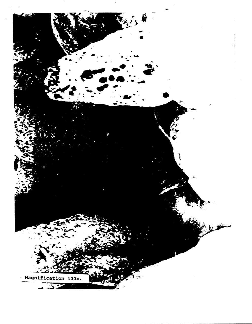
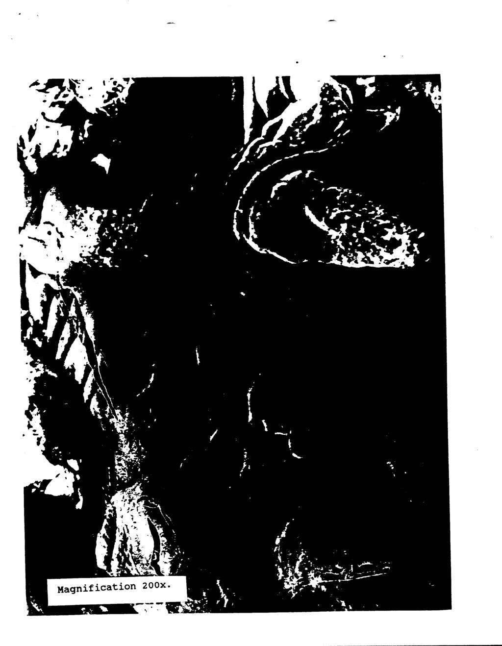
8 The photographs enclosed show numerous strands of cilia dispersed everywhere on the epithelial layer. In Figure 1, the cilia are shown to be uniformly distributed along the surface. It seems as though, four or five cilia have bound together or are intertwined. In Figure 2, papillae are shown to be disrupted. Perhaps the disruption may have taken place when the cilia collapsed during the Peldri 11 dehydration. The papillae seem to have holes on their surfaces. A higher magnification of a papillae is shown in Figure 3. This photograph shows a papillae with numerous cilia on its surface. The remainder of the photographs taken are located in the appendix including the original scanning electron microscope photographs. From these photographs, we are able to conclude that there are cilia on the surface of the mantle of the Cypraea caputserpentis, and in some areas, there distribution is uniform, as shown in Figure 1. Although the papillae located on the epithelia look smooth in Figure 2, there are many other papillae exhibiting many cilia (Figure 3). The disruption of the cells seen in many photographs located in the appendix lead us to acknowledge the need for many experiments using different techniques of dehydration and drying of these -delicate cells. A good technique which should be tried is the substitution of critical point drying instead of the Peldri 11. A comparison of the two would help in determining the best way for preservation of natural looking cells. Another technique which should also be tested is to monitor the specimen when soaking in 100% Peldri 11 solution, in order to not let the specimen to become dry.
9 APPENDIX

10

11
12

13

14

15
16

17 Magnification 500x.

18 -.- Magnification 400x.
19 Magnification 500x.
20 * - Magnification 700x.

21
22
Scanning Electron Microscopy of Thiobacilli
 Arch. Microbiol. 99, 323-329 (1974) 0 by Springer-Verlag 1974 Scanning Electron Microscopy of Thiobacilli Grown on Colloïdal Sulfur J. Baldensperger", L. J. Guarraia**, and W. J. Humphreys*** Department
NERVE ENDINGS OF THE ORGAN OF CORTI AUTHORS: DOMINGGUS MANGAPE DEPARTMENT OF OTOLARYNGOLOGY HIROSHIMA UNIVERSITY
 Ear Res Jpn 13 NERVE ENDINGS OF THE ORGAN OF CORTI AUTHORS: DOMINGGUS MANGAPE YASUO HARADA NOBUHARU TAGASHIRA DEPARTMENT OF OTOLARYNGOLOGY HIROSHIMA UNIVERSITY SCHOOL OF MEDICINE. Nerve endings of the
Scanning electron microscopy of pulmonary alveolar capillary vessels
 Thorax (1973), 28, 222. Scanning electron microscopy of pulmonary alveolar capillary vessels I. G. S. ALEXANDER', B. C. RITCHIE, and J. E. MALONEY Departments of Anatomy and Medicine, Monash University,
Yara Saddam. Amr Alkhatib. Ihsan
 1 Yara Saddam Amr Alkhatib Ihsan NOTE: Yellow highlighting=correction/addition to the previous version of the sheet. Histology (micro anatomy) :- the study of tissues and how they are arranged into organs.
The Ruth & Ted Braun Awards for Writing Excellence at Saginaw Valley State University
 The Ruth & Ted Braun Awards for Writing Excellence at Saginaw Valley State University Analysis of Reproductive Structures of Venustachona ellipsiformis and Pyganodon lacustris by Scanning and Transmission
Elastic Skeleton of Intracranial Cerebral Aneurysms in Rats
 1722 Elastic Skeleton of Intracranial Cerebral Aneurysms in Rats Naohiro Yamazoe, MD, Nobuo Hashimoto, MD, Haruhiko Kikuchi, MD, and Fumitada Hazama, MD In an attempt to clarify the developmental mechanism
AET-treated normal red cells (PNH-like cells)
") J. clin. Path., 1971, 24, 677-684 Electron microscope study of PNH red cells and AET-treated normal red cells (PNH-like cells) S. M. LEWIS, G. LAMBERTENGHI, S. FERRONE, AND G. SIRCHIA From the Department
J. clin. Path., 1971, 24, 677-684 Electron microscope study of PNH red cells and AET-treated normal red cells (PNH-like cells) S. M. LEWIS, G. LAMBERTENGHI, S. FERRONE, AND G. SIRCHIA From the Department
Chemical Level Of Organization
 Chemical Level Of Organization List the Four Chemical Elements that Make Up Most of the Body s Mass Oxygen Carbon Hydrogen Nitrogen Distinguish Between Organic and Inorganic Compounds Organic Compounds:
Surface morphology of the gastroduodenal mucosa in duodenal ulceration
 Gut, 1984, 25, 1203-1210 Surface morphology of the gastroduodenal mucosa in duodenal ulceration H W STEER From the Southampton General Hospital, Southampton SUMMARY Endoscopic biopsies from the duodenal
Ultrastructure of Connective Tissue Cells of Giant African Snails Achatina fulica (Bowdich)
") Kasetsart J. (Nat. Sci.) 36 : 285-290 (2002) Ultrastructure of Connective Tissue Cells of Giant African Snails Achatina fulica (Bowdich) Viyada Seehabutr ABSTRACT The connective tissue sheath of cerebral
Kasetsart J. (Nat. Sci.) 36 : 285-290 (2002) Ultrastructure of Connective Tissue Cells of Giant African Snails Achatina fulica (Bowdich) Viyada Seehabutr ABSTRACT The connective tissue sheath of cerebral
Scanning Electron Microscopical Observation on the Penetration Mechanism of Fowl Spermatozoa into the Ovum in the Process of Fertilization
 J. Fac. Fish. Anim. Husb., Hiroshima Univ. (1976), 15: 85-92 Scanning Electron Microscopical Observation on the Penetration Mechanism of Fowl Spermatozoa into the Ovum in the Process of Fertilization Shunsaku
one Organelle Function
 5 (a) Complete the table by giving one function of each organelle. 8 Areas outside the box will not be scanned for marking Organelle Function Mitochondrion Rough endoplasmic Smooth endoplasmic Golgi body
The Ruth & Ted Braun Awards for Writing Excellence at Saginaw Valley State University
 The Ruth & Ted Braun Awards for Writing Excellence at Saginaw Valley State University Methods of Spermatogenesis in the Freshwater Mussels Pyganodon lacustris and Venustochonda ellipsiformis as Examined
A close look at pollen grains
 A close look at pollen grains Elizabeth M A Hirst Duncan Shaw/SPL The photographs on pages 10-11 show pollen grains from several different plant species. They were made using a scanning electron microscope.
Surface characteristics of human articular cartilagea scanning electron microscope study
 J. Anat. (1971), 108, 1, pp. 23-30 23 With 16 figures Printed in Great Britain Surface characteristics of human articular cartilagea scanning electron microscope study IAN C. CLARKE BioEngineering Unit,
Epithelium Characteristics cont. 2. Apical Surface
 Epithelium Characteristics cont. 2. Apical Surface always has one exposed (apical) surface Some surfaces are smooth & slick, others may have: microvilli fingerlike extensions of the plasma membrane; increase
Scanning Electron Microscopic Observations on the Sperm Penetration through the Zona Pellucida of Mouse Oocytes Fertilized in vitro
 Scanning Electron Microscopic Observations on the Sperm Penetration through the Zona Pellucida of Mouse Oocytes Fertilized in vitro Masatsugu MOTOMURA and Yutaka TOYODA School of Veterinary Medicine and
Prepared By Student. Dania Abed Al-majeed. Rahma Raad Hanna. Balqees Mohammed Aasim. Dania Hisham. Rasha Rafiee
 Prepared By Student Rahma Raad Hanna Balqees Mohammed Aasim Dania Hisham Dania Abed Al-majeed Rasha Rafiee Epithelia Epithelia can be derived from ectoderm, mesoderm or endoderm -ectoderm gives rise to
Human HIV (1+2) antigen&antibody ELISA Kit
 antigen&antibody ELISA Kit") Human HIV (1+2) antigen&antibody ELISA Kit Catalog Number. CSB-E18042h For the qualitative determination of human HIV (1+2) antibody and P24 antigen concentrations in serum, plasma. This package insert
Human HIV (1+2) antigen&antibody ELISA Kit Catalog Number. CSB-E18042h For the qualitative determination of human HIV (1+2) antibody and P24 antigen concentrations in serum, plasma. This package insert
Chapter 05. *Lecture Outline. PowerPoints prepared by Melanie Waite-Altringer Biology Faculty Member of Anoka-Ramsey Community College
 Chapter 05 *Lecture Outline *See separate Image PowerPoint slides for all figures and tables pre-inserted into PowerPoint without notes. PowerPoints prepared by Melanie Waite-Altringer Biology Faculty
POLLEN-WALL PROTEINS: ELECTRON- MICROSCOPIC LOCALIZATION OF ACID PHOSPHATASE IN THE INTINE OF CROCUS VERNUS
 J. Cell Sci. 8, 727-733 (197O 727 Printed in Great Britain POLLEN-WALL PROTEINS: ELECTRON- MICROSCOPIC LOCALIZATION OF ACID PHOSPHATASE IN THE INTINE OF CROCUS VERNUS R.B. KNOX* AND J. HESLOP-HARRISONf
ALL PHOTOS ARE IDENTIFIED IN THE LOWER RIGHT CORNER WITH THE MAGNIFICATION POWER THAT THE PHOTO WAS TAKEN WITH. SCAN - THIS IS A VERY LOW POWER IMAGE
 ALL PHOTOS ARE IDENTIFIED IN THE LOWER RIGHT CORNER WITH THE MAGNIFICATION POWER THAT THE PHOTO WAS TAKEN WITH. SCAN - THIS IS A VERY LOW POWER IMAGE THAT WE USE WHEN A SAMPLE IS SO BIG THAT YOU CAN T
Tissues. Tissues - Overview. Bio 101 Laboratory 3. Epithelial Tissues and Integument
 Bio 101 Laboratory 3 Epithelial Tissues and Integument 1 Tissues Tissues to be examined under the microscope Epithelial Tissue Integument Connective Tissue **We will be doing muscle and nervous tissues
B17 instructions for 227. April 15, 2011
 Microviewer 227: Comparative Digestive Systems Introduction This set is one of a series of lessons examining comparative life function systems. In these sets, you will examine slides of different animals,
ELECTRON MICROSCOPE STUDIES OF THE MICROVILLI OF HELA CELLS
 ELECTRON MICROSCOPE STUDIES OF THE MICROVILLI OF HELA CELLS HAROLD W. FISHER and T. W. COOPER From the Biophysics Laboratories, the University of Rhode Island, Kingston, Rhode Island 0W2881 ABSTRACT Microvilli
Electron Microscopy. dishes in Eagle minimum essential medium with 10% serum to a density that allowed them to grow in a C02
 JOURNAL OF BACTERIOLOGY, Mar. 1978, p. 1452-1456 0021-9193/78/0133-1452$02.00/0 Copyright 1978 American Society for Microbiology Vol. 133, No. 3 Printed in U.S.A. Positive Detection of Mycoplasma Contamination
Organs of the Respiratory System Laboratory Exercise 52
 Organs of the Respiratory System Laboratory Exercise 52 Background The organs of the respiratory system include the nose, nasal cavity, sinuses, pharynx, larynx, trachea, bronchial tree, and lungs. They
Lecture Overview. Marieb s Human Anatomy and Physiology. Chapter 4 Tissues: The Living Fabric Epithelial Tissues Lecture 9. Introduction to Tissues
 Marieb s Human Anatomy and Physiology Marieb Hoehn Chapter 4 Tissues: The Living Fabric Epithelial Tissues Lecture 9 Lecture Overview Introduction to Tissues Epithelial Tissues Location General characteristics
Gonad development in the sea cucumber Holothuria scabra Jaeger, 1833
 15 Gonad development in the sea cucumber Holothuria scabra Jaeger, 1833 Abstract Mélanie Demeuldre 1 and Igor Eeckhaut 1 The purpose of this study is to describe the gonads of small Holothuria scabra.
ON THE PRESENCE OF A CILIATED COLUMNAR EPITHELIAL CELL TYPE WITHIN THE BOVINE CERVICAL MUCOSA 1
 ON THE PRESENCE OF A CILIATED COLUMNAR EPITHELIAL CELL TYPE WITHIN THE BOVINE CERVICAL MUCOSA 1 R. I. Wordinger, 2 J. B. Ramsey, I. F. Dickey and I. R. Hill, Jr. Clemson University, Clemson, South Carolina
INVESTIGATION OF THE ULTRAFINE STRUCTURE OF THE KIDNEY BY MEANS OF SCANNING ELECTRON MICROSCOPE
 THE KURUME MEDICAL JOURNAL 1975 Vol.22, No.3, P.135-141 INVESTIGATION OF THE ULTRAFINE STRUCTURE OF THE KIDNEY BY MEANS OF SCANNING ELECTRON MICROSCOPE I. THE GLOMERULUS SHINSHI NODA Department of Urology,
Epithelium tissue system
 Epithelium tissue system Histology : is the study of the microscopic anatomy (microanatomy) of cells and tissues of plants and animals. It is commonly performed by examining cells and tissues under a light
SensoLyte pnpp Alkaline Phosphatase Assay Kit *Colorimetric*
 SensoLyte pnpp Alkaline Phosphatase Assay Kit *Colorimetric* Catalog # 72146 Kit Size 500 Assays (96-well plate) Optimized Performance: This kit is optimized to detect alkaline phosphatase activity Enhanced
Histology Notes -Part 1: Epithelial Tissues
 Introduction Group of cells w/ similar structure & function = TISSUE Four Basic Tissue Types 1. Epithelial-covers 2. Connective-supports 3. Muscular*-produces movement (will discuss in the muscular system
FIXATION OF TISSUES MODULE 5.1 INTRODUCTION OBJECTIVES 5.2 AIMS OF FIXATION 5.3 PRINCIPLE OF FIXATION. Notes
 MODULE Fixation of Tissues 5 FIXATION OF TISSUES 5.1 INTRODUCTION It is a process by which the cells or tissues are fixed in chemical and partly physical state so that they can withstand subsequent treatment
DECILIATION IN THE PUERPERAL FALLOPIAN TUBE*
 FERTILITY AND STERILITY Copyright < 1978 The American Fertility Society Vol. 29, No.1, January 1978 Printed in U.SA. DECILIATION IN THE PUERPERAL FALLOPIAN TUBE* KENICHI SEKI, M.D.t J. RAWSON, PH.D.t CARLTON
ACERTAIN degree of correlation between
 Electron Microscopy of Human Plasma Lipoprotein Separated by Ultracentrifugation By D. E. BEISCHER, PH.D. The particle size distribution of the Sf 30 "class" of serum lipoprotein was determined by the
HYDROSALPINX SIMPLEX AS SEEN BY THE SCANNING ELECTRON MICROSCOPE*
 FERTILITY AND STERILITY Copyright," 1977 The American Fertility Society Vol. 28, No.9, September 1977 Printed in U.s.A. HYDROSALPINX SIMPLEX AS SEEN BY THE SCANNING ELECTRON MICROSCOPE* EVA PATEK, M.D.t
Endothelial cell damage in human and rabbit corneas stored in K-Sol without antioxidants
 British Journal of Ophthalmology, 1989, 73, 803-808 Endothelial cell damage in human and rabbit corneas stored in K-Sol without antioxidants T SANJEEVA REDDY, EMILY D VARNELL, ROGER W BEUERMAN, NICOLAS
Scanning Electron Microscopy of the Lingual Dorsal Surface of the Beagle Dog. Shin-ichi IWASAKI and Koichi SAKATA
 Okajimas Folia Anat. Jpn., 62 (1) : 1-14, May 1985 Scanning Electron Microscopy of the Lingual Dorsal Surface of the Beagle Dog By Shin-ichi IWASAKI and Koichi SAKATA Department of Oral Anatomy, Nippon
Cellular Transport Worksheet
 Cellular Transport Worksheet Name Section A: Cell Membrane Structure 1. Label the cell membrane diagram. You ll need to draw lines to some of the structures. **Draw cholesterol molecules in the membrane.**
Human TSH ELISA Kit. User Manual
 Human TSH ELISA Kit User Manual Catalog number: GTX15585 GeneTex Table of Contents A. Product Description... 2 B. Kit Components... 3 C. Additional Required Materials (not included)... 3 D. Reagent Preparation...
THE DISCOCYTE-ECHINOCYTE TRANSFORMATION AS AN INDEX OF HUMAN RED CELL TRAUMA 1
 THE DSCOCYTE-ECHNOCYTE TRANSFORMATON AS AN NDEX OF HUMAN RED CELL TRAUMA KETH L. BLACK AND RCHARD D. JONES, Division of Surgical Research, Saint Luke's Hospital, Cleveland, Ohio Abstract. Scanning electron
Thursday, October 16 th
 Thursday, October 16 th Good morning. Those of you needing to take the Enzymes and Energy Quiz will start very soon. Students who took the quiz Wednesday: Please QUIETLY work on the chapter 6 reading guide.
Tissues. Tissues - Overview. Bio211 Laboratory 2. Epithelial and Connective Tissues
 Bio211 Laboratory 2 Epithelial and Connective Tissues 1 Tissues Tissues to be examined under the microscope Epithelial Tissue (p. 79 Lab Manual) [TODAY] Connective Tissue (p. 93 Lab Manual) [TODAY] Muscle/Nervous
A adipose cells. B capillary. C epithelium
 EPITHELIA Objective The objective of this class is to observe how different epithelia vary in terms of cell shape, size and number of cell layers enabling them to be well adapted for functions in different
SURGICAL PATHOLOGY - HISTOLOGY STAINING MANUAL - NERVE TISSUE Page: 1 of 3 BODIAN'S METHOD - NERVE FIBERS PURPOSE: For demonstrating nerve fibers.
 SURGICAL PATHOLOGY - HISTOLOGY Date: STAINING MANUAL - NERVE TISSUE Page: 1 of 3 BODIAN'S METHOD - NERVE FIBERS PURPOSE: For demonstrating nerve fibers. PRINCIPLE: Protargol-S (silver proteinate) is used
Slide 154: Pancreas, H&E
 Slide 154: Pancreas, H&E the pancreas, located adjacent to the duodenum, is a mixed exocrine and endocrine gland; it is usually readily identifiable by the presence of the interspersed endocrine pancreatic
ENHANCEMENT OF THE GRANULATION OF ADRFNERGIC STORAGE VESICLES IN DRUG-FREE SOLUTION
 ENHANCEMENT OF THE GRANULATION OF ADRFNERGIC STORAGE VESICLES IN DRUG-FREE SOLUTION TAKASHI IWAYAMA and J. B. FURNESS. From the Department of Zoology, University of Melbourne, Victoria, Australia. Dr.
ELLEN ROTER DIRKSEN. From the Cancer Research Institute, University of California San Francisco, San Francisco, California 94143
 CILIOGENESIS IN THE MOUSE OVIDUCT A Scanning Electron Microscope Study ELLEN ROTER DIRKSEN. From the Cancer Research Institute, University of California San Francisco, San Francisco, California 94143 INTRODUCTION
Microscopic Study of Histological Changes the Use of Ileal Mucosa as a Bladder (Radical Cystectomy - Case Report)
") Microscopic Study of Histological Changes the Use of Ileal Mucosa as a Bladder (Radical Cystectomy - Case Report) Sareh Najaf Asaadi, Hassan Morovvati, Ahmad Reza Taftachi International Journal of Advanced
Microscopic Study of Histological Changes the Use of Ileal Mucosa as a Bladder (Radical Cystectomy - Case Report) Sareh Najaf Asaadi, Hassan Morovvati, Ahmad Reza Taftachi International Journal of Advanced
Scanning Electron Microscopy of the Small Intestine of a Normal Unsuckled Calf and a Calf with Enteric Colibacillosis
 Vet. Pathol. 15; 400-406 (1978) Scanning Electron Microscopy of the Small Intestine of a Normal Unsuckled Calf and a Calf with Enteric Colibacillosis G. R. PEARSON. E. F. LOGAN and G. P. BRENNAN Departmcnt
Scanning electron microscope characterization of abrasion in human teeth
 Dental Journal Mahidol Dental Journal Original Article Scanning electron microscope characterization of abrasion in human teeth B.Sc., D.D.S., Specialty Certificate in Oral Pathology, M.S. Department of
Lecture Overview. Chapter 4 Epithelial Tissues Lecture 9. Introduction to Tissues. Epithelial Tissues. Glandular Epithelium
 Visual Anatomy & Physiology First Edition Martini & Ober Chapter 4 Lecture 9 Lecture Overview Introduction to Tissues Location General characteristics Functions Classification Glandular Epithelium 2 Where
Histology of the Eye
 Histology of the Eye Objectives By the end of this lecture, the student should be able to describe: The general structure of the eye. The microscopic structure of:»cornea.»retina. EYE BULB Three coats
1. (a) (i) Ability to distinguish points (close together); 1 (ii) Electrons have a shorter wavelength; 1
 (i) Ability to distinguish points (close together); 1 (ii) Electrons have a shorter wavelength; 1") 1. (a) (i) Ability to distinguish points (close together); 1 Electrons have a shorter wavelength; 1 (b) (i) Golgi / nucleus / mitochondrion / endoplasmic reticulum / chromosome / larger ribosomes; R Membrane
1. (a) (i) Ability to distinguish points (close together); 1 Electrons have a shorter wavelength; 1 (b) (i) Golgi / nucleus / mitochondrion / endoplasmic reticulum / chromosome / larger ribosomes; R Membrane
Study of different tissues Abnormal cells and tissues can be compared to normal tissues to identify disease, such as cancer Being able to know and
 CHAPTER 4 Study of different tissues Abnormal cells and tissues can be compared to normal tissues to identify disease, such as cancer Being able to know and recognize normal tissues under the microscope
CoQ10(Coenzyme Q10) ELISA Kit
 ELISA Kit") CoQ10(Coenzyme Q10) ELISA Kit Catalogue No.: EU0196 Size: 48T/96T Reactivity: Universal Detection Range: 0.781-50ng/ml Sensitivity:
CoQ10(Coenzyme Q10) ELISA Kit Catalogue No.: EU0196 Size: 48T/96T Reactivity: Universal Detection Range: 0.781-50ng/ml Sensitivity:
Effect of Parainfluenza-3 Virus Neuraminidase on the Structure of the Gel Phase of Bovine Nasal Secretion
 INFECTION AND IMMUNITY, Oct. 1974, p. 877-882 Copyright 0 1974 American Society for Microbiology Vol. 10, No. 4 Printed in U.S.A. Effect of Parainfluenza-3 Virus Neuraminidase on the Structure of the Gel
Pollen Slide Mounting Protocol
 Pollen Slide Mounting Protocol Materials: Syn-Matrix mounting medium Microcentrifuge Microscope slides Slide coverslips (18mm x 18mm) Coverslip podium (see Figure 1) Capillary tubes Dissecting microscope
INTERNATIONAL BIRD STRIKE COMMITTEE Amsterdam, April 2000 NEW TECHNIQUE FOR PREPARATION OF FEATHER REMAINS
 INTERNATIONAL BIRD STRIKE COMMITTEE IBSC25/WP-I2 Amsterdam, 17-21 April 2000 NEW TECHNIQUE FOR PREPARATION OF FEATHER REMAINS R.K. Sharma & Chinmay Joshi Department of Zoology, Bareilly College, P.O.Box
LOW-ANGLE X-RAY DIFFRACTION AND ELECTRON-MICROSCOPE STUDIES OF ISOLATED CELL MEMBRANES
 J. Cell Sci. I, 287-296 (1966) 287 Printed in Great Britain LOW-ANGLE X-RAY DIFFRACTION AND ELECTRON-MICROSCOPE STUDIES OF ISOLATED CELL MEMBRANES J. B. FINEAN, R. COLEMAN, W. G. GREEN* AND A. R. LIMBRICK
(b) Stomach s function 1. Dilution of food materials 2. Acidification of food (absorption of dietary Fe in small intestine) 3. Partial chemical digest
 Stomach s function 1. Dilution of food materials 2. Acidification of food (absorption of dietary Fe in small intestine) 3. Partial chemical digest") (1) General features a) Stomach is widened portion of gut-tube: between tubular and spherical; Note arranged of smooth muscle tissue in muscularis externa. 1 (b) Stomach s function 1. Dilution of food
(1) General features a) Stomach is widened portion of gut-tube: between tubular and spherical; Note arranged of smooth muscle tissue in muscularis externa. 1 (b) Stomach s function 1. Dilution of food
Biology. Dr. Khalida Ibrahim
 Dr. Khalida Ibrahim Biology Histology: Histology: is the study of the tissues of the body. Tissue: group of similar cells combined to perform a common function. The human body is composed of only 4 basic
Three Dimensional Architecture of Collagen Fibrils in the Corpus cavernosum of the Crab-eating Monkey
 Okajimas Folia Anat. Jpn., 73(4): 185-194, October, 1996 Three Dimensional Architecture of Collagen Fibrils in the Corpus cavernosum of the Crab-eating Monkey By Takashi NAKANO Department of Anatomy, Aichi
MYOFIBRILLAR STRUCTURAL CHANGES CAUSED BY MARINATION WITH CALCIUM PHOSPHATE OR CALCIUM CHLORIDE AND SODIUM PYROPHOSPHATE
 Cattlemen s Day 2002 MYOFIBRILLAR STRUCTURAL CHANGES CAUSED BY MARINATION WITH CALCIUM PHOSPHATE OR CALCIUM CHLORIDE AND SODIUM PYROPHOSPHATE T. E. Lawrence, A. T. Waylan, and C. L. Kastner Summary Ultrastructural
Tissues. How do cells form tissues?
 Tissues How do cells form tissues? Using cell junctions Tissues Epithelial tissue Connective tissue Muscle tissue Nervous tissue Epithelial Tissue Closely packed cells in continuous sheets connected by
MULTIPLE CHOICE. Choose the one alternative that best completes the statement or answers the question.
 Exam Name MULTIPLE CHOICE. Choose the one alternative that best completes the statement or answers the question. 1) A nanometer would be a suitable unit of measurement for which of the following? 1) A)
Human Alpha 1 microglobulin ELISA Kit
 Human Alpha 1 microglobulin ELISA Kit Catalogue No.: EH4144 Size: 48T/96T Reactivity: Human Range:0.625-40ng/ml Sensitivity:
To provide you with necessary knowledge and skills to accurately perform 3 HIV rapid tests and to determine HIV status.
 Module 9 Performing HIV Rapid Tests Purpose To provide you with necessary knowledge and skills to accurately perform 3 HIV rapid tests and to determine HIV status. Pre-requisite Modules Module 3: Overview
Bacterial Structure and Function
 Bacterial Structure and Function Charles Okolie, PhD. Room 311 (on level 4), First College Building, Landmark University okolie.charles@lmu.edu.ng Tel: Ext: Mobile: 08060241166 Structure of Bacteria The
» Croscarmellose Sodium is a cross linked polymer of carboxymethylcellulose sodium.
 BRIEFING Croscarmellose Sodium, NF 22 page 2856 and page 702 of PF 30(2) [Mar. Apr. 2004]. A modification is made in the test for Degree of substitution to correct the endpoint color to agree with the
Skin Structure of the Hawaiian Monk Seal (Monachus schauinslandi)i
i") Pacific Science (1975), Vol. 29, No.2, p. 153-157 Printed in Great Britain Skin Structure of the Hawaiian Monk Seal (Monachus schauinslandi)i G. C. WHITTOW,2 J. SZEKERCZES,2 E. KRIDLER,3 AND D. L. OLSEN3
Pacific Science (1975), Vol. 29, No.2, p. 153-157 Printed in Great Britain Skin Structure of the Hawaiian Monk Seal (Monachus schauinslandi)i G. C. WHITTOW,2 J. SZEKERCZES,2 E. KRIDLER,3 AND D. L. OLSEN3
Epithelia will be discussed according to the following scheme: Type Number of layers Shape Line drawing. Squamous Cuboidal Columnar
 Epithelia Epithelia will be discussed according to the following scheme: Type Number of layers Shape Line drawing Simple Squamous Cuboidal Columnar Covering and Lining epithelium Pseudostratified Stratified
INVESTIGATIVE OPHTHALMOLOGY
 January 1972 Volume 11, Number 1 INVESTIGATIVE OPHTHALMOLOGY Scanning electron microscopy of corneal graft rejection: Epithelial rejection, endothelial rejection, and formation of posterior graft membranes
Rubella Latex Agglutination Test
 Rubella Latex Agglutination Test Cat. No.:DLAT1088 Pkg.Size:30T Intended use The Rubella Latex Agglutination Test is a rapid latex particle agglutination test for the qualitative and semi-quantitative
Notes to complete gas exchange in mammals
 Notes to complete gas exchange in mammals Mass flow of air to respiratory surface this is achieved through the mechanics of ventilation (breathing). This ensures a regular supply of air into and out of
RG12T. For In Vitro Diagnostic Use. Passive Particle-Agglutination Test for Detection of Antibodies to HTLV-
 RG12T Read this insert carefully before performing the assay and keep for future reference. The reliability of assay procedures other than those described in this package insert cannot be guaranteed. For
Mesenchymal Stem Cells Reshape and Provoke Proliferation of Articular. State Key Laboratory of Bioreactor Engineering, East China University of
 Mesenchymal Stem Cells Reshape and Provoke Proliferation of Articular Chondrocytes by Paracrine Secretion Lei Xu, Yuxi Wu, Zhimiao Xiong, Yan Zhou, Zhaoyang Ye *, Wen-Song Tan * State Key Laboratory of
THE MICROSCOPE PAST: 40 YEARS AGO
 THE MICROSCOPE Vol 58:2, pp 87-91 (2010) THE MICROSCOPE PAST: 40 YEARS AGO Glycol Methacrylate, an Embedding Medium to Study the Ultrastructure of Cotton Treated with Swelling Agents S.M. Betrabet* and
and glycoprotein in human bronchial glands
 Thorax, 1981, 36, 108-115 Ultrastructural localisation of lactoferrin and glycoprotein in human bronchial glands DENISE BOWES, A E CLARK, AND B CORRIN From Midhurst Medical Research Institute, Midhurst,
Distribution of the Pores of Epithelial Basement Membrane in the Rat Small Intestine
 FULL PAPER Anatomy Distribution of the Pores of Epithelial Basement Membrane in the Rat Small Intestine Takashi TAKEUCHI 1) and Tatsuo GONDA 1) 1) Institute of Experimental Animals, Shimane Medical University,
Figure 4.1. Using Figure 4.1, identify the following: 1) The region that contains adipose tissue is indicated by letter. Diff: 2 Page Ref: 115
 The region that contains adipose tissue is indicated by letter. Diff: 2 Page Ref: 115") Essentials of Anatomy and Physiology, 9e (Marieb) Chapter 4 Skin and Body Membranes Short Answer Figure 4.1 Using Figure 4.1, identify the following: 1) The region that contains adipose tissue is indicated
Essentials of Anatomy and Physiology, 9e (Marieb) Chapter 4 Skin and Body Membranes Short Answer Figure 4.1 Using Figure 4.1, identify the following: 1) The region that contains adipose tissue is indicated
MagniSort Mouse CD4 Naive T cell Enrichment Kit Catalog Number: RUO: For Research Use Only. Not for use in diagnostic procedures.
 Page 1 of 2 MagniSort Mouse CD4 Naive T cell Enrichment Kit RUO: For Research Use Only. Not for use in diagnostic procedures. Mouse splenocytes were unsorted (left) or sorted with the MagniSort Mouse CD4
Serum Amyloid A ELISA
 Serum Amyloid A ELISA For the quantitative determination of serum amyloid A (SAA) in serum plasma For Research use Only. Not for Use in Diagnostic Procedures Please see Appendix A for Reference Serum information
MORPHOLOGIC CHARACTERISTICS OF THE CHEMICALLY INDUCED ACROSOME REACTION IN HUMAN SPERMATOZOA*
 FERTILITY AND STERILITY Copyright 1979 The American Fertility Society Vol. 32, No.1, July 1979 Printed in U.SA. MORPHOLOGIC CHARACTERISTICS OF THE CHEMICALLY INDUCED ACROSOME REACTION IN HUMAN SPERMATOZOA*
Scanning electron microscopic study of the tongue in the rainbow lorikeet (Trichoglossus haematodus)
") Okajimas Folia Anat. Jpn., 88(1): 17 21, May, 2011 Scanning electron microscopic study of the tongue in the rainbow lorikeet (Trichoglossus haematodus) By Shoichi EMURA 1, Toshihiko OKUMURA 2 and Huayue
Okajimas Folia Anat. Jpn., 88(1): 17 21, May, 2011 Scanning electron microscopic study of the tongue in the rainbow lorikeet (Trichoglossus haematodus) By Shoichi EMURA 1, Toshihiko OKUMURA 2 and Huayue
CELLS ARE A BAG OF GOO
 Fifth Grade, 2013 CELLS ARE A BAG OF GOO By Michael E. Knotts, Curt M. Peterson, and Diana Anderson 2013 by Michael E. Knotts, Curt M. Peterson, and Diana Anderson, All Rights Reserved Objective and Overview:
Explain the reason for this difference in resolving power.
 1. (a) An electron microscope has a much greater resolving power than an optical microscope. (i) Explain the meaning of the term resolving power. Explain the reason for this difference in resolving power.
psittaci by Silver-Methenamine Staining and
 JOURNAL OF BACTERIOLOGY, July 1972, p. 267-271 Copyright 1972 American Society for Microbiology Vol. 111, No. 1 Printed in U.S.A. Location of Polysaccharide on Chlamydia psittaci by Silver-Methenamine
The Digestive System Laboratory
 The Digestive System Laboratory 1 The Digestive Tract The alimentary canal is a continuous tube stretching from the mouth to the anus. Liver Gallbladder Small intestine Anus Parotid, sublingual, and submaxillary
Changing Your Trach Tube
 Patient & Family Guide 2017 Changing Your Trach Tube www.nshealth.ca Changing Your Trach Tube Your entire trach tube should be changed about once a month, or sooner if you notice an odour (smell) or get
INVESTIGATIVE OPHTHALMOLOGY. Corneal and conjunctival changes in dysproteinemia
 August 1969 Volume 8, Number 4 INVESTIGATIVE OPHTHALMOLOGY Corneal and conjunctival changes in dysproteinemia 7?. M. H. Pinkerton and David M. Robertson A case of dysproteinemia with corneal and conjunctival
MagniSort Mouse CD4 T cell Enrichment Kit Catalog Number: RUO: For Research Use Only. Not for use in diagnostic procedures.
 Page 1 of 2 MagniSort Mouse CD4 T cell Enrichment Kit RUO: For Research Use Only. Not for use in diagnostic procedures. Mouse splenocytes were unsorted (left) or sorted with the MagniSort Mouse CD4 T cell
There are enzymes in biological washing powders. Biological washing powder has to be used at temperatures below 45 C.
 There are enzymes in biological washing powders. Biological washing powder has to be used at temperatures below 45 C. The enzymes in biological washing powders do not work on the stains on clothes at temperatures
TRANSIL ASSAY KITS. Frequently Asked Questions (FAQs)
") TRANSIL ASSAY KITS Frequently Asked Questions (FAQs) 1. 2. 3. What is the principle of TRANSIL technology? What membrane is on the surface of the What are the key advantages of the TRANSIL technology?
TRANSIL ASSAY KITS Frequently Asked Questions (FAQs) 1. 2. 3. What is the principle of TRANSIL technology? What membrane is on the surface of the What are the key advantages of the TRANSIL technology?
Figure 4.25: SEM Photo of Hyaluronic Acid Specimen, High Load, 22X
 Figure 4.25 shows the specimen from test CS18 at a magnification of 22X. Hyaluronic acid, dissolved at a concentration of 0.375 wt.% in buffered saline solution, was the lubricant in this test, with high
Organ Cultures of Respiratory Epithelium Infected with Rhinovirus or Parainfluenza Virus Studied in a Scanning Electron Microscope
 INFECTION AND IMMUNITY, JUly 1972, p. 68-76 Copyright 1972 American Society for Microbiology Vol. 6, No. 1 Printed in U.S.A. Organ Cultures of Respiratory Epithelium Infected with Rhinovirus or Parainfluenza
MagniSort Human CD4 Memory T cell Enrichment Kit Catalog Number: RUO: For Research Use Only. Not for use in diagnostic procedures.
 Page 1 of 2 MagniSort Human CD4 Memory T cell Enrichment Kit RUO: For Research Use Only. Not for use in diagnostic procedures. Normal human peripheral blood mononuclear cells were unsorted (left) or sorted
Microscopic Anatomy of Inferior Medullary Velum Of Cerebellum
 32 J Anat. Soc. India 51(1) 32-34 (2002) Microscopic Anatomy of Of Cerebellum Arora, N.K. Department of Anatomy, Government Medical College, Chandigarh INDIA. Abstract. A study of the inferior medullary