Objectives: To assess the distribution of bone erosions in the feet of patients
|
|
|
- Wendy Williamson
- 5 years ago
- Views:
Transcription
1 Gout on CT: a symmetric arthropathy. Anthony J Doyle (corresponding author) Objectives: To assess the distribution of bone erosions in the feet of patients with gout using computed tomography (CT) and thereby to test the hypothesis that gout is an asymmetric arthropathy. Methods: CT scans of both feet were obtained from 25 patients with chronic gout. CT scans were scored for bone erosion using a semi-quantitative method based on the rheumatoid arthritis MRI scoring system (RAMRIS). CT bone erosion was assessed at 22 bones in each foot (total 1,100 bones) by two independent radiologists. Symmetry was assessed by two methods: a) comparing right and left foot scores for each patient; b) calculating the proportion of paired joints with or without erosions. Results: Observer agreement was excellent (intra-class correlation coefficient 0.92). In the group overall, the difference in scores between the feet was not significant (Student T test p=0.8). In 17 of 25 patients, the difference in erosion scores between the two feet was less than the inter-observer difference. In 24 of 25 patients, the proportion of paired joints was greater than 0.5, indicating symmetric disease. Conclusions: Erosive disease from gout is, in fact, a symmetric process in our patient group. This finding is contrary to the established view of gout as an asymmetric arthritis and lends new insight into the behaviour of this common disease. 1
2 Introduction Gout is a common arthropathy worldwide and is often described in medical textbooks and review articles as being asymmetric [1-6]. However, there has been little formal research into the distribution of gouty lesions seen on imaging, particularly when using quantitative scoring systems. We used a set of high-resolution CT scans obtained from patients with gout and scored by two independent observers to analyse the distribution of erosive bone disease in the group, especially with regard to right/left symmetry. Patients and Methods Patients Twenty-five adult patients with gout were recruited from rheumatology outpatient clinics in Auckland, New Zealand. The Northern Y Regional ethics committee approved the study and patients provided written informed consent according to the Declaration of Helsinki. As previously described [7], all patients had a history of acute gout according to ACR diagnostic criteria [8], and were excluded if they were experiencing an acute gout flare at the time of assessment, had lower limb amputation or diabetes mellitus. Information regarding age, gender, ethnicity, disease duration, presence of subcutaneous tophi, and other clinical characteristics of gout was recorded. Plain radiographs of the feet and blood for serum urate testing were also obtained on the day of assessment. CT scans The CT scans of the feet and ankles were performed on a Philips Brilliance 16-slice scanner (Philips Medical Systems, Best, The Netherlands). The patients were positioned supine with the knees bent to 90 degrees and the 2
3 feet dorsiflexed 45 degrees. Both feet were scanned together with the CT gantry vertical. The range covered was from 5cm above the ankle joint to the ends of the toes. All scans were performed with the same image protocol; acquisition at 16 x 0.75mm, reconstructed on a bone algorithm, 768 matrix, to 0.8mm slices with a 0.4mm increment. (kvp 140, 120 mas/ slice). Additional reconstructions were done on a soft tissue algorithm, 512 matrix, also to a 0.8mm slice with a 0.4mm increment. The images were viewed as 0.8mm slices on a Philips CT workstation and reconstructed to 3mm slices for viewing on Picture Archiving Communication System (PACS). CT scans were analyzed for bone erosion by two independent musculoskeletal radiologists (AD and LB) who were blinded to the clinical details, plain radiographic damage scores and each other s CT erosion scores. The radiologists used the overlapping thin slices to interactively generate multiplanar reformations on standard PACS workstations (Impax version 4, Agfa-Gevaert, Belgium; Osirix version 3.6, Osirix Foundation, Geneva, Switzerland). Bone erosion was assessed using reformatted images in the anatomic axial, sagittal and coronal planes. Erosions on CT were defined as focal areas of loss of cortex with sharply defined margins, seen in two planes, with cortical break seen in at least one plane. Bone erosion was scored using a semi-quantitative method based on the rheumatoid arthritis MRI scoring system (RAMRIS) [7]; each bone was scored separately on a scale from 0-10, based on the proportion of eroded bone compared to the "assessed bone volume", judged on all available images-0: no erosion; 1: 1-10% of bone eroded; 2; 11-20%, etc. For long bones and large tarsal bones, the "assessed bone volume" was from the articular surface (or its best 3
4 estimated position if absent) to a depth of 1 cm. Bone erosion was assessed at 22 bones in each foot (44 bones/patient) and in a total of 1,100 bones. These sites were selected to include all bones in the foot and ankle except for the great toe distal phalanx and the lesser toe phalanges. The following bones were scored; distal and proximal portions of the 1 st proximal phalanx, 1 st -5 th metatarsal (MT) heads, 1 st -5 th MT bases, lateral, middle and medial cuneiforms, navicular, cuboid, anterior process of calcaneus, proximal calcaneus, distal talus, proximal talus and distal tibia. The erosion scores for right and left feet in each patient and in the group as a whole were compared for each observer and for the averaged scores of both observers using the Student T test. For the paired joint analysis, each joint was recorded as normal if the erosion score was zero and as diseased if the score was not zero. The number of matching pairs was then divided by the total number of joints counted; symmetry is equivalent to this ratio being 0.5 or greater. Results The clinical characteristics of the patients have been reported in detail previously [9]. In brief, 19 (75%) were male, 17 (68%) were non-polynesian and 8 (32%) were of Maori or Pacific ancestry. The median (range) age was 60 (37 83) years and disease duration was 21 (1 50) years. Thirteen (52%) patients had clinical evidence of tophaceous disease, 11 (44%) had microscopically proven disease and 22 (88%) were on regular urate-lowering therapy (21 on allopurinol, 1 on probenecid). The median (range) serum urate was 0.35 ( ) mmol/l. The median (range) gout radiographic damage score for the feet was 17 (0 70). 4
5 The overall erosion scores for right and left feet were very similar for each observer (Table 1). The observer intra-class correlation coefficient was 0.92, indicating excellent agreement. The observer intra-class correlation coefficient (95% CI) for the overall erosion scores was 0.92 (0.82,0.96); for the right feet 0.91(0.83, 0.95) and for the left feet 0.92(0.84,0.96) indicating excellent observer agreement. No significant difference was seen between the scores for right and left feet by observer (p=0.8 observer 1 and 0.7 observer 2); neither was there any difference between the averaged observer scores for right and left feet (p=0.7). Sub analysis of the erosion scores for the fore foot, mid foot and hind foot showed no right/left difference in any of these areas. (Table 2). A typical example of a patient with moderately advanced symmetric gout is shown in Figure 1. The median (range) total CT erosion score (22 sites/patient) was 29 ( ). The average (SD) difference between observer 1 and observer 2 scores for each foot was 5.22 (4.4). We used this average observer difference as a threshold for a significant right/left difference between the two feet. The average (SD) right/left combined observer score difference was 5.4 (6.4). In eight of 25 patients (32%), the right/left difference was greater than 5.22 points and deemed asymmetric (Table 3). In each of these, the observers scored the asymmetry in same direction. Four patients had higher scores on the right, and four on the left. In the remaining 17 (68%) patients, the difference score was 4 or less; in 12 (48%), it was 2.5 or less. Even in the patient with the highest degree of asymmetry, erosions were visible in similar sites on each foot (Figure 2). 5
6 The clinical features of the asymmetric and symmetric groups are given in Table 4. Patients in the asymmetric group were younger, but there was no significant difference in gender, disease duration, presence of tophi on physical examination or serum urate level. There was no difference in the mean total CT erosion score in the asymmetric group compared with the symmetric group (47.3(32.8) vs. 36.4(30.0), p=0.44). Another mathematically based but less strict definition of symmetry was described by Helliwell in 2000 [10]. In this definition, a pair of joints is counted as symmetric if arthritis is recorded as being either present or absent bilaterally. Overall symmetry is defined as being present when the ratio of symmetric pairs of joints to the total number of pairs of joints observed is equal to or greater than 0.5. Using this definition, 24 of our 25 patients had symmetric disease. The only patient with asymmetric disease had a right/left score difference of 16. 6
7 Discussion The traditional teaching in imaging of gout is that it is an asymmetrical arthritis, affecting one side of the body more than the other. Our results indicate that, at least in this patient group, the distribution of gouty erosions in the feet on CT was, in fact, quite symmetric in the vast majority of cases. This was regardless of gender, duration of disease, serum urate and total erosion score. The group with asymmetric disease was a little over ten years younger on average, a result whose significance is uncertain. Previous studies have shown that CT and MRI are more reliable than radiographs for demonstrating erosions in both rheumatoid arthritis and gout, [11-13]. More recently, our group has validated a CT scoring method for gout [7] in which it was discovered that around 65% of the erosion score could be attributed to proximal joints not well seen on radiographs. It is probably reasonable to assume that the CT findings here can be taken as a reasonably true depiction of the actual erosive disease distribution. Other work shows that erosive disease shown on imaging does correlate with clinical features of gout including tophus burden and functional indices such as grip strength [9,12]. It is likely that the bilaterally symmetric nature of the disease shown here will eventually be reflected clinically. Several different explanations may account for the variance between our findings and the traditional view of gout as an asymmetric arthritis in imaging terms. One is that the degree of symmetry simply has not been formally investigated previously; there is very little quantitative data in the literature regarding radiographic findings in gout. Previous authors may have been 7
8 relying on the usual initial clinical presentation of gout as a monoarthritis being a feature that must inevitably be reflected radiographically. Another possible explanation is that the disease begins in an asymmetric fashion and progresses toward symmetry with time. The radiographic signs of gout are said to emerge after 5-10 years disease duration [14] and our patients had an average disease duration of about 20 years. However, disease duration was the same for the symmetric and asymmetric groups, a feature that does not favour duration alone as the determinant of symmetry. The most likely explanation, based on this and our group s previous work showing a high prevalence of mid and hind-foot disease [7], is that most patients with gout do have involved joints that are asymptomatic and that simply have not been detected with older imaging techniques, in particular plain radiographs. As more research is performed into the features of gout on modalities such as CT, MRI and ultrasound where sensitivity is higher than that of radiographs, it may become clearer as to whether most gouty arthritis begins as a symmetric process or evolves into one. The current study, however, strongly suggests that the majority of patients with established gout do in fact have bilaterally symmetric disease. 8
9 Table 1. Erosion scores by foot and observer. Data are presented as mean (SD). Right foot Left foot p Observer (16.1) 20.5 (16.3) 0.8 Observer (15.1) 20.0 (17.5) 0.7 9
10 Table 2. Fore, mid and hind foot scores. Data are pooled from both readers and presented as mean (SD). Right foot Left foot p Fore foot 6.4(6.0) 6.7(6.5) 0.9 Mid foot 10.1(7.7) 10.8(10.1) 0.8 Hind foot 3.2(3.7) 2.8(2.9)
11 Table 3. Difference in erosion scores in asymmetric cases Case Mean Observer Observer Side greater left right right left left right left right 11
12 Table 4. Clinical features of symmetric and asymmetric groups. Symmetric Asymmetric p Age, years, mean (SD) 64(10.3) 53(11.0) 0.02 Male sex, n (%) 13 (76%) 6 (75%) 1.0 Disease duration, years, mean (SD) 22(15.2) 21(10.0) 0.8 Tophi on physical examination, n (%) 8(47) 5(63) 0.7 Serum urate, mmol/l, mean (SD) 0.36(0.1) 0.36(0.2) 0.9 CT erosion score, mean (SD) 36.4(30.0) 47.3(32.8)
13 References 1. Watt I, Middlemiss H. The radiology of gout. Clin Radiol 1975; 26: Gentili A. Advanced Imaging of Gout. Seminars in Musculoskeletal Radiology 2003; 7: Monu JUV, Pope TL. Gout: a clinical and radiologic review. Radiol Clin N Am 2004; 42: Resnick D, Kransdorf MJ. Gouty Arthritis. In: Bone and Joint Imaging. Philadelphia: Elsevier Saunders; 2005: Dhanda S, Jagmohan P, Tian QS. A re-look at an old disease: A multimodality review on gout. Clin Radiol 2011; 66: Schumacher HR, Chen LX. Gout and other Crystal-associated Arthropathies. In: Harrison s Principles of Internal Medicine, 18 th edition. Access Medicine, McGraw-Hill; Dalbeth N, Doyle A, Boyer L, et al. Development of a computed tomography method of scoring bone erosion in patients with gout: validation and clinical implications. Rheumatology 2011;50: Wallace SL, Robinson H, Masi AT, Decker JL, McCarty DJ, Yu TF. Preliminary criteria for the classification of the acute arthritis of primary gout. Arthritis Rheum 1977;20: Dalbeth N, Clark B, McQueen F, Doyle A, Taylor W. Validation of a radiographic damage index in chronic gout. Arthritis Rheum 2007;57: Helliwell PS, Hetthen J, Sokoll K, Green M, Archeson A, Lubrano E, Veale D, Emery P. Joint symmetry in early and late rheumatoid and 13
14 psoriatic arthritis: Comparison with a Mathematical Model. Arthritis Rheum 2000; 43: Perry D, Stewart N, Benton N et al. Detection of erosions in the rheumatoid hand; a comparative study of multidetector computerized tomography versus magnetic resonance scanning. J Rheumatol 2005;32: Dalbeth N, Clark B, Gregory K et al. Mechanisms of bone erosion in gout: a quantitative analysis using plain radiography and computed tomography. Ann Rheum Dis 2009;68: Carter JD, Kedar RP, Anderson S et al. An analysis of MRI and ultrasound imaging in patients with gout who have normal plain radiographs. Rheumatology 2009;48: Bloch C, Hermann G, Yu TF. A radiologic reevaluation of gout: a study of 2000 patients. Am J Roentgenol 1980, 134;

 Figure 1B Mid foot")

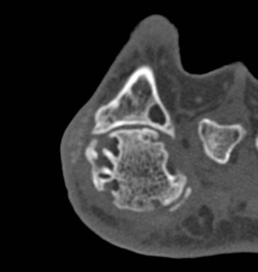
15 Figure 1A Fore foot erosions in patient with symmetric gout (score right=40, left=41) Figure 1B Mid foot erosions in patient with symmetric gout (score right=40, left=41) 15

 are slightly larger")

16 Figure 2A: Fore foot erosions in patient with asymmetric gout (score right=30, left=57) are slightly larger on left Figure 2B: Mid foot erosions in patient with asymmetric gout (score right=30, left=57) are slightly larger on left 16

 are slightly larger")
17 Figure 2B: Hind foot erosions in patient with asymmetric gout (score right=30, left=57) are slightly larger on left 17
Disclosures. Ultrasonography. Conventional radiography (XR) Magnetic resonance imaging. Conventional CT 10/27/2013
 Magnetic resonance imaging. Conventional CT 10/27/2013") What I can see in my gout patient that I couldn t before: the role of advanced imaging in gout diagnosis and monitoring Disclosures ND is supported by the Health Research Council of New Zealand I have
What I can see in my gout patient that I couldn t before: the role of advanced imaging in gout diagnosis and monitoring Disclosures ND is supported by the Health Research Council of New Zealand I have
Urate crystal deposition and bone erosion in gout: inside-out or outside-in? A dual-energy computed tomography study
 Towiwat et al. Arthritis Research & Therapy (2016) 18:208 DOI 10.1186/s13075-016-1105-z RESEARCH ARTICLE Urate crystal deposition and bone erosion in gout: inside-out or outside-in? A dual-energy computed
Gout and Hyperuricemia
 Gout and Hyperuricemia 100 Access publication Sep. 2016 Computed tomography manifestation of gouty arthritis Hu Yabin 1, Yang Qing 1, Cao Qiang 2, Ren Jianan 1, Yang Qing * Objective: Of the most commonly
A case of extensive synovial involvement by tophaceous gout
 A case of extensive synovial involvement by tophaceous gout Nausheen Khan, MB BS, FCRad (D) Irma van de Werke, MB ChB, FRCR Farzanah Ismail, MB ChB, FCRad (D) Department of Radiology, Kalafong Hospital,
Spondyloarthritis: A Gouty Display
 Spondyloarthritis: A Gouty Display Preetam Gongidi 1*, Shawn Gough-Fibkins 2 1. Nova Southeastern University College of Osteopathic Medicine, Fort Lauderdale, FL, USA 2. Broward General Medical Center,
Institutional review board approval was obtained prior to the start of this study.
 Lower Limb Alignment and Length Measurements - Comparison of Computed Tomography, Upright Full-Length Conventional Radiography and Upright Biplanar Linear-Low Dose X-ray Scanner Poster No.: C-1382 Congress:
Radiologic manifestations of gout in MRI
 Radiologic manifestations of gout in MRI Poster No.: C-1053 Congress: ECR 2012 Type: Authors: Keywords: DOI: Educational Exhibit D. Gorostiza Laborda, A. Urresola Olabarrieta, B. Canteli Padilla, F. Perez-Ruiz,
The EULAR OMERACT rheumatoid arthritis MRI reference image atlas: the wrist joint
 i23 The EULAR OMERACT rheumatoid arthritis MRI reference image atlas: the wrist joint B Ejbjerg, F McQueen, M Lassere, E Haavardsholm, P Conaghan, P O Connor, P Bird, C Peterfy, J Edmonds, M Szkudlarek,
Dual energy CT in diagnosis of Gout
 Dual energy CT in diagnosis of Gout Poster No.: R-0060 Congress: RANZCR ASM 2013 Type: Educational Exhibit Authors: F. Tabatabaie Moghadam, A. Moghaddam, F. Ghazanfari ; 1 1 1 2 2 Brisbane/AU, Melbourne/AU
Hi RES Extremity - (04/18/2011) CTDI: ~13 mgy per acquisition Used for evaluation of: Ankle Elbow Hand Wrist Foot /Calcaneous Toes Fingers
 CTDI: ~13 mgy per acquisition Used for evaluation of: Ankle Elbow Hand Wrist Foot /Calcaneous Toes Fingers") P a g e 1 Hi RES Extremity - (04/18/2011) CTDI: ~13 mgy per acquisition Used for evaluation of: Ankle Elbow Hand Wrist Foot /Calcaneous Toes Fingers Billing: 1. CT Upper/Lower Extremity of concern without
P a g e 1 Hi RES Extremity - (04/18/2011) CTDI: ~13 mgy per acquisition Used for evaluation of: Ankle Elbow Hand Wrist Foot /Calcaneous Toes Fingers Billing: 1. CT Upper/Lower Extremity of concern without
Rheumatology Cases for the Internist
 Rheumatology Cases for the Internist Marc C. Hochberg, MD, MPH Professor of Medicine Head, Division of Rheumatology and Clinical Immunology Vice Chair, Department of Medicine University of Maryland School
Section 4: Tarsal Coalitions
 Case H (Figure 2): PedCat CBCT transverse plane reconstruction of right Lisfranc midfoot dislocation compared to normal left foot. Clinical Relevance of the PedCat Study: The weight bearing CBCT study
The radiologist and the raiders of the lost image
 The radiologist and the raiders of the lost image Poster No.: P-0072 Congress: ESSR 2014 Type: Educational Poster Authors: M. J. Ereño Ealo, E. Montejo Rodrigo, B. Sancho, E. Pastor; Galdakao/ES Keywords:
Journal reading. Introduction. Introduction. Ottawa Ankle Rules. Method
 Journal reading Presenter: PGY 林聖傑 Supervisor: Dr. 林俊龍 102.12.23 The accuracy of ultrasound evaluation in foot and ankle trauma Salih Ekinci, MD American Journal of Emergency Medicine 31 (2013) 1551 1555
Radiologic Imaging Magnetic Resonance Imaging (MRI)
") Radiologic Imaging X-ray has always been the golden rule in diagnosing and treating podiatric patients. Unfortunately, for some patients the diagnosis is not as evident. That is when we need to utilize
Radiologic Imaging X-ray has always been the golden rule in diagnosing and treating podiatric patients. Unfortunately, for some patients the diagnosis is not as evident. That is when we need to utilize
PROPHECY. Preoperative Navigation Guides ANKLE CT SCAN PROTOCOL
 PROPHECY Preoperative Navigation Guides ANKLE CT SCAN PROTOCOL 90 FIGURE 1 Examples FIGURE 1 Examples of neutral ankle positioning. PROPHECY Ankle CT Scan Protocol PROPHECY INBONE and PROPHECY INFINITY
Radiographic Assessment of Pediatric Foot Alignment: Self-Assessment Module
 1.5 CME AJR Integrative Imaging LIFELONG LEARNING FOR RADIOLOGY Radiographic Assessment of Pediatric Foot Alignment: Self-Assessment Module Mahesh M. Thapa 1,2, Sumit Pruthi 1,2, Felix S. Chew 2 ABSTRACT
Articular disease of the hand - the target joint approach
 Articular disease of the hand - the target joint approach Poster No.: C-1817 Congress: ECR 2016 Type: Educational Exhibit Authors: R. R. Domingues Madaleno 1, A. P. Pissarra 1, I. Abreu 2, A. Canelas 1,
CLINICAL PRESENTATION AND RADIOLOGY QUIZ QUESTION
 Donald L. Renfrew, MD Radiology Associates of the Fox Valley, 333 N. Commercial Street, Suite 100, Neenah, WI 54956 11/24/2012 Radiology Quiz of the Week # 100 Page 1 CLINICAL PRESENTATION AND RADIOLOGY
Section 3: Foot Subluxations and Dislocations
 Section 3: Foot Subluxations and Dislocations Case Study F: Lisfranc s Midfoot Dislocation Clinical History: J.K. a 28 year old female presents complaining of a painful right foot. She sustained an acute
Cover Page. The handle holds various files of this Leiden University dissertation.
 Cover Page The handle http://hdl.handle.net/1887/40654 holds various files of this Leiden University dissertation. Author: Stomp, W. Title: MR imaging in early rheumatoid arthritis : techniques and applications
Crystal Deposition Disease and Psoriatic Arthritis
 74 Crystal Deposition Disease and Psoriatic Arthritis Philip J. O Connor, MRCP, FRCR, FFSEM (UK) 1,2 1 Department of Radiology, Leeds Teaching Hospitals, Chapel Allerton Hospital, Leeds, United Kingdom
Dual-energy computed tomography compared with ultrasound in the diagnosis of gout
 RHEUMATOLOGY Rheumatology 2014;53:173 179 doi:10.1093/rheumatology/ket341 Advance Access publication 17 October 2013 Original article Dual-energy computed tomography compared with ultrasound in the diagnosis
Imaging. Ultrasound imaging for the rheumatologist XXXVI. Sonographic assessment of the foot in gout patients
 Imaging Ultrasound imaging for the rheumatologist XXXVI. Sonographic assessment of the foot in gout patients E. Filippucci 1, G. Meenagh 2, A. Delle Sedie 3, G. Sakellariou 4, A. Iagnocco 5, L. Riente
ELENI ANDIPA General Hospital of Athens G. Gennimatas
 ELENI ANDIPA General Hospital of Athens G. Gennimatas Technological advances over the last years have caused a dramatic improvement in ultrasound quality and resolution An established imaging modality
HI-Res Extremity Sensation 16
 Page 1 Routine Extremity - (2/14/2013) CTDI: ~20 mgy per acquisition Used for evaluation of: Humerus Forearm Femur Knee Tib/Fib Billing: 1. CT Upper/Lower Extremity of concern without contrast, with contrast,
A Pictorial Review of Congenital Tarsal Coalition
 A Pictorial Review of Congenital Tarsal Coalition Poster No.: C-2305 Congress: ECR 2011 Type: Educational Exhibit Authors: J. Jethwa, M. Tapp; Torquay/UK Keywords: Musculoskeletal joint, Musculoskeletal
MSK CT Extremities: Positioning and Reformations
 MSK CT Extremities: Positioning and Reformations Hand: Patient lying in prone position, with affected arm extended above head. Place body off centered in effort to set affected hand in isocenter. Hand
International Journal of Research in Health Sciences ISSN: Available online at: Case Study
 International Journal of Research in Health Sciences ISSN: 2321-7251 Available online at: http://www.ijrhs.org/ Case Study Foreign body granuloma mimicking a soft tissue neoplasm *Rohan Sawant, Abhishek
CLINICAL PRESENTATION AND RADIOLOGY QUIZ QUESTION
 Donald L. Renfrew, MD Radiology Associates of the Fox Valley, 333 N. Commercial Street, Suite 100, Neenah, WI 54956 12/01/2012 Radiology Quiz of the Week # 101 Page 1 CLINICAL PRESENTATION AND RADIOLOGY
Ethan M. Braunstein, M.D. 1, Steven A. Goldstein, Ph.D. 2, Janet Ku, M.S. 2, Patrick Smith, M.D. 2, and Larry S. Matthews, M.D. 2
 Skeletal Radiol (1986) 15:27-31 Skeletal Radiology Computed tomography and plain radiography in experimental fracture healing Ethan M. Braunstein, M.D. 1, Steven A. Goldstein, Ph.D. 2, Janet Ku, M.S. 2,
CLINICAL PRESENTATION AND RADIOLOGY QUIZ QUESTION
 Donald L. Renfrew, MD Radiology Associates of the Fox Valley, 333 N. Commercial Street, Suite 100, Neenah, WI 54956 12/29/2012 Radiology Quiz of the Week # 105 Page 1 CLINICAL PRESENTATION AND RADIOLOGY
pedcat Clinical Case Studies
 pedcat Clinical Case Studies C u r v e B e a m 1 7 5 T i t u s A v e, S u i t e 3 0 0 W a r r i n g t o n, P A 1 8 9 7 6 267-4 8 3-8081 w w w. c u r v e b e a m. c o m PedCAT: Clinical Evidence of diagnostic
In Vitro Analysis of! Foot and Ankle Kinematics:! Robotic Gait Simulation. William R. Ledoux
 In Vitro Analysis of! Foot and Ankle Kinematics:! Robotic Gait Simulation William R. Ledoux RR&D Center of Excellence for Limb Loss Prevention and Prosthetic Engineering, VA Puget Sound Departments of
Message of the Month for GPs June 2013
 Message of the Month for GPs June 2013 Dr Winn : Consultant Musculoskeletal Radiologist, Manchester Royal Infirmary Imaging of the musculoskeletal system Musculoskeletal pain is a common problem in the
BLUE SKY SCHOOL OF PROFESSIONAL MASSAGE AND THERAPEUTIC BODYWORK Musculoskeletal Anatomy & Kinesiology KNEE & ANKLE MUSCLES
 BLUE SKY SCHOOL OF PROFESSIONAL MASSAGE AND THERAPEUTIC BODYWORK Musculoskeletal Anatomy & Kinesiology KNEE & ANKLE MUSCLES MSAK201-I Session 3 1) REVIEW a) THIGH, LEG, ANKLE & FOOT i) Tibia Medial Malleolus
radiologymasterclass.co.uk
 http://radiologymasterclass.co.uk Hip X-ray anatomy - Normal AP (anterior-posterior) Shenton's line is formed by the medial edge of the femoral neck and the inferior edge of the superior pubic ramus Loss
Ultrasound Evaluation of Masses
 Ultrasound Evaluation of Masses Jon A. Jacobson, M.D. Professor of Radiology Director, Division of Musculoskeletal Radiology University of Michigan Disclosures: Consultant: Bioclinica Advisory Panel: GE,
B. CT protocols for the spine
 B. CT protocols for the spine Poster No.: A-003 Congress: ECR 2010 Type: Invited Speaker Topic: Neuro Authors: B. Tins; Oswestry/UK Keywords: CT, spine, diagnostic imaging protocol DOI: 10.1594/ecr2010/A-003
The value of weight-bearing functional CT scans
 The value of weight-bearing functional scans In musculoskeletal medicine, advanced imaging like computed axial tomography () scanning, has become invaluable to the evaluation and management of patients
S tructural joint damage, a major outcome in
 i3 An introduction to the EULAR OMERACT rheumatoid arthritis MRI reference image atlas M Østergaard, J Edmonds, F McQueen, C Peterfy, M Lassere, B Ejbjerg, P Bird, P Emery, H Genant, P Conaghan... This
Characterizing scaphoid nonunion deformity using 2-D and 3-D imaging techniques ten Berg, P.W.L.
 UvA-DARE (Digital Academic Repository) Characterizing scaphoid nonunion deformity using 2-D and 3-D imaging techniques ten Berg, P.W.L. Link to publication Citation for published version (APA): ten Berg,
Practical CT and MRI Anthony J. Fischetti, DVM, MS, DACVR Department Head of Diagnostic Imaging The Animal Medical Center, New York OBJECTIVE:
 Practical CT and MRI Anthony J. Fischetti, DVM, MS, DACVR Department Head of Diagnostic Imaging The Animal Medical Center, New York OBJECTIVE: This lecture describes the most common indications for referred
Osteitis: a retrospective feasibility study comparing single-source dual-energy CT to MRI in selected patients with suspected acute gout
 Skeletal Radiol (2017) 46:185 190 DOI 10.1007/s00256-016-2533-1 SCIENTIFIC ARTICLE Osteitis: a retrospective feasibility study comparing single-source dual-energy CT to MRI in selected patients with suspected
New insights into an old disease: advanced imaging in the diagnosis and management of gout
 1 Department of Molecular Medicine and Pathology, University of Auckland, Auckland, New Zealand 2 Department of Radiology, Auckland City Hospital and Specialist Radiology and MRI, Auckland, New Zealand
~, /' ~::'~ EXTENSOR HALLUCIS LONGUS. Leg-anterolateral :.:~ / ~\,
 TIBIALIS ANTERIOR Lateral condyle of tibia, upper half of lateral surface of tibia, interosseous membrane Medial side and plantar surface of medial cuneiform bone, and base of first metatarsal bone Dorsiflexes
Emerging Applications in Musculoskeletal CT Imaging
 Emerging pplications in Musculoskeletal CT Imaging y K Murali MD(RD), PDCC, Director of Interventional Radiology, G. Francis DMRD, DN (RD), Consultant Radiologist, and R. Madan, MS, MD, Consultant Radiologist,
Rheumatoid arthritis, seronegative spondylarthritides and gout. György Nagy
 Rheumatoid arthritis, seronegative spondylarthritides and gout György Nagy Dec 4, 2017 Rheumatoid arthritis Rheumatoid arthritis Chronic, progressive, autoimmune disorder of the joints with extra-articular
Semiology of arthopathies and its complications
 Semiology of arthopathies and its complications Poster No.: C-1029 Congress: ECR 2013 Type: Educational Exhibit Authors: M. Caba Cuevas, E. M. Ocón Alonso, D. M. Castaño Palacio, I. Zabala Martín-Gil,
PRELIMINARY CRITERIA FOR THE CLASSIFICATION OF THE ACUTE ARTHRITIS OF PRIMARY GOUT
 895 PRELIMINARY CRITERIA FOR THE CLASSIFICATION OF THE ACUTE ARTHRITIS OF PRIMARY GOUT STANLEY L. WALLACE, HARRY ROBINSON, ALFONSE T. MASI, JOHN L. DECKER, DANIEL J. McCARTY. and T SAI-FAN Yo The American
THE JOURNAL OF NUCLEAR MEDICINE Vol. 56 No. 3 March 2015 Rauscher et al.
 Supplemental Figure 1 Correlation analysis of tracer between and subsequent as assessed by SUV max in focal lesions (A). x-axis displays quantitative values as obtained by, and y-axis displays corresponding
BERGEN COMMUNITY COLLEGE Division of Science and Health Radiography Program. STUDENT COURSE of STUDY and SYLLABUS for LECTURE Spring 2016
 A. COURSE INFORMATION Course Title: Specialty Imaging and Therapeutic Modalities Course Code: RAD 275 Section: 001 Credits: 1.0 (1 hour per week for 15 weeks= 15 lecture hours) Pre-requisites: Co-requisites:
Case Report Painful Os Peroneum Syndrome: Underdiagnosed Condition in the Lateral Midfoot Pain
 Case Reports in Radiology Volume 2016, Article ID 8739362, 4 pages http://dx.doi.org/10.1155/2016/8739362 Case Report Painful Os Peroneum Syndrome: Underdiagnosed Condition in the Lateral Midfoot Pain
Ultrasound of Mid and Hindfoot Pathology
 Ultrasound of Mid and Hindfoot Pathology Levon N. Nazarian, M.D. Professor of Radiology Thomas Jefferson University Hospital Disclosures None relevant to this presentation Educational Objective Following
My Technique for Adjusting the Excessively Pronated Foot
 My Technique for Adjusting the Excessively Pronated Foot by Mark N. Charrette, DC One can think of Chiropractic in terms of science, art, and philosophy. The art or application of Chiropractic technique
CT OF THE ANKLE: WHAT YOU AND YOUR SURGEON WANT TO SEE
 CT OF THE ANKLE: WHAT YOU AND YOUR SURGEON WANT TO SEE Associate Professor, Department of Radiology, Musculoskeletal Section 600 Highland Ave, E3/311 Madison, WI 53792-3252 (office) 608-262-9385 (e-fax)
Cover Page. The handle holds various files of this Leiden University dissertation.
 Cover Page The handle http://hdl.handle.net/1887/2978 holds various files of this Leiden University dissertation. Author: Krabben, Annemarie Title: Predictive factors for the development and disease course
Therapeutic Foot Care Certificate Program Part I: Online Home Study Program
 Therapeutic Foot Care Certificate Program Part I: Online Home Study Program 1 Anatomy And Terminology Of The Lower Extremity Joan E. Edelstein, MA, PT, FISPO Associate Professor of Clinical Physical Therapy
MRI of Diabetic foot - appearances and mimics, a pictorial review
 MRI of Diabetic foot - appearances and mimics, a pictorial review Poster No.: C-0526 Congress: ECR 2012 Type: Educational Exhibit Authors: R. Dutta, M. George; Singapore/SG Keywords: Musculoskeletal joint,
Screening for and Assessment of Osteonecrosis in Oncology Patients. Sue C. Kaste, DO SPR Postgraduate Course 2015
 Screening for and Assessment of Osteonecrosis in Oncology Patients Sue C. Kaste, DO SPR Postgraduate Course 2015 The author declares no potential conflicts of interest or financial disclosures Osteonecrosis
CT Imaging at the Point-of-Care
 ENGLISH True Dedication The new Planmed Verity Extremity CT Scanner revolutionizes extremity CT imaging. The compact unit brings 3D imaging at emergency departments, orthopedic clinics or trauma centers
Role of diagnostic ultrasonography in detecting gouty arthritis
 The Egyptian Rheumatologist (2013) 35, 71 75 Egyptian Society for Joint Diseases and Arthritis The Egyptian Rheumatologist www.rheumatology.eg.net www.sciencedirect.com ORIGINAL ARTICLE Role of diagnostic
 17.2 A-P Lower Leg Measure: A-P at mid-lower leg Protection: Apron draped over pelvis SID: 40 Table top No Tube Angle Film: 7 x17 I.D. down or diagonal 14 x 17 www.fisiokinesiterapia.biz A-P Lower Leg
Imaging the musculoskeletal system. An Introduction
 Imaging the musculoskeletal system An Introduction Objectives Discuss: commonly used imaging modalities in the musculoskeletal system normal imaging anatomy in the extremities fracture description Imaging
MRI IN NONOSSEOUS ABNORMALITIES OF THE FOREFOOT: A PICTORIAL REVIEW
 MRI IN NONOSSEOUS ABNORMALITIES OF THE FOREFOOT: A PICTORIAL REVIEW I Delgado, P Melloni, M Veintemillas, R Valls, M Vilagran, A Valera UDIAT. Sabadell (Barcelona). Spain. PURPOSE To catalog the wide spectrum
MRI of Pediatric Ankle and Foot. Mahesh Thapa, MD Associate Professor Seattle Children s University of Washington School of Medicine
 MRI of Pediatric Ankle and Foot Mahesh Thapa, MD Associate Professor Seattle Children s University of Washington School of Medicine Disclosures Under contract with Lippincott Williams and Wilkins (LWW)
A Comparative Study of Ultrasonographic Findings with Clinical and Radiological Findings of Painful Osteoarthritis of the Knee Joint
 Med. J. Cairo Univ., Vol. 84, No. 3, December: 97-, www.medicaljournalofcairouniversity.net A Comparative Study of Ultrasonographic Findings with Clinical and Radiological Findings of Painful Osteoarthritis
Manifestations of rheumatoid arthritis: epidural pannus and atlantoaxial subluxation resulting in basilar invagination.
 Thomas Jefferson University Jefferson Digital Commons Department of Rehabilitation Medicine Faculty Papers Department of Rehabilitation Medicine 1-1-2012 Manifestations of rheumatoid arthritis: epidural
Title: Low omega-3 fatty acid levels associate with frequent gout attacks a case
 Title: Low omega-3 fatty acid levels associate with frequent gout attacks a case control study Authors: A Abhishek 1, Ana M Valdes 1, Michael Doherty 1 Affiliation: Academic Rheumatology, University of
RADIOGRAPHY OF THE ANKLE and LOWER LEG
 RADIOGRAPHY OF THE ANKLE and LOWER LEG Patient Position: ANKLE AP Projection Part Position: True Slight to place foot s long axis Center to Central Ray: to IR Midway Note: Ankle joint is to tips of malleoli
Arthritis Research & Therapy 2009, 11:232 (doi: /ar2687)
") Available online http://arthritis-research.com/content/11/3/232 Review Gout Imaging of gout: findings and utility Fernando Perez-Ruiz 1, Nicola Dalbeth 2, Aranzazu Urresola 3, Eugenio de Miguel 4 and Naomi
Available online http://arthritis-research.com/content/11/3/232 Review Gout Imaging of gout: findings and utility Fernando Perez-Ruiz 1, Nicola Dalbeth 2, Aranzazu Urresola 3, Eugenio de Miguel 4 and Naomi
Clinical Biomechanics
 Clinical Biomechanics 26 (2011) 90 94 Contents lists available at ScienceDirect Clinical Biomechanics journal homepage: www.elsevier.com/locate/clinbiomech Functional and biomechanical characteristics
Cover Page. The handle holds various files of this Leiden University dissertation.
 Cover Page The handle http://hdl.handle.net/1887/29578 holds various files of this Leiden University dissertation. Author: Krabben, Annemarie Title: Predictive factors for the development and disease course
MONOSODIUM URATE CRYSTALS IN THE KNEE JOINTS OF PATIENTS WITH ASYMPTOMATIC NONTOPHACEOUS GOUT
 148 MONOSODIUM URATE CRYSTALS IN THE KNEE JOINTS OF PATIENTS WITH ASYMPTOMATIC NONTOPHACEOUS GOUT JOHN S. BOMALASKI, GERONIMO LLUBERAS, and H. RALPH SCHUMACHER, JR. We aspirated synovial fluid from the
Hypertrophic Osteoarthropathy
 September 2005 Hypertrophic Osteoarthropathy Roxanne Landesman, Harvard Medical School Year III Hypothetical Patient A patient presents with persistent right ankle pain, and no history of trauma. As the
The Lower Limb VII: The Ankle & Foot. Anatomy RHS 241 Lecture 7 Dr. Einas Al-Eisa
 The Lower Limb VII: The Ankle & Foot Anatomy RHS 241 Lecture 7 Dr. Einas Al-Eisa Ankle joint Synovial, hinge joint Allow movement of the foot in the sagittal plane only (1 degree of freedom): dorsiflexion:
Shoulder Position: Supine arm in the neutral position. Collateral arm above head Indication: fracture humerus, fracture scapula
 Shoulder Position: Supine arm in the neutral position. Collateral arm above head Indication: fracture humerus, fracture scapula No instrumentation With metal or cast KV/ Effective mas/rotation time 140/300/1.0
WITH. The Next Step in Office MRI
 WITH The Next Step in Office MRI Introducing S-scan the Next Step in Office MRI Based on extensive customer feedback and years of engineering, Esaote has designed the S-scan with exp Technology, an optimized
1. To review the diagnosis of gout and its differential. 2. To understand the four stages of gout
 Objectives 1. To review the diagnosis of gout and its differential GOUT 2. To understand the four stages of gout 3. To develop an approach for the acute treatment of gout Anthony Lim 9/13/12 Cycle 3 4.
Gout. Crystal deposition disease: Imaging perspectives. Crystal associated arthropathies. Clinical Stages of Gout 07/06/60
 Crystal associated arthropathies Crystal deposition disease: Imaging perspectives Warapat Virayavanich, MD Ramathibodi hospital, Mahidol University Commonly seen arthropathy MSU (gout) CPPD HADD Uncommon
Introduction to Human Osteology Chapter 3: Hands and Feet
 Introduction to Human Osteology Chapter 3: Hands and Feet Roberta Hall Kenneth Beals Holm Neumann Georg Neumann Gwyn Madden Revised in 1978, 1984, and 2008 Bones of the Hand Eight carpal bones, in two
IMAGING TECHNIQUES CHAPTER 4. Imaging techniques
 IMAGING TECHNIQUES Imaging techniques 23 4.1. Conventional radiographic findings Conventional radiography, tomography, arthrography and stress views have traditionally been used for imaging the ankle and
UvA-DARE (Digital Academic Repository) Treatment of osteochondral defects of the talus van Bergen, C.J.A. Link to publication
 Treatment of osteochondral defects of the talus van Bergen, C.J.A. Link to publication") UvA-DARE (Digital Academic Repository) Treatment of osteochondral defects of the talus van Bergen, C.J.A. Link to publication Citation for published version (APA): van Bergen, C. J. A. (2014). Treatment
UvA-DARE (Digital Academic Repository) Treatment of osteochondral defects of the talus van Bergen, C.J.A. Link to publication Citation for published version (APA): van Bergen, C. J. A. (2014). Treatment
A radiographic and CT scoring system for quantification of the healing process in delayed-unions of long bone fractures: a feasibility study
 A radiographic and CT scoring system for quantification of the healing process in delayed-unions of long bone fractures: a feasibility study Poster No.: C-1980 Congress: ECR 2013 Type: Scientific Exhibit
A radiological classification system for talocalcaneal coalition based on a multi-planar imaging study using CT and MRI
 Insights Imaging (2013) 4:563 567 DOI 10.1007/s13244-013-0267-3 ORIGINAL ARTICLE A radiological classification system for talocalcaneal coalition based on a multi-planar imaging study using CT and MRI
Methods of Counting Ribs on Chest CT: The Modified Sternomanubrial Approach 1
 Methods of Counting Ribs on Chest CT: The Modified Sternomanubrial Approach 1 Kyung Sik Yi, M.D., Sung Jin Kim, M.D., Min Hee Jeon, M.D., Seung Young Lee, M.D., Il Hun Bae, M.D. Purpose: The purpose of
Fracture risk in unicameral bone cyst. Is magnetic resonance imaging a better predictor than plain radiography?
 Acta Orthop. Belg., 2011, 77, 230-238 ORIGINAL STUDY Fracture risk in unicameral bone cyst. Is magnetic resonance imaging a better predictor than plain radiography? Nathalie PiREAU, Antoine DE GHELDERE,
Naviculo-Medial Cuneiform Coalition:
 Naviculo-Medial Cuneiform Coalition: Radiological Features 1 Yun Sun Choi, M.D., Sung Moon Kim, M.D. 2, Kyung Tae Lee, M.D. 3, Ki Won Young, M.D. 3, Sang Jin Bae, M.D. 2, Joong Mo Ahn, M.D. 4, Myung Jin
Case 57 What is the diagnosis? Insidious onset forefoot pain in a 50 year old female for last 3 months.
 Case 57 What is the diagnosis? Insidious onset forefoot pain in a 50 year old female for last 3 months. Diagnosis: II MTP instability Demographics of MT instability Lesser MTP joint instability occurs
Spiral Ct with Three-Dimensional and Multiplanar Reconstruction in the Diagnosis of Anterior Chest Wall Joint and Bone Disorders
 Acta Radiologica ISSN: 0284-1851 (Print) 1600-0455 (Online) Journal homepage: https://www.tandfonline.com/loi/iard20 Spiral Ct with Three-Dimensional and Multiplanar Reconstruction in the Diagnosis of
Update - Imaging of the Spondyloarthropathies. Spondyloarthropathies. Spondyloarthropathies
 Update - Imaging of the Spondyloarthropathies Donald J. Flemming, M.D. Dept of Radiology Penn State Hershey Medical Center Spondyloarthropathies Family of inflammatory arthritides of synovium and entheses
FieldStrength. Achieva 3.0T enables cutting-edge applications, best-in-class MSK images
 FieldStrength Publication for the Philips MRI Community Issue 33 December 2007 Achieva 3.0T enables cutting-edge applications, best-in-class MSK images Palo Alto Medical Clinic Sports Medicine Center employs
CT SCAN PROTOCOL. Shoulder
 CT SCAN PROTOCOL Shoulder Purpose and Summary CT images made with this protocol are used to provide the orthopedic surgeon with a detailed 3D anatomical reconstruction of the patient s scapula and proximal
CT assessment of children with in-toeing gait
 CT assessment of children with in-toeing gait Poster No.: C-0234 Congress: ECR 2011 Type: Authors: Keywords: DOI: Educational Exhibit C. L. Holland 1, A. Kamil 2, A. Puttanna 2 ; 1 Walsall/UK, 2 Birmingham/
Chronic Tophaceous Gout in Multiple Spines: A Case Report and Literature Review
 Journal of Rheumatic Diseases Vol. 22, No. 4, August, 2015 http://dx.doi.org/10.4078/jrd.2015.22.4.250 Case Report Chronic Tophaceous Gout in Multiple Spines: A Case Report and Literature Review Kyoung
Proximal tibial bony and meniscal slopes are higher in ACL injured subjects than controls: a comparative MRI study
 Proximal tibial bony and meniscal slopes are higher in ACL injured subjects than controls: a comparative MRI study Ashraf Elmansori, Timothy Lording, Raphaël Dumas, Khalifa Elmajri, Philippe Neyret, Sebastien
Stability of Ankle Fracture-Dislocations following Successful Closed Reduction
 Stability of Ankle Fracture-Dislocations following Successful Closed Reduction Andrew P. Matson 1, MD Cynthia L. Green 1, PhD Shepard R. Hurwitz 2, MD Robert D. Zura 3, MD 1. Duke University School of