Shoulder Position: Supine arm in the neutral position. Collateral arm above head Indication: fracture humerus, fracture scapula
|
|
|
- Shona Melanie Russell
- 5 years ago
- Views:
Transcription
1 Shoulder Position: Supine arm in the neutral position. Collateral arm above head Indication: fracture humerus, fracture scapula No instrumentation With metal or cast KV/ Effective mas/rotation time 140/300/ /300/1.0 (sec) Detector collimation (mm) Slice thickness (mm) Pitch Extended CT scale box off optional Kernel Bone (B=70) Soft tissue(b=30) Bone (B=45) Soft tissue(b=30) Reconstruction Increment (mm) Start position Above clavicle Above clavicle Finish position Lower pole of scapula or entire fracture Lower pole of scapula or entire fracture Reconstruction Plane/Window Axial (bone) Sagittal of the glenoid (bone/soft tissue) Coronal of the glenoid(bone) Axial (bone) Sagittal of the glenoid (bone/soft tissue) Coronal of the glenoid(bone) Reconstruction interval 2.0 mm thick x 2.0 mm interval 2.0 mm thick x 2.0 mm interval 3D reconstruction For scapula fracture : glenoid surface (figure1) : (Optional) rotation of the shoulder joint For humerus fracture : rotation of the shoulder joint(figure2) For scapula fracture : glenoid surface (figure1) : (Optional) rotation of the shoulder joint For humerus fracture : rotation of the shoulder (figure2) 1

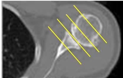
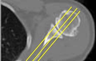
2 Shoulder Coverage Sagittal reconstruction Coronal reconstruction 2

3 3D for Humerus fracture 3D of glenoid surface in scapula and glenoid fracture 3
4 Wrist Position: Prone arm over head : In scaphoid fracture with screw, align the screw follow the beam (figure 1) No instrumentation With metal or cast KV/ Effective mas/rotation time 120/300/ /300/1.0 (sec) Detector collimation (mm) 0.3 (x 16) 0.3 (x 16) Slice thickness (mm) Pitch Extended CT scale box off optional Kernel Bone (U=90) Soft tissue(u=40) U=40 Reconstruction Increment (mm) Start position Proximal metacarpals Proximal metacarpals Finish position 2 cm of distal radius (or through fracture) 2 cm of distal radius (or through fracture) Reconstruction Plane/Window Axial (soft, bone) Sagittal (bone) Coronal (bone) * along the scaphoid in scaphoid fracture Axial (soft, bone) Sagittal (bone) Coronal (bone) * along the scaphoid in scaphoid fracture Reconstruction interval 1.0 mm thick x 1.0 mm interval 1.0 mm thick x 1.0 mm interval 3D reconstruction For scaphoid /carpal fracture : (No need) For distal radius fracture : Rotation of the wrist bone (figure2)/and wrist with tendons (figure3) to see relationship of fracture and tendon : En face view of the radius (figure4) For scaphoid or carpal fracture : (No need) For distal radius fracture : Rotation of the wrist bone (figure2)/and wrist with tendons (figure3) to see relationship of fracture and tendon : En face view of the radius (figure4) 4


5 Position scaphoid fracture with metal Coverage 3D Bone & tendons 5

6 En face view of radius 6
7 Pelvis Position: Supine, toes together Indications: Pelvic fracture, sacral fracture, acetabular fracture, lumbrosacral junction injury, complication of arthroplasty No instrumentation With metal or cast KV/ Effective mas/rotation time 120/300/ /400/1.0 (sec) Detector collimation (mm) 0.6 (x 64) 0.6 (x 64) Slice thickness (mm) Pitch Extended CT scale box off optional Kernel Bone (B=70) Soft tissue(b=30) B=45 Reconstruction Increment (mm) Start position Iliac crest (Figure1) Iliac crest (Figure1) Finish position Below lesser trochanter Below lesser trochanter Reconstruction Plane/Window Axial (soft, bone) Coronal (bone) Sagittal (bone) (affected side from outside to inside) Axial (soft, bone) Coronal (bone) Sagittal (bone) (affected side from outside to inside) Reconstruction interval 2.0 mm thick x 2.0 mm interval 2.0 mm thick x 2.0 mm interval 3D reconstruction 1. Pelvic inlet view (figure2) 2. Pelvic outlet view ( figure3) 3. Profile of affected acetabulum (figure4) 4. Inside of the affected ilium(figure5) 1. Pelvic inlet view (figure2) 2. Pelvic outlet view (figure3) 3. Profile of affected acetabulum (figure4) 4. Inside of the affected ilium(figure5) 7
")


8 Coverage (Figure1) Pelvic inlet view (figure2) Pelvic outlet view ( figure3) 8


9 Profile of affected acetabulum (figure 4) Inside of the affected ilium(figure5) 9
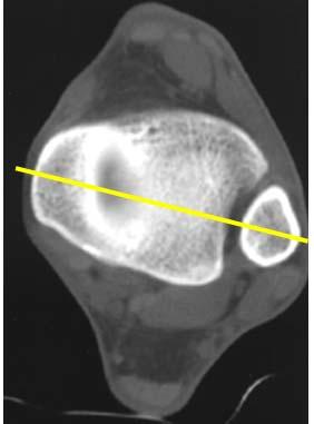
10 Knee Position: Supine. Keep knee AP Indications: Tibial plateau fracture, condylar fracture No instrumentation With metal or cast KV/ Effective mas/rotation time 120/300/ /300/1.0 (sec) Detector collimation (mm) Slice thickness (mm) Pitch Extended CT scale box off optional Kernel Bone (B=70) Soft tissue(b=30) B=30 Reconstruction Increment (mm) Start position Above femoral epicondyle Above femoral epicondyle Finish position Below the fracture Below the fracture Reconstruction Plane/Window Axial (soft, bone) Coronal (bone) Sagittal (bone) (soft) Axial (soft, bone) Coronal (bone) Sagittal (bone) (soft) Reconstruction interval 2.0 mm thick x 2.0 mm interval 2.0 mm thick x 2.0 mm interval 3D reconstruction 1. Rotation of the knee 2. En face view of the articular surface (remove femur) 1. Rotation of the knee 2. En face view of the articular surface (remove femur) 10


11 Coverage 3D en face view of the tibial plateau 11
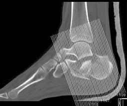
12 Ankle and foot Position: Supine, foot in neutral position Indications: Calcaneal fracture, distal tibia fracture No instrumentation With metal or cast KV/ Effective mas/rotation time 120/300/ /300/1.0 (sec) Detector collimation (mm) 0.6 (x64) 0.6(x64) Slice thickness (mm) Bone/Soft tissue 0.6/0.75 Bone/Soft tissue 0.6/0.75 Pitch Extended CT scale box off optional Kernel Bone (B=70) Soft tissue(b=30) B=45 Reconstruction Increment (mm) Start position Above distal tibia Above distal tibia Finish position Through calcaneus Through calcaneus Reconstruction Plane/Window Axial (soft, bone) Coronal (soft,bone)(figure 1) Sagittal (soft, bone) (figure 2) For calcaneal fracture Coronal oblique (bone) (fig 3) Axial oblique (bone) (fig 4) Axial (soft, bone) Coronal (soft,bone)(figure1) Sagittal (soft,bone) (figure 2) For calcaneal fracture Coronal oblique (bone) (figure 3) Axial oblique (bone) (figure 4) Reconstruction interval 2.0 mm thick x 2.0 mm interval 2.0 mm thick x 2.0 mm interval 3D reconstruction 1. Rotation of the ankle with tendon ( to see the relationship of fracture and tendon) 1. Rotation of the ankle with tendon ( to see the relationship of fracture and tendon) 12
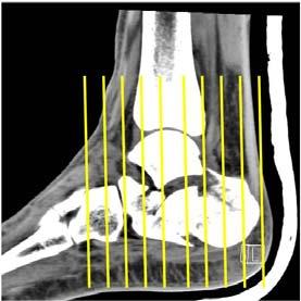
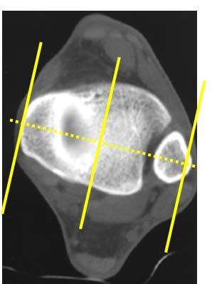
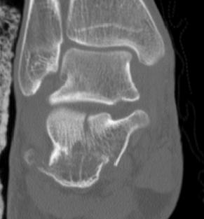
13 Coronal reconstruction Sagittal reconstruction Coronal oblique calcaneus (perpendicular to the subtalar joint) 13


14 Coronal oblique calcaneus (perpendicular to the subtalar joint) 3D view of the tendon 14
Radiographic Positioning Summary (Basic Projections RAD 222)
") Lower Extremity Radiographic Positioning Summary (Basic Projections RAD 222) AP Pelvis AP Hip (Unilateral) (L or R) AP Femur Mid and distal AP Knee Lateral Knee Pt lies supine on table Align MSP to Center
Lower Extremity Radiographic Positioning Summary (Basic Projections RAD 222) AP Pelvis AP Hip (Unilateral) (L or R) AP Femur Mid and distal AP Knee Lateral Knee Pt lies supine on table Align MSP to Center
Hi RES Extremity - (04/18/2011) CTDI: ~13 mgy per acquisition Used for evaluation of: Ankle Elbow Hand Wrist Foot /Calcaneous Toes Fingers
 CTDI: ~13 mgy per acquisition Used for evaluation of: Ankle Elbow Hand Wrist Foot /Calcaneous Toes Fingers") P a g e 1 Hi RES Extremity - (04/18/2011) CTDI: ~13 mgy per acquisition Used for evaluation of: Ankle Elbow Hand Wrist Foot /Calcaneous Toes Fingers Billing: 1. CT Upper/Lower Extremity of concern without
P a g e 1 Hi RES Extremity - (04/18/2011) CTDI: ~13 mgy per acquisition Used for evaluation of: Ankle Elbow Hand Wrist Foot /Calcaneous Toes Fingers Billing: 1. CT Upper/Lower Extremity of concern without
Biology 218 Human Anatomy. Adapted from Martini Human Anatomy 7th ed. Chapter 7 The Skeletal System Appendicular Division
 Adapted from Martini Human Anatomy 7th ed. Chapter 7 The Skeletal System Appendicular Division Introduction The appendicular skeleton includes: Pectoral girdle Shoulder bones Upper limbs Pelvic girdle
MSK CT Extremities: Positioning and Reformations
 MSK CT Extremities: Positioning and Reformations Hand: Patient lying in prone position, with affected arm extended above head. Place body off centered in effort to set affected hand in isocenter. Hand
Radiology Positioning Practical Test #2 Table (By Jung Park):
:") Radiology Positioning Practical Test #2 Table (By Jung Park): (Lower Extremity): patient is fully gowned / no artifacts / properly shielded (exposure for femur and below : hold still, don t move ) (exposure
Radiology Positioning Practical Test #2 Table (By Jung Park): (Lower Extremity): patient is fully gowned / no artifacts / properly shielded (exposure for femur and below : hold still, don t move ) (exposure
The Appendicular Skeleton
 8 The Appendicular Skeleton PowerPoint Lecture Presentations prepared by Jason LaPres Lone Star College North Harris 8-1 The Pectoral Girdle The Pectoral Girdle Also called shoulder girdle Connects the
Pectoral (Shoulder) Girdle
 Girdle") Chapter 8 Skeletal System: Appendicular Skeleton Pectoral girdle Pelvic girdle Upper limbs Lower limbs 8-1 Pectoral (Shoulder) Girdle Consists of scapula and clavicle Clavicle articulates with sternum
Chapter 8 Skeletal System: Appendicular Skeleton Pectoral girdle Pelvic girdle Upper limbs Lower limbs 8-1 Pectoral (Shoulder) Girdle Consists of scapula and clavicle Clavicle articulates with sternum
10/12/2010. Upper Extremity. Pectoral (Shoulder) Girdle. Clavicle (collarbone) Skeletal System: Appendicular Skeleton
 Girdle. Clavicle (collarbone) Skeletal System: Appendicular Skeleton") Skeletal System: Appendicular Skeleton Pectoral girdle Pelvic girdle Upper limbs Lower limbs 8-1 Pectoral (Shoulder) Girdle Consists of scapula and clavicle Clavicle articulates with sternum (Sternoclavicular
Skeletal System: Appendicular Skeleton Pectoral girdle Pelvic girdle Upper limbs Lower limbs 8-1 Pectoral (Shoulder) Girdle Consists of scapula and clavicle Clavicle articulates with sternum (Sternoclavicular
HI-Res Extremity Sensation 16
 Page 1 Routine Extremity - (2/14/2013) CTDI: ~20 mgy per acquisition Used for evaluation of: Humerus Forearm Femur Knee Tib/Fib Billing: 1. CT Upper/Lower Extremity of concern without contrast, with contrast,
Country Health SA Medical Imaging
 Country Health SA Medical Imaging REMOTE OPERATORS POSITIONING GUIDE Contents Image Evaluation Page 4 Positioning Guides Section 1 - THORAX 1.1 Chest Page 5 1.2 Bedside Chest Page 7 1.3 Ribs Page 8 Section
Chapter 8. The Appendicular Skeleton. Lecture Presentation by Lee Ann Frederick University of Texas at Arlington Pearson Education, Inc.
 Chapter 8 The Appendicular Skeleton Lecture Presentation by Lee Ann Frederick University of Texas at Arlington An Introduction to the Appendicular Skeleton The Appendicular Skeleton 126 bones Allows us
Chapter 7: Skeletal System: Gross Anatomy
 Chapter 7: Skeletal System: Gross Anatomy I. General Considerations A. How many bones in an average adult skeleton? B. Anatomic features of bones are based on II. Axial Skeleton A. Skull 1. Functionally
Chapter 8B. The Skeletal System: Appendicular Skeleton. The Appendicular Skeleton. Clavicle. Pectoral (Shoulder) Girdle
 Girdle") The Appendicular Skeleton Chapter 8B The Skeletal System: Appendicular Skeleton 126 bones Pectoral (shoulder) girdle Pelvic (hip) girdle Upper limbs Lower limbs Functions primarily to facilitate movement
The Appendicular Skeleton Chapter 8B The Skeletal System: Appendicular Skeleton 126 bones Pectoral (shoulder) girdle Pelvic (hip) girdle Upper limbs Lower limbs Functions primarily to facilitate movement
SKELETAL SYSTEM 206. AXIAL SKELETON 80 APPENDICULAR SKELETON 126 (see Figure 6.1) Clavicle. Clavicle. Pectoral girdles. Scapula. Scapula.
 Clavicle. Clavicle. Pectoral girdles. Scapula. Scapula.") SKELETAL SYSTEM 206 AXIAL SKELETON 80 APPENDICULAR SKELETON 126 (see Figure 6.1) Pectoral girdles 4 Clavicle Scapula 2 2 Clavicle Scapula Humerus 2 Humerus Upper limbs 60 Radius 2 Ulna Carpal bones Metacarpal
SKELETAL SYSTEM 206 AXIAL SKELETON 80 APPENDICULAR SKELETON 126 (see Figure 6.1) Pectoral girdles 4 Clavicle Scapula 2 2 Clavicle Scapula Humerus 2 Humerus Upper limbs 60 Radius 2 Ulna Carpal bones Metacarpal
Technical guide of musculoskeletal CT: Fundamental aspects that a radiographer needs to know for daily practice.
 Technical guide of musculoskeletal CT: Fundamental aspects that a radiographer needs to know for daily practice. Poster No.: C-1829 Congress: ECR 2014 Type: Educational Exhibit Authors: C. Fraga Piñeiro,
ORTHOSCAN MOBILE DI POSITIONING GUIDE
 ORTHOSCAN MOBILE DI POSITIONING GUIDE Table of Contents SHOULDER A/P of Shoulder... 4 Tangential (Y-View) of Shoulder... 5 Lateral of Proximal Humerus... 6 ELBOW A/P of Elbow... 7 Extended Elbow... 8 Lateral
Amy Warenda Czura, Ph.D. 1 SCCC BIO130 Lab 7 Appendicular Skeleton & Articulations
 The Skeletal System II: Appendicular Skeleton and Articulations Exercises 11, 13 (begins: page 145 in 9 th and 10 th editions) Exercises 10, 11 (begins: page 147 in 11 th edition, page 149 in 12 th edition)
A. Incorrect! The appendicular skeleton includes bones of the shoulder, arm, hand, pelvis, leg and foot.
 Anatomy and Physiology - Problem Drill 08: The Skeletal System III No. 1 of 10 1. Which of the following statements about the appendicular skeleton is correct? A. The appendicular skeleton includes bones
Lab Activity 9. Appendicular Skeleton Martini Chapter 8. Portland Community College BI 231
 Lab Activity 9 Appendicular Skeleton Martini Chapter 8 Portland Community College BI 231 Appendicular Skeleton Upper & Lower extremities Shoulder Girdle Pelvic Girdle 2 Humerus 3 Humerus: Proximal End
Exercise 11. The Appendicular Skeleton
 Exercise 11 The Appendicular Skeleton The Appendicular Skeleton The appendicular skeleton contains 126 bones. Consists of the upper and lower limbs, the pectoral girdles, and the pelvic girdles. The pectoral
Anatomy and Physiology 2016
 Anatomy and Physiology 2016 O = Temporal line I = coronoid process (Mandible) A = elevates mandible (chewing) O = galea aponeurotica (layer of dense fibrous tissue which covers the upper part of the cranium)
PRE-LAB EXERCISES. Before we get started, look up the definitions of these common bone marking terms: Canal: Condyle: Facet: Fissure:
 1 PRE-LAB EXERCISES When studying the skeletal system, the bones are often sorted into two broad categories: the axial skeleton and the appendicular skeleton. This lab focuses on the appendicular skeleton,
FLT105 12/02.
 FLT105 12/02 www.biometmerck.co.uk Disclaimer Biomet Merck Ltd, as the manufacturer of this device, does not practice medicine and does not recommend this or any other surgical technique for use on a
The Appendicular Skeleton
 8 The Appendicular Skeleton PowerPoint Lecture Presentations prepared by Jason LaPres Lone Star College North Harris An Introduction to the Appendicular Skeleton Learning Outcomes 8-1 Identify the bones
Vascular CT Protocols
 Vascular CT Protocols V 1D: Chest and abdominal CT angiogram (aortic dissection protocol) V 1T: Chest CT angiogram (aortic trauma protocol) V 2: Abdominal and pelvis CT angiogram (aortic aneurysm protocol)
Surgical Care at the District Hospital. EMERGENCY & ESSENTIAL SURGICAL CARE
 Surgical Care at the District Hospital 1 18 Orthopedic Trauma Key Points 2 18.1 Upper Extremity Injuries Clavicle Fractures Diagnose fractures from the history and by physical examination Treat with a
Chapter 8 The Skeletal System: The Appendicular Skeleton. Copyright 2009 John Wiley & Sons, Inc.
 Chapter 8 The Skeletal System: The Appendicular Skeleton Appendicular Skeleton It includes bones of the upper and lower limbs Girdles attach the limbs to the axial skeleton The pectoral girdle consists
Goniometry. Wrist Flexion: Pt seated with forearm resting on table (use olecranon process & midline of ulna as reference for stationary arm)
") Goniometry Wrist Flexion: Pt seated with forearm resting on table (use olecranon process & midline of ulna as reference for stationary arm) Wrist Extension: Pt seated with forearm resting on table (Goniometer
Goniometry Wrist Flexion: Pt seated with forearm resting on table (use olecranon process & midline of ulna as reference for stationary arm) Wrist Extension: Pt seated with forearm resting on table (Goniometer
The Skeletal System THE APPENDICULAR SKELETON
 The Skeletal System THE APPENDICULAR SKELETON The appendicular skeleton consists of the girdles and the skeleton of the limbs. The upper (anterior) limbs are attached to the pectoral (shoulder) girdle
Upper Extremity Page Lower Extremity Special Cases
 MSK MRI PROTOCOLS Contents Upper Extremity Shoulder Elbow Wrist Finger Thumb Lower Extremity Hip Pelvis Thigh Knee Lower Extremity/Shin Ankle Foot Special Cases Soft Tissue Mass Metal Protocol Page MSK
Biology 152 Appendicular Skeleton Anatomy Objectives
 Biology 152 Appendicular Skeleton Anatomy Objectives We will learn proper bone names, left/right/medial, and the parts of bones in this exercise. Start by learning the names of the bones. As you gain comfort
Biology 218 Human Anatomy
 Chapter 8 Adapted from Tortora 10 th ed. LECTURE OUTLINE A. Introduction (p. 203) 1. The appendicular skeleton contains 126 bones that form: i. two pectoral (shoulder) girdles two upper limbs i one pelvic
Chapter 8 The Skeletal System: The Appendicular Skeleton. Copyright 2009 John Wiley & Sons, Inc.
 Chapter 8 The Skeletal System: The Appendicular Skeleton Appendicular Skeleton The primary function is movement It includes bones of the upper and lower limbs Girdles attach the limbs to the axial skeleton
Bone List Anatomy
 1 Frontal Bone Skull 2 Parietal Bone Skull 3 Occipital Bone Skull 4 Temporal Bone Skull 5 Coronal Suture Skull 6 Sagittal Suture Skull 7 Squamous suture Skull 8 Lambdoid Suture Skull 9 Surpaorbital Ridge
Important Parts of Bones
 Important Parts of Bones For 2015 Know: Humerus (posterior) Clavical Femur (Anterior) Foot Hand Mandible Os Coxa Scapula Skull (Anterior, Inferior, Lateral) Sternum Humerus (posterior) A. olecranon fossa
Musculoskeletal MR Protocols
 Musculoskeletal MR Protocols Joint-based protocols MSK 1: Shoulder MRI MSK 1A: Shoulder MR arthrogram MSK 1AB: Shoulder MR arthrogram (instability protocol) MSK 2: Elbow MRI MSK 2A: Elbow MR arthrogram
Principles of Anatomy and Physiology
 Principles of Anatomy and Physiology 14 th Edition CHAPTER 8 The Skeletal System: The Appendicular Skeleton The Appendicular Skeleton The 126 bones of the appendicular skeleton are primarily concerned
CHAPTER 8 LECTURE OUTLINE
 CHAPTER 8 LECTURE OUTLINE I. INTRODUCTION A. The appendicular skeleton includes the bones of the upper and lower extremities and the shoulder and hip girdles. B. The appendicular skeleton functions primarily
Dr.Israa H. Mohsen. Lecture 5. The vertebral column
 Anatomy Lecture 5 Dr.Israa H. Mohsen The vertebral column The vertebral column a flexible structure consisting of 33 vertebrae holds the head and torso upright, serves as an attachment point for the legs,
Radiography Protocols
 Radiography Protocols Upper Limb Second through Fifth Digits (Standard 3 views) First Digit (Thumb) (Standard 3 views) Hand (Standard 3 views) Wrist (Standard 4 views) Forearm (Standard 2 views) Elbow
Anatomy & Physiology Skeletal System Worksheet
 1. Name the five functions of the skeleton. c) d) e) Anatomy & Physiology Skeletal System Worksheet 2. The term for the shaft of a bone is:. 3. The bony struts found in spongy bone are called. 4. In ossification,
Upper Extremity Page Lower Extremity Special Cases
 MSK MRI PROTOCOLS Contents Upper Extremity Page Shoulder Elbow Wrist Finger Thumb Lower Extremity Hip Pelvis Thigh Knee Lower Extremity/Shin Ankle Foot Special Cases Soft Tissue Mass Metal Protocol MSK
PH-2 Whole Body Phantom PBU-50. PH-2B CT Whole Body Phantom PBU-60. Contents. Point of X-ray Photography Head radiography P.1 ~ P.5 P.6 ~ P.
 PH-2 Whole Body Phantom PBU-50 PH-2B CT Whole Body Phantom PBU-60 Production supervision: Kyoto College of Medical Science Photography cooperation: Kyoto College of Medical Science Toshifumi Kasai Keiko
RADIOGRAPHY OF THE WRIST
 RADIOGRAPHY OF THE WRIST Patient Position: WRIST PA Projection, elbow in same plane Part Position: Hand ; fingers centered to IR Central Ray: Structures Shown: NOTE: Optional AP projection best demonstrates
UPPER EXTREMITY
 UPPER EXTREMITY 11-24-17 MSK TIPS: Ensure extremity of interest is as isocenter as possible SHIM all Fat sat scans!! Only use 4ch wrist if additional coverage needed If Contrast needed: Multihance.1mmol/kg
Bone Flashcards for 10a
 Bone Flashcards for 0a CLAVICLE (collar bone). Sternal extremity (end) flat end. Acromial extremity (end) rounded end. SCAPULA (shoulder blade). Right or left scapula?. Superior border (superior margin).
BLUE SKY SCHOOL OF PROFESSIONAL MASSAGE AND THERAPEUTIC BODYWORK. Musculoskeletal Anatomy & Kinesiology I TERMINOLOGY, STRUCTURES, & SKELETAL OVERVIEW
 BLUE SKY SCHOOL OF PROFESSIONAL MASSAGE AND THERAPEUTIC BODYWORK Musculoskeletal Anatomy & Kinesiology I TERMINOLOGY, STRUCTURES, & SKELETAL OVERVIEW MSAK101-I Session 1 Learning Objectives: 1. Define
11/25/2012. Chapter 7 Part 2: Bones! Skeletal Organization. The Skull. Skull Bones to Know Cranium
 Chapter 7 Part 2: Bones! 5) Distinguish between the axial and appendicular skeletons and name the major parts of each 6) Locate and identify the bones and the major features of the bones that compose the
Exercise Science Section 2: The Skeletal System
 Exercise Science Section 2: The Skeletal System An Introduction to Health and Physical Education Ted Temertzoglou Paul Challen ISBN 1-55077-132-9 Role of the Skeleton Protection Framework Attachments for
TORNIER BLUEPRINT. 3D Planning + PSI SCAN PROTOCOL
 TORNIER BLUEPRINT 3D Planning + PSI SCAN PROTOCOL Contents 3 Introduction 3 Patient preparation 3 Scanning instructions 4 Image instructions 5 Scanning parameters 6 Technical instructions 2 BLUEPRINT 3D
Axial skeleton bones and markings
 Axial skeleton bones and markings Skull Cranial bones Frontal x 1 Supraorbital foramen Occipital x 1 Foramen magnum Occipital condyles Superior nuchal line Inferior nuchal line Anterior cranial fossa External
Perpendicular Plate Zygomatic Bone. Mental Foramen Mandible
 Glabella Frontal Middle Nasal Concha Nasal Lacrimal Perpendicular Plate Zygomatic Inferior Nasal Concha Maxilla Mental Mandible Skull (anterior view) Squamosal Suture Coronal Suture Frontal Parietal Nasal
NHS Training for Physiotherapy Support Workers. Workbook 11 The articular system
 NHS Training for Physiotherapy Support Workers Workbook 11 The articular system Contents Workbook 11 The articular system 1 11.1 Aim 3 11.2 Learning outcomes 3 11.3 The articular system 4 11.4 Individual
radiologymasterclass.co.uk
 http://radiologymasterclass.co.uk Hip X-ray anatomy - Normal AP (anterior-posterior) Shenton's line is formed by the medial edge of the femoral neck and the inferior edge of the superior pubic ramus Loss
Biology 2401 The Skeletal System
 Biology 2401 The Skeletal System Purpose: The lab will describe the microscopic and gross anatomy of bone, identify bones of the body, and identify important bone markings. I. Overview of the Skeleton
Proteus XR/f Patient positioning guide
 Proteus XR/f Patient positioning guide PROTEUS XR/F Now a single digital x-ray room accommodates nearly all your radiographic studies. With extended tube coverage and wireless detectors, Proteus XR/f gives
FUNCTIONAL ANATOMY WEEKLY NOTES:
 FUNCTIONAL ANATOMY WEEKLY NOTES: Pages 2-11 Week 1 Head and Neck Pages 12-39 Week 2 Trunk Pages 40-52 Week 3 Hip Joint Pages 53-60 Week 4 Knee Joint Pages 61-70 Week 5 Ankle Joint Pages 71-82 Week 6 Foot
AP1 Lab 4 - Appendicular Skeleton
 Project 1 Learn the Bone Names AP1 Lab 4 - Appendicular Skeleton Use Figure 7.1 and the hanging skeletons to learn the whole bones of the arms and legs. Don t learn the features of each bone yet just be
In-Depth Foundations: Anatomy Terms to Know
 Be familiar with / able to identify and define all the following parts. The Spine Cranium Vertebrae Cervical, Thoracic, Lumbar Sacrum Coccyx Bones of Upper Body Cranium Mastoid process; Occipital condyle,
Ch. 5 - Skeletal System
 Ch. 5 - Skeletal System Bones are living, ever-changing structures. This allows them grow and adapt to new situations that the body encounters. The functions of the skeletal system: 1) support bones are
Copyright 2003 Pearson Education, Inc. publishing as Benjamin Cummings. Dr. Nabil Khouri MD, MSc, Ph.D
 Dr. Nabil Khouri MD, MSc, Ph.D Pelvic Girdle (Hip) Organization of the Lower Limb It is divided into: The Gluteal region The thigh The knee The leg The ankle The foot The thigh and the leg have compartments
Dieter Marquardt Medizintechnik GmbH 08 Cannulated Screws
 Cannulated Screws Product Name: Cannulated Screw Ø 3.5 Screw diameter: 3.5 mm Core diameter: 2.5 mm Head diameter: 5.0 mm Thread: short or full Cannulation: 1.35 mm Inner hexagon: 2.5 mm Screw Length:
bio4165 lab quiz 1 Posterior View Anterior View Lateral View Anterior View bio fall.quarter lab.quiz.1...page.1 of 6
 B A Posterior View D C E Lateral View bio.4165...fall.quarter.2005...lab.quiz.1...page.1 of 6 F I G 35 Posterior View H bio.4165...fall.quarter.2005...lab.quiz.1...page.2 of 6 J Posterior View L K Inferior
Table of Compensation for Injury 1/2015
 Table of Compensation for 1/2015 Valid from 01.04.2015 1. CRANIAL INJURIES 1.1 Cranial bone fractures: a) fracture of cranial dome 10 b) fracture of cranial base 15 c) fracture of cranial dome and base
Chapter 8: The Appendicular Skeleton
 Chapter 8: The Appendicular Skeleton In Chapter 8, we complete our tour of the skeleton that began in Chapter 7. As with Chapter 7, much of this material is best learned in lab, but we will outline some
Spring Written By: J. E. Sutton. Contents: I. Overview of the Skeleton: II. Appendicular Skeleton III. Axial Skeleton IV.
 Spring 2012 Written By: J. E. Sutton Contents: I. Overview of the Skeleton: II. Appendicular Skeleton III. Axial Skeleton IV. Articulations Overview of the Skeleton: I. Orientation to Human Skeleton: a.
Types of Plates 1. New Dynamic Compression Plate: Diaphyseal fracture: Radius, Ulna, Humerus, Rarely tibia
 Types of Plates 1. New Dynamic Compression Plate: DCP Diaphyseal fracture: Radius, Ulna, Humerus, Rarely tibia 1. Undercut adjacent to the holes low contact: less stress shield 2. Undercut at the undersurface
Basic Radiographic Principles Part II
 Basic Radiographic Principles Part II Kristopher Avant, D.O. October 19 th, 2016 I have no disclosures relevant to the material presented in this discussion. Good Stuff!!! 1 Really? Really! Musculoskeletal
Anatomage Table Instructors Guide- Lower Limb
 The Lower Limb Anatomage Table Instructors Guide- Lower Limb Table of Contents Lower Limb 1- The Skeletal System...3 1: Hip Bone...3 2: Hip Joint and Femur...4 3: Patella and Knee Joint...7 4: Tibia, Fibula,
Surgical Privileges Form Orthopedic Surgery CLINICAL PRIVILEGES REQUEST Applicant s Name:. Scope of Practice:... License No. (If Any):... Facility:..
:... Facility:..") Name of applicant Surgical Privileges Form Orthopedic Surgery CLINICAL PRIVILEGES REQUEST Applicant s Name:. Scope of Practice:... License No. (If Any):..... Facility:.. Requested Recommended Not Privileges
Name of applicant Surgical Privileges Form Orthopedic Surgery CLINICAL PRIVILEGES REQUEST Applicant s Name:. Scope of Practice:... License No. (If Any):..... Facility:.. Requested Recommended Not Privileges
Multiapical Deformities p. 97 Osteotomy Concepts and Frontal Plane Realignment p. 99 Angulation Correction Axis (ACA) p. 99 Bisector Lines p.
 p. 99 Bisector Lines p.") Normal Lower Limb Alignment and Joint Orientation p. 1 Mechanical and Anatomic Bone Axes p. 1 Joint Center Points p. 5 Joint Orientation Lines p. 5 Ankle p. 5 Knee p. 5 Hip p. 8 Joint Orientation Angles
Normal Lower Limb Alignment and Joint Orientation p. 1 Mechanical and Anatomic Bone Axes p. 1 Joint Center Points p. 5 Joint Orientation Lines p. 5 Ankle p. 5 Knee p. 5 Hip p. 8 Joint Orientation Angles
Multiple Choice Identify the letter of the choice that best completes the statement or answers the question.
 RA202 positioning class three- EXM Multiple Choice Identify the letter of the choice that best completes the statement or answers the question. 1. Which of the following hand projections would be used
Lab-1. Miss. Lina Al-Onazy & samar Al-Wgeet =)
") Lab-1 Introduction The human skeleton is composed of 300 bones at birth and by the time adulthood is reached, some bones have fused together to give a total of 206 bones in the body. The human skeleton
Lab-1 Introduction The human skeleton is composed of 300 bones at birth and by the time adulthood is reached, some bones have fused together to give a total of 206 bones in the body. The human skeleton
Appendicular Skeleton. Prof. Abdulameer Al-Nuaimi
 Appendicular Skeleton Prof. Abdulameer Al-Nuaimi a.alnuaimi@sheffield.ac.uk abdulameerh@yahoo.com Hi Prof, It is great to hear from you, I really enjoyed your teaching last year. You taught me the hardest
Chapter 8 Outline. Pectoral Girdle Upper Limb Pelvic Girdle Lower Limb Aging of the Appendicular Skeleton Development of the Appendicular Skeleton
 Chapter 8 Outline Pectoral Girdle Upper Limb Pelvic Girdle Lower Limb Aging of the Appendicular Skeleton Development of the Appendicular Skeleton Figure 8.1 Appendicular Skeleton Pectoral Girdle Clavicle
BONE CHALLENGE DANIL HAMMOUDI.MD
 BONE CHALLENGE DANIL HAMMOUDI.MD Bone Basic functions? A. support B. protection C. movement assistance in D. RBC formation-hemopoiesis E. mineral homeostasis +importance of calcium F. energy supply -yellow
Balanced Body Movement Principles
 Balanced Body Movement Principles How the Body Works and How to Train it. Module 3: Lower Body Strength and Power Developing Strength, Endurance and Power The lower body is our primary source of strength,
 17.2 A-P Lower Leg Measure: A-P at mid-lower leg Protection: Apron draped over pelvis SID: 40 Table top No Tube Angle Film: 7 x17 I.D. down or diagonal 14 x 17 www.fisiokinesiterapia.biz A-P Lower Leg
Trauma fixation choices chart your fracture
 Trauma fixation choices chart your fracture This brochure is intended for informational and educational purposes only. It is the responsibility of operating physicians to determine and utilize the appropriate
The Skeletal System in Action!! The Skeletal System in Action!
 Skeletal System The Skeletal System in Action!! The Skeletal System in Action! 5 Functions of the Skeletal System 1. Movement: Skeletal system provides points of attachment for muscles. Your legs and arms
Index. Note: Page numbers of article titles are in boldface type.
 Note: Page numbers of article titles are in boldface type. A Abscess, epidural, 822 824 Achilles tendon rupture, 894 895, 981 982 Acromioclavicular separations, shoulder pain in, 751 753 Adhesive capsulitis,
Appendicular Skeleton. Dr. Carmen E. Rexach Anatomy 35 Mt. San Antonio College
 Appendicular Skeleton Dr. Carmen E. Rexach Anatomy 35 Mt. San Antonio College Pectoral girdle clavicle scapula Upper limb brachium antebrachium carpus manus Pelvic girdle oscoxae Lower limb femoral region
Upper Limb Imaging Requirements
 Imaging Requirements Upper Limb Imaging Requirements Instructions for Measurement Radiography and CT Scans Please read before commencing radiography Stanmore Implants 210 Centennial Avenue Centennial Park
TORNIER. scan protocol V2.1. Tornier Upper Extremities
 TORNIER TM scan protocol V2.1 Tornier Upper Extremities Contents 4 Introduction 4 Patient Preparation 4 Scanning Instructions 5 Image Instructions 6 Scanning Parameters 7 Technical Instructions Scan Protocol
Hands PA; Obl. Lat.; Norgaard s Thumb AP; Lat. PA. PA; Lat.: Obls.; Elongated PA with ulnar deviation
 Projections Region Basic projections Additional / Modified projections Upper Limbs Hands PA; Obl. Lat.; Norgaard s Thumb ; Lat. PA Fingers PA; Lat. Wrist PA; Lat. Obls. Scaphoid Lunate Trapezium Triquetral
CT SCAN PROTOCOL. Shoulder
 CT SCAN PROTOCOL Shoulder Purpose and Summary CT images made with this protocol are used to provide the orthopedic surgeon with a detailed 3D anatomical reconstruction of the patient s scapula and proximal
PEDIATRIC CASTING AND SPLINTING HEATHER KONG, M.D. SHRINERS HOSPITAL FOR CHILDREN PORTLAND OCTOBER 7, 2017
 PEDIATRIC CASTING AND SPLINTING HEATHER KONG, M.D. SHRINERS HOSPITAL FOR CHILDREN PORTLAND OCTOBER 7, 2017 DISCLOSURES I have no financial relationship with any company or product discussed in this presentation.
Anatomy. Anatomy deals with the structure of the human body, and includes a precise language on body positions and relationships between body parts.
 Anatomy deals with the structure of the human body, and includes a precise language on body positions and relationships between body parts. Proper instruction on safe and efficient exercise technique requires
Human Anatomy, First Edition McKinley & O'Loughlin
 Human Anatomy, First Edition McKinley & O'Loughlin Chapter 8 : Appendicular Skeleton 8-1 Appendicular Skeleton Includes the bones of the upper and lower limbs. The girdles of bones that attach the upper
Musculoskeletal Applications for CT. Tal Laor, MD Cincinnati Children s Hospital University of Cincinnati College of Medicine
 Musculoskeletal Applications for CT Tal Laor, MD Cincinnati Children s Hospital University of Cincinnati College of Medicine I have no commercial disclosures. Why CT? Complimentary to other modalities
8 1 Basic Principles. Palpating Muscle Bellies. Palpating Bony Prominences
 8 1 Basic Principles Fig. 1.4 Locating the medial epicondyle. Tip: The shape of bony prominences can be visualized by looking at their morphology. However, variations are expected to be encountered quite
Operative Dictations in Orthopedic Surgery
 Operative Dictations in Orthopedic Surgery Springer ISBN-13: 9781461474784 Table of Contents Part One Pediatrics Chapter 1 Posterior Spinal Fusion with Instrumentation Chapter 2 In Situ Fusion L5 to S1
Lab Exercise #04 The Skeletal System Student Performance Objectives
 Lab Exercise #04 The Skeletal System Student Performance Objectives The material that you are required to learn in this exercise can be found in either the lecture text or the supplemental materials provided
PROFESSIONAL RELIABLE TRAUMA MANUAL. Health is Important
 PROFESSIONAL RELIABLE TRAUMA MANUAL Health is Important 1 Vision In the past, the Treasure Voyages of Zheng He started the epoch-making communication and fusion by sea between the Oriental and the Occidental.
THE NANCY NAIL. The End Caps ADVANTAGES OF NANCY NAIL
 NANCY NAIL THE NANCY NAIL Nancy nails are manufactured from a specific titanyum alloy with proprietary surface treatment, which provides increased fatigue resistance. Six nail diameters (1.5 mm 2.0 mm
Anatomy of the Musculoskeletal System
 Anatomy of the Musculoskeletal System Kyle E. Rarey, Ph.D. Department of Anatomy & Cell Biology and Otolaryngology University of Florida College of Medicine Outline of Presentation Vertebral Column Upper
RADIOGRAPHY OF THE ELBOW & HUMERUS
 RADIOGRAPHY OF THE ELBOW & HUMERUS Patient Position: ELBOW AP Projection in same plane Part Position: Hand in ; patient Centered to Humeral epicondyles Central Ray: Structures Shown: AP Elbow Criteria
Pearson Education Limited Edinburgh Gate Harlow Essex CM20 2JE England and Associated Companies throughout the world
 Pearson Education Limited Edinburgh Gate Harlow Essex CM20 2JE England and Associated Companies throughout the world Visit us on the World Wide Web at: www.pearsoned.co.uk Pearson Education Limited 204