Medical Imaging (MRI) By: Engr. Joseph Ronald Cañedo
|
|
|
- Peregrine McKinney
- 5 years ago
- Views:
Transcription

1
2 Medical Imaging (MRI) By: Engr. Joseph Ronald Cañedo
3 MRI History July 3, was the first MRI exam ever performedon ahumanbeing. Ittookalmostfivehourstoproduceoneimage. Dr. Raymond Damadian, a physician and scientist, along with colleagues Dr. Larry Minkoff and Dr. MichaelGoldsmith, labored tirelessly for seven long years to reach this point. Late 1982 to present - MRI can image in seconds whatusedtotakehours.
4 Magnetic resonance imaging (MRI) is primarily used in medical imaging to visualize the structure and function of the body. It provides detailed images of the body inany plane. MR has much greater soft tissue contrast than Computed tomography (CT) making it especially useful in neurological, musculoskeletal, cardiovascular and oncolological diseases.
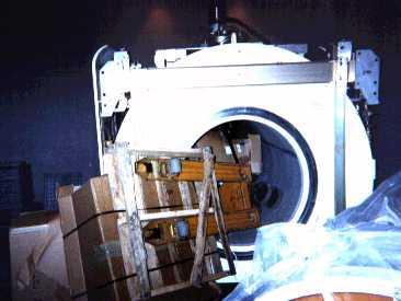
5 MRI Scan The basic design used in most is agiant cube. The cube in atypical system might be 7feet tall by 7feet wide by 10 feet long (2 mby 2m by 3 m), although new models are rapidly shrinking. There is a horizontal tube running through the magnet from front to back. This tube is known asthe bore of themagnet.

6

7

8
9 Magnetic Resonance The biggest and most important component in an MRI systemis the magnet. The magnet in an MRI system is rated using aunit of measureknownasatesla. Another unit of measure commonly used with magnets isthe gauss (1tesla=10,000gauss). The magnets in use today in MRI are in the 0.5-tesla to 2.0-teslarange,or5,000to20,000gauss. Magnetic fields greater than 2 tesla have not been approved for use in medical imaging, though much more powerful magnets -- up to 60 tesla -- are used in research.
10 Magnetic Resonance The MRI suite can be avery dangerous place if strictprecautions are not observed. Metal objects can become dangerous projectiles if they are taken into the scan room. The magnetic force exerted on an object increases exponentially as it nears the magnet.
11
12 MRI Safety Prior to allowing a patient or support staff member into the scan room, he or she is thoroughly screened for metal objects. (External metalobjects) InternalMetalObjectsisalsohazardous. Metallicfragments inthe eye Pacemakers Aneurysmclips(brain) Dentalimplants Orthopedicimplants
13 MRI Safety There areno known biologicalhazardsto humans from being exposed to magnetic fields of the strengthused inmedicalimagingtoday. Most facilities prefer not to image pregnant women. This is due to the fact that there has not been much research done in the area of biological effects on adeveloping fetus. The first trimester in a pregnancy is the most critical becausethat isthetime of the mostrapid cellular reproductionand division.
14 MRI Magnet There are three basic types of magnets used in MRIsystems: 1. Resistive magnets consist of many windings or coils of wire wrapped around acylinder or bore through which an electric current is passed. This causes a magnetic field to be generated. If the electricity is turned off, the magnetic field dies out. These magnets are lower in cost to construct than a superconducting magnet (see below), but require huge amounts of electricity (up to 50 kilowatts) to operate because of the natural resistance in the wire. To operate this type of magnet above about the0.3-tesla levelwould beprohibitivelyexpensive.
15 MRI Magnet There are three basic types of magnets used in MRIsystems: 2. A permanent magnet is just that -- permanent. Its magnetic field is always there and always on full strength, so it costs nothing to maintain the field. The major drawback is that these magnets are extremely heavy: They weigh many, many tons at the 0.4-tesla level. Astronger field would require a magnet so heavy it would be difficult to construct. Permanent magnets are getting smaller, but are still limitedto low fieldstrengths.
16 MRI Magnet There are three basic types of magnets used in MRI systems: 3. Superconducting magnets are by far the most commonly used. Asuperconducting magnet is somewhat similar to a resistive magnet--coilsor windingsof wirethrough which acurrent of electricity is passed create the magnetic field. The important difference is that the wire is continually bathed in liquid helium at degrees below zero. his almost unimaginable cold causes the resistance in the wire to drop to zero, reducing the electrical requirement for the system dramatically and making it much more economical to operate. Superconductive systems are still very expensive, but they can easily generate 0.5-tesla to 2.0-teslafields,allowingformuchhigher-qualityimaging.
17 How MRI Works: Atoms The human body is made up of untold billions of atoms, the fundamental building blocks of all matter. The nucleus of an atom spins, or precesses,on an axis. A top that is spinning slightly off the vertical axis is precessing about the verticalaxis. A hydrogen atom precesses about a magneticfield.
18 How MRI Works: Atoms Imagine billions of nuclei all randomly spinning or precessingin everydirection. There are many different types of atoms in the body, but for the purposes of MRI, only concernedwith the hydrogen atom. Itisan ideal atomfor MRI becauseitsnucleushas a single proton andalarge magnetic moment. The large magnetic moment means that, when placed in amagnetic field, the hydrogen atom has a strong tendency to line up with the directionofthemagneticfield.
19 How MRI Works: Atoms All of the hydrogen protons will align with the magnetic field in one direction or the other. The vast majority cancel each other out, but, as shown here, in any sample there is one or two"extra"protons.
20 MRI Machine The MRI machine applies an RF (radiofrequency) pulse that isspecificonlytohydrogen. The system directs the pulse toward the area of the body wewant toexamine. The pulse causes the protons in that area to absorb the energy required to make them spin, or precess, in a differentdirection. Thisisthe"resonance" part ofmri. The RF pulse forces them (only the one or two extra unmatched protons per million) to spin at a particular frequency,inaparticular direction. The specific frequency of resonance is called the Larmour frequency and is calculated based on the particular tissue beingimagedandthestrengthofthemainmagnetic field.

21 MRI Images This MRI scan shows the upper torso in side view so that the bones of the spine are evident.


22 MRI Images This image shows the tumor growth in afemale's brain, sliced here in lateral view.
 and with aperson of similar age having Alzheimer's Disease (right), allimagedat the")
23 MRI Images This image set is comparing ayoung individual (left) with an athletic male in his 80's (center) and with aperson of similar age having Alzheimer's Disease (right), allimagedat the samelevel.

24
25 MRI Advantages MRI is ideal for: Diagnosing multiple sclerosis (MS) Diagnosing tumors of the pituitary gland and brain Diagnosing infections in the brain, spine or joints Visualizing torn ligaments in the wrist, knee and ankle Visualizing shoulder injuries Diagnosing tendonitis Evaluating masses in the soft tissues of the body Evaluating bone tumors, cysts and bulging or herniated discs in the spine Diagnosing strokes in their earliest stages
26 MRI Disadvantages Itdoeshavedrawbacks. There are many people who cannot safely be scanned with MRI (for example,because they have pacemakers), andalso peoplewhoare toobig tobescanned. There are many claustrophobic people in the world, and being in an MRI machine can be a very disconcerting experienceforthem. The machine makes atremendous amountof noise duringa scan. The noise sounds like acontinual, rapid hammering. Patients are given earplugs or stereo headphones to muffle the noise (inmost MRI centers you can even bring your own cassette or CD to listen to). The noise is due to the rising electrical current in thewires of the gradient magnets being opposed by the main magnetic field. The stronger the main field,thelouderthegradientnoise.
27 MRI Disadvantages Itdoeshavedrawbacks. MRI scans require patients to hold very still for extended periods of time. MRI exams can range in length from 20 minutes to 90 minutes or more. Even very slight movement of the part being scanned can cause very distortedimagesthatwill havetoberepeated. Orthopedic hardware (screws, plates, artificial joints) in the area of ascan can cause severe artifacts (distortions) on the images. The hardware causes a significant alteration in the main magnetic field. Remember, a uniformfieldiscriticaltogoodimaging. MRI systems are very, very expensive to purchase, and thereforetheexamsarealsovery expensive.
28 CT vs. MRI A computed tomography (CT) scanner uses X- rays, atype of ionizing radiation, to acquire its images, making it a good tool for examining tissue composed of elements of a relatively higher atomic number than the tissue surrounding them, such as bone and calcifications (calcium based) within the body (carbon based flesh), or of structures (vessels, bowel). MRI, on the other hand, uses non-ionizing radio frequency (RF) signals to acquire its images and is bestsuitedfor non-calcifiedtissue.
29 CT vs. MRI CT may be enhanced by use of contrast agents containing elements of a higher atomic number than the surrounding flesh such as iodineor barium. Contrast agents for MRI are those which have paramagnetic properties. Both CT and MRI scanners can generate multiple two-dimensional cross-sections (slices) of tissue and three-dimensional reconstructions.
30 CT vs. MRI MRI is also best suited for cases when a patient is to undergo the exam several times successively in the short term, because, unlike CT, it does not expose the patient to the hazardsof ionizing radiation.
31 Future of MRI The future of MRI seems limited only by our imagination. Thistechnology isstillin itsinfancy, comparatively speaking. It has been in widespread use for less than 20 years (comparedwith over100years forx-rays). Very small scanners for imaging specific body parts are being developed. For instance, ascanner that you simply place your arm, knee or foot in are currently in use in some areas. Our ability to visualize the arterial and venous system is improving all the time. Functional brain mapping (scanning aperson's brain while he or she is performing a certain physical task such as squeezing aball, or looking at a particular type of picture) is helping researchers better understandhow thebrain works. Research is under way ina few institutions to image the ventilation dynamics of the lungs through the use of hyperpolarized helium-3 gas. The development of new, improved ways to image strokes in theirearlieststagesisongoing.
32
Magnetic Resonance Imaging (MRI)
") (MRI) Disclaimer This film is an educational resource only and should not be used to make a decision on MRI. All such decisions must be made in consultation with a physician or licensed healthcare provider.
(MRI) Disclaimer This film is an educational resource only and should not be used to make a decision on MRI. All such decisions must be made in consultation with a physician or licensed healthcare provider.
Syllabus References. Resources. Video: MRI Introduction
 MRI Lesson Outline Syllabus References 9.6.4.2.5 Define precessing and relate the frequency of the precessing to the composition of the nuclei and the strength of the applied external magnetic field 9.6.4.2.6
Radiologic Imaging Magnetic Resonance Imaging (MRI)
") Radiologic Imaging X-ray has always been the golden rule in diagnosing and treating podiatric patients. Unfortunately, for some patients the diagnosis is not as evident. That is when we need to utilize
Radiologic Imaging X-ray has always been the golden rule in diagnosing and treating podiatric patients. Unfortunately, for some patients the diagnosis is not as evident. That is when we need to utilize
Magnetic Resonance Imaging on Soft Tissue. Jiten K. Mistry Calvin Gan
 Magnetic Resonance Imaging on Soft Tissue 1 Jiten K. Mistry Calvin Gan Outline Background of Medical Imaging Introduction to MRI How MRI works MRI of Soft Tissue Benefits & Risks Recent Advances 2 The
Magnetic Resonance Imaging (MRI) Dynamic Pelvic Floor
 Dynamic Pelvic Floor") Scan for mobile link. Magnetic Resonance Imaging (MRI) Dynamic Pelvic Floor Dynamic pelvic floor magnetic resonance imaging (MRI) is a noninvasive test that uses a powerful magnetic field, radio waves
Scan for mobile link. Magnetic Resonance Imaging (MRI) Dynamic Pelvic Floor Dynamic pelvic floor magnetic resonance imaging (MRI) is a noninvasive test that uses a powerful magnetic field, radio waves
P2 Visual - Perception
 P2 Visual - Perception 2014 SOSE Neuroimaging of high-level visual functions gyula.kovacs@uni-jena.de 11/09/06 Functional magnetic resonance imaging (fmri) The very basics What is fmri? What is MRI? The
Imaging Patient Education. Magnetic Resonance Imaging (MRI)
") Magnetic Resonance Imaging (MRI) What you should know about your Body MRI exam: Purpose: Magnetic Resonance Imaging (MRI) uses radio waves and a strong magnetic field to provide clear and detailed images
Magnetic Resonance Imaging (MRI) What you should know about your Body MRI exam: Purpose: Magnetic Resonance Imaging (MRI) uses radio waves and a strong magnetic field to provide clear and detailed images
Magnetic Resonance Imaging (MRI) - Knee
 - Knee") Scan for mobile link. Magnetic Resonance Imaging (MRI) - Knee Magnetic resonance imaging (MRI) of the knee uses a powerful magnetic field, radio waves and a computer to produce detailed pictures of the
Scan for mobile link. Magnetic Resonance Imaging (MRI) - Knee Magnetic resonance imaging (MRI) of the knee uses a powerful magnetic field, radio waves and a computer to produce detailed pictures of the
MR Angiography (MRA)
") MR Angiography (MRA) What is MR Angiography? What are some common uses of the procedure? How should I prepare? What does the equipment look like? How does the procedure work? How is the procedure performed?
MR Angiography (MRA) What is MR Angiography? What are some common uses of the procedure? How should I prepare? What does the equipment look like? How does the procedure work? How is the procedure performed?
Magnetic Resonance Imaging (MRI) - Spine
 - Spine") Scan for mobile link. Magnetic Resonance Imaging (MRI) - Spine Magnetic resonance imaging (MRI) of the spine uses radio waves, a magnetic field and a computer to produce detailed pictures of the spine
Scan for mobile link. Magnetic Resonance Imaging (MRI) - Spine Magnetic resonance imaging (MRI) of the spine uses radio waves, a magnetic field and a computer to produce detailed pictures of the spine
Magnetic Resonance Imaging (MRI) - Body
 - Body") Scan for mobile link. Magnetic Resonance Imaging (MRI) - Body What is MRI of the Body? Magnetic resonance imaging (MRI) is a noninvasive medical test that physicians use to diagnose and treat medical conditions.
Scan for mobile link. Magnetic Resonance Imaging (MRI) - Body What is MRI of the Body? Magnetic resonance imaging (MRI) is a noninvasive medical test that physicians use to diagnose and treat medical conditions.
Magnetic Resonance, Functional (fmri) - Brain
 - Brain") Scan for mobile link. Magnetic Resonance, Functional (fmri) - Brain Functional magnetic resonance imaging (fmri) measures the metabolic changes that occur within the brain. It may be used to examine the
Scan for mobile link. Magnetic Resonance, Functional (fmri) - Brain Functional magnetic resonance imaging (fmri) measures the metabolic changes that occur within the brain. It may be used to examine the
mri sequences 8267BD21D03EEA3AE7926DD1904E7425 Mri Sequences 1 / 6
 Mri Sequences 1 / 6 2 / 6 3 / 6 Mri Sequences An MRI sequence is a number of radiofrequency pulses and gradients that result in a set of images with a particular appearance. This article presents a simplified
Magnetic Resonance Imaging (MRI) - Head
 - Head") Scan for mobile link. Magnetic Resonance Imaging (MRI) - Head Magnetic resonance imaging (MRI) of the head uses a powerful magnetic field, radio waves and a computer to produce detailed pictures of the
Scan for mobile link. Magnetic Resonance Imaging (MRI) - Head Magnetic resonance imaging (MRI) of the head uses a powerful magnetic field, radio waves and a computer to produce detailed pictures of the
ANNOUNCING THE NEW STONY BROOK UNIVERSITY OUTPATIENT IMAGING CENTER
 ANNOUNCING THE NEW STONY BROOK UNIVERSITY OUTPATIENT IMAGING CENTER PROVIDING THE MOST ADVANCED DIAGNOSTICS FOR THE HIGHEST QUALITY OF CARE Call Our Dedicated Line: (631) 638-2121 When you need the benefit
Safety Issue of Static Magnetic Field (1)
") MRI Safety Safety Issue of Static Magnetic Field (1) Currently, typical MRI utilizes static field with strengths of 0.5 to 3 Tesla (FDA guideline is max 4 T for infants < 1 month and 8 T for others) 1
MRI Safety Safety Issue of Static Magnetic Field (1) Currently, typical MRI utilizes static field with strengths of 0.5 to 3 Tesla (FDA guideline is max 4 T for infants < 1 month and 8 T for others) 1
Radiology. General radiology department. X-ray
 The radiology directorate provides a diagnostic, interventional and therapeutic service for its local population, and a tertiary service for the region. It also provides support to some national work such
Magnetic Resonance Cholangiopancreatography (MRCP)
") Magnetic Resonance Cholangiopancreatography (MRCP) What is Magnetic Resonance Cholangiopancreatography (MRCP)? What are some common uses of the procedure? How should I prepare for the procedure? What does
Magnetic Resonance Cholangiopancreatography (MRCP) What is Magnetic Resonance Cholangiopancreatography (MRCP)? What are some common uses of the procedure? How should I prepare for the procedure? What does
Magnetic Resonance Imaging (MRI) - Body
 - Body") Scan for mobile link. Magnetic Resonance Imaging (MRI) - Body Magnetic resonance imaging (MRI) of the body uses a powerful magnetic field, radio waves and a computer to produce detailed pictures of the
Scan for mobile link. Magnetic Resonance Imaging (MRI) - Body Magnetic resonance imaging (MRI) of the body uses a powerful magnetic field, radio waves and a computer to produce detailed pictures of the
Magnetic Resonance Imaging (MRI) Scans. Patient Information
 Scans. Patient Information") Magnetic Resonance Imaging (MRI) Scans Patient Information Author ID: LW/ST Leaflet Number: Rad 004 Version: 5 Name of Leaflet: Magnetic Resonance Imaging (MRI) Scans Date Produced: December 2017 Review
Magnetic Resonance Imaging (MRI) Scans Patient Information Author ID: LW/ST Leaflet Number: Rad 004 Version: 5 Name of Leaflet: Magnetic Resonance Imaging (MRI) Scans Date Produced: December 2017 Review
Northumbria Healthcare NHS Foundation Trust. Breast MRI. Issued by the Breast Team
 Northumbria Healthcare NHS Foundation Trust Breast MRI Issued by the Breast Team What is an MRI Scan? MRI stands for magnetic resonance imaging; it is a non-invasive medical imaging test with no radiation
Your Appointment has been scheduled for:
 Please read this important information about the imaging study being scheduled by your health care provider. Pacific Coast Imaging provides this information as a service to enhance the patient experience
Children's (Pediatric) MRI for Appendicitis
 MRI for Appendicitis") Scan for mobile link. Children's (Pediatric) MRI for Appendicitis Children's magnetic resonance imaging (MRI) for appendicitis uses a powerful magnetic field, radio waves and a computer to produce detailed
Scan for mobile link. Children's (Pediatric) MRI for Appendicitis Children's magnetic resonance imaging (MRI) for appendicitis uses a powerful magnetic field, radio waves and a computer to produce detailed
Magnetic Resonance Imaging (MRI) - Cardiac (Heart)
 - Cardiac (Heart)") Scan for mobile link. Magnetic Resonance Imaging (MRI) - Cardiac (Heart) Cardiac magnetic resonance imaging (MRI) uses a powerful magnetic field, radio waves and a computer to produce detailed pictures
Scan for mobile link. Magnetic Resonance Imaging (MRI) - Cardiac (Heart) Cardiac magnetic resonance imaging (MRI) uses a powerful magnetic field, radio waves and a computer to produce detailed pictures
X-ray (Radiography) - Bone
 - Bone") Scan for mobile link. X-ray (Radiography) - Bone Bone x-ray uses a very small dose of ionizing radiation to produce pictures of any bone in the body. It is commonly used to diagnose fractured bones or
Scan for mobile link. X-ray (Radiography) - Bone Bone x-ray uses a very small dose of ionizing radiation to produce pictures of any bone in the body. It is commonly used to diagnose fractured bones or
Mri Of The Musculoskeletal System
 We have made it easy for you to find a PDF Ebooks without any digging. And by having access to our ebooks online or by storing it on your computer, you have convenient answers with mri of the musculoskeletal
Psychologists who map the brain s fissures (grooves on the brain which appear as a deep fold) and inner recesses
 and inner recesses") Also called psychobiologists Psychologists who map the brain s fissures (grooves on the brain which appear as a deep fold) and inner recesses Methods include: 1) Recording 2) Stimulating 3) Lesioning 4)
Also called psychobiologists Psychologists who map the brain s fissures (grooves on the brain which appear as a deep fold) and inner recesses Methods include: 1) Recording 2) Stimulating 3) Lesioning 4)
Magnetic Resonance, Functional (fmri) - Brain
 - Brain") Magnetic Resonance, Functional (fmri) - Brain What is Functional MR Imaging (fmri) - Brain? Magnetic resonance imaging (MRI) is a noninvasive medical test that helps physicians diagnose and treat medical
Magnetic Resonance, Functional (fmri) - Brain What is Functional MR Imaging (fmri) - Brain? Magnetic resonance imaging (MRI) is a noninvasive medical test that helps physicians diagnose and treat medical
Non-Invasive Techniques
 Non-Invasive Techniques Key: Does not hurt the organism Psychology 372 Physiological Psychology Steven E. Meier, Ph.D. Listen to the audio lecture while viewing these slides or view the video presentation
Non-Invasive Techniques
 Many Procedures Non-Invasive Techniques Key: Does not hurt the organism Psychology 372 Physiological Psychology Steven E. Meier, Ph.D. Listen to the audio lecture while viewing these slides or view the
Magnetic Resonance Imaging (MRI) - Body
 - Body") Magnetic Resonance Imaging (MRI) - Body What is MRI of the Body? Magnetic resonance imaging (MRI) is a noninvasive medical test that helps physicians diagnose and treat medical conditions. MR imaging uses
Magnetic Resonance Imaging (MRI) - Body What is MRI of the Body? Magnetic resonance imaging (MRI) is a noninvasive medical test that helps physicians diagnose and treat medical conditions. MR imaging uses
MRI and CT of the CNS
 MRI and CT of the CNS Dr.Maha ELBeltagy Assistant Professor of Anatomy Faculty of Medicine The University of Jordan 2018 Computed Tomography CT is used for the detection of intracranial lesions. CT relies
Stroke Imaging Basics. Jeremy Hopkin M.D.
 Stroke Imaging Basics Jeremy Hopkin M.D. Goals Introduce the basic physical properties of imaging used in stroke. Understand why each modality is used in the setting of stroke. Understand some strengths
COMENIUS-Project: SM&CLIL Radiation & Medicine
 Medical imaging refers to the techniques and processes used to create images of the human body (or parts thereof) for clinical purposes. Thanks to modern mathematics and computer technology, medical imaging
FOR CMS (MEDICARE) MEMBERS ONLY NATIONAL COVERAGE DETERMINATION (NCD) FOR MAGNETIC RESONANCE IMAGING:
 MEMBERS ONLY NATIONAL COVERAGE DETERMINATION (NCD) FOR MAGNETIC RESONANCE IMAGING:") National Imaging Associates, Inc. Clinical guidelines BONE MARROW MRI Original Date: July 2008 Page 1 of 5 CPT Codes: 77084 Last Review Date: September 2014 NCD 220.2 MRI Last Effective Date: July 2011
National Imaging Associates, Inc. Clinical guidelines BONE MARROW MRI Original Date: July 2008 Page 1 of 5 CPT Codes: 77084 Last Review Date: September 2014 NCD 220.2 MRI Last Effective Date: July 2011
A Patient s Guide to Diffuse Idiopathic Skeletal Hyperostosis (DISH)
") A Patient s Guide to Diffuse Idiopathic Skeletal Hyperostosis (DISH) 6565 Fannin Street Houston, TX 77030 Phone: 713-790-3333 DISCLAIMER: The information in this booklet is compiled from a variety of sources.
A Patient s Guide to Diffuse Idiopathic Skeletal Hyperostosis (DISH) 6565 Fannin Street Houston, TX 77030 Phone: 713-790-3333 DISCLAIMER: The information in this booklet is compiled from a variety of sources.
Magnetic Resonance Imaging (MRI) Safety
 Safety") Scan for mobile link. Magnetic Resonance Imaging (MRI) Safety What is MRI and how does it work? Magnetic resonance imaging, or MRI, is a way of obtaining detailed images of organs and tissues throughout
Scan for mobile link. Magnetic Resonance Imaging (MRI) Safety What is MRI and how does it work? Magnetic resonance imaging, or MRI, is a way of obtaining detailed images of organs and tissues throughout
MR Angiography 1. What is MR Angiography? What are some common uses of the procedure? August 17, 2007
 http://www.radiologyinfo.org MR Angiography (MRA) This procedure is reviewed by a physician with expertise in the area presented and is further reviewed by committees from the American College of Radiology
FOR CMS (MEDICARE) MEMBERS ONLY NATIONAL COVERAGE DETERMINATION (NCD) FOR MAGNETIC RESONANCE IMAGING:
 MEMBERS ONLY NATIONAL COVERAGE DETERMINATION (NCD) FOR MAGNETIC RESONANCE IMAGING:") National Imaging Associates, Inc. Clinical guidelines SINUS MRI Original Date: November 2007 Page 1 of 5 CPT Codes: 70540, 70542, 70543 Last Review Date: July 2014 NCD 220.2 MRI Last Effective Date: July
National Imaging Associates, Inc. Clinical guidelines SINUS MRI Original Date: November 2007 Page 1 of 5 CPT Codes: 70540, 70542, 70543 Last Review Date: July 2014 NCD 220.2 MRI Last Effective Date: July
Having MR Small Bowel (MR Enterography)
") Having MR Small Bowel (MR Enterography) Information for Patients In this leaflet: Introduction 2 What is an MR Small Bowel?..2 What do I need to do to before my scan?....2 Where do I go when I arrive at
Having MR Small Bowel (MR Enterography) Information for Patients In this leaflet: Introduction 2 What is an MR Small Bowel?..2 What do I need to do to before my scan?....2 Where do I go when I arrive at
MRI Patient Screening and History
 Griffin Imaging, LLC Fax:: (770) 229-4632 Specializing In Open MRI, CT & Ultrasound MRI Patient Screening and History Patient Information Sheet PATIENT NAME: AGE: WEIGHT: SEX: MALE FEMALE REFERRED BY DOCTOR:
Basics of MRI Part I
 Basics of MRI Part I Mathew J. Dixon, D.O. Chairman Department of Radiology Memorial Health University Medical Center Savannah, GA Objectives Brief History Concept of MRI Creation of a Magnetic Field Concepts
Magnetic Resonance Imaging (MRI) - Prostate
 - Prostate") Scan for mobile link. Magnetic Resonance Imaging (MRI) - Prostate Magnetic resonance imaging (MRI) of the prostate uses a powerful magnetic field, radio waves and a computer to produce detailed pictures
Scan for mobile link. Magnetic Resonance Imaging (MRI) - Prostate Magnetic resonance imaging (MRI) of the prostate uses a powerful magnetic field, radio waves and a computer to produce detailed pictures
Neuroimaging and Assessment Methods
 Psych 2200, Lecture 5 Experimental Design and Brain Imaging Methods Tues Sept 15, 2015 Revised TA office hours (Sam), today 4-5p, and wed 11:30-1:30. I will not have office hours this thurs but you should
A Patient s Guide to Popliteal Cysts
 A Patient s Guide to Popliteal Cysts Suite 11-13/14/15 Mount Elizabeth Medical Center 3 Mount Elizabeth Singapore, 228510 Phone: (65) 6738 2628 Fax: (65) 6738 2629 DISCLAIMER: The information in this booklet
ACR MRI Accreditation Program. ACR MRI Accreditation Program Update. Educational Objectives. ACR accreditation. History. New Modular Program
 ACR MRI Accreditation Program Update Donna M. Reeve, MS, DABR, DABMP Department of Imaging Physics University of Texas M.D. Anderson Cancer Center Educational Objectives Present requirements of the new
Patient Education. Magnetic Resonance Imaging (MRI)
") Patient Education Magnetic Resonance Imaging (MRI) Imaging Patient Education What you should know about your Body MRI exam. Purpose: Magnetic Resonance Imaging (MRI) uses radio waves and a strong magnetic
Patient Education Magnetic Resonance Imaging (MRI) Imaging Patient Education What you should know about your Body MRI exam. Purpose: Magnetic Resonance Imaging (MRI) uses radio waves and a strong magnetic
 Page 1 of 7 Title Authored By Course No Contact Hour 1 Diagnostic Magnetic Resonance Imaging (MRI) Susan Ullmann BS, MT DMRI040109 Purpose This course describes principles, clinical uses, and techniques
Magnetic Resonance Imaging (MRI) - Breast
 - Breast") Scan for mobile link. Magnetic Resonance Imaging (MRI) - Breast What is MRI of the Breast? Magnetic resonance imaging (MRI) is a noninvasive medical test that physicians use to diagnose and treat medical
Scan for mobile link. Magnetic Resonance Imaging (MRI) - Breast What is MRI of the Breast? Magnetic resonance imaging (MRI) is a noninvasive medical test that physicians use to diagnose and treat medical
*smith&nephew. MRI Safety Information & Parameters for Smith & Nephew Orthopaedics AG. Knee Implants
 Knee Implants MRI Safety Information & Parameters for Smith & Nephew Orthopaedics AG Knee Implants *smith&nephew Supporting healthcare professionals for over 150 years Summary All knee implants of Smith
Magnetic Resonance Imaging (MRI) - Breast
 - Breast") Scan for mobile link. Magnetic Resonance Imaging (MRI) - Breast Magnetic resonance imaging (MRI) of the breast uses a powerful magnetic field, radio waves and a computer to produce detailed pictures of
Scan for mobile link. Magnetic Resonance Imaging (MRI) - Breast Magnetic resonance imaging (MRI) of the breast uses a powerful magnetic field, radio waves and a computer to produce detailed pictures of
HSC Physics. Module 9.6. Medical Physics
 HSC Physics Module 9.6 Medical Physics Contextual Outline 9.6 Medical Physics (28 indicative hours) The use of other advances in technology, developed from our understanding of the electromagnetic spectrum,
SpineFAQs. Neck Pain Diagnosis and Treatment
 SpineFAQs Neck Pain Diagnosis and Treatment Neck pain is a common reason people visit their doctor. Neck pain typically doesn't start from a single injury. Instead, the problem usually develops over time
created by high-voltage devices Examples include medical and dental x-rays, light, microwaves and nuclear energy
 What is radiation? Radiation is energy emitted from a source, that travels through space and can penetrate matter. Listed below are two types that we are exposed to and contribute to our overall radiation
Introduction. Cardiac Imaging Modalities MRI. Overview. MRI (Continued) MRI (Continued) Arnaud Bistoquet 12/19/03
 MRI (Continued) Arnaud Bistoquet 12/19/03") Introduction Cardiac Imaging Modalities Arnaud Bistoquet 12/19/03 Coronary heart disease: the vessels that supply oxygen-carrying blood to the heart, become narrowed and unable to carry a normal amount
Introduction Cardiac Imaging Modalities Arnaud Bistoquet 12/19/03 Coronary heart disease: the vessels that supply oxygen-carrying blood to the heart, become narrowed and unable to carry a normal amount
The main causes of cervical radiculopathy include degeneration, disc herniation, and spinal instability.
 SpineFAQs Cervical Radiculopathy Neck pain has many causes. Mechanical neck pain comes from injury or inflammation in the soft tissues of the neck. This is much different and less concerning than symptoms
FOR CMS (MEDICARE) MEMBERS ONLY NATIONAL COVERAGE DETERMINATION (NCD) FOR MAGNETIC RESONANCE IMAGING:
 MEMBERS ONLY NATIONAL COVERAGE DETERMINATION (NCD) FOR MAGNETIC RESONANCE IMAGING:") National Imaging Associates, Inc. Clinical guidelines TEMPOROMANDIBULAR JOINT (TMJ) MRI Original Date: May 23, 2003 Page 1 of 5 CPT Code: 70336 Last Review Date: May 2016 NCD 220.2 MRI Last Effective Date:
National Imaging Associates, Inc. Clinical guidelines TEMPOROMANDIBULAR JOINT (TMJ) MRI Original Date: May 23, 2003 Page 1 of 5 CPT Code: 70336 Last Review Date: May 2016 NCD 220.2 MRI Last Effective Date:
GUIDELINE 2-G-004 (formerly APPENDIX 3) HSREB (March 2005) Page 1
 HSREB (March 2005) Page 1") HSREB (March 2005) Page 1 Guidelines for Ethics Approval of Research Protocols Involving Human Exposure to Magnetic Resonance Imaging Required Wording in Informed Consent Documentation; The Food & Drug
HSREB (March 2005) Page 1 Guidelines for Ethics Approval of Research Protocols Involving Human Exposure to Magnetic Resonance Imaging Required Wording in Informed Consent Documentation; The Food & Drug
*smith&nephew. MRI Safety Information & Parameters for Smith & Nephew Orthopaedics AG. Shoulder Implants
 Shoulder Implants MRI Safety Information & Parameters for Smith & Nephew Orthopaedics AG Shoulder Implants *smith&nephew Supporting healthcare professionals for over 150 years Summary All shoulder implants
Methods for assessing the brain basis of developmental disorders
 Announcements LIGN171: Child Language Acquisition http://ling.ucsd.edu/courses/lign171 Final Exam will be a take-home exam Format similar to the short assignment (no multiple choice, etc.) Will be handed
Urography. What is Urography?
 Scan for mobile link. Urography Urography uses imaging and contrast material to evaluate or detect blood in urine, kidney or bladder stones, and cancer in the urinary tract. Urography with conventional
COGNITIVE SCIENCE 17. Peeking Inside The Head. Part 1. Jaime A. Pineda, Ph.D.
 COGNITIVE SCIENCE 17 Peeking Inside The Head Part 1 Jaime A. Pineda, Ph.D. Imaging The Living Brain! Computed Tomography (CT)! Magnetic Resonance Imaging (MRI)! Positron Emission Tomography (PET)! Functional
A Patient s Guide to Popliteal Cysts
 A Patient s Guide to Popliteal Cysts 1436 Exchange Street Middlebury, VT 05753 Phone: 802-388-3194 Fax: 802-388-4881 cvo@champlainvalleyortho.com DISCLAIMER: The information in this booklet is compiled
Emory University School of Medicine Department of Psychiatry and Behavioral Sciences Informed Consent Form (MRI Version)
") Emory University School of Medicine Department of Psychiatry and Behavioral Sciences Informed Consent Form (MRI Version) Title: The Neurobiology of Uncertainty Principal Investigator: Gregory S. Berns,
Emory University School of Medicine Department of Psychiatry and Behavioral Sciences Informed Consent Form (MRI Version) Title: The Neurobiology of Uncertainty Principal Investigator: Gregory S. Berns,
MRI Guidelines for the Axonics Sacral Neuromodulation System
 MRI Guidelines for the Axonics Sacral Neuromodulation System For Healthcare Professionals Rx only Note: This document is a supplement to the physician manuals provided with the Axonics SNM System. This
How Much Tesla Is Too Much?
 How Much Tesla Is Too Much? Johnny U. V. Monu, MB, BS; MSc Professor of Radiology and Orthopedics University of Rochester School of Medicine Rochester, New York Historical Timeline Clinical Imaging 1970
Announcements. Final Exam will be a take-home exam. Format similar to the short assignment (no multiple choice, etc.)
") Announcements Final Exam will be a take-home exam Format similar to the short assignment (no multiple choice, etc.) Will be handed out at end of last class period (Thursday June 5 th ) Due by 6 pm June
Announcements Final Exam will be a take-home exam Format similar to the short assignment (no multiple choice, etc.) Will be handed out at end of last class period (Thursday June 5 th ) Due by 6 pm June
 The spine is made of a column of bones. Each bone, or vertebra, is formed by a round block of bone, called a vertebral body. A bony ring attaches to the back of the vertebral body. When the vertebra bones
Comanche County Medical Center to Bring World Class Imaging to the Area
 Comanche County Medical Center to Bring World Class Imaging to the Area April 23, 2008-9 am Van Dyke, Texas - - - The CCMC Board of Directors unanimously voted to move forward with purchase of several
Patient Positioning Aid: Project Proposal
 Patient Positioning Aid: Project Proposal Funded By the RERC Team 10: Christen Thomsen Ashley Reeners Bhavin Patel Andrew Harris Executive Summary: The patient positioning aid will allow persons with disabilities
Your surgeon will order pre-operative testing before you have surgery.
 Tests You May Need Prior to Surgery Your surgeon will order pre-operative testing before you have surgery. These tests give your surgeon valuable information regarding your current health condition. Below
Bone Densitometry. What is a Bone Density Scan (DXA)? What are some common uses of the procedure?
? What are some common uses of the procedure?") Scan for mobile link. Bone Densitometry What is a Bone Density Scan (DXA)? Bone density scanning, also called dual-energy x-ray absorptiometry (DXA) or bone densitometry, is an enhanced form of x-ray technology
Scan for mobile link. Bone Densitometry What is a Bone Density Scan (DXA)? Bone density scanning, also called dual-energy x-ray absorptiometry (DXA) or bone densitometry, is an enhanced form of x-ray technology
CONVENTIONAL AND OPEN MRI EXAMS
 CONVENTIONAL AND OPEN MRI EXAMS PATIENT GUIDE WHAT TO EXPECT DURING YOUR MRI EXAM Magnetic Resonance Imaging (MRI) is one of the most accurate ways to examine the soft tissue areas of the body. This test
FieldStrength. Achieva 3.0T enables cutting-edge applications, best-in-class MSK images
 FieldStrength Publication for the Philips MRI Community Issue 33 December 2007 Achieva 3.0T enables cutting-edge applications, best-in-class MSK images Palo Alto Medical Clinic Sports Medicine Center employs
MUSCULOSKELETAL DISORDERS: THE BIGGEST JOB SAFETY PROBLEM. What Are Musculoskeletal Disorders
 MUSCULOSKELETAL DISORDERS: THE BIGGEST JOB SAFETY PROBLEM What Are Musculoskeletal Disorders Every year more than 1.8 million workers in the United States suffer painful back and repetitive strain injuries,
MRI Scan. Patient Information. MRI Department Cobalt Imaging Centre. Registered Charity No:
 MRI Scan Patient Information MRI Department Cobalt Imaging Centre www.cobalthealth.co.uk Registered Charity No: 1090790 This leaflet aims to answer questions about having an MRI scan What is an MRI scan?
Medical imaging X-ray, CT, MRI, scintigraphy, SPECT, PET Györgyi Műzes
 Medical imaging X-ray, CT, MRI, scintigraphy, SPECT, PET Györgyi Műzes Semmelweis University, 2nd Dept. of Medicine Medical imaging: definition technical process of creating visual representations about
Introduction to Radiology
 Introduction - Lecture 1 436 Teams Introduction to Radiology Objectives Introduce the various Medical Imaging Modalities. Understand the basics of image generation. Relate imaging to gross anatomy. Appreciate
ACR MRI Accreditation: Medical Physicist Role in the Application Process
 ACR MRI Accreditation: Medical Physicist Role in the Application Process Donna M. Reeve, MS, DABR, DABMP Department of Imaging Physics University of Texas M.D. Anderson Cancer Center Educational Objectives
Positron Emission Tomography Computed Tomography (PET/CT)
") Positron Emission Tomography Computed Tomography (PET/CT) What is Positron Emission Tomography Computed Tomography (PET/CT) Scanning? What are some common uses of the procedure? How should I prepare for
Positron Emission Tomography Computed Tomography (PET/CT) What is Positron Emission Tomography Computed Tomography (PET/CT) Scanning? What are some common uses of the procedure? How should I prepare for
MRI scan. Radiology Department Patient Information Leaflet
 MRI scan Radiology Department Patient Information Leaflet Introduction This leaflet is for people having an MRI scan. MRI stands for magnetic resonance imaging. It gives information on the scan, and the
A patient s step by step guide to Magnetic Resonance Imaging
 R_ESSENZA_eng.indd 4 01.04.2008 15:56:20 Uhr A patient s step by step guide to Magnetic Resonance Imaging R_ESSENZA_eng.indd 5 01.04.2008 15:56:22 Uhr 1 Patient Guide What is an MRI scan? Magnetic resonance
Appendix A: Introduction to Imaging Modalities for Which Data Were Collected in the 2017 Imaging Inventory
 Appendix A: Introduction to Imaging Modalities for Which Data Were Collected in the 207 Imaging Inventory Computed Tomography Computed tomography (CT) employs X-rays as a source of ionizing radiation,
https://smartcare.adam.com/popup.aspx?locid=3050&font=12
 Page 1 of 5 Head MRI Definition A head MRI (magnetic resonance imaging) is an imaging test that uses powerful magnets and radio waves to create pictures of the brain and surrounding nerve tissues. It does
Option D: Medicinal Chemistry
 Option D: Medicinal Chemistry Basics - unstable radioactive nuclei emit radiation in the form of smaller particles alpha, beta, positron, proton, neutron, & gamma are all used in nuclear medicine unstable
Ergonomics / Back Safety
 120 White Bridge Rd. Nashville, TN 37209 Ergonomics / Back Safety We often think of back safety as only being important for people in heavy physical jobs. The fact is that back safety is important for
Life saver 9. Life saver 9. Life saver 9. Life saver 8. Mobility of device 3. Mobility of device 1. PET scan. Ultra Violet Lamp. MRI scanner.
 Ultra Violet Lamp MRI scanner Used to stop jaundice in babies helping the tiny liver clean the blood. Also used to treat people with TB, Lupus or even treat acne. Mobility of device 3 Uses magnets to change
Basics of MR Imaging. Dynamic MRI. MRI Closed. The bed rotates from Upright to Recumbent, stopping at any angle in between.
 Basics of MR Imaging Dynamic MRI MRI Closed The bed rotates from Upright to Recumbent, stopping at any angle in between MRI Open Patient with Low Back Pain After Surgery Extremity MRI Sagittal T2 WI of
JiangSu Magspin Instrument Co., Ltd
 Page 1 of 5 Magspin ARMOUS - 0.25T Dedicated Open Extremity MRI System The Beginning of a New Era in Extremity MR Imaging Superior Sitting Position MR Imaging ARMOUS is the long awaited Dedicated Open
BHARAT SEVAK SAMAJ NATIONAL DEVELOPMENT AGENCY, PROMOTED BY GOVERNMENT OF INDIA BSS DIPLOMA IN MRI SCAN
 BHARAT SEVAK SAMAJ NATIONAL DEVELOPMENT AGENCY, PROMOTED BY GOVERNMENT OF INDIA CENTRAL BOARD OF EXAMINATIONS BSS NATION TIONAL VOCA OCATION TIONAL EDUCATION MISSION BSS DIPLOMA IN MRI SCAN TWO YEAR COURSE
University College Hospital. Having an MRI arthrogram. Imaging Department
 University College Hospital Having an MRI arthrogram Imaging Department If you would like this leaflet in another format, for example: large print or audio, or in translation please contact us on 020 3456
Orthopaedic and Spine Institute 21 Spurs Lane, Suite 245, San Antonio, TX Tel#
 Orthopaedic and Spine Institute 21 Spurs Lane, Suite 245, San Antonio, TX 78240 www.saspine.com Tel# 210-487-7463 PATIENT GUIDE TO SHOULDER INSTABILITY LABRAL (BANKART) REPAIR / CAPSULAR SHIFT WHAT IS
Radiology Reference Guide
 Radiology Reference Guide Your skeleton gives your body structure and support. It is made of living bone cells, living tissues, blood vessels, mineral deposits and water. Your skeleton also protects delicate
Breast Magnetic Resonance Imaging (MRI) Westmead Breast Cancer Institute
 Westmead Breast Cancer Institute") Breast Magnetic Resonance Imaging (MRI) Westmead Breast Cancer Institute What is breast MRI? Breast MRI is a technique that uses a magnetic field to create an image of the breast tissue, using hundreds
Breast Magnetic Resonance Imaging (MRI) Westmead Breast Cancer Institute What is breast MRI? Breast MRI is a technique that uses a magnetic field to create an image of the breast tissue, using hundreds
General Nuclear Medicine
 General Nuclear Medicine What is General Nuclear Medicine? What are some common uses of the procedure? How should I prepare? What does the equipment look like? How does the procedure work? How is the procedure
Imaging the musculoskeletal system. An Introduction
 Imaging the musculoskeletal system An Introduction Objectives Discuss: commonly used imaging modalities in the musculoskeletal system normal imaging anatomy in the extremities fracture description Imaging
CARDIAC MRI. Cardiovascular Disease. Cardiovascular Disease. Cardiovascular Disease. Overview
 CARDIAC MRI Dr Yang Faridah A. Aziz Department of Biomedical Imaging University of Malaya Medical Centre Cardiovascular Disease Diseases of the circulatory system, also called cardiovascular disease (CVD),
Annual Associate Safety Module Magnetic Resonance Imaging (MRI)
") Annual Associate Safety Module Magnetic Resonance Imaging (MRI) MRI Safety and YOU! Rev 12/16 Magnetic Resonance Imaging (MRI) MRI uses a large magnet with a powerful magnetic field, radiofrequencies (RF
Annual Associate Safety Module Magnetic Resonance Imaging (MRI) MRI Safety and YOU! Rev 12/16 Magnetic Resonance Imaging (MRI) MRI uses a large magnet with a powerful magnetic field, radiofrequencies (RF
X-ray (Radiography) - Chest
 - Chest") Scan for mobile link. X-ray (Radiography) - Chest Chest x-ray uses a very small dose of ionizing radiation to produce pictures of the inside of the chest. It is used to evaluate the lungs, heart and chest
Scan for mobile link. X-ray (Radiography) - Chest Chest x-ray uses a very small dose of ionizing radiation to produce pictures of the inside of the chest. It is used to evaluate the lungs, heart and chest
Cardiac MR -Complimentary -Competitor -Conqueror?
 Cardiac MR -Complimentary -Competitor -Conqueror? Dr Girish Dwivedi MRCP (UK), PhD (UK), FASE Staff Cardiologist, Assistant Professor in Medicine University of Ottawa Heart Institute University of Ottawa,