How Much Tesla Is Too Much?
|
|
|
- Polly Leonard
- 5 years ago
- Views:
Transcription

1 How Much Tesla Is Too Much? Johnny U. V. Monu, MB, BS; MSc Professor of Radiology and Orthopedics University of Rochester School of Medicine Rochester, New York
2 Historical Timeline Clinical Imaging 1970 s MRI clinical imaging at 0.6 T 1980 s 1.5 T imaging for clinical use T scanner Ohio state University T was approved for clinical use T scanners University of Chicago 2007 U of Minnesota, MGH and NIH all have 7.0 T MRI for research
3 Overview Currently there are 11T, 9T 7T and 4T MRI in research facilities all over the world 7 Tesla scanners operate in up to 35 research clinics world wide The more powerful the magnet - - The better the images - The faster the imaging time - The more patient through put T seem to be the upper limit for current technology
4 Where We Are Nomenclature - <1.0 T Low field - > 1.0T < 3.0T High field - > 3.0T Ultra High field 1.5T systems are still the work horse for MR imaging 3T MRI scanners are already saturating the developed world The vendors have started planning marketing to Africa


5 High Field Strength MRI Scanners 3.0T 1.5T Outwardly very similar
6 7.0T Scanner smaller foot print
7 Regulation and Use The FDA has limited the power of the MRI for clinical use to 8T Europe (IEC) limited MRI power to 4T but tends to follow the lead of the US 2007 Theysohn et al evaluated 102 patients on whole body ultra high field (7.0T) MRI systems Magn Reson Mater Phy : Chang et al. used 11.7T magnet to perform MRI microcopic exam of the meniscal root ligaments Skeletal Radiol :1395
8 Higher Field MRI Scanner- Advantages Has higher signal to noise Has higher contrast to noise - Use of less contrast agent Images have higher spatial resolution - Ability to image smaller structures with improved resolution - Thinner slices can be obtained Images can be acquired at faster times - Less motion degradation


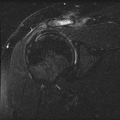
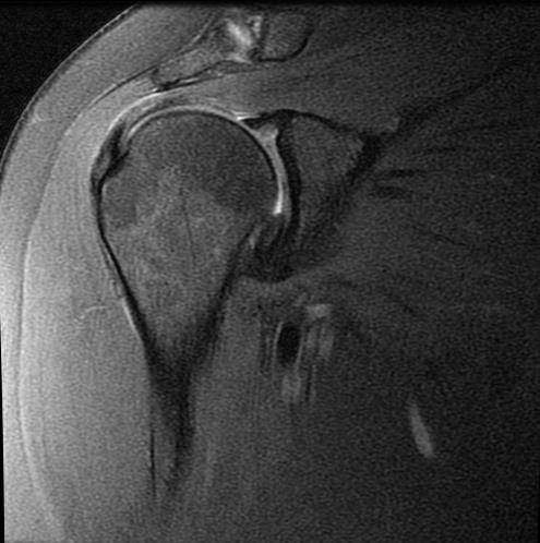
9 Advantages Higher SNR and Improved Resolution 1.5T 3T


10 Advantages Higher SNR and Improved Resolution 1.5T 3T


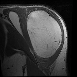
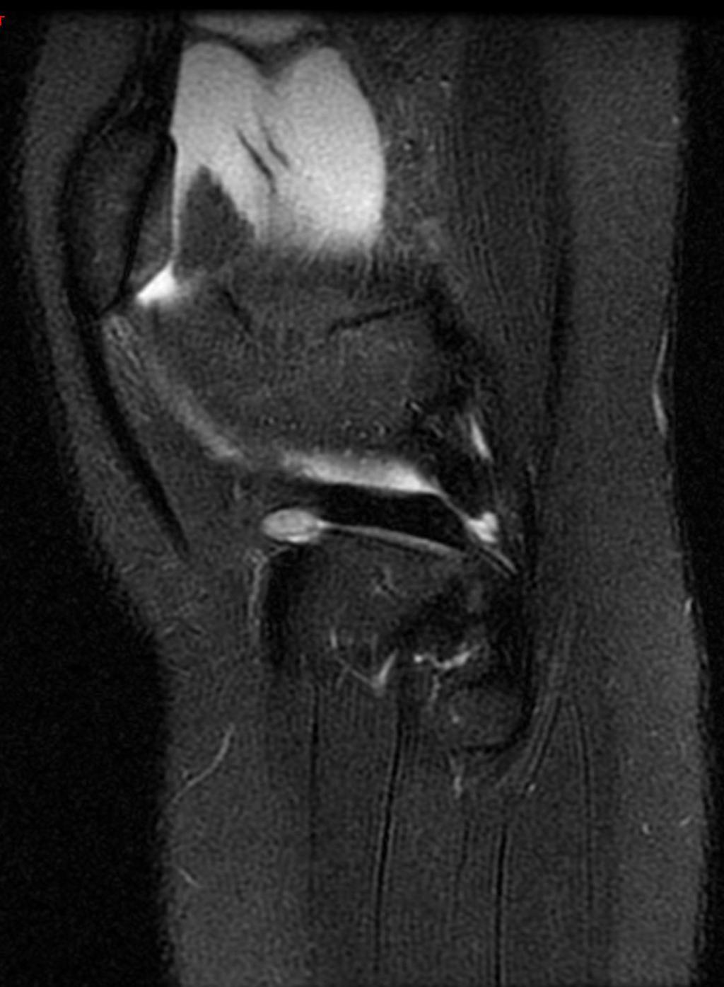
11 Advantages Higher SNR 1.5T 3T TR3383/TE46/ET 3 TR3500/TE37/ET 8


12 Advantages Higher SNR 1.5T 3T TR 1383/TE 10/ET 3 TR 2000/TE 19/ET 6


13 Advantages Higher SNR 1.5T 3T TR 3300/TE 54/ET 12 TR 3500/TE 36/ET 8


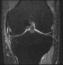
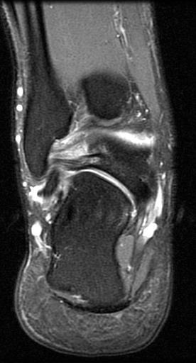
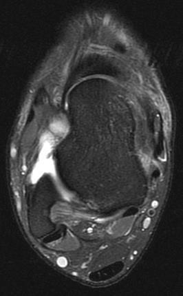
14 Patient with Resolving Lateral Friction Syndrome 3.0 T Knee Image 1.5 T Knee Image
15 Fat Saturation Better at 3T


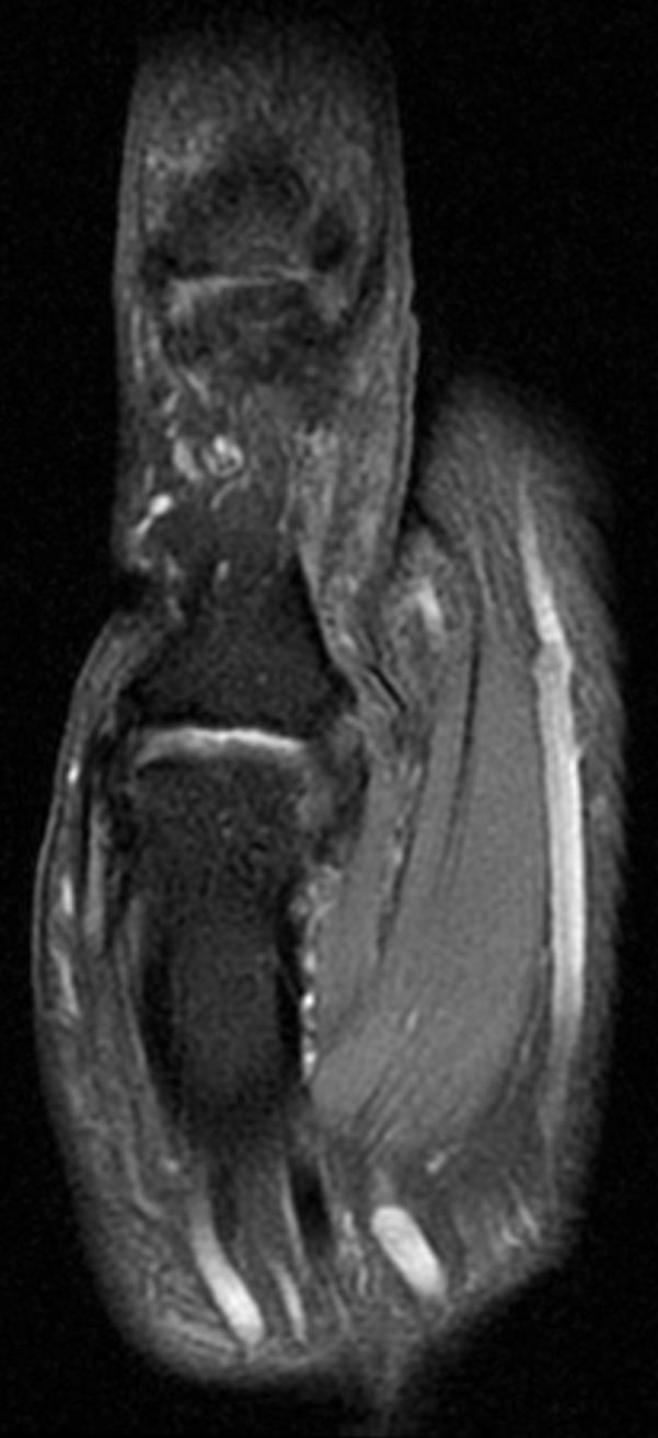
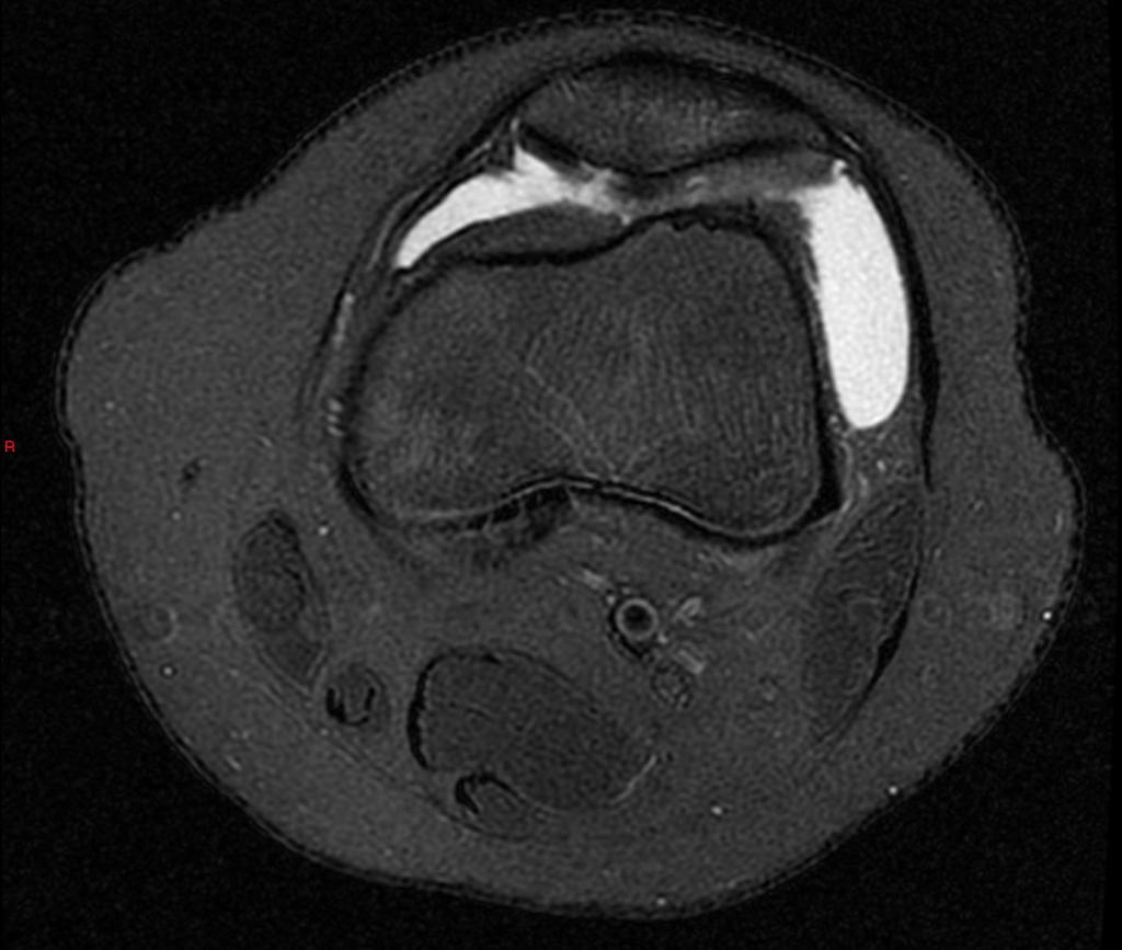
16 Smaller Structures Better Visualized Intrinsic Wrist Ligaments SL ligament and LT ligament clearly seen


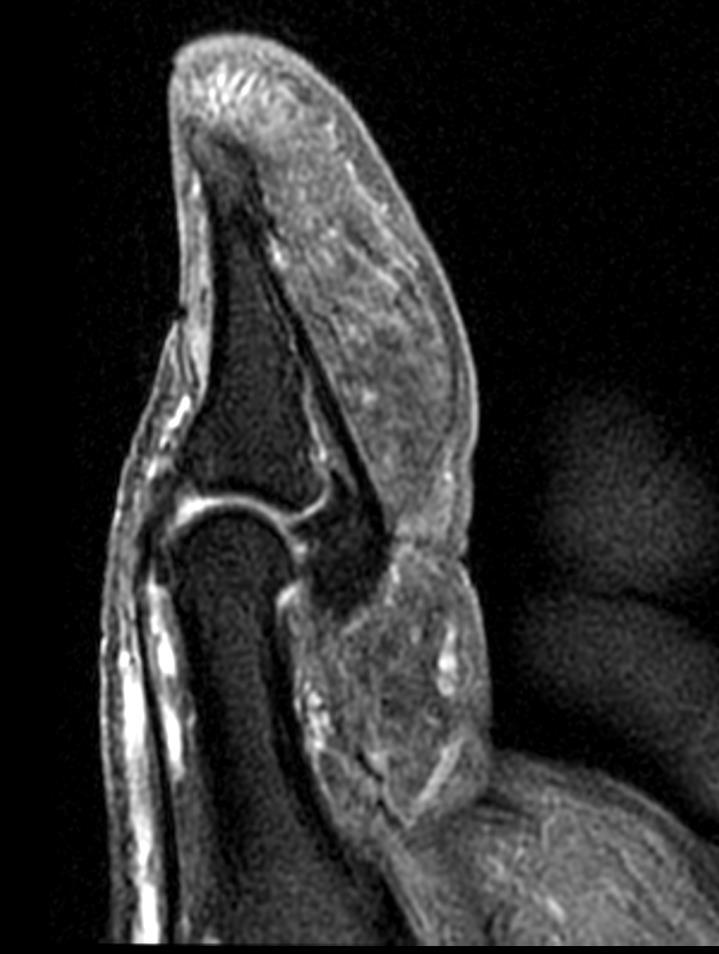
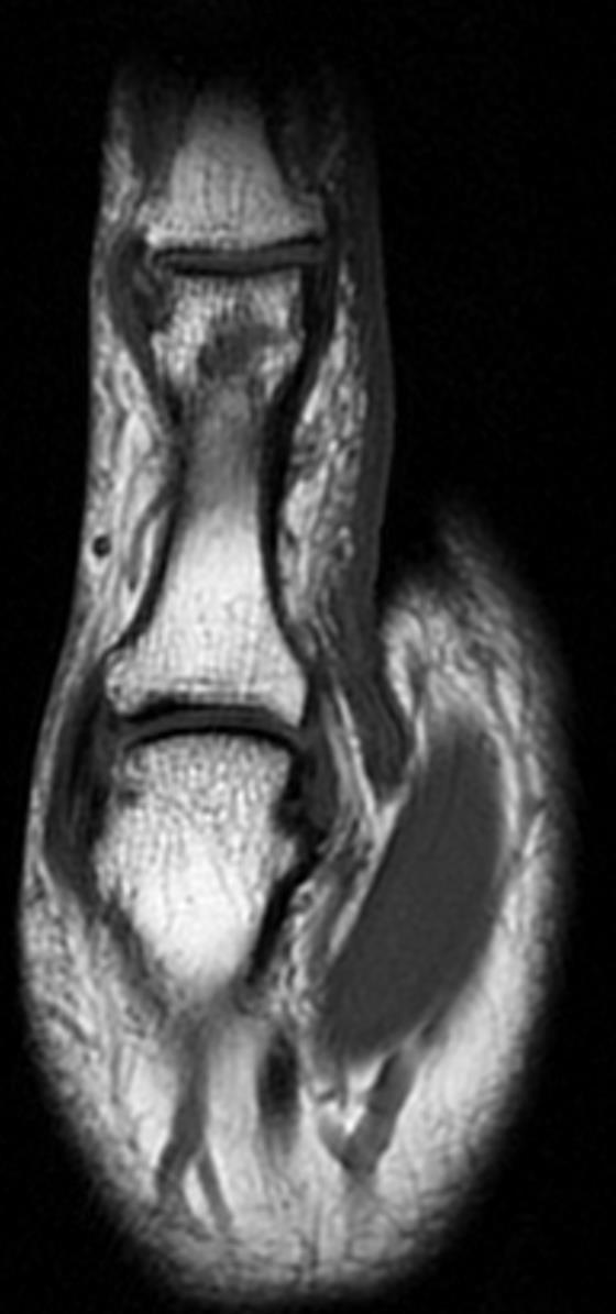
17 Improved Resolution Deltoid Ligament Plantar Fascia


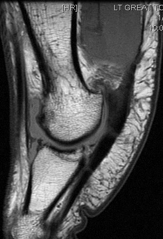
18 Mass Great Toe Bluish colored mass with skin discoloration Underside of the great toe


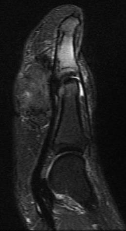
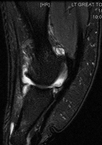
19 T1W, IntW+Fat and Post Contr T1W +Fat Sat
20 UltraHigh Field Imaging Advantageous In - Sports medicine type patients with injury to tendons, ligaments and cartilage Small parts imaging such as small joints of hands and feet Some structures sub-optimally imaged in the 1.5T scanner such as cartilage better imaged at 3T or higher Fine detail studies such as evaluating bony trabecular in osteoporosis Functional MRI and MRI spectroscopy better at higher field strengths
21 Reality Check Scanning parameters must be optimized to realize the promises of Trade off between high resolution scanning versus faster scanning Higher Field strength scanners not efficient at acquiring T1W SE images Consider using FSE T1W sequences
22 Complaints at UltraHigh Field Scanners Acoustic noise increased Nausea Vertigo Flashing lights Headaches Metallic taste in mouth Induced currents inside the body Heat deposited within tissue higher (SAR issues) Missile effects around the scanner
23 Other Inherent Limitations Stronger magnetic field Electronic device advisory more stringent Susceptibility effects more pronounced - Field inhomogeneity - Chemical shift Motion degradation more severe Hardware availability e.g. Coils are limited - Ability to build coils in house - Some vendors maintain coil monopoly
24 Cost of Imaging at at Higher Fields Quality of images are not the same - patient sacrifices quality when scanned in 1.5T 3T scanner costs about ($ m) twice as much as a 1.5T scanner ($ m) to purchase Special shielding required higher field scanners - increased installation cost Maintenance cost is increased Most 3T scanners owned by medical schools and bigger corporations and research institutions Replenishment of cryogens more frequent at higher fields
25 In Summary - Higher MRI Scanner Images have higher resolution - Ability to image smaller structures with improved resolution - Thinner slices can be obtained Images can be acquired at faster times - Less motion degradation - More body area can be covered For numerous studies diagnostic ability of the radiologist not significantly altered irrespective of scanner type for now Costs for a higher field scanners significantly more than that for a 1.5T scanner
26 Thank you
27 Suggested Reading 9-, 10- or even 11-Tesla - does higher mean better? Professor Siegfried Trattnig MD, Clinic for Radiodiagnostics, Medical University of Vienna Theysohnet al. Subjective acceptance of 7 Tesla MRI for human imaging, Magn Reson Mater Phy (2008) 21:63 72 In Vivo High-Resolution 7 Tesla MRI Shows Early and Diffuse Cortical Alterations in CADASIL (8): e Stafford RJ.High Field MRI: Technology, Applications, Safety, and Limitations Chang et al. Morphologic characterization of meniscal root ligaments in the human knee with magnetic resonance microscopy at 11.7 and 3 T. Skeletal Radiol (2014) 43:
28
29 7.0T Scanner smaller foot print
30 Advantages 3T Scanner Higher Tissue Contrast Has higher contrast to noise. Tissue contrast is determined by a number of variables including - the TR and TE chosen by the scan operator - the T1 and T2 relaxation times of the tissues being studied - the use of fat saturation Since the T1 relaxation times have increased at 3.0T - the TR must be longer to achieve the same type of contrast seen at 1.5T. - Similarly, the TE should be slightly shorter to account for decreases in T2 relaxation times In gradient echo examinations, - the flip angle should be lower to account for the increased T1 relaxation times Since T2* effects are doubled at 3.0T versus 1.5T, TE needs to be shorter at 3.0T to produce similar contrast for those sequences
31 Disadvantages - Safety Issues Specific Absorption Rate (SARs) issues - Increased heating of tissues - Becomes an issue when imaging large structures not a real problem in MSK except for spine imaging - Most units will adjust to approved parameters before scanning Same disadvantages as other MRI scanners otherwise

32 Advantages Higher SNR and Improved Resolution 1.5T 3T
33 3T MRI Scanner-Advantages Has higher signal to noise Has higher contrast to noise - Use of less contrast agent Images have higher spatial resolution - Ability to image smaller structures with improved resolution - Thinner slices can be obtained Images can be acquired at faster times - Less motion degradation

34 Fat Saturation Better at 3T T2W T1W


35 Improved Resolution Improved visualization of ligaments and cartilage

36 Deltoid Lesion

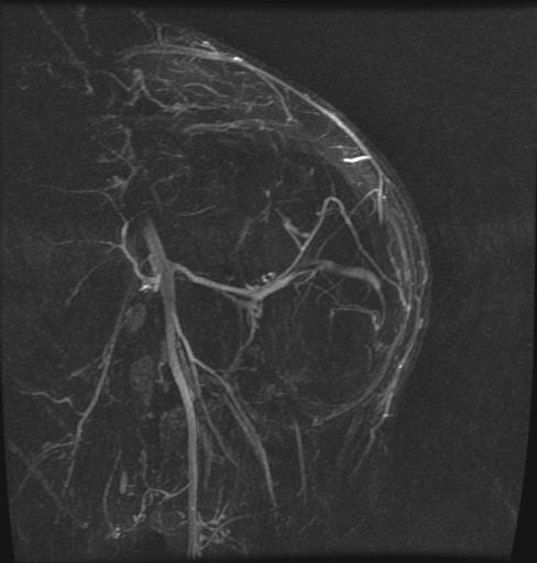
37 Vascular Map
38 Overview 1999 Ohio state University 8.0T scanner 9.4 T scanners University of Chicago U of Minnesota, MGH and NIH all have 7.0 T MRI for research T seem to be the upper limit for current technology


39 Enchondroma Index Finger Observe articular cartilage
40 Advantages 3T Scanner Higher Signal to Noise Ratio In theory 3T signal to noise ratio [SNR] 2x that of 1.5T - Generally the intrinsic SNR available in a MRI experiment is a function of the strength of the main magnetic field the volume of tissue being imaged the radiofrequency coil being used - However image quality at 3.0T influenced by changes to tissue relaxation times sensitivity to magnetic susceptibility the chemical shift difference between fat and water

41 High Field Strength MRI Scanner - 3Tesla (3T) Unit 3T MRI scanner has - Double the strength of a 1.5T scanner x Earth s magnetic field Research tool for over 20 years Clinical use for nearly 15 years
42 High Field MRI Scanner- Advantages Has higher signal to noise Has higher contrast to noise Images have higher spatial resolution Images can be acquired at faster times


43 Smaller Structures Better Visualized - Collateral Ligaments


44 Great Toe - osteoarthritis Bony spurs [arrow] more easily seen
45 Ulna Collateral Ligament Injury
46 Cartillage fragment and defect
47 Posterior Talo-fibular Ligament


48 Advantages Higher SNR 1.5T 3T
ACR MRI Accreditation Program. ACR MRI Accreditation Program Update. Educational Objectives. ACR accreditation. History. New Modular Program
 ACR MRI Accreditation Program Update Donna M. Reeve, MS, DABR, DABMP Department of Imaging Physics University of Texas M.D. Anderson Cancer Center Educational Objectives Present requirements of the new
Why Talk About Technique? MRI of the Knee:
 Why Talk About Technique? MRI of the Knee: Part 1 - Imaging Techniques Mark Anderson, M.D. University of Virginia Health Sciences Center Charlottesville, Virginia Always had an interest teach our fellows
High Field MR of the Spine
 Department of Radiology University of California San Diego 3T for MR Applications Advantages High Field MR of the Spine Increased signal-to-noise Better fat suppression Increased enhancement with gadolinium
In vivo diffusion tensor imaging (DTI) of articular cartilage as a biomarker for osteoarthritis
 of articular cartilage as a biomarker for osteoarthritis") In vivo diffusion tensor imaging (DTI) of articular cartilage as a biomarker for osteoarthritis Jose G. Raya 1, Annie Horng 2, Olaf Dietrich 2, Svetlana Krasnokutsky 3, Luis S. Beltran 1, Maximilian F.
In vivo diffusion tensor imaging (DTI) of articular cartilage as a biomarker for osteoarthritis Jose G. Raya 1, Annie Horng 2, Olaf Dietrich 2, Svetlana Krasnokutsky 3, Luis S. Beltran 1, Maximilian F.
Musculoskeletal Imaging at 3T with Simultaneous Use of Multipurpose Loop Coils
 Clinical Orthopedic Imaging Musculoskeletal Imaging at 3T with Simultaneous Use of Multipurpose Loop Coils Elena Ferrer 1 ; Rafael Coronado Santos 2 1 Radiology Department, Clínica Creu Blanca, Barcelona,
FieldStrength. Achieva 3.0T enables cutting-edge applications, best-in-class MSK images
 FieldStrength Publication for the Philips MRI Community Issue 33 December 2007 Achieva 3.0T enables cutting-edge applications, best-in-class MSK images Palo Alto Medical Clinic Sports Medicine Center employs
RECENT ADVANCES IN CLINICAL MR OF ARTICULAR CARTILAGE
 In Practice RECENT ADVANCES IN CLINICAL MR OF ARTICULAR CARTILAGE By Atsuya Watanabe, MD, PhD, Director, Advanced Diagnostic Imaging Center and Associate Professor, Department of Orthopedic Surgery, Teikyo
Echelon Oval provides a robust suite of leading musculoskeletal imaging capabilities for detailed assessment of all anatomy for your most challenging
 Echelon Oval provides a robust suite of leading musculoskeletal imaging capabilities for detailed assessment of all anatomy for your most challenging cases. Hitachi Medical Systems America, Inc. 1959 Summit
MRI of Cartilage. D. BENDAHAN (PhD)
") MRI of Cartilage D. BENDAHAN (PhD) Centre de Résonance Magnétique Biologique et Médicale UMR CNRS 7339 Faculté de Médecine de la Timone 27, Bd J. Moulin 13005 Marseille France david.bendahan@univ-amu.fr
MRI of Cartilage D. BENDAHAN (PhD) Centre de Résonance Magnétique Biologique et Médicale UMR CNRS 7339 Faculté de Médecine de la Timone 27, Bd J. Moulin 13005 Marseille France david.bendahan@univ-amu.fr
Musculoskeletal MR Protocols
 Musculoskeletal MR Protocols Joint-based protocols MSK 1: Shoulder MRI MSK 1A: Shoulder MR arthrogram MSK 1AB: Shoulder MR arthrogram (instability protocol) MSK 2: Elbow MRI MSK 2A: Elbow MR arthrogram
This presentation is the intellectual property of the author. Contact them for permission to reprint and/or distribute.
 MRI of the Knee Jennifer Swart, M.D. Musculoskeletal Radiology South Texas Radiology Group Outline Coils, Patient Positioning Acquisition Parameters, Planes and Pulse Sequences Knee Arthrography Normal
This presentation is the intellectual property of the author. Contact them at for permission to reprint and/or distribute.
 MRI of the Knee Jennifer Swart, M.D. Musculoskeletal Radiology South Texas Radiology Group Financial Disclosure Dr. Jennifer Swart has no relevant financial relationships with commercial interests to disclose.
ACR MRI Accreditation: Medical Physicist Role in the Application Process
 ACR MRI Accreditation: Medical Physicist Role in the Application Process Donna M. Reeve, MS, DABR, DABMP Department of Imaging Physics University of Texas M.D. Anderson Cancer Center Educational Objectives
Mri Of The Musculoskeletal System
 We have made it easy for you to find a PDF Ebooks without any digging. And by having access to our ebooks online or by storing it on your computer, you have convenient answers with mri of the musculoskeletal
醫用磁振學 MRM 肌肉骨骼磁振造影簡介 肌肉骨骼磁振造影. 本週課程內容 General Technical Considerations 肌肉骨骼磁振造影簡介 盧家鋒助理教授國立陽明大學生物醫學影像暨放射科學系
 本週課程內容 http://www.ym.edu.tw/~cflu 肌肉骨骼磁振造影簡介 醫用磁振學 MRM 肌肉骨骼磁振造影 盧家鋒助理教授國立陽明大學生物醫學影像暨放射科學系 alvin4016@ym.edu.tw MRI of the musculoskeletal system (5th/6th edition) Editor: Thomas H. Berquist MD 2 General
MR imaging of the knee in marathon runners before and after competition
 Skeletal Radiol (2001) 30:72 76 International Skeletal Society 2001 ARTICLE W. Krampla R. Mayrhofer J. Malcher K.H. Kristen M. Urban W. Hruby MR imaging of the knee in marathon runners before and after
Radiologic Imaging Magnetic Resonance Imaging (MRI)
") Radiologic Imaging X-ray has always been the golden rule in diagnosing and treating podiatric patients. Unfortunately, for some patients the diagnosis is not as evident. That is when we need to utilize
Radiologic Imaging X-ray has always been the golden rule in diagnosing and treating podiatric patients. Unfortunately, for some patients the diagnosis is not as evident. That is when we need to utilize
Sport Specific MRI. The symptoms of the majority, if not all sports injuries are experienced when upright, and weight-bearing
 Sport Specific MRI The symptoms of the majority, if not all sports injuries are experienced when upright, and weight-bearing A complete, accurate MRI assessment can only be made when in the position of
Comparative study of imaging at 3.0 T versus 1.5 T of the knee
 Skeletal Radiol (2009) 38:761 769 DOI 10.1007/s00256-009-0683-0 SCIENTIFIC ARTICLE Comparative study of imaging at 3.0 T versus 1.5 T of the knee Scott Wong & Lynne Steinbach & Jian Zhao & Christoph Stehling
dgemric Effectively Predicts Cartilage Damage Associated with Femoroacetabular Impingement
 Riccardo Lattanzi 1,2 Catherine Petchprapa 2 Daniele Ascani 1 Roy I. Davidovitch 3 Thomas Youm 3 Robert J. Meislin 3 Michael. Recht 2 1 The Bernard and Irene Schwartz Center for Biomedical Imaging, New
Cardiac MRI at 7T Syllabus contribution: Matthew Robson
 Cardiac MRI at 7T Syllabus contribution: Matthew Robson Field strength escalation has occurred over the last 10 years. Whilst 1.5T remains the most prevalent field strength the loss of 1T and the popularity
ACR Breast MRI Accreditation Program - DRAFT
 ACR Breast MRI Accreditation Program - DRAFT Donna M. Reeve, MS, DABR, DABMP Department of Imaging Physics Educational Objectives Provide an overview of the ACR Breast MRI Accreditation Program (BMRAP)
Cover Page. The handle holds various files of this Leiden University dissertation.
 Cover Page The handle http://hdl.handle.net/1887/35124 holds various files of this Leiden University dissertation. Author: Wokke, Beatrijs Henriette Aleid Title: Muscle MRI in Duchenne and Becker muscular
Orthopedic Hardware Imaging Part II: MRI v. Metal
 Orthopedic Hardware Imaging Trent Roth, MD And Lauren Ladd, MD Indiana University School of Medicine IU Health Physicians-Radiology Recap: Imaging Techniques Radiography Standard for initial and surveillance
3/22/2017. MR Protocol Review Clinical Opportunities for Physicists. AAPM Newsletter March/April Technical to Clinical Transition - How?
 AAPM Newsletter March/April 2017 Clinical Opportunities for Physicists Anshuman Panda, Ph.D. AAPM Spring Clinical Meeting New Orleans, LA March 2017 No conflict of interest to declare Physics Summit on
Original Research JOURNAL OF MAGNETIC RESONANCE IMAGING 22: (2005)
") JOURNAL OF MAGNETIC RESONANCE IMAGING 22:788 793 (2005) Original Research STIR vs. T1-Weighted Fat-Suppressed Gadolinium- Enhanced MRI of Bone Marrow Edema of the Knee: Computer-Assisted Quantitative Comparison
JOURNAL OF MAGNETIC RESONANCE IMAGING 22:788 793 (2005) Original Research STIR vs. T1-Weighted Fat-Suppressed Gadolinium- Enhanced MRI of Bone Marrow Edema of the Knee: Computer-Assisted Quantitative Comparison
MRI Assessments of Cartilage
 SNR IMPACTS THE ACCURACY AND PRECISION OF KNEE ARTICULAR CARTILAGE T2 RELAXATION TIME MEASUREMENTS B.J. Dardzinski 1, E. Schneider 2 1 Merck Sharp & Dohme Corp., West Point, PA USA 2 Imaging Institute,
The Key to Confidence
 WITH The Key to Confidence The Key to Confidence More detail, better accuracy, greater confidence The G-scan Brio is a revolutionary MRI approach for all musculoskeletal applications, which allows you
Safety Issue of Static Magnetic Field (1)
") MRI Safety Safety Issue of Static Magnetic Field (1) Currently, typical MRI utilizes static field with strengths of 0.5 to 3 Tesla (FDA guideline is max 4 T for infants < 1 month and 8 T for others) 1
MRI Safety Safety Issue of Static Magnetic Field (1) Currently, typical MRI utilizes static field with strengths of 0.5 to 3 Tesla (FDA guideline is max 4 T for infants < 1 month and 8 T for others) 1
Sensitivity and Specificity in Detection of Labral Tears with 3.0-T MRI of the Shoulder
 Magee and Williams MRI for Detection of Labral Tears Musculoskeletal Imaging Clinical Observations C M E D E N T U R I C L I M G I N G JR 2006; 187:1448 1452 0361 803X/06/1876 1448 merican Roentgen Ray
MR Advance Techniques. Vascular Imaging. Class II
 MR Advance Techniques Vascular Imaging Class II 1 Vascular Imaging There are several methods that can be used to evaluate the cardiovascular systems with the use of MRI. MRI will aloud to evaluate morphology
6/23/2009. Inversion Recovery (IR) Techniques and Applications. Variations of IR Technique. STIR, FLAIR, TI and TI Null. Applications of IR
 Techniques and Applications. Variations of IR Technique. STIR, FLAIR, TI and TI Null. Applications of IR") The Anatomy of Basic R Pulse Sequences Inversion Recovery () Techniques and Applications Chen Lin, PhD Indiana University School of edicine & Clarian Health Partners agnetization Preparation Section Chemical
The Anatomy of Basic R Pulse Sequences Inversion Recovery () Techniques and Applications Chen Lin, PhD Indiana University School of edicine & Clarian Health Partners agnetization Preparation Section Chemical
BioMatrix Tuners: CoilShim
 MAGNETOM Vida special issue Head and Neck Imaging Clinical 11 BioMatrix Tuners: CoilShim Miriam R. Keil, Ph.D.; Jörg Rothard; Carmel Hayes, Ph.D. Siemens Healthineers, Erlangen, Germany A cervical spine
Cardiovascular MR Imaging at 3 T: Opportunities, Challenges, and Solutions 1
 TECHNICAL ADVANCEMENTS IN CARDIAC MR IMAGING 1612 Cardiovascular MR Imaging at 3 T: Opportunities, Challenges, and Solutions 1 Prabhakar Rajiah, MD, FRCR Michael A. Bolen, MD Abbreviations: BOLD = blood
The value of weight-bearing functional CT scans
 The value of weight-bearing functional scans In musculoskeletal medicine, advanced imaging like computed axial tomography () scanning, has become invaluable to the evaluation and management of patients
Meniscal Tears: Role of Axial MRI Alone and in Combination with Other Imaging Planes
 Nefise Cagla Tarhan 1,2 Christine. Chung 1 urea Valeria Rosa Mohana-orges 1 Tudor Hughes 1 Donald Resnick 1 Received September 30, 2003; accepted after revision February 2, 2004. 1 Department of Radiology,
Comparative study of high resolusion ultrasonography and magnetic resonance imaging in diagnosing traumatic knee injuries & pathologies
 Original article: Comparative study of high resolusion ultrasonography and magnetic resonance imaging in diagnosing traumatic knee injuries & pathologies Dr. Rakesh Gujjar*, Dr. R. P. Bansal, Dr. Sandeep
Comparison of 1.5 T Tesla and 3.0 T Tesla Magnetic Resonance Imaging for Evaluating Local Extension of Endometrial Cancer
 Showa Univ J Med Sci 27 1, 21 28, March 2015 Original Comparison of 1.5 T Tesla and 3.0 T Tesla Magnetic Resonance Imaging for Evaluating Local Extension of Endometrial Cancer Naomi YAGI, Masanori HIROSE,
Meniscus T2 Relaxation Time at Various Stages of Knee Joint Degeneration
 Meniscus T2 Relaxation Time at Various Stages of Knee Joint Degeneration Richard Kijowski, Michael Fazio, Benjamin Beduhn, and Fang Liu Department of Radiology University of Wisconsin School of Medicine
Imaging the musculoskeletal system. An Introduction
 Imaging the musculoskeletal system An Introduction Objectives Discuss: commonly used imaging modalities in the musculoskeletal system normal imaging anatomy in the extremities fracture description Imaging
MRI PEDIATRIC PROTOCOLS (Updated 6/19/2018)
") MRI PEDIATRIC PROTOCOLS (Updated 6/19/2018) *Please get or let us know where radiologist can review plain films. *For Texas Orthopedics and other Docs requesting only MSK section read for their pediatric
MRI PEDIATRIC PROTOCOLS (Updated 6/19/2018) *Please get or let us know where radiologist can review plain films. *For Texas Orthopedics and other Docs requesting only MSK section read for their pediatric
Tissue-engineered medical products Evaluation of anisotropic structure of articular cartilage using DT (Diffusion Tensor)-MR Imaging
-MR Imaging") Provläsningsexemplar / Preview TECHNICAL REPORT ISO/TR 16379 First edition 2014-03-01 Tissue-engineered medical products Evaluation of anisotropic structure of articular cartilage using DT (Diffusion Tensor)-MR
Provläsningsexemplar / Preview TECHNICAL REPORT ISO/TR 16379 First edition 2014-03-01 Tissue-engineered medical products Evaluation of anisotropic structure of articular cartilage using DT (Diffusion Tensor)-MR
Imaging of Articular Cartilage
 Clinical Imaging of Articular Cartilage Imaging of Articular Cartilage Prof. Dr. K. Verstraete Ghent University Introduction : Articular Cartilage Histology and biochemical composition Review of Imaging
JMSCR Vol 05 Issue 07 Page July 2017
 www.jmscr.igmpublication.org Impact Factor 5.84 Index Copernicus Value: 83.27 ISSN (e)-2347-176x ISSN (p) 2455-0450 DOI: https://dx.doi.org/10.18535/jmscr/v5i7.84 Anatomical Differences Between T2 WI FSE
A Patient s Guide to Diffuse Idiopathic Skeletal Hyperostosis (DISH)
") A Patient s Guide to Diffuse Idiopathic Skeletal Hyperostosis (DISH) 6565 Fannin Street Houston, TX 77030 Phone: 713-790-3333 DISCLAIMER: The information in this booklet is compiled from a variety of sources.
A Patient s Guide to Diffuse Idiopathic Skeletal Hyperostosis (DISH) 6565 Fannin Street Houston, TX 77030 Phone: 713-790-3333 DISCLAIMER: The information in this booklet is compiled from a variety of sources.
Worldwide Leading Academic Center Gets a Lift in Advanced MR Imaging
 Worldwide Leading Academic Center Gets a Lift in Advanced MR Imaging As one of the largest and most authoritative scientific medical centers in Europe, Erasmus University Medical Center (Rotterdam, Netherlands),
Knee Articular Cartilage in an Asymptomatic Population : Comparison of T1rho and T2 Mapping
 TR_002 Technical Reports Knee Articular Cartilage in an Asymptomatic Population : Comparison of T1rho and T2 Mapping Min A Yoon 1,*, Suk-Joo Hong 1, Chang Ho Kang 2, Baek Hyun Kim 3 1 Korea University
CT ARTHROGRAPHY It s not Always About the
 CT ARTHROGRAPHY It s not Always About the Magnet Kirkland W. Davis, M.D. University of Wisconsin Department of Radiology Disclosures Financial FDA IA Gd! What Is CT Arthrography? (CTR) Arthrogram: imaging
Magnetic Resonance Imaging on Soft Tissue. Jiten K. Mistry Calvin Gan
 Magnetic Resonance Imaging on Soft Tissue 1 Jiten K. Mistry Calvin Gan Outline Background of Medical Imaging Introduction to MRI How MRI works MRI of Soft Tissue Benefits & Risks Recent Advances 2 The
MRI FAQs. After reading these documents and checking your protocols, you can apply online here: https://acredit.acr.org.
 MRI FAQs Application - General Q. My facility plans to apply for ACR MRI Accreditation, where do I start? A. Start by reading the following documents, available on the ACR website: The Diagnostic Modality
Real-Time MRI of Joint Movement With TrueFISP
 JOURNAL OF MAGNETIC RESONANCE IMAGING 15:710 715 (2002) Technical Note Real-Time MRI of Joint Movement With TrueFISP Harald H. Quick, MSc, 1 * Mark E. Ladd, PhD, 1 Matthias Hoevel, MD, 2 Silke Bosk, RT,
Imaging Symposium. Tuesday, 28 March Date: Teaching & Learning Centre, Queen Elizabeth University Hospital. Venue:
 Imaging Symposium Date: Venue: Tuesday, 28 March 2017 Teaching & Learning Centre, Queen Elizabeth University Hospital Biographies Professor Siegfried Trattnig Prof. Dr. Siegfried Trattnig graduated from
Abdominal MRI Techniques in Pediatric Oncology
 Abdominal MRI Techniques in Pediatric Oncology Jonathan R. Dillman, M.D. Assistant Professor Departments of Radiology & Urology Section of Pediatric Radiology C.S. Mott Children s Hospital Disclosures
Magnetic Resonance Angiography
 Magnetic Resonance Angiography 1 Magnetic Resonance Angiography exploits flow enhancement of GR sequences saturation of venous flow allows arterial visualization saturation of arterial flow allows venous
JiangSu Magspin Instrument Co., Ltd
 Page 1 of 5 Magspin ARMOUS - 0.25T Dedicated Open Extremity MRI System The Beginning of a New Era in Extremity MR Imaging Superior Sitting Position MR Imaging ARMOUS is the long awaited Dedicated Open
ACR Accreditation Update in MRI
 ACR Accreditation Update in MRI Whole Body Systems Extremity (MSK) Ron Price Vanderbilt University Medical Center Nashville, TN Dedicated Breast MRI Accreditation Update 1. ACR MRI Accreditation: Overview,
mri sequences 8267BD21D03EEA3AE7926DD1904E7425 Mri Sequences 1 / 6
 Mri Sequences 1 / 6 2 / 6 3 / 6 Mri Sequences An MRI sequence is a number of radiofrequency pulses and gradients that result in a set of images with a particular appearance. This article presents a simplified
Non-Contrast MRA. How and When 1996! Why Non-Contrast MRA? Angiography: What are our goals? Inflow Techniques Differences in excitation hx
 A major teaching hospital of Harvard Medical School Angiography: What are our goals? Non-Contrast MRA: How and When Neil M. Rofsky, M.D. Professor of Radiology, Harvard Medical School Director of MRI &
*smith&nephew. MRI Safety Information & Parameters for Smith & Nephew Orthopaedics AG. Knee Implants
 Knee Implants MRI Safety Information & Parameters for Smith & Nephew Orthopaedics AG Knee Implants *smith&nephew Supporting healthcare professionals for over 150 years Summary All knee implants of Smith
Musculoskeletal Imaging What to order? Brian Cole, MD
 Musculoskeletal Imaging What to order? Brian Cole, MD my background: 1994 University of Illinois 1998 MD University of Illinois College of Medicine 1999-2003 Diagnostic Radiology Mayo Clinic 2004 Fellowship
SSSR. 1. Nov Ankle. Postoperative Imaging of Cartilage Repair. and Lateral Ligament Reconstruction
 Ankle Postoperative Imaging of Cartilage Repair and Lateral Ligament Reconstruction Andrea B. Rosskopf, MD University Hospital Balgrist Imaging of Cartilage Repair Why? To assess the technical success
External Distal Radius Fixator. Supplement to the 8 mm rod fixator system
 External Distal Radius Fixator. Supplement to the 8 mm rod fixator system Surgical technique This publication is not intended for distribution in the USA. Instruments and implants approved by the AO Foundation
Cartilage Repair Options
 Imaging of Cartilage Repair Carl S. Winalski, MD Imaging Institute Department of Biomedical Engineering Cleveland Clinic Cartilage Repair Options Direct repair Marrow stimulation Autologous transplantation
Case Report: Knee MR Imaging of Haemarthrosis in a Case of Haemophilia A
 Clinical > Pediatric Imaging Case Report: Knee MR Imaging of Haemarthrosis in a Case of Haemophilia A M. A. Weber, J. K. Kloth University Hospital Heidelberg, Department of Diagnostic and Interventional
Message of the Month for GPs June 2013
 Message of the Month for GPs June 2013 Dr Winn : Consultant Musculoskeletal Radiologist, Manchester Royal Infirmary Imaging of the musculoskeletal system Musculoskeletal pain is a common problem in the
A quality control program for MR-guided focused ultrasound ablation therapy
 JOURNAL OF APPLIED CLINICAL MEDICAL PHYSICS, VOLUME 3, NUMBER 2, SPRING 2002 A quality control program for MR-guided focused ultrasound ablation therapy Tao Wu* and Joel P. Felmlee Department of Radiology,
Low-Profile Wrist Fixator. For stabilization of fractures of the distal radius.
 Low-Profile Wrist Fixator. For stabilization of fractures of the distal radius. Technique Guide Part of the External Fixation System Low-Profile Wrist Fixator Indications Intended for stabilization of
1Pulse sequences for non CE MRA
 MRI: Principles and Applications, Friday, 8.30 9.20 am Pulse sequences for non CE MRA S. I. Gonçalves, PhD Radiology Department University Hospital Coimbra Autumn Semester, 2011 1 Magnetic resonance angiography
Magnetic Resonance Imaging
 Magnetic Resonance Imaging The purpose of structured education is to provide the opportunity for individuals to develop mastery of discipline-specific knowledge that, when coupled with selected clinical
Speed, Comfort and Quality with NeuroDrive
 Speed, Comfort and Quality with NeuroDrive Echelon Oval provides a broad range of capabilities supporting fast, accurate diagnosis of brain conditions and injuries. From anatomical depiction to vascular
Role of Diffusion WIs and T 2 * GRE Pulse Sequences in Dubious Vertebral Marrow Pathological Lesions
 Journal of the Egyptian Nat. Cancer Inst., Vol. 19, No. 4, December: 254-262, 2007 Role of Diffusion WIs and T 2 * GRE Pulse Sequences in Dubious Vertebral Marrow Pathological Lesions OMAR M. OSMAN, M.D.*;
An excellent fit to expand the imaging center, Prodiva 1.5T
 An excellent fit to expand the imaging center, Prodiva 1.5T FieldStrength MRI magazine User experiences - December 2017 www.philips.com/fieldstrength High quality MR imaging combined with a simplified,
The Essentials Tissue Characterization and Knobology
 The Essentials Tissue Characterization and Knobology Randy E. Moore, DC, RDMS RMSK No relevant financial relationships Ultrasound The New Standard of Care Musculoskeletal sonography has become the standard
Powered by. Dedicated MRI
 Powered by Dedicated MRI Provides the latest software and hardware upgrade configuration powered by exp technology: boosting productivity, increasing image quality, and adding new acquisition techniques.
Personal use only. MRI Metal Artifact Reduction: Shoulder Implants and Arthroplasty. Reto Sutter, MD
 MRI Metal Artifact Reduction: Shoulder Implants and Arthroplasty Reto Sutter, MD University Hospital Balgrist Zurich University of Zurich Cor PD fat sat 56-year old male patient with positive lift-off
Medium External Fixator Humeral Shaft Frame. Modular frame for upper extremity use.
 Medium External Fixator Humeral Shaft Frame. Modular frame for upper extremity use. Technique Guide Part of the Medium External Fixation System MRI Information Synthes Medium External Fixation devices
Emerging Applications in Musculoskeletal CT Imaging
 Emerging pplications in Musculoskeletal CT Imaging y K Murali MD(RD), PDCC, Director of Interventional Radiology, G. Francis DMRD, DN (RD), Consultant Radiologist, and R. Madan, MS, MD, Consultant Radiologist,
Prevalence of Meniscal Radial Tears of the Knee Revealed by MRI After Surgery
 Downloaded from www.ajronline.org by 46.3.207.114 on 12/22/17 from IP address 46.3.207.114. Copyright RRS. For personal use only; all rights reserved Thomas Magee 1 Marc Shapiro David Williams Received
ORIGINAL ARTICLE. ROLE OF MRI IN EVALUATION OF TRAUMATIC KNEE INJURIES Saurabh Chaudhuri, Priscilla Joshi, Mohit Goel
 ROLE OF MRI IN EVALUATION OF TRAUMATIC KNEE INJURIES Saurabh Chaudhuri, Priscilla Joshi, Mohit Goel 1. Associate Professor, Department of Radiodiagnosis & imaging, Bharati Vidyapeeth Medical College and
FOR CMS (MEDICARE) MEMBERS ONLY NATIONAL COVERAGE DETERMINATION (NCD) FOR MAGNETIC RESONANCE IMAGING:
 MEMBERS ONLY NATIONAL COVERAGE DETERMINATION (NCD) FOR MAGNETIC RESONANCE IMAGING:") National Imaging Associates, Inc. Clinical guidelines BONE MARROW MRI Original Date: July 2008 Page 1 of 5 CPT Codes: 77084 Last Review Date: September 2014 NCD 220.2 MRI Last Effective Date: July 2011
National Imaging Associates, Inc. Clinical guidelines BONE MARROW MRI Original Date: July 2008 Page 1 of 5 CPT Codes: 77084 Last Review Date: September 2014 NCD 220.2 MRI Last Effective Date: July 2011
Advanced musculoskeletal MRI at ultra-high field (7 T)
") PERSPECTIVE Advanced musculoskeletal MRI at ultra-high field (7 T) MRI at ultra-high field (7 T) offers high signal-to-noise ratio, which can be used beneficially in several different musculoskeletal applications.
PERSPECTIVE Advanced musculoskeletal MRI at ultra-high field (7 T) MRI at ultra-high field (7 T) offers high signal-to-noise ratio, which can be used beneficially in several different musculoskeletal applications.
Expanding therapy options for
 MR systems Sonalleve MR-HIFU Expanding therapy options for women s health and oncology 2 Discover the freedom of patient-friendly and non-invasive therapy options Sonalleve MR-HIFU is an innovative therapy
Role of Magnetic Resonance Imaging in Patients with Knee Trauma
 Original Research Article Role of Magnetic Resonance Imaging in Patients with Knee Trauma Bhautik Kapadia 1, Bhumika Suthar 2* 1 Associate Professor, 2 Assistant Professor, Department of Radiodiagnosis,
UCLA UCLA UCLA 7/10/2015. The need for MRI in radiotherapy. Multiparametric MRI reflects a more complete picture of the tumor biology
 Ke Sheng, Ph.D., DABR Professor of Radiation Oncology University of California, Los Angeles The need for MRI in radiotherapy T1 FSE CT Tumor and normal tissues in brain, breast, head and neck, liver, prostate,
Magnetic resonance imaging of the distal limb of the standing horse Keywords: Introduction Equine MRI limb scanner Scanning procedure
 4 EQUINE VETERINARY EDUCATION Equine vet. Educ. (00) () 4-8 Satellite Article Magnetic resonance imaging of the distal limb of the standing horse T. S. MAIR*, J. KINNS, R. D. JONES AND N. M. BOLAS Bell
WITH. The Next Step in Office MRI
 WITH The Next Step in Office MRI Introducing S-scan the Next Step in Office MRI Based on extensive customer feedback and years of engineering, Esaote has designed the S-scan with exp Technology, an optimized
MY PATIENT HAS KNEE PAIN. David Levi, MD Chief, Division of Musculoskeletal l limaging Atlantic Medical Imaging
 MY PATIENT HAS KNEE PAIN David Levi, MD Chief, Division of Musculoskeletal l limaging Atlantic Medical Imaging Causes of knee pain Non traumatic Trauma Osteoarthritis Patellofemoral pain Menisci or ligaments
Rad Tech 4643 MRI Torso and Extremities
 Rad Tech 4643 MRI Torso and Extremities Prostate Cancer Leiomyoma Retroverted Anteverted Ovarian Cyst Gone Wrong Fibroid (Leiomyoma) IUD Ovary Hysterectomy? What are we to see when imaging a female pelvis
Introduction. Cardiac Imaging Modalities MRI. Overview. MRI (Continued) MRI (Continued) Arnaud Bistoquet 12/19/03
 MRI (Continued) Arnaud Bistoquet 12/19/03") Introduction Cardiac Imaging Modalities Arnaud Bistoquet 12/19/03 Coronary heart disease: the vessels that supply oxygen-carrying blood to the heart, become narrowed and unable to carry a normal amount
Introduction Cardiac Imaging Modalities Arnaud Bistoquet 12/19/03 Coronary heart disease: the vessels that supply oxygen-carrying blood to the heart, become narrowed and unable to carry a normal amount
5/28/2015. The need for MRI in radiotherapy. Multiparametric MRI reflects a more complete picture of the tumor biology
 Ke Sheng, Ph.D., DABR Professor of Radiation Oncology University of California, Los Angeles The need for MRI in radiotherapy T1 FSE CT Tumor and normal tissues in brain, breast, head and neck, liver, prostate,
Essentials of Clinical MR, 2 nd edition. 99. MRA Principles and Carotid MRA
 99. MRA Principles and Carotid MRA As described in Chapter 12, time of flight (TOF) magnetic resonance angiography (MRA) is commonly utilized in the evaluation of the circle of Willis. TOF MRA allows depiction
The Effects of Music intervention on Functional connectivity. Supplemental Information
 Yang et al. 0 The Effects of Music intervention on Functional connectivity strength of Brain in Schizophrenia Supplemental Information Mi Yang,#, Hui He #, Mingjun Duan,, Xi Chen, Xin Chang, Yongxiu Lai,
2014 AAPM 56 th Annual Meeting
 2014 AAPM 56 th Annual Meeting SAM Diagnostic Radiology MR Safety - Deep Brain Stimulator and Other Neurostimulators Yunhong Shu, Ph.D. Mayo Clinic, Rochester, MN Outline Background of Neurostimulator
Publication for the Philips MRI Community
 FieldStrength Publication for the Philips MRI Community Issue 38 Summer 2009 Pediatric MSK imaging benefits from tailored scan protocols Vanderbilt University Children s Hospital builds dedicated scans
SIR MICHELANGELO REFALO
 SIR MICHELANGELO REFALO SIXTH FORM Half Yearly Exam 2014 Name: Subject: Physical Education Intermediate Time: 2 Hours Answer ALL questions Question 1: (The Skeleton) The human skeleton is made up of 206
FOR CMS (MEDICARE) MEMBERS ONLY NATIONAL COVERAGE DETERMINATION (NCD) FOR MAGNETIC RESONANCE IMAGING:
 MEMBERS ONLY NATIONAL COVERAGE DETERMINATION (NCD) FOR MAGNETIC RESONANCE IMAGING:") National Imaging Associates, Inc. Clinical guidelines SINUS MRI Original Date: November 2007 Page 1 of 5 CPT Codes: 70540, 70542, 70543 Last Review Date: July 2014 NCD 220.2 MRI Last Effective Date: July
National Imaging Associates, Inc. Clinical guidelines SINUS MRI Original Date: November 2007 Page 1 of 5 CPT Codes: 70540, 70542, 70543 Last Review Date: July 2014 NCD 220.2 MRI Last Effective Date: July
Oak foundation for donating the 3T Siemens Verio scanner. Board of directors BBH and Frh Hospitals for supporting the
 Knee pain and inflammation in the infrapatellar fat pad estimated by conventional and dynamic contrast-enhanced magnetic resonance imaging in obese patients with osteoarthritis: a crosssectional study
Original Report. The Reverse Segond Fracture: Association with a Tear of the Posterior Cruciate Ligament and Medial Meniscus
 Eva M. Escobedo 1 William J. Mills 2 John. Hunter 1 Received July 10, 2001; accepted after revision October 1, 2001. 1 Department of Radiology, University of Washington Harborview Medical enter, 325 Ninth
Page 1 of 6. Appendix 1
 Page 1 Appendix 1 Rotation Objectives and Schedule 1. Introductory Month 4 weeks 2. Total Joints 4 weeks a. Diagnosis and management of hip and knee arthritis b. Indications for surgery c. Implant selection;
KNEE ALIGNMENT SYSTEM (KAS) MRI Protocol
 MRI Protocol") KNEE ALIGNMENT SYSTEM (KAS) MRI Protocol Sample referral sticker Referral Sticker Insert here Corin 17 Bridge Street Pymble NSW Australia 2073 P: +61 (0)2 9497 7400 F: +61 (0)2 9497 7498 E: KAS.customerservice@coringroup.com
KNEE ALIGNMENT SYSTEM (KAS) MRI Protocol Sample referral sticker Referral Sticker Insert here Corin 17 Bridge Street Pymble NSW Australia 2073 P: +61 (0)2 9497 7400 F: +61 (0)2 9497 7498 E: KAS.customerservice@coringroup.com