Magnetic Resonance Imaging on Soft Tissue. Jiten K. Mistry Calvin Gan
|
|
|
- Nicholas Barnett
- 6 years ago
- Views:
Transcription
1 Magnetic Resonance Imaging on Soft Tissue 1 Jiten K. Mistry Calvin Gan
2 Outline Background of Medical Imaging Introduction to MRI How MRI works MRI of Soft Tissue Benefits & Risks Recent Advances 2
3 The Need for Medical Imaging A method required to view and diagnose the human body noninvasively Could aid and lead to surgery and treatment of the patient Cause and Effect Cause Internal Tissue Effect Received signal 3

4 History of Medical Imaging The Medical Imaging industry was said to be conceived by Wilhelm Conrad Roentgen in 1895 His discovery of X-rays began to allow physicians to use primitive x-ray machines to view broken bones and contrast-enhanced structures in the body such as the GI system. Further development of Medical Imaging was fairly insignificant until the later half of the 20th century, when computer technology was beginning to play a large role in the instruments developed Clarity of the image created as well as the speed at which the image is rendered have been the two properties pulling development in the industry X ray of Roentgen s wife s hand
5 Computed Tomography (CT) Scan Developed during the late 1960 s Uses X-ray images to re-create a cross sectional (axial) image of the body, in part or whole Body is bombarded with low intensity x-rays and sensors detect the amount of x-rays that rebound Study blood vessels Identify masses and tumors, including cancer Guide a surgeon to the right area during a biopsy 5
6 Ultrasound First introduced in the 1960 s A probe is used to send sound waves into the body Sounds waves reflect off of internal organs and tissues Probe records reflected waves machine then calculates distance of tissue from probe based on echo return time and speed of sound in tissue (5,005 ft/s) 6
7 Where do these techniques fall short? CT scan Can only provide axial plane image Unable to distinguish between different types of tissues Ultrasound Also unable to distinguish between different tissues Why do previous methods have this problem? Provide images that show the density of the object Tissues in the human body have a fairly uniform density, Very difficult to distinguish between different types of tissues 7
8 Different Types of Tissues Deep vs. Superficial tissue Ligaments vs. Tendons Organs vs. surrounding tissue How do we identify these tissues separately? 8
9 9
10 Why MRI? Unlike other more common Medical Imaging Techniques, MRI creates images from signals received from resonating protons within the matter The data range we can have for signals from protons is quite large, allowing us to more easily differentiate between different matter which are closely related 10
11 Soft Tissue MRI is excellent for imaging of soft tissue Why? Rather then using the different densities for different tissues (which does not work) we instead look at the vastly different magnetic properties of the proton in the tissue we are observing, which can allow us to determine other properties of the 11 tissue
12 Magnetic Properties of Soft Tissue T1 (ms) T2 (ms) Putamen 747 ± ± 4 Caudate nucleus 822 ± ± 4 Thalamus 703 ± ± 4 Cortical gray matter 871 ± ± 2 Corpus callosum 509 ± ± 8 White matter 515 ± ± 5 Cerebrospinal fluid 1900 ± ± 3 Artery 1205 ± ± 5.3 Vein 1335 ± ±
13 Magnetic Properties of Soft Tissue 13

14 What is MRI? A noninvasive diagnostic technique to produce images of inside the human body Ability to provide functional as well as anatomical information 14
15 What is MRI? Equipment: MRI scanner 1. Magnet: largest and most expensive part i) Is superconducting, liquid helium is cooled to 4K 2. RF System: transmitting and receiving of signals 3. Gradients: Spatial encoding, determine plane for imaging Computer: Configures gradients Input from RF system is sent to a monitor 15

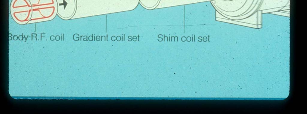
16 16 Image courtesy of Dr. Noseworthy

17 17 Image courtesy of Dr. Noseworthy


18 Magnetic Fields Region of space Produced in the presence of a magnet or moving electric current The direction of a magnetic field at any point is towards the south pole of a bar magnet Positive particles follow the path of magnetic field The direction in which a compass needle points 18
19 Magnetic Fields Nikola Tesla (July 10, 1856 January 7, 1943) Tesla: SI unit for measuring the magnetic field (B) Source Superconducting (laboratory) magnet Strong conventional (laboratory) magnet Field Magnitude (T) 30 2 MRI scanners use fields ranging from T can operate up to 9.4 teslas World s largest MRI machine Bar magnet & surface of the sun 1e-2 Surface of the earth µ Inside the human brain (nerve impulses) 1e Serway & Beichner, 2000
20 How MRI Works 1. A strong magnetic field align protons 2. i) RF pulses excite these protons ii) Upon realignment, each proton emits distinct signals 3. The signals are then detected and read, producing an image of inside the body Video: 20

21 21 1. A strong magnetic field align protons

22 How MRI Works 2 i) RF pulses excite these proton 22
23 How MRI Works What are the time constants T1 and T2? 2 ii) Upon realignment, each proton emits distinct signals T1 and T2 are magnetic timing parameters which differ from tissue to tissue 23

24 How MRI Works T1-Weighted T2-Weighted 24
25 How MRI Works 25

26 How MRI Works 3. The signals are then detected and read, producing an image of inside the body, using K space 26 Burger et al., 2001

27 Case Studies Rotator Cuff Tear - These muscles keep the humeral head centered on the glenoid cavity - traumatic injury and wearing down may lead to the tear - the white arrow points to the supraspinatus tendon

28 Case Studies The rotator cuff tear is identified as loose, degenerated, and frayed tissue around the cuff edge Pain occurs over the deltoid muscle and when the arm is raised The red arrow points to the torn edge of the rotator cuff 28

29 Case Studies Pituitary Tumour The Pituitary gland is a small endocrine/hormone gland which is located at the base of the brain. Growths or tumours in the pituitary can secrete excessive amounts of hormone Symptoms: Headache, visual disturbance, clinical effects of excessive hormone secretion. 29

30 Case Studies Multiple Sclerosis Demyelinating disease that affects the CNS MRI reveals plagues in the white matter of the brain and spinal cord. 30

31 Case Studies 31 T1-Weighted


32 Case Studies Normal vs Torn ACL 32

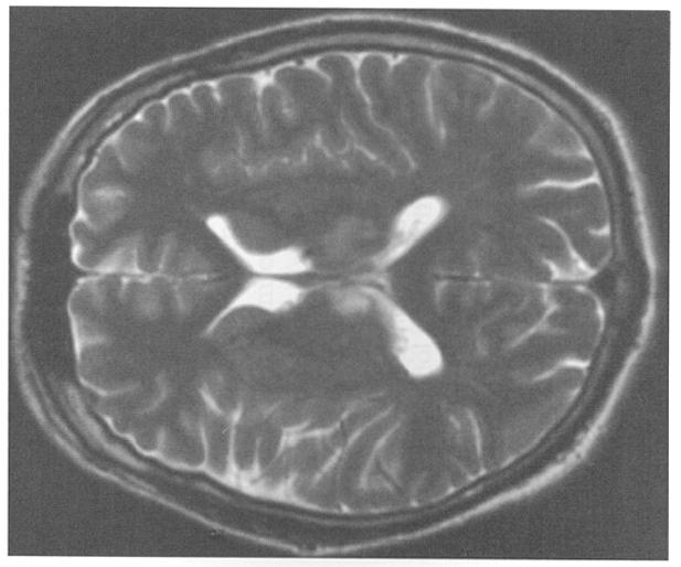
33 Case Studies This image is an axial section of the head of an elderly man. What has caused the abnormality shown? 33

34 Case Studies young female patient had experienced amenorrhoea What can you conclude from this MRI image? 34
35 MRI Disadvantages High Initial cost, translates to a high examination cost Metallic orthopedic hardware cause distortions in images as they interfere with magnetic field Require patient to hold still for up to 90 minutes Could cause distortion in images 35
36 MRI Disadvantages cont. Individuals with pacemakers or large non stainless steel implants are unsafe to use MRI machine Creates a large noise during operation 36
37 Future of MRI Ultra Low Field MRI 37 MRI machine which uses a Magnetic Field of a few microtesla to create an image Signals are measured by SQUIDS (superconducting quantum interference devices) the most sensitive detectors of magnetic flux Allows paramedics to obtain quick MRI images on the go (crappy quality but can easily diagnose injury)
38 References Bister, Jeffrey. Clinical Applications of Medical Imaging. Plenum Press. New York, New York, 1986 Young, Stuart W. Nuclear Magnetic Resonance Imaging. Basic Principles. Raven Press Books, Ltd. New York, New York,
Syllabus References. Resources. Video: MRI Introduction
 MRI Lesson Outline Syllabus References 9.6.4.2.5 Define precessing and relate the frequency of the precessing to the composition of the nuclei and the strength of the applied external magnetic field 9.6.4.2.6
Imaging Patient Education. Magnetic Resonance Imaging (MRI)
") Magnetic Resonance Imaging (MRI) What you should know about your Body MRI exam: Purpose: Magnetic Resonance Imaging (MRI) uses radio waves and a strong magnetic field to provide clear and detailed images
Magnetic Resonance Imaging (MRI) What you should know about your Body MRI exam: Purpose: Magnetic Resonance Imaging (MRI) uses radio waves and a strong magnetic field to provide clear and detailed images
Non-Invasive Techniques
 Non-Invasive Techniques Key: Does not hurt the organism Psychology 372 Physiological Psychology Steven E. Meier, Ph.D. Listen to the audio lecture while viewing these slides or view the video presentation
Non-Invasive Techniques
 Many Procedures Non-Invasive Techniques Key: Does not hurt the organism Psychology 372 Physiological Psychology Steven E. Meier, Ph.D. Listen to the audio lecture while viewing these slides or view the
Radiologic Imaging Magnetic Resonance Imaging (MRI)
") Radiologic Imaging X-ray has always been the golden rule in diagnosing and treating podiatric patients. Unfortunately, for some patients the diagnosis is not as evident. That is when we need to utilize
Radiologic Imaging X-ray has always been the golden rule in diagnosing and treating podiatric patients. Unfortunately, for some patients the diagnosis is not as evident. That is when we need to utilize
Magnetic Resonance Imaging (MRI)
") (MRI) Disclaimer This film is an educational resource only and should not be used to make a decision on MRI. All such decisions must be made in consultation with a physician or licensed healthcare provider.
(MRI) Disclaimer This film is an educational resource only and should not be used to make a decision on MRI. All such decisions must be made in consultation with a physician or licensed healthcare provider.
Ultrasound - Musculoskeletal
 Ultrasound - Musculoskeletal What is Ultrasound Imaging of the Musculoskeletal System? Ultrasound imaging, also called ultrasound scanning or sonography, involves exposing part of the body to high-frequency
Abdominal Ultrasound
 Abdominal Ultrasound What is Ultrasound Imaging of the Abdomen? What are some common uses of the procedure? How should I prepare? What does the equipment look like? How does the procedure work? How is
Falls clinic tests explained
 Falls clinic tests explained Day Hospital RDaSH Doncaster Community Integrated Services Have you had one or more falls recently? It happens to more people than you think. It can be a common problem and
*smith&nephew. MRI Safety Information & Parameters for Smith & Nephew Orthopaedics AG. Shoulder Implants
 Shoulder Implants MRI Safety Information & Parameters for Smith & Nephew Orthopaedics AG Shoulder Implants *smith&nephew Supporting healthcare professionals for over 150 years Summary All shoulder implants
Introduction. Cardiac Imaging Modalities MRI. Overview. MRI (Continued) MRI (Continued) Arnaud Bistoquet 12/19/03
 MRI (Continued) Arnaud Bistoquet 12/19/03") Introduction Cardiac Imaging Modalities Arnaud Bistoquet 12/19/03 Coronary heart disease: the vessels that supply oxygen-carrying blood to the heart, become narrowed and unable to carry a normal amount
Introduction Cardiac Imaging Modalities Arnaud Bistoquet 12/19/03 Coronary heart disease: the vessels that supply oxygen-carrying blood to the heart, become narrowed and unable to carry a normal amount
Diagnostic Imaging
 www.fisiokinesiterapia.biz Diagnostic Imaging Diagnostic Imaging is no longer limited to radiography. Major technological advancements have lead to the use of new and improved imaging technologies. The
RADIOLOGY (MEDICAL IMAGING)
") RADIOLOGY (MEDICAL IMAGING) Radiology is the study of the diagnosis of disease by the use of radiant energy (radiation). In the past this meant the use of X-rays to make an image. Today many other forms
RADIOLOGY (MEDICAL IMAGING) Radiology is the study of the diagnosis of disease by the use of radiant energy (radiation). In the past this meant the use of X-rays to make an image. Today many other forms
MRI and CT of the CNS
 MRI and CT of the CNS Dr.Maha ELBeltagy Assistant Professor of Anatomy Faculty of Medicine The University of Jordan 2018 Computed Tomography CT is used for the detection of intracranial lesions. CT relies
Ultrasound imaging is a noninvasive medical test that helps physicians diagnose and treat medical conditions.
 CAROTID ULTRASOUND What is Carotid Ultrasound Imaging? Ultrasound imaging, also called ultrasound scanning or sonography, involves exposing part of the body to highfrequency sound waves to produce pictures
Psychologists who map the brain s fissures (grooves on the brain which appear as a deep fold) and inner recesses
 and inner recesses") Also called psychobiologists Psychologists who map the brain s fissures (grooves on the brain which appear as a deep fold) and inner recesses Methods include: 1) Recording 2) Stimulating 3) Lesioning 4)
Also called psychobiologists Psychologists who map the brain s fissures (grooves on the brain which appear as a deep fold) and inner recesses Methods include: 1) Recording 2) Stimulating 3) Lesioning 4)
Chapter Overview. Chapter 1. Anatomy. Physiology
 Chapter Overview Chapter 1 An Introduction to the Human Body Define Anatomy and Physiology Levels of Organization Characteristics of Living Things Homeostasis Anatomical Terminology 1 2 Anatomy Describes
HSC Physics. Module 9.6. Medical Physics
 HSC Physics Module 9.6 Medical Physics Contextual Outline 9.6 Medical Physics (28 indicative hours) The use of other advances in technology, developed from our understanding of the electromagnetic spectrum,
X-ray (Radiography) - Bone
 - Bone") Scan for mobile link. X-ray (Radiography) - Bone Bone x-ray uses a very small dose of ionizing radiation to produce pictures of any bone in the body. It is commonly used to diagnose fractured bones or
Scan for mobile link. X-ray (Radiography) - Bone Bone x-ray uses a very small dose of ionizing radiation to produce pictures of any bone in the body. It is commonly used to diagnose fractured bones or
Improving Methods for Breast Cancer Detection and Diagnosis. The National Cancer Institute (NCI) is funding numerous research projects to improve
 is funding numerous research projects to improve") CANCER FACTS N a t i o n a l C a n c e r I n s t i t u t e N a t i o n a l I n s t i t u t e s o f H e a l t h D e p a r t m e n t o f H e a l t h a n d H u m a n S e r v i c e s Improving Methods for
CANCER FACTS N a t i o n a l C a n c e r I n s t i t u t e N a t i o n a l I n s t i t u t e s o f H e a l t h D e p a r t m e n t o f H e a l t h a n d H u m a n S e r v i c e s Improving Methods for
Life saver 9. Life saver 9. Life saver 9. Life saver 8. Mobility of device 3. Mobility of device 1. PET scan. Ultra Violet Lamp. MRI scanner.
 Ultra Violet Lamp MRI scanner Used to stop jaundice in babies helping the tiny liver clean the blood. Also used to treat people with TB, Lupus or even treat acne. Mobility of device 3 Uses magnets to change
Do you think the ultrasound experiments on mice were justified?
 Q1.(a) Explain what ultrasound is. (b) Ultrasound is used for pre-natal scanning. This is much safer than using X-rays. However, doctors were only sure ultrasound was safe after experiments on mice. Do
A Patient s Guide to Anatomic Total Shoulder Replacement (Standard Shoulder Shoulder)
") A Patient s Guide to Anatomic Total Shoulder Replacement (Standard Shoulder Shoulder) Introduction Yearly, there are approximately 50,000 shoulder replacements performed annually in the USA. Anatomic shoulder
A Patient s Guide to Anatomic Total Shoulder Replacement (Standard Shoulder Shoulder) Introduction Yearly, there are approximately 50,000 shoulder replacements performed annually in the USA. Anatomic shoulder
Ways to Study Brain Structures and Functioning. Can physically trace connections. Ablation. Is the most primitive Can be done with any structures
 Ways to Study Brain Structures and Functioning Can physically trace connections Is the most primitive Can be done with any structures Ablation Can remove a piece of the brain and see what happens If the
COMENIUS-Project: SM&CLIL Radiation & Medicine
 Medical imaging refers to the techniques and processes used to create images of the human body (or parts thereof) for clinical purposes. Thanks to modern mathematics and computer technology, medical imaging
*smith&nephew. MRI Safety Information & Parameters for Smith & Nephew Orthopaedics AG. Knee Implants
 Knee Implants MRI Safety Information & Parameters for Smith & Nephew Orthopaedics AG Knee Implants *smith&nephew Supporting healthcare professionals for over 150 years Summary All knee implants of Smith
Introduction to the Course and the Techniques. Jeffry R. Alger, PhD Ahmanson-Lovelace Brain Mapping Center Department of Neurology
 Introduction to the Course and the Techniques Jeffry R. Alger, PhD Ahmanson-Lovelace Brain Mapping Center Department of Neurology (jralger@ucla.edu) CTSI Neuroimaging April 2014 Rationale for the Course
An abdominal ultrasound produces a picture of the organs and other structures in the upper abdomen.
 Scan for mobile link. Ultrasound - Abdomen Ultrasound imaging of the abdomen uses sound waves to produce pictures of the structures within the upper abdomen. It is used to help diagnose pain or distention
Chapter 6 Section 1. The Nervous System: The Basic Structure
 Chapter 6 Section 1 The Nervous System: The Basic Structure Essential Question: How does studying the biology of the brain give us an understanding of our behavior? Draw or type 2 things you already know
Human Systems. Technology - Ultrasounds
 Human Systems Technology - Ultrasounds What is General Ultrasound Imaging? Ultrasound imaging, also called ultrasound scanning or sonography, involves exposing part of the body to high-frequency sound
Chapter 6. Body and Behavior
 Chapter 6 Body and Behavior Section 1 The Nervous System: The Basic Structure How the nervous system works Central nervous system (CNS)- the brain and spinal cord Spinal cord- nerves that run up and down
Hepatobiliary investigations
 Hepatobiliary investigations Hepatobiliary Services Information for patients Liver i Stomach Pancreas Gall bladder Introduction You have been referred to the Hepatobiliary Unit. We specialise in procedures
Scans in Neurofibromatosis
 Scans in Neurofibromatosis A scan creates an image or picture of internal organs of the body such as bone or soft tissue. Scans are used by doctors to help to identify the cause of your symptoms. Your
Biological Research Strategies and Hormones
 Biological Research Strategies and Hormones WHS AP Psychology Unit 3: Biological Psychology Essential Task 3-6: Detail historic and contemporary research strategies and technologies that support research
Patient Education. Magnetic Resonance Imaging (MRI)
") Patient Education Magnetic Resonance Imaging (MRI) Imaging Patient Education What you should know about your Body MRI exam. Purpose: Magnetic Resonance Imaging (MRI) uses radio waves and a strong magnetic
Patient Education Magnetic Resonance Imaging (MRI) Imaging Patient Education What you should know about your Body MRI exam. Purpose: Magnetic Resonance Imaging (MRI) uses radio waves and a strong magnetic
OPTION I TEST REVIEW
 IB PHYSICS 3 Name: Period: Date: DEVIL PHYSICS BADDEST CLASS ON CAMPUS OPTION I TEST REVIEW s2. This question is about defects of hearing. The graph below shows an audiogram for a person who has not been
Principles of Ultrasound. Cara C. Prideaux, M.D. University of Utah PM&R Sports Medicine Fellow March 14, 2012
 Principles of Ultrasound Cara C. Prideaux, M.D. University of Utah PM&R Sports Medicine Fellow March 14, 2012 None Disclosures Outline Introduction Benefits and Limitations of US Ultrasound (US) Physics
Ultrasound - Prostate
 Scan for mobile link. Ultrasound - Prostate Ultrasound of the prostate uses sound waves to produce pictures of a man s prostate gland and to help diagnose symptoms such as difficulty urinating or an elevated
Esophageal cancer. What is esophageal cancer? Esophageal cancer is a disease in which malignant (cancer) cells form in the tissues of the esophagus.
 cells form in the tissues of the esophagus.") Esophageal Cancer Esophageal cancer What is esophageal cancer? What are risk factors? Signs and symptoms Tests for esophageal cancer Stages of esophageal cancer Treatment options What is esophageal cancer?
Esophageal Cancer Esophageal cancer What is esophageal cancer? What are risk factors? Signs and symptoms Tests for esophageal cancer Stages of esophageal cancer Treatment options What is esophageal cancer?
Organization of the nervous system. The withdrawal reflex. The central nervous system. Structure of a neuron. Overview
 Overview The nervous system- central and peripheral The brain: The source of mind and self Neurons Neuron Communication Chemical messengers Inside the brain Parts of the brain Split Brain Patients Organization
Lower Extremity Arterial Disease
 Lower Extremity Arterial Disease Circulating the Facts About Peripheral Disease Brought to you by the Education Committee of the Society for 1 www.svnnet.org Peripheral Artery Disease (PAD) Many people
General information about prostate cancer
 Prostate Cancer General information about prostate cancer Key points Prostate cancer is a disease in which malignant (cancer) cells form in the tissues of the prostate. Signs of prostate cancer include
Children's (Pediatric) Ultrasound - Abdomen
 Ultrasound - Abdomen") Scan for mobile link. Children's (Pediatric) Ultrasound - Abdomen Children s (pediatric) ultrasound imaging of the abdomen is a safe, noninvasive test that uses sound waves to produce a clear picture of
Scan for mobile link. Children's (Pediatric) Ultrasound - Abdomen Children s (pediatric) ultrasound imaging of the abdomen is a safe, noninvasive test that uses sound waves to produce a clear picture of
ACR MRI Accreditation Program. ACR MRI Accreditation Program Update. Educational Objectives. ACR accreditation. History. New Modular Program
 ACR MRI Accreditation Program Update Donna M. Reeve, MS, DABR, DABMP Department of Imaging Physics University of Texas M.D. Anderson Cancer Center Educational Objectives Present requirements of the new
Biceps Tendon Rupture
 Disclaimer This movie is an educational resource only and should not be used to manage Orthopaedic Health. All decisions about Biceps Tendon Rupture must be made in conjunction with your Physician or a
DISTINGUISHING BETWEEN ACUTE AND CHRONIC ROTATOR CUFF INJURIES IN WORKERS COMPENSATION PATIENTS
 DISTINGUISHING BETWEEN ACUTE AND CHRONIC ROTATOR CUFF INJURIES IN WORKERS COMPENSATION PATIENTS Lyndon B. Gross M.D. Ph.D. The Orthopedic Center of St. Louis SHOULDER PAIN Third most common musculoskeletal
A Patient s Guide to Quadrilateral Space Syndrome
 A Patient s Guide to Quadrilateral Space Syndrome Orthopedic and Sports Medicine 825 South 8th Street, #550 Minneapolis, MN 55404 Phone: 612-333-5000 Fax: 612-333-6922 DISCLAIMER: The information in this
Medical imaging X-ray, CT, MRI, scintigraphy, SPECT, PET Györgyi Műzes
 Medical imaging X-ray, CT, MRI, scintigraphy, SPECT, PET Györgyi Műzes Semmelweis University, 2nd Dept. of Medicine Medical imaging: definition technical process of creating visual representations about
Certification Review. Module 28. Medical Coding. Radiology
 Module 28 is the study of x-rays, using radiant energy and other imaging techniques, such as resonance imaging or ultrasound, to diagnose illnesses and diseases. Vocabulary Barium enema (BE): lower gastrointestinal
General Information Key Points
 The content of this booklet was adapted from content originally published by the National Cancer Institute. Male Breast Cancer Treatment (PDQ ) Patient Version. Updated September 29,2017. https://www.cancer.gov/types/breast/patient/male-breast-treatment-pdq
Your surgeon will order pre-operative testing before you have surgery.
 Tests You May Need Prior to Surgery Your surgeon will order pre-operative testing before you have surgery. These tests give your surgeon valuable information regarding your current health condition. Below
Prostate Cancer. What is prostate cancer?
 Scan for mobile link. Prostate Cancer Prostate cancer is a tumor of the prostate gland, which is located in front of the rectum and below the bladder. Your doctor may perform a physical exam, prostate-specific
Radiology. General radiology department. X-ray
 The radiology directorate provides a diagnostic, interventional and therapeutic service for its local population, and a tertiary service for the region. It also provides support to some national work such
MRI GUIDELINES FOR INSPIRE THERAPY
 MRI GUIDELINES FOR INSPIRE THERAPY Clinician s Manual ONLY The following is a trademark of Inspire Medical Systems, Inc.: Inspire Introduction Read the information in this manual prior to conducting an
Lecture 1. Lecture 1: The Different Modalities
 Lecture 1 Lecture 1: The Different Modalities In this Lecture Understanding the difference between the different modalities available Learn when to chose the appropriate modality Trust me, during the next
Imaging the musculoskeletal system. An Introduction
 Imaging the musculoskeletal system An Introduction Objectives Discuss: commonly used imaging modalities in the musculoskeletal system normal imaging anatomy in the extremities fracture description Imaging
A Patient s Guide to Labral Tears
 A Patient s Guide to Labral Tears 20295 NE 29th Place, Ste 300 Aventura, FL 33180 Phone: (786) 629-0910 Fax: (786) 629-0920 admin@instituteofsports.com DISCLAIMER: The information in this booklet is compiled
MRI GUIDELINES FOR INSPIRE THERAPY
 MRI GUIDELINES FOR INSPIRE THERAPY Clinician s Manual The following is a trademark of Inspire Medical Systems, Inc.: Inspire This product and/or the use of this product in a method may be covered by one
Arteriogram An X-ray of an artery after the injection of dye.
 A Abscess A localized collection of pus in any part of the body, usually surrounded by inflamed tissue. Anesthetic An agent that causes loss of sensation with or without the loss of consciousness. Angiography,
Restoring Tumorous MRI Brain Images
 Restoring Tumorous MRI Brain Images Yadavan Varatharajah Summer Ventures 2007 Appalachian State University Abstract Brain tumors are caused by many environmental, lifestyle, and genetic factors. This project
A Patient s Guide to Cuff (Rotator) Tear Arthropathy
 Tear Arthropathy") A Patient s Guide to Cuff (Rotator) Tear Arthropathy 20295 NE 29th Place, Ste 300 Aventura, FL 33180 Phone: (786) 629-0910 Fax: (786) 629-0920 admin@instituteofsports.com DISCLAIMER: The information in
A Patient s Guide to Cuff (Rotator) Tear Arthropathy 20295 NE 29th Place, Ste 300 Aventura, FL 33180 Phone: (786) 629-0910 Fax: (786) 629-0920 admin@instituteofsports.com DISCLAIMER: The information in
Introduction to Radiology
 Introduction - Lecture 1 436 Teams Introduction to Radiology Objectives Introduce the various Medical Imaging Modalities. Understand the basics of image generation. Relate imaging to gross anatomy. Appreciate
Computed Tomography (CT) - Sinuses
 - Sinuses") Scan for mobile link. Computed Tomography (CT) - Sinuses Computed tomography (CT) of the sinuses uses special x-ray equipment to evaluate the paranasal sinus cavities hollow, air-filled spaces within the
Scan for mobile link. Computed Tomography (CT) - Sinuses Computed tomography (CT) of the sinuses uses special x-ray equipment to evaluate the paranasal sinus cavities hollow, air-filled spaces within the
Manchester Adult Cochlear Implant Programme
 Manchester Royal Infirmary Manchester Adult Cochlear Implant Programme Information for Patients and Professionals Contents Introduction 3 The normal ear 4 The cochlear implant 5 The assessment procedure
Bio11: The Nervous System. Body control systems. The human brain. The human brain. The Cerebrum. What parts of your brain are you using right now?
 Bio11: The Nervous System Body control systems Nervous system Quick Sends message directly to target organ Endocrine system Sends a hormone as a messenger to the target organ Can target several organs
MRI Scan. Patient Information. MRI Department Cobalt Imaging Centre. Registered Charity No:
 MRI Scan Patient Information MRI Department Cobalt Imaging Centre www.cobalthealth.co.uk Registered Charity No: 1090790 This leaflet aims to answer questions about having an MRI scan What is an MRI scan?
General Nuclear Medicine
 General Nuclear Medicine What is General Nuclear Medicine? What are some common uses of the procedure? How should I prepare? What does the equipment look like? How does the procedure work? How is the procedure
Shoulder Joint Replacement
 Shoulder Joint Replacement Although shoulder joint replacement is less common than knee or hip replacement, it is just as successful in relieving joint pain. Shoulder replacement surgery was first performed
MR Angiography 1. What is MR Angiography? What are some common uses of the procedure? August 17, 2007
 http://www.radiologyinfo.org MR Angiography (MRA) This procedure is reviewed by a physician with expertise in the area presented and is further reviewed by committees from the American College of Radiology
ROTATOR CUFF INJURIES / IMPINGEMENT SYNDROME
 ROTATOR CUFF INJURIES / IMPINGEMENT SYNDROME Shoulder injuries are common in patients across all ages, from young, athletic people to the aging population. Two of the most common problems occur in the
Children's (Pediatric) MRI for Appendicitis
 MRI for Appendicitis") Scan for mobile link. Children's (Pediatric) MRI for Appendicitis Children's magnetic resonance imaging (MRI) for appendicitis uses a powerful magnetic field, radio waves and a computer to produce detailed
Scan for mobile link. Children's (Pediatric) MRI for Appendicitis Children's magnetic resonance imaging (MRI) for appendicitis uses a powerful magnetic field, radio waves and a computer to produce detailed
1. Processes nutrients and provides energy for the neuron to function; contains the cell's nucleus; also called the soma.
 1. Base of brainstem; controls heartbeat and breathing 2. tissue destruction; a brain lesion is a naturally or experimentally caused destruction of brain tissue 3. A thick band of axons that connects the
Sensitivity and Specificity in Detection of Labral Tears with 3.0-T MRI of the Shoulder
 Magee and Williams MRI for Detection of Labral Tears Musculoskeletal Imaging Clinical Observations C M E D E N T U R I C L I M G I N G JR 2006; 187:1448 1452 0361 803X/06/1876 1448 merican Roentgen Ray
DIGITAL IMAGE PROCESSING IN ULTRASOUND IMAGES
 DIGITAL IMAGE PROCESSING IN ULTRASOUND IMAGES Kamaljeet Kaur Computer Science & Engineering Department Guru Nanak Dev Engg. College, Ludhiana. Punjab-India meetk.89@gmail.com ABSTRACT-- Image processing
Stroke Imaging Basics. Jeremy Hopkin M.D.
 Stroke Imaging Basics Jeremy Hopkin M.D. Goals Introduce the basic physical properties of imaging used in stroke. Understand why each modality is used in the setting of stroke. Understand some strengths
How Are Shoulder Problems Diagnosed? How Are Shoulder Problems Treated? What Are the Most Common Shoulder Problems? What Are Shoulder Problems?
 How Are Shoulder Problems Diagnosed? Doctors diagnose shoulder problems by using: Medical history. Physical examination. Tests such as x rays, ultrasound, and magnetic resonance imaging (MRI) How Are Shoulder
P2 Visual - Perception
 P2 Visual - Perception 2014 SOSE Neuroimaging of high-level visual functions gyula.kovacs@uni-jena.de 11/09/06 Functional magnetic resonance imaging (fmri) The very basics What is fmri? What is MRI? The
A Patient s Guide to Labral Tears
 A Patient s Guide to Labral Tears Sports-related injuries require specialized care to promote optimum healing. Whether you are a weekend jogger or tennis player, a professional soccer player or marathon
Breast Cancer. What is breast cancer?
 Scan for mobile link. Breast Cancer Breast cancer is a malignant tumor in or around breast tissue. It usually begins as a lump or calcium deposit that develops from abnormal cell growth. Most breast lumps
ANNOUNCING THE NEW STONY BROOK UNIVERSITY OUTPATIENT IMAGING CENTER
 ANNOUNCING THE NEW STONY BROOK UNIVERSITY OUTPATIENT IMAGING CENTER PROVIDING THE MOST ADVANCED DIAGNOSTICS FOR THE HIGHEST QUALITY OF CARE Call Our Dedicated Line: (631) 638-2121 When you need the benefit
Physical Principles of Ultrasound
 Physical Principles of Ultrasound Grateful appreciation to Richard A. Lopchinsky, MD, FACS and Nancy H. Van Name, RDMS, RTR, and MarleneKattaron, RDMS 2000 UIC All Rights Reserved. Course Objectives Identify
Practical CT and MRI Anthony J. Fischetti, DVM, MS, DACVR Department Head of Diagnostic Imaging The Animal Medical Center, New York OBJECTIVE:
 Practical CT and MRI Anthony J. Fischetti, DVM, MS, DACVR Department Head of Diagnostic Imaging The Animal Medical Center, New York OBJECTIVE: This lecture describes the most common indications for referred
Dental Cone Beam CT. What is Dental Cone Beam CT?
 Scan for mobile link. Dental Cone Beam CT Dental cone beam computed tomography (CT) is a special type of x-ray equipment used when regular dental or facial x-rays are not sufficient. Your doctor may use
ACR MRI Accreditation: Medical Physicist Role in the Application Process
 ACR MRI Accreditation: Medical Physicist Role in the Application Process Donna M. Reeve, MS, DABR, DABMP Department of Imaging Physics University of Texas M.D. Anderson Cancer Center Educational Objectives
for Heart-Health Scanning
 Magnetocardiography for Heart-Health Scanning CardioMag Imaging, Inc. 1 Basic Principles of Magnetocardiography (MCG) The cardiac electric activity that produces a voltage difference on the body surface
Shoulder Instability
 J F de Beer, K van Rooyen, D Bhatia Shoulder Instability INSTABILITY means that the shoulder dislocates completely (dislocation) or partially (subluxation). Anatomy The shoulder consists of a ball (humeral
 The spine is made of a column of bones. Each bone, or vertebra, is formed by a round block of bone, called a vertebral body. A bony ring attaches to the back of the vertebral body. When the vertebra bones
What Are Shoulder Problems?
 What Are the Parts of the Shoulder? The shoulder joint is made up of bones held in place by muscles, tendons, and ligaments. Tendons are tough cords of tissue that hold the shoulder muscles to bones. They
SpineFAQs. Neck Pain Diagnosis and Treatment
 SpineFAQs Neck Pain Diagnosis and Treatment Neck pain is a common reason people visit their doctor. Neck pain typically doesn't start from a single injury. Instead, the problem usually develops over time
External Distal Radius Fixator. Supplement to the 8 mm rod fixator system
 External Distal Radius Fixator. Supplement to the 8 mm rod fixator system Surgical technique This publication is not intended for distribution in the USA. Instruments and implants approved by the AO Foundation
Medical Use of Radioisotopes
 Medical Use of Radioisotopes Therapy Radioisotopes prove to be useful in the application of brachytherapy, the procedure for using temporary irradiation close to the area of disease (i.e. cancer) 10% Medical
Calcific Tendonitis of the Shoulder
 A Patient s Guide to Calcific Tendonitis of the Shoulder 2350 Royal Boulevard Suite 200 Elgin, IL 60123 Phone: 847.931.5300 Fax: 847.931.9072 DISCLAIMER: The information in this booklet is compiled from
Duplex Ultrasound. A Detailed Look at Your Blood Vessels
 Duplex Ultrasound A Detailed Look at Your Blood Vessels What Is Duplex Ultrasound? Ultrasound is a test that uses sound waves to create detailed pictures of the inside of your body. Duplex ultrasound is
Your child is having an MRI scan without sedation or general anaesthetic
 Your child is having an MRI scan without sedation or general anaesthetic Information for families Great Ormond Street Hospital for Children NHS Foundation Trust 2 This information sheet from Great Ormond
Arthroscopy / MRI Correlation Conference. Department of Radiology, Section of MSK Imaging Department of Orthopedic Surgery 7/19/16
 Arthroscopy / MRI Correlation Conference Department of Radiology, Section of MSK Imaging Department of Orthopedic Surgery 7/19/16 Case 1: 29 YOM with recurrent shoulder dislocations Glenoid Axial T1FS
Breast Cancer. What is breast cancer?
 Scan for mobile link. Breast Cancer Breast cancer is a malignant tumor in or around breast tissue. It usually begins as a lump or calcium deposit that develops from abnormal cell growth. Most breast lumps
APPLICATION AND DEPLOYMENT OF ADVANCED NDE TECHNIQUES IN HIGH PRESSURE VESSELS
 APPLICATION AND DEPLOYMENT OF ADVANCED NDE TECHNIQUES IN HIGH PRESSURE VESSELS Jeffrey P. Milligan, Daniel T. Peters, Structural Integrity Associates, Inc., USA Many advances in Non-Destructive Examination
relieve pressure on the lungs treat symptoms such as shortness of breath and pain determine the cause of excess fluid in the pleural space.
 Scan for mobile link. Thoracentesis Thoracentesis uses imaging guidance and a needle to help diagnose and treat pleural effusions, a condition in which the space between the lungs and the inside of the
Magnetic Resonance Imaging (MRI) Dynamic Pelvic Floor
 Dynamic Pelvic Floor") Scan for mobile link. Magnetic Resonance Imaging (MRI) Dynamic Pelvic Floor Dynamic pelvic floor magnetic resonance imaging (MRI) is a noninvasive test that uses a powerful magnetic field, radio waves
Scan for mobile link. Magnetic Resonance Imaging (MRI) Dynamic Pelvic Floor Dynamic pelvic floor magnetic resonance imaging (MRI) is a noninvasive test that uses a powerful magnetic field, radio waves