Meningeal carcinomatosis presenting with leukoencephalopathylike imaging findings
|
|
|
- Dina Hodges
- 5 years ago
- Views:
Transcription
30050-9 DOI: https://doi.org/10.1016/j.ensci.2018.11.")
1 Accepted Manuscript Meningeal carcinomatosis presenting with leukoencephalopathylike imaging findings Hiroshi Tsuji, Shioya Ayako, Norio Takayashiki, Toshiyuki Irie, Satoshi Itoi, Taisuke Kodama, Yuki Kaji, Ryota Matsuoka, Ryota Mashiko, Yasushi Shibata, Akiko Ishii, Yuko Siato, Akira Tamaoka PII: S (18) DOI: Reference: ENSCI 160 To appear in: eneurologicalsci Received date: 31 October 2018 Accepted date: 17 November 2018 Please cite this article as: Hiroshi Tsuji, Shioya Ayako, Norio Takayashiki, Toshiyuki Irie, Satoshi Itoi, Taisuke Kodama, Yuki Kaji, Ryota Matsuoka, Ryota Mashiko, Yasushi Shibata, Akiko Ishii, Yuko Siato, Akira Tamaoka, Meningeal carcinomatosis presenting with leukoencephalopathy-like imaging findings. Ensci (2018), j.ensci This is a PDF file of an unedited manuscript that has been accepted for publication. As a service to our customers we are providing this early version of the manuscript. The manuscript will undergo copyediting, typesetting, and review of the resulting proof before it is published in its final form. Please note that during the production process errors may be discovered which could affect the content, and all legal disclaimers that apply to the journal pertain.
2 Case Report Meningeal carcinomatosis presenting with leukoencephalopathy-like imaging findings Hiroshi Tsuji, MD, PhD a,1,* Shioya Ayako, MD, PhD b,1 ; Norio Takayashiki, MD, PhD c ; Toshiyuki Irie, MD, PhD d ; Satoshi Itoi, MD e ; Taisuke Kodama, MD e ; Yuki Kaji, MD e ; Ryota Matsuoka, MD c ; Ryota Mashiko, MD, PhD f ; Yasushi Shibata, MD, PhD f ; Akiko Ishii, MD, PhD 1 ; Yuko Siato, MD, PhD g ; Akira Tamaoka, MD, PhD a a Department of Neurology, University of Tsukuba, Tenodai, Tsukuba-city, Japan b Department of Neurology, Tsukuba University Mito Kyodo General Hospital, Miyamachi, Mito-city, Japan c Department of Pathology, Tsukuba University Mito Kyodo General Hospital, Miyamachi, Mito-city, Japan d Department of Radiology, Tsukuba University Mito Kyodo General Hospital, Miyamachi, Mito-city, Japan e Department of Medicine, Tsukuba University Mito Kyodo General Hospital, Miyamachi, Mito-city, Japan f Department of Neurosurgery, Tsukuba University Mito Kyodo General Hospital, Miyamachi, Mito-city, Japan g Department of Laboratory Medicine, National Center Hospital, National Center of Neurology and Psychiatry, Ogawahigashimachi, Kodaira-city, Japan * Corresponding author at: Department of Neurology, University of Tsukuba, Tenodai, Tsukuba-city, Japan. Sources of support This work was supported by a KAKENHI grant (17K15887). 1 H. Tsuji and A. Shioya contributed equally to this study.
3 Abstract Meningeal carcinomatosis is a unique and rare form of metastasis observed in patients with malignant tumours. Diagnosis is simple when the primary lesion of the malignant tumour is clear, and when multiple miliary lesions are confirmed via cranial contrast MRI; however, many patients exhibit atypical imaging findings. In the present report, we discuss the case of a 72-year-old man who presented with subacute consciousness impairment and MRI findings suggestive of progressive, bilateral leukoencephalopathy-like lesions around the ventricles. Idiopathic hydrocephalus was initially suspected due to increased cerebrospinal fluid (CSF) pressure accompanied by normal cell counts. Although the patient underwent a ventriculoperitoneal shunt operation, his symptoms did not improve. Whole-body CT revealed findings suggestive of adenocarcinoma in the left lung. Paraneoplastic syndrome was suspected, and he was treated with three courses of high-dose intravenous methylprednisolone. However, his neurological symptoms did not improve, and he died 2 months after admission. The patient was ultimately diagnosed with meningeal carcinomatosis due to lung adenocarcinoma upon autopsy. In this case, we suspected that the white matter lesions observed on MRI resulted from secondary hydrocephalus due to obstruction of the CSF circulation. This is the first reported case of progressive leukoencephalopathy-like imaging findings in a patient with meningeal carcinomatosis. Keywords: Meningeal carcinomatosis; carcinomatous encephalitis; miliary brain metastases; lung cancer; leukoencephalopathy
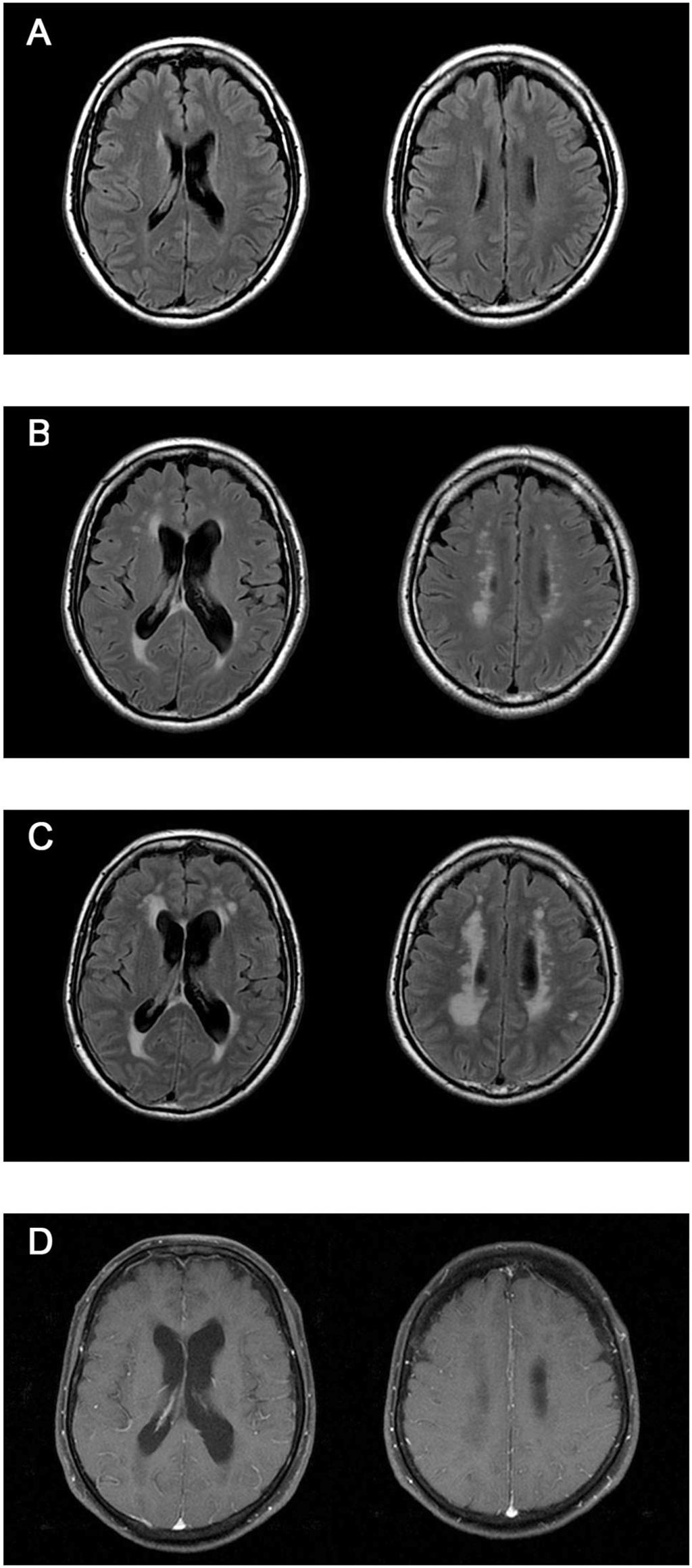
4 1. Introduction Meningeal carcinomatosis, also known as carcinomatous encephalitis or miliary brain metastasis, is a unique and rare form of metastasis observed in patients with malignant tumours [1]. Diagnosis is simple when the primary lesion of the malignant tumour is clear, and when multiple miliary lesions are confirmed via cranial contrast MRI; however, many patients exhibit atypical imaging findings [2]. In the present report, we discuss the case of a patient whose primary symptom was subacute impaired consciousness. Although meningeal carcinomatosis due to lung adenocarcinoma was ultimately diagnosed at autopsy, imaging revealed seemingly progressive leukoencephalopathy-like lesions. 2. Case A 72-year-old man presented with chief concerns of confusion and progressive gait disorder. Nine years earlier, the patient underwent cranial MRI for headache at a different hospital. However, as no abnormalities were detected, he was diagnosed with tension headache, which naturally receded (Fig. 1A). Gait disturbances began to manifest 3 months prior to his initial visit. One month prior to the initial visit, he was hospitalized due to deteriorating ambulatory function, nonsensical speech, and easy irritability. Upon admission, the patient appeared agitated and confused: He could not accurately state his own medical history and began to walk in his will. Blood counts and biochemical findings were normal. Tests for serum HIV antibodies, HTLV-1 antibodies, and syphilis were negative. Initial cerebrospinal fluid (CSF) pressure was elevated (34 cmh 2 O), while CSF protein, glucose, and cell count values were 21 mg/dl, 64 mg/dl, and 2/mm 3, respectively. Bacterial, fungal, and tuberculosis cultures were negative, as were PCR findings for herpes virus and John Cunningham (JC virus). CSF cytology was regarded as class I. High-signal areas were observed around the lateral ventricles on cranial fluid-attenuated inversion recovery (FLAIR) images (Fig. 1C). However, no such lesions were observed on gadolinium-enhanced images (Fig. 1D). Enlargement of the bilateral cerebral ventricles was observed, relative to the cranial MRI obtained 9 years
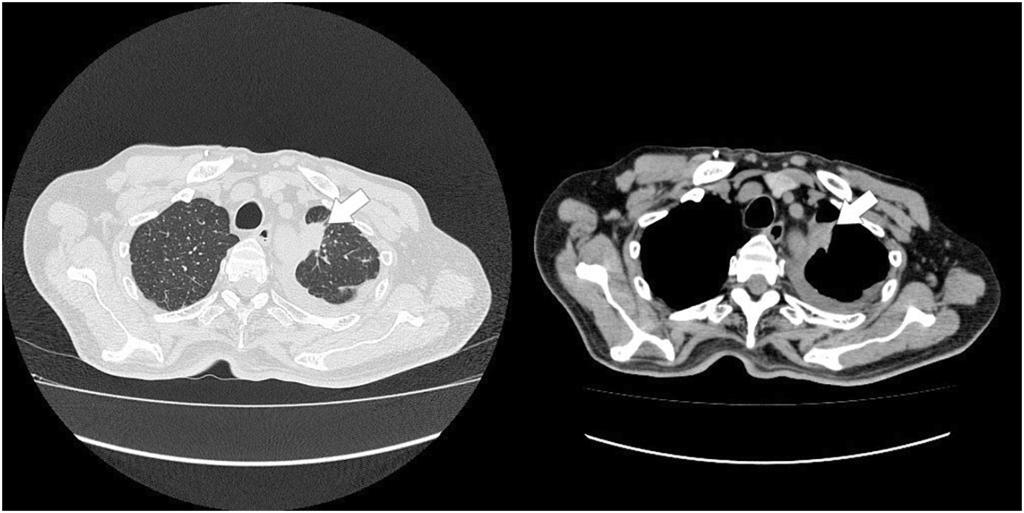
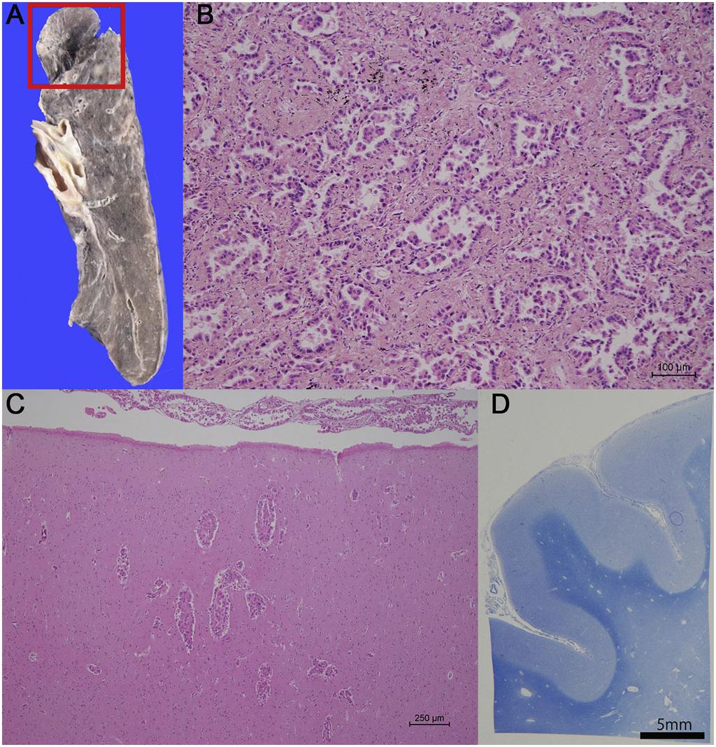
5 earlier (Fig. 1A and C). Relative to the cranial MRI obtained 3 months prior to admission, the patient s most recent images revealed enlargement of the high-intensity lesions surrounding the lateral ventricles (Fig. 1B and C). The patient was diagnosed with hydrocephalus due to bilateral lateral ventricle enlargement (Fig. 1C) and increased CSF pressure. Although he underwent ventriculoperitoneal shunt surgery 2 weeks later, his symptoms did not improve, and his overall physical condition had deteriorated. Whole-body CT findings were suggestive of lung adenocarcinoma in the S1 area of the left lung (Fig. 2A) Due to the patient s poor physical status, further testing of the lung adenocarcinoma could not be conducted. Paraneoplastic syndrome was suspected. The patient was treated with three courses of high-dose intravenous methylprednisolone. However, his neurological symptoms did not improve, and he died 2 months after admission. Autopsy revealed a nodular lesion in the left upper lung (Fig. 3A), and lung adenocarcinoma was confirmed via light microscopy (Fig. 3B). Brain weight was 1,410 g, and the cerebral pia mater was clouded. We also observed infiltration of cancer cells into the pia mater and Virchow-Robin space, along with partial microscopic metastasis into the cortex (Fig. 3C). Decreased myelin staining and axonal loss were observed in the white matter, although no infiltration of cancer cells had occurred (Fig. 3D). Based on these findings, the patient was diagnosed with meningeal carcinomatosis due to lung adenocarcinoma. 3. Discussion Contrast MRI is useful for diagnostic imaging in patients with meningeal carcinomatosis. Typically, multiple lesions are observed on T2-weighted images, and gadolinium-enhanced images of the affected regions are used to confirm the diagnosis [2]. However, in patients with atypical imaging findings, diagnosis becomes extremely difficult. Previous reports have described cases in which simple cranial MRI revealed no abnormalities, despite the presence of multiple metastatic lesions on contrast MRI [3]. Conversely, other reports have described cases in which abnormalities were observed on simple T2-weighted images only [4]. Moreover, some reports have demonstrated that abnormalities need not be present on cranial MRI, in which case diagnoses can only be verified via autopsy [5].
6 Enlargement of the white matter lesions surrounding the lateral ventricles was considered to have occurred due to hydrocephalus, as the autopsy did not reveal direct infiltration of the lung adenocarcinoma into the white matter (Fig. 3D). Indeed, previous reports have indicated that both enlargement of the lateral ventricles and high T2 and FLAIR signals in the white matter surrounding the lateral ventricles can be observed on cranial MRI in patients with hydrocephalus [6]. In the present case, the patient was ultimately diagnosed with meningeal carcinomatosis due to lung adenocarcinoma upon autopsy. Cranial MR images were obtained prior to onset, enabling us to observe increases in the extent of white matter lesions around the ventricles over time (Fig. 1). Notably, no previous reports have described leukoencephalopathy-like imaging findings in a patient with meningeal carcinomatosis. As such, this case is of critical instructional value for oncologists and other clinicians. Conflicts of interest The authors declare that there is no conflict of interest. Acknowledgments We would like to thank Editage ( for English language editing.
7 References [1] Madow L, Alpers BJ. Encephalitic form of metastatic carcinoma. AMA Arch Neurol Psychiatry 1951;65: [2] Iguchi Y, Mano K, Goto Y, Nakano T, Nomura F, Shimokata T, et al. Miliary brain metastases from adenocarcinoma of the lung: MR imaging findings with clinical and post-mortem histopathologic correlation. Neuroradiology 2007;49: [3] Fukuda Y, Homma T, Kohga H, Uki J, Shisa H. A lung cancer case with numerous calcified metastatic nodules of the brain. Neuroradiology 1988;30: [4] Nemzek W, Poirier V, Salamat MS, Yu T. Carcinomatous encephalitis (miliary metastases): lack of contrast enhancement. Am J Neuroradiol 1993;14: [5] Nakamura H, Toyama M, Uezu K, Nakamoto A, Toda T, Saito A. Case 1. Lung cancer with miliary brain metastases undetected by imaging studies. J Clin Oncol 2001;19: [6] Zimmerman RD, Fleming CA, Lee BC, Saint-Louis LA, Deck MD. Periventriculars hyperintensity as seen by magnetic resonance: prevalence and significance. Am J Roentgenol 1986;146:
8 Fig. 1 Cranial MRI scans at different time points. A. No major abnormalities were observed on fluid-attenuated inversion recovery (FLAIR) images obtained 9 years prior to admission. B. Three months prior to admission, hyperintense lesions appeared in the region surrounding the lateral ventricles. C. Enlargement of the lesions surrounding the cerebral ventricles was observed at the time of the patient s admission, along with increases in ventricle size. D. No abnormalities were detected on contrast-enhanced MR images. Fig. 2 Chest CT after several days of hospitalization revealed a tumour shadow in the left S1 region (arrow), as well as accumulation of pleural fluid. Fig. 3 Pathological findings. A mm nodular lesion with irregular edges was observed directly below the pleura in left upper lobe (square). B. Light microscopy revealed an adenocarcinoma displaying papillary and acinar proliferation. C. Infiltration of tumour cells into the pia mater and Virchow-Robin spaces. Microscopic metastases in the cortex (haematoxylin & eosin staining). D. Decreased staining intensity of the pia mater in the white matter (Klüver-Barrera staining).
9 Highlights Meningeal carcinomatosis is often associated with atypical imaging findings. Progressive leukoencephalopathy-like imaging findings were observed in our patient Autopsy revealed meningeal carcinomatosis due to lung adenocarcinoma Similar imaging findings have yet to be reported for meningeal carcinomatosis
10 Figure 1
11 Figure 2
12 Figure 3
S (18) doi: /j.ensci Reference: ENSCI 122
 doi: /j.ensci Reference: ENSCI 122") Accepted Manuscript A case of cerebellar ataxia associated with VZV infection Hirofumi Matsuyama, Takekazu Ohi PII: S2405-6502(18)30012-1 DOI: doi:10.1016/j.ensci.2018.04.003 Reference: ENSCI 122 To appear
Accepted Manuscript A case of cerebellar ataxia associated with VZV infection Hirofumi Matsuyama, Takekazu Ohi PII: S2405-6502(18)30012-1 DOI: doi:10.1016/j.ensci.2018.04.003 Reference: ENSCI 122 To appear
Accepted Manuscript. Comparison of costs and outcomes of patients presenting with a rare brainstem syndrome. Devin E. Prior, Vijay Renga
 Accepted Manuscript Comparison of costs and outcomes of patients presenting with a rare brainstem syndrome Devin E. Prior, Vijay Renga PII: S2405-6502(18)30036-4 DOI: https://doi.org/10.1016/j.ensci.2018.11.004
Accepted Manuscript. Pituitary metastasis of breast cancer mimicking IgG4-related hypophysitis
 Accepted Manuscript Pituitary metastasis of breast cancer mimicking IgG4-related hypophysitis Kanako Nose, Toshiyasu Ogata, Jun Tsugawa, Tooru Inoue, Kazuki Nabeshima, Yoshio Tsuboi PII: S2405-6502(18)30046-7
doi: /j.jns
 doi: 10.1016/j.jns.2014.05.055 Title: Sixth nerve palsy associated with obstruction in Dorello s canal, accompanied by nodular type muscular sarcoidosis Author: Ayako Shioya 1), Hiroshi Takuma 1), Masanari
Leptomeningeal metastasis: management and guidelines. Emilie Le Rhun Lille, FR Zurich, CH
 Leptomeningeal metastasis: management and guidelines Emilie Le Rhun Lille, FR Zurich, CH Definition of LM LM is defined as the spread of tumor cells within the leptomeninges and the subarachnoid space
Neuroradiology of AIDS
 Neuroradiology of AIDS Frank Minja,, HMS IV Gillian Lieberman MD September 2002 AIDS 90% of HIV patients have CNS involvement 1 10% of AIDS patients present first with neurological symptoms 2 73-80% of
Gefitinib and High-Dose Fractionated Radiotherapy for Carcinomatous Encephalitis from Non-Small Cell Lung Carcinoma
 Gefitinib and High-Dose Fractionated Radiotherapy for Carcinomatous Encephalitis from Non-Small Cell Lung Carcinoma The Harvard community has made this article openly available. Please share how this access
Title: Painless jaundice as an initial presentation of lung adenocarcinoma
 Title: Painless jaundice as an initial presentation of lung adenocarcinoma Authors: Irene Andaluz García, Irene González Partida, Javier Lucas Ramos, Jorge Yebra Carmona DOI: 10.17235/reed.2018.5587/2018
Meningeal thickening in MRI: from signs to etiologies
 Meningeal thickening in MRI: from signs to etiologies Poster No.: C-1979 Congress: ECR 2016 Type: Educational Exhibit Authors: A. Hssine, N. Mallat, M. Limeme, H. Zaghouani, S. Majdoub, H. Amara, D. Bakir,
Hiroyuki Hanakawa, Nobuya Monden, Kaori Hashimoto, Aiko Oka, Isao Nozaki, Norihiro Teramoto, Susumu Kawamura
 Accepted Manuscript Radiation-induced laryngeal angiosarcoma: Case report Hiroyuki Hanakawa, Nobuya Monden, Kaori Hashimoto, Aiko Oka, Isao Nozaki, Norihiro Teramoto, Susumu Kawamura PII: S2468-5488(18)30005-5
Accepted Manuscript. Cerebral fat embolism syndrome in sickle cell disease without evidence of shunt
 Accepted Manuscript Cerebral fat embolism syndrome in sickle cell disease without evidence of shunt Cody L. Nathan, Whitley W. Aamodt, Tanuja Yalamarti, Calli Dogon, Paul Kinniry PII: S2405-6502(18)30044-3
Central Nervous System Immune Reconstitution Disease: Pathology
 Central Nervous System Immune Reconstitution Disease: Pathology F.Gray, H.Adle-Biassette, F.Héran, G. Pialoux, A.Moulignier, APHP Hôpital Lariboisière Université Paris VII Introduction of HAART, which
Primary Central Nervous System Lymphoma with Lateral Ventricle Involvement
 The Open Medical Imaging Journal, 2012, 6, 103-107 103 Open Access Primary Central Nervous System Lymphoma with Lateral Ventricle Involvement Yumi Oie 1,*, Kazuhiro Murayama 1, Shinya Nagahisa 2, Masato
Hydrocephalus 1/16/2015. Hydrocephalus. Functions of Cerebrospinal fluid (CSF) Flow of CSF
 Flow of CSF") Hydrocephalus Hydrocephalus Ruth Arms, MSN, CNS-BC, SCRN Hydrocephalus is the buildup of fluid in the cavities (ventricles) deep within the brain. The excess fluid increases the size of the ventricles
Hydrocephalus Hydrocephalus Ruth Arms, MSN, CNS-BC, SCRN Hydrocephalus is the buildup of fluid in the cavities (ventricles) deep within the brain. The excess fluid increases the size of the ventricles
Chest Radiology Interpretation: Findings of Tuberculosis
 Chest Radiology Interpretation: Findings of Tuberculosis Get out your laptops, smart phones or other devices pollev.com/chestradiology Case #1 1 Plombage Pneumonia Cancer 2 Reading the TB CXR Be systematic!
Human Herpes Virus-6 Limbic Encephalitis
 Case Studies [1] March 19, 2013 Case history: A 32-year-old Caucasian female with newly diagnosed acute myeloid leukemia (AML) was treated with induction chemotherapy and attained complete remission. She
Case Report. Herpes simplex virus encephalitis presenting as frontal lobe hemorrhage
 1 Case Report Herpes simplex virus encephalitis presenting as frontal lobe hemorrhage Authors: Shila, MD, *Jessica Erfan, MPAS, PA-C, Ray Bogitch, MD, Jefferson T. Miley, MD Department of Neurology, Dell
Early View Article: Online published version of an accepted article before publication in the final form.
 : Online published version of an accepted article before publication in the final form. Journal Name: International Journal of Case Reports and Images (IJCRI) Type of Article: Clinical Images Title: Urinary
Case 7391 Intraventricular Lesion
 Case 7391 Intraventricular Lesion Bastos Lima P1, Marques C1, Cabrita F2, Barbosa M2, Rebelo O3, Rio F1. 1Neuroradiology, 2Neurosurgery, 3Neuropathology, Coimbra University Hospitals, Portugal. University
1 Maiser. 5-Fluorouracil (5-FU) Induced Acute Toxic Leukoencephalopathy. Samuel Maiser, MD. Department of Neurology, University of Minnesota
 Induced Acute Toxic Leukoencephalopathy. Samuel Maiser, MD. Department of Neurology, University of Minnesota") 1 Maiser 5-Fluorouracil (5-FU) Induced Acute Toxic Leukoencephalopathy Samuel Maiser, MD Department of Neurology, University of Minnesota Case This is a 57-year-old right-handed male with a history of
1 Maiser 5-Fluorouracil (5-FU) Induced Acute Toxic Leukoencephalopathy Samuel Maiser, MD Department of Neurology, University of Minnesota Case This is a 57-year-old right-handed male with a history of
LOSS OF CONSCIOUSNESS & ASSESSMENT. Sheba Medical Center Acute Medicine Department MATTHEW WRIGHT
 LOSS OF CONSCIOUSNESS & ASSESSMENT Sheba Medical Center Acute Medicine Department MATTHEW WRIGHT OUTLINE Causes Head Injury Clinical Features Complications Rapid Assessment Glasgow Coma Scale Classification
Menigitidis. Dr Rodney Itaki Lecturer Anatomical Pathology Discipline
 Menigitidis Dr Rodney Itaki Lecturer Anatomical Pathology Discipline University of Papua New Guinea Division of Pathology School of Medicine & Health Sciences Review Normal Microanatomy Image Ref: www.histology-world.com
Scottish Surveillance of Healthcare Associated Infection Programme (SSHAIP) Health Protection Scotland (HPS) SSI Surveillance Protocol 7th Edition
 Health Protection Scotland (HPS) SSI Surveillance Protocol 7th Edition") 1 Contents Female reproductive system operations (Abdominal hysterectomy and Caesarean section)... 3 Intra-abdominal infections... 3 Endometritis... 4 Other infections of the female reproductive tract...
1 Contents Female reproductive system operations (Abdominal hysterectomy and Caesarean section)... 3 Intra-abdominal infections... 3 Endometritis... 4 Other infections of the female reproductive tract...
Title: unusual case report of inflammatory. fibrous polyps in the upper gastrointestinal tract. Authors: Baifang Wang, Guoqing Xiang, Jia Zhu
 Title: An unusual case report of inflammatory fibrous polyps in the upper gastrointestinal tract Authors: Baifang Wang, Guoqing Xiang, Jia Zhu DOI: 10.17235/reed.2018.5734/2018 Link: PubMed (Epub ahead
Comment on Association of bullous pemphigoid with malignancy: A systematic review and meta-analysis
 Accepted Manuscript Comment on Association of bullous pemphigoid with malignancy: A systematic review and meta-analysis Maglie Roberto, MD, Antiga Emiliano, MD, PhD, Caproni Marzia, MD, PhD PII: S0190-9622(17)32812-8
Accepted Manuscript. Classical features of Zollinger-Ellison syndrome, in images. Ali Alshati, MD, Toufic Kachaamy, MD
 Accepted Manuscript Classical features of Zollinger-Ellison syndrome, in images Ali Alshati, MD, Toufic Kachaamy, MD PII: S0016-5107(19)30069-0 DOI: https://doi.org/10.1016/j.gie.2019.01.026 Reference:
Accepted Manuscript. Preoperative CEA in Patients with Colorectal Metastases Matters. Benny Weksler, MBA, MD
 Accepted Manuscript Preoperative CEA in Patients with Colorectal Metastases Matters Benny Weksler, MBA, MD PII: S0022-5223(19)30068-6 DOI: https://doi.org/10.1016/j.jtcvs.2019.01.016 Reference: YMTC 14019
Case Report Intracranial Capillary Hemangioma in the Posterior Fossa of an Adult Male
 Case Reports in Radiology Volume 2016, Article ID 6434623, 4 pages http://dx.doi.org/10.1155/2016/6434623 Case Report Intracranial Capillary Hemangioma in the Posterior Fossa of an Adult Male Jordan Nepute,
Five Most Common Problems in Surgical Neuropathology
 Five Most Common Problems in Surgical Neuropathology If the brain were so simple that we could understand it, we would be so simple that we couldn t Emerson Pugh What is your greatest difficulty in neuropathology?
Interstitial syndrome
 Interstitial syndrome Ground-glass attenuation Miliary and nodular images linear images Etienne Leroy Terquem Pierre L Her SPI / ISP Soutien Pneumologique International / International Support for Pulmonology
Accepted Manuscript. Editorial. Responsive neurostimulation for epilepsy: more than stimulation. Jayant N. Acharya
 Accepted Manuscript Editorial Responsive neurostimulation for epilepsy: more than stimulation Jayant N. Acharya PII: S2467-981X(18)30022-2 DOI: https://doi.org/10.1016/j.cnp.2018.06.002 Reference: CNP
A challenging neurological complication in a young HIV-infected woman
 A challenging neurological complication in a young HIV-infected woman Ianache Irina-Cristiana Vi tor Ba es Clini al Hospital for Infectious and Tropical Diseases Bucharest - HIV/AIDS department Assessment
The central nervous system
 Sectc.qxd 29/06/99 09:42 Page 81 Section C The central nervous system CNS haemorrhage Subarachnoid haemorrhage Cerebral infarction Brain atrophy Ring enhancing lesions MRI of the pituitary Multiple sclerosis
Intrahepatic Sarcomatoid Cholangiocarcinoma with Portal Vein Thrombosis: A Case Report 1
 Intrahepatic Sarcomatoid Cholangiocarcinoma with Portal Vein Thrombosis: A Case Report 1 Jae-Hoon Lim, M.D., Jin Woong Kim, M.D., Suk Hee Heo, M.D., Yong Yeon Jeong, M.D., Heoung Keun Kang, M.D. A 53-year-old
MRI imaging in meningeal diseases
 Original article MRI imaging in meningeal diseases 1Dr. Narendrakumar M Shah, 2 Dr Vaishali D M 1Associate professor, Department of Radiodiagnosis, SDM Medical college, Dharwad 2Consultant radiologist,
Meningitis. A fact sheet for patients and carers
 A fact sheet for patients and carers Meningitis This fact sheet provides information on meningitis. Our fact sheets are designed as general introductions to each subject and are intended to be concise.
Accepted Manuscript. Ultrasound and adnexal pathology: what is the evidence? Wouter Froyman, MD, Lil Valentin, MD, PhD, Dirk Timmerman, MD, PhD
 Accepted Manuscript Ultrasound and adnexal pathology: what is the evidence? Wouter Froyman, MD, Lil Valentin, MD, PhD, Dirk Timmerman, MD, PhD PII: S0002-9378(16)30461-6 DOI: 10.1016/j.ajog.2016.07.027
IMAGING OF INTRACRANIAL INFECTIONS
 IMAGING OF INTRACRANIAL INFECTIONS Dr Carolina Kachramanoglou LYSHOLM DEPARTMENT OF NEURORADIOLOGY NATIONAL HOSPITAL FOR NEUROLOGY AND NEUROSURGERY Plan Introduce MR sequences that are useful in the diagnosis
Diffusion-Weighted and Conventional MR Imaging Findings of Neuroaxonal Dystrophy
 AJNR Am J Neuroradiol 25:1269 1273, August 2004 Diffusion-Weighted and Conventional MR Imaging Findings of Neuroaxonal Dystrophy R. Nuri Sener BACKGROUND AND PURPOSE: Neuroaxonal dystrophy is a rare progressive
Case Report Three Cases of Neoplastic Meningitis Initially Diagnosed with Infectious Meningitis in Emergency Department
 Case Reports in Emergency Medicine Volume 2013, Article ID 561475, 4 pages http://dx.doi.org/10.1155/2013/561475 Case Report Three Cases of Neoplastic Meningitis Initially Diagnosed with Infectious Meningitis
Case Report. Introduction
 doi: 10.5761/atcs.cr.17-00135 Case Report A Case of Paraneoplastic Limbic Encephalitis in a Patient with Invasive Thymoma with Anti-Glutamate Receptor Antibody-Positive Cerebrospinal Fluid: A Case Report
Multifocal inflammatory leukoencephalopathy induced by accidental consumption of levamisole: A case report
 Iranian Journal of Neurology Case Report Ir J neurol 2012; 11(2): 65-69 Multifocal inflammatory leukoencephalopathy induced by accidental consumption of levamisole: A case report Received: 25 Oct 2011
Case Report pissn / eissn J Korean Soc Radiol 2015;73(5):
:") Case Report pissn 1738-2637 / eissn 2288-2928 http://dx.doi.org/10.3348/jksr.2015.73.5.337 Uncommon Manifestations of Scrub Typhus Encephalitis in Two Cases: Clinical and Magnetic Resonance Imaging Findings
Case Report pissn 1738-2637 / eissn 2288-2928 http://dx.doi.org/10.3348/jksr.2015.73.5.337 Uncommon Manifestations of Scrub Typhus Encephalitis in Two Cases: Clinical and Magnetic Resonance Imaging Findings
Paraneoplastic limbic encephalitis in a patient with rectal adenocarcinoma: A rare entity
 Hooda et al. 6 CASE REPORT PEER REVIEWED OPEN ACCESS Paraneoplastic limbic encephalitis in a patient with rectal adenocarcinoma: A rare entity Kusum Hooda, Nishant Gupta, Charu Chanana, Pranav Sharma,
MRI Findings from a Case of Fulminating Adult-onset Measles Encephalitis
 CASE REPORT MRI Findings from a Case of Fulminating Adult-onset Measles Encephalitis Kazutaka Jin 1, Shigeru Sato 2, Ryuji Saito 2, Ayumu Ohnuma 2, Hiroshi Nomura 2 and Yasuto Itoyama 3 Abstract We report
MR Imaging of Leptomeningeal Metastases: Comparison of Three Sequences
 AJNR Am J Neuroradiol 23:817 821, May 2002 MR Imaging of Leptomeningeal Metastases: Comparison of Three Sequences Sanjay K. Singh, Norman E. Leeds, and Lawrence E. Ginsberg BACKGROUND AND PURPOSE: Recent
Paraneoplastic limbic encephalitis in Hodgkin s Lymphoma. Marc Wein, HMS III Dr. Gillian Lieberman, MD September 17, 2007
 Paraneoplastic limbic encephalitis in Hodgkin s Lymphoma Marc Wein, HMS III Dr. Gillian Lieberman, MD September 17, 2007 Our patient s CC: Disorientation and insomnia HPI: 27 year old man AW with no PMH
Brain Pain Infections of the CNS
 FRIDAY, OCTOBER 28, 2016 Brain Pain Infections of the CNS Suyash Mohan MD, PDCC Assistant Professor of Radiology & Neurosurgery Division of Neuroradiology, Department of Radiology Perelman School of Medicine
The Paraneoplastic Limbic Encephalitis: MRI Characterization of a Deceiving Neurological Disorder
 Journal of the Egyptian Nat. Cancer Inst., Vol. 20, No. 4, December: 403-409, 2008 The Paraneoplastic Limbic Encephalitis: MRI Characterization of a Deceiving Neurological Disorder ALAA M. ELORABY, M.D.
Grand-round meeting for Dementia - A patient with rapidly progressing dementia. Dr. Ho Ka Shing Tuen Mun Hospital
 Grand-round meeting for Dementia - A patient with rapidly progressing dementia Dr. Ho Ka Shing Tuen Mun Hospital Mr. Wong, 60 years old Security guard Ex-smoker for over 20 years, non-drinker Premorbid
Role of MRI in acute disseminated encephalomyelitis
 Original Research Article Role of MRI in acute disseminated encephalomyelitis Shashvat Modiya 1*, Jayesh Shah 2, C. Raychaudhuri 3 1 1 st year resident, 2 Associate Professor, 3 HOD and Professor Department
Accepted Manuscript. Will the fourth dimension guide us toward the perfect Norwood arch reconstruction? Minoo N. Kavarana, MD, FACS
 Accepted Manuscript Will the fourth dimension guide us toward the perfect Norwood arch reconstruction? Minoo N. Kavarana, MD, FACS PII: S0022-5223(19)30714-7 DOI: https://doi.org/10.1016/j.jtcvs.2019.03.040
Pleural effusion as an initial manifestation in a patient with primary pulmonary monoclonal B-cell lymphocyte proliferative disease
 Du et al. Respiratory Research (2018) 19:247 https://doi.org/10.1186/s12931-018-0941-6 LETTER TO THE EDITOR Pleural effusion as an initial manifestation in a patient with primary pulmonary monoclonal B-cell
pulmonary metastasis 80EE4727C6037E7F69A9981B7E55A238 Pulmonary Metastasis 1 / 6
 Pulmonary Metastasis 1 / 6 2 / 6 3 / 6 Pulmonary Metastasis Metastatic tumors in the lungs are cancers that developed at other places in the body (or other parts of the lungs). They then spread through
Medulloblastoma with atypical dynamic imaging changes: A case report with literature review
 Accepted Manuscript Medulloblastoma with atypical dynamic imaging changes: A case report with literature review Shuang-Shuang Song, Jian-Hong Wang, Wei-Wei Fu, Ying Li, Qing-Lan Sui, Xue-Jun Liu PII: S1878-8750(17)30950-6
Case Report A Case of Primary Submandibular Gland Oncocytic Carcinoma
 Case Reports in Otolaryngology Volume 2013, Article ID 384238, 4 pages http://dx.doi.org/10.1155/2013/384238 Case Report A Case of Primary Submandibular Gland Oncocytic Carcinoma Kunihiko Tokashiki, Kiyoaki
Benign brain lesions
 Benign brain lesions Diagnostic and Interventional Radiology Hung-Wen Kao Department of Radiology, Tri-Service General Hospital, National Defense Medical Center Computed tomography Hounsfield unit (HU)
A common case definition for PML
 A common case definition for PML Transatlantic workshop: Drug-related Progressive Multifocal Leukoencephalopathy (PML) 25.-26.7.2011, EMA, London Paul-Ehrlich-Institut Dr. Dirk Mentzer, MD Paul-Ehrlich-Str.
Opportunistic infections in the era of cart, still a problem in resource-limited settings
 Opportunistic infections in the era of cart, still a problem in resource-limited settings Cristiana Oprea Victor Babes Clinical Hospital for Infectious and Tropical Diseases, Bucharest, Romania Assessment
Endosaccular aneurysm occlusion with Guglielmi detachable coils for obstructive hydrocephalus caused by a large basilar tip aneurysm Case report
 Neurosurg Focus 7 (4):Article 5, 1999 Endosaccular aneurysm occlusion with Guglielmi detachable coils for obstructive hydrocephalus caused by a large basilar tip aneurysm Case report Akira Watanabe, M.D.,
Detection of Leptomeningeal CNS Metastases in Children
 Detection of Leptomeningeal CNS Metastases in Children Noah D. Sabin, M.D. Julie H. Harreld M.D. Kathleen J. Helton M.D. Zoltan Patay M.D., Ph.D. St. Jude Children s Research Hospital Memphis, TN Leptomeningeal
General Identification. Name: 江 X X Age: 29 y/o Gender: Male Height:172cm, Weight: 65kg Date of admission:95/09/27
 General Identification Name: 江 X X Age: 29 y/o Gender: Male Height:172cm, Weight: 65kg Date of admission:95/09/27 Chief Complaint Sudden onset of seizure for several minutes Present illness This 29-year
Accepted Manuscript. Unexpected high incidence of hepatocellular carcinoma in patients with hepatitis C in the era of DAAs: too alarming?
 Accepted Manuscript Unexpected high incidence of hepatocellular carcinoma in patients with hepatitis C in the era of DAAs: too alarming? Qing-Lei Zeng, Zhi-Qin Li, Hong-Xia Liang, Guang-Hua Xu, Chun-Xia
Original Article. Emergency Department Evaluation of Ventricular Shunt Malfunction. Is the Shunt Series Really Necessary? Raymond Pitetti, MD, MPH
 Original Article Emergency Department Evaluation of Ventricular Shunt Malfunction Is the Shunt Series Really Necessary? Raymond Pitetti, MD, MPH Objective: The malfunction of a ventricular shunt is one
Case Report High Grade Glioma Mimicking Voltage Gated Potassium Channel Complex Associated Antibody Limbic Encephalitis
 Case Reports in Neurological Medicine, Article ID 458790, 4 pages http://dx.doi.org/10.1155/2014/458790 Case Report High Grade Glioma Mimicking Voltage Gated Potassium Channel Complex Associated Antibody
MULTI-SYSTEM SARCOIDOSIS CAUSING PANHYPOPITUITARISM: RAPID IMPROVEMENT WITH CORTICOSTEROID THERAPY Rashid Mahboob, MD; Ali A.
 ENDOCRINE PRACTICE Rapid Electronic Article in Press Rapid Electronic Articles in Press are preprinted manuscripts that have been reviewed and accepted for publication, but have yet to be edited, typeset
May He Rest in Peace
 May He Rest in Peace Neurologic Complications of AIDS Medical Knowledge Fiesta 2012 Paul K. King MD pkingmd@yahoo.com Objectives definition of HIV/AIDS what are the neurologic complications of AIDS how
General History. 林陳 珠 Female 69 years old 住院期間 : ~ Chief Complaint : sudden loss of conscious 5 minutes in the morning.
 General History 林陳 珠 Female 69 years old 住院期間 : 93.5.8~93.5.15 Chief Complaint : sudden loss of conscious for 2-52 5 minutes in the morning. General History DM under regular medical control for 10 years.
The predicament of cancer presenting during pregnancy
 The predicament of cancer presenting during pregnancy Poster No.: C-3001 Congress: ECR 2010 Type: Educational Exhibit Topic: Radiographers Authors: D. O'Mahony, G. Murphy, G. Wilson, M. T. Keogan; Dublin/IE
Accepted Manuscript. The Aorta in Repaired Tetralogy of Fallot: A Potential Source of Late Danger? Joseph B. Clark, MD
 Accepted Manuscript The Aorta in Repaired Tetralogy of Fallot: A Potential Source of Late Danger? Joseph B. Clark, MD PII: S0022-5223(18)32548-0 DOI: 10.1016/j.jtcvs.2018.09.044 Reference: YMTC 13498 To
Helpful Information for evaluation of new neurological symptoms in patients receiving TYSABRI
 Helpful Information for evaluation of new neurological symptoms in patients receiving TYSABRI This information is provided as an educational resource for healthcare providers and should be considered current
Prevalence of venous sinus stenosis in Pseudotumor cerebri(ptc) using digital subtraction angiography (DSA)
 using digital subtraction angiography (DSA)") Prevalence of venous sinus stenosis in Pseudotumor cerebri(ptc) using digital subtraction angiography (DSA) Dr.Mohamed hamdy ibrahim MBBC,MSc,MD, PhD Neurology Degree Kings lake university (USA). Fellow
Prevalence of venous sinus stenosis in Pseudotumor cerebri(ptc) using digital subtraction angiography (DSA) Dr.Mohamed hamdy ibrahim MBBC,MSc,MD, PhD Neurology Degree Kings lake university (USA). Fellow
Case Scenario 1. The patient agreed to a CT guided biopsy of the left upper lobe mass. This was performed and confirmed non-small cell carcinoma.
 Case Scenario 1 An 89 year old male patient presented with a progressive cough for approximately six weeks for which he received approximately three rounds of antibiotic therapy without response. A chest
Occult Cerebrospinal Fluid Fistula between Ventricle and Extra-Ventricular Position of the Ventriculoperitoneal Shunt Tip
 197 Occult Cerebrospinal Fluid Fistula between Ventricle and Extra-Ventricular Position of the Ventriculoperitoneal Shunt Tip Ching-Yi Lee 1, Chieh-Tsai Wu 1, Kuang-Lin Lin 2, Hsun-Hui Hsu 3 Abstract-
Management of carcinomatous meningitis in a patient with advanced lung adenocarcinoma
 Management of carcinomatous meningitis in a patient with advanced lung adenocarcinoma Clinical Case Presentation Emilie Le Rhun Centre Hospitalier Régional et Universitaire (CHRU) de Lille Neurochirurgie
Does the lung nodule look aggressive enough to warrant a more extensive operation?
 Accepted Manuscript Does the lung nodule look aggressive enough to warrant a more extensive operation? Michael Kuan-Yew Hsin, MBChB, MA, FRCS(CTh), David Chi-Leung Lam, MD, PhD, FRCP (Edin, Glasg & Lond)
MR neuroimaging of HIV infected patients : A pictorial review
 MR neuroimaging of HIV infected patients : A pictorial review Poster No.: R-0198 Congress: 2014 CSM Type: Scientific Exhibit Authors: P. F. Kwan, R. Thomas, A. Dixon; SOUTH YARRA/AU Keywords: Neuroradiology
Utility of radiologic imaging in the diagnosis and follow up of normal pressure hydrocephalus
 Utility of radiologic imaging in the diagnosis and follow up of normal pressure hydrocephalus Poster No.: C-0312 Congress: ECR 2015 Type: Scientific Exhibit Authors: C. Rodríguez, D. Marquina, A. Mir Torres,
Diagnostic Value of Contrast-Enhanced Fluid- Attenuated Inversion Recovery MR Imaging of Intracranial Metastases
 AJNR Am J Neuroradiol 25:761 765, May 2004 Diagnostic Value of Contrast-Enhanced Fluid- Attenuated Inversion Recovery MR Imaging of Intracranial Metastases Nil Ercan, Serap Gultekin, Halil Celik, Turgut
Brain abscess rupturing into the lateral ventricle causing meningitis: a case report
 Brain abscess rupturing into the lateral ventricle causing meningitis: a case report Endry Martinez, and Judith Berger SBH Health System, 4422 Third Ave, Bronx, NY 10457 Key words: brain abscess, rupture
Question 1 History. Likely Diagnosis Differential. Further Investigation or Management. Requires Paediatric Surgical referral for laparotomy
 Question 1 Male newborn spilling green tinged vomit day 1 of life Imaging Abdominal X-Rays performed on 03/05/2012 Upper and lower gastrointestinal contrast studies performed on 03/05/2012 Abdominal X-Rays
Cysts Arachnoid Cyst (also called Leptomeningeal Cyst)
") Cysts This article was provided to us by David Schiff, MD, Co-Director of the Neuro-Oncology Center and Professor of Neurology, Neurosurgery, and Medicine at the University of Virginia, Charlottesville.
Cysts This article was provided to us by David Schiff, MD, Co-Director of the Neuro-Oncology Center and Professor of Neurology, Neurosurgery, and Medicine at the University of Virginia, Charlottesville.
Brain AVM with Accompanying Venous Aneurysm with Intracerebral and Intraventricular Hemorrhage
 Cronicon OPEN ACCESS EC PAEDIATRICS Case Report Brain AVM with Accompanying Venous Aneurysm with Intracerebral and Intraventricular Hemorrhage Dimitrios Panagopoulos* Neurosurgical Department, University
The Journal of Thoracic and Cardiovascular Surgery
 Accepted Manuscript Chronic type A dissection: when to operate? Francois Dagenais, MD PII: S0022-5223(18)33131-3 DOI: https://doi.org/10.1016/j.jtcvs.2018.11.032 Reference: YMTC 13781 To appear in: The
Non-Traumatic Neuro Emergencies
 Department of Radiology University of California San Diego Non-Traumatic Neuro Emergencies John R. Hesselink, M.D. Nontraumatic Neuroemergencies 1. Acute focal neurological deficit 2. Worst headache of
Transient asymptomatic white matter lesions following Epstein-Barr virus encephalitis
 Case report http://dx.doi.org/10.3345/kjp.2011.54.9.389 Korean J Pediatr 2011;54(9):389-393 Transient asymptomatic white matter lesions following Epstein-arr virus encephalitis Yoon Young Jang, MD, Kye
Dr Paul Holmes Guy s and St Thomas NHS Foundation Trust, London
 Dr Paul Holmes Guy s and St Thomas NHS Foundation Trust, London HIV and Lumbar punctures in 2018 Paul Holmes Consultant Neurologist Guy s and St Thomas Hospitals I have no competing interests Summary of
Title: Lower gastrointestinal bleeding as a form of presentation in an adult case of Abernethy syndrome
 Title: Lower gastrointestinal bleeding as a form of presentation in an adult case of Abernethy syndrome Authors: Aida Suárez Sánchez, Lorena Solar García, Carmen María García Bernardo, Alberto Miyar de
Accepted Manuscript. Radiotracer localization: Finding a nodule in the haystack. Jules Lin, MD
 Accepted Manuscript Radiotracer localization: Finding a nodule in the haystack Jules Lin, MD PII: S0022-5223(18)32260-8 DOI: 10.1016/j.jtcvs.2018.08.018 Reference: YMTC 13340 To appear in: The Journal
High Signal Intensity of the Infundibular Stalk on Fluid-Attenuated Inversion Recovery MR
 High Signal Intensity of the Infundibular Stalk on Fluid-Attenuated Inversion Recovery MR Yutaka Araki, Ryuichirou Ashikaga, Satoru Takahashi, Jun Ueda, and Osamu Ishida PURPOSE: To determine the MR imaging
Extent of lymphadenectomy for esophageal squamous cell cancer: interpreting the post-hoc analysis of a randomized trial
 Accepted Manuscript Extent of lymphadenectomy for esophageal squamous cell cancer: interpreting the post-hoc analysis of a randomized trial Vaibhav Gupta, MD PII: S0022-5223(18)33169-6 DOI: https://doi.org/10.1016/j.jtcvs.2018.11.055
A Guide to the Radiologic Evaluation of Extra-Axial Hemorrhage
 July 2013 A Guide to the Radiologic Evaluation of Extra-Axial Hemorrhage John Dickson, Harvard Medical School Year III Agenda 1. Define extra-axial hemorrhage and introduce its subtypes 2. Review coup
Accepted Article. Massive gastrointestinal pneumatosis in a patient with celiac disease and superior mesenteric artery syndrome
 Accepted Article Massive gastrointestinal pneumatosis in a patient with celiac disease and superior mesenteric artery syndrome Aleix Martínez-Pérez, Ramón Trullenque-Juan, Sandra Santarrufina-Martínez,
A Case of Pancreatic Carcinoma with Bilateral Hilar
 Shinshu Med J, 66⑵:151~155, 2018 A Case of Pancreatic Carcinoma with Bilateral Hilar 18 F-FDG and 67 Ga Hyperaccumulation Satoshi Kawakami 1 )*, Yasunari Fujinaga 1), Shin Yanagisawa 1) Masumi Kadoya 1),
Laura Tormoehlen, M.D. Neurology and EM-Toxicology Indiana University
 Laura Tormoehlen, M.D. Neurology and EM-Toxicology Indiana University Disclosures! No conflicts of interest to disclose Neuroimaging 101! Plain films! Computed tomography " Angiography " Perfusion! Magnetic
Neurology Clerkship Learning Objectives
 Neurology Clerkship Learning Objectives Clinical skills Perform a neurological screening examination of the cranial nerves, motor system, reflexes, and sensory system under the observation and guidance
ECMM Excellence Centers Quality Audit
 ECMM Excellence Centers Quality Audit Person in charge: Department: Head of Department: Laboratory is accredited according to ISO 15189 (Medical Laboratories Requirements for quality and competence) Inspected
Ischemic Stroke as the Initial Manifestation of Neurosyphilis in a Young Adult Patient Positive for Human Immunodeficiency Virus
 Ischemic Stroke as the Initial Manifestation of Neurosyphilis in a Young Adult Patient Positive for Human Immunodeficiency Virus Ayaka Numao a, Keisuke Suzuki a, Hidehiro Takekawa a, Toshiki Nakamura b,
Outline. Neuroradiology. Diffusion Imaging in. Clinical Applications of. Basics of Diffusion Imaging. Basics of Diffusion Imaging
 Clinical Applications of Diffusion Imaging in Neuroradiology No disclosures Stephen F. Kralik Assistant Professor of Radiology Indiana University School of Medicine Department of Radiology and Imaging
WHAT IS DEMENTIA? An acquired syndrome of decline in memory and other cognitive functions sufficient to affect daily life in an alert patient
 DEMENTIA WHAT IS DEMENTIA? An acquired syndrome of decline in memory and other cognitive functions sufficient to affect daily life in an alert patient Progressive and disabling Not an inherent aspect of