New Imaging Concepts in Central Nervous System Neoplasms
|
|
|
- Everett Thomas
- 5 years ago
- Views:
Transcription
1 New Imaging Concepts in Central Nervous System Neoplasms Maarten Lequin Department of Pediatric Radiology Wilhelmina Children s Hospital/University Medical Center Utrecht
2 New Imaging Concepts in Central Nervous System Neoplasms What are the new concepts? How will it change our radiological thinking? Diagnosis, imaging protocols, treatment strategies When to use? Conventional and additional advanced MR sequences

3 New Imaging Concepts in Central Nervous System Neoplasms What are the new concepts? Transcriptional profiling new brain tumor subtypes
4 New Imaging Concepts in Central Nervous System Neoplasms What are the new concepts? Transcriptional profiling new brain tumor subtypes Subtyping better understanding of tumor behaviour
5 New Imaging Concepts in Central Nervous System Neoplasms What are the new concepts? Transcriptional profiling new brain tumor subtypes Subtyping better understanding of tumor behaviour Subtyping predict better outcome and may influence treatment strategy choices
6 New Imaging Concepts in Central Nervous System Neoplasms What are the new concepts? Transcriptional profiling new brain tumor subtypes Subtyping better understanding of tumor behaviour Subtyping predict better outcome and may influence treatment strategy choices Subtyping new concept of radiological thinking
7 New Imaging Concepts in Central Nervous System Neoplasms What are the new concepts? Transcriptional profiling new brain tumor subtypes Subtyping better understanding of tumor behaviour Subtyping predict better outcome and may influence treatment strategy choices Subtyping new concept of radiological thicking Predict subtype better assess outcome and treatment strategy choices


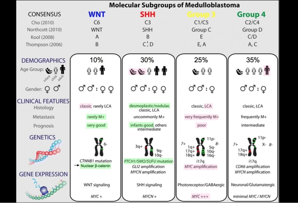
8 Molecular tumor subtypes MR image characteristics Molecular markers Targeted treatment Prognosis


9 Molecular subtypes in Brain tumors
10 From 2007 to 2016 WHO Classification of CNS tumors some major changes Formulating concept of how CNS tumor diagnoses are structured in the molecular era Major restructuring of diffuse gliomas, with incorporation of genetically defined entities Major restructuring of medulloblastomas, with incorporation of genetically defined entities Incorporation of a genetically defined ependymoma variant Major restructuring of other embryonal tumors, with incorporation of genetically defined entities and removal of the term primitive neuroectodermal tumor Novel approach distinguishing pediatric look-alikes, including designation of novel, genetically defined entity Courtesy of Dr. David Louis
 genes. Paul A.")
11 (A) Unsupervised hierarchical clustering of human 1.0 exon array expression data from 103 primary medulloblastomas using 1,450 high standard deviation (SD) genes. Paul A. Northcott et al. JCO 2011;29: by American Society of Clinical Oncology
12
13


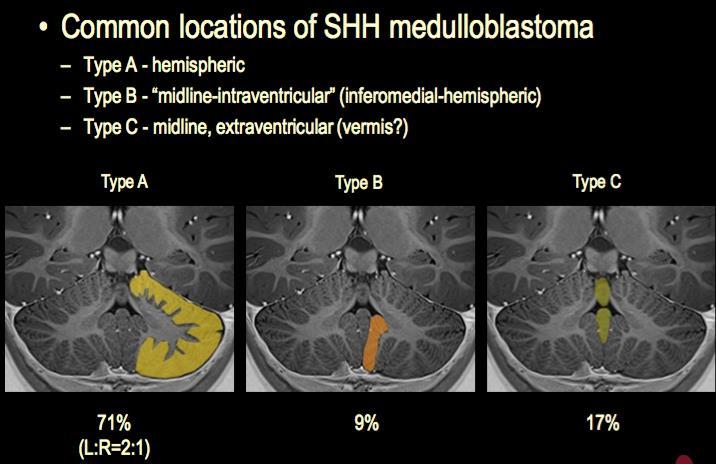
14 Putative points of origins of WNT-subgroup medulloblastoma in sagittal, transverse, and coronal planes. 69% 31% 75% 19% Z. Patay et al. AJNR Am J Neuroradiol 2015;36: by American Society of Neuroradiology
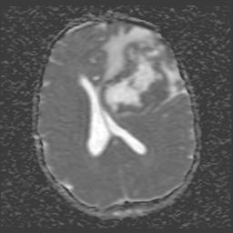
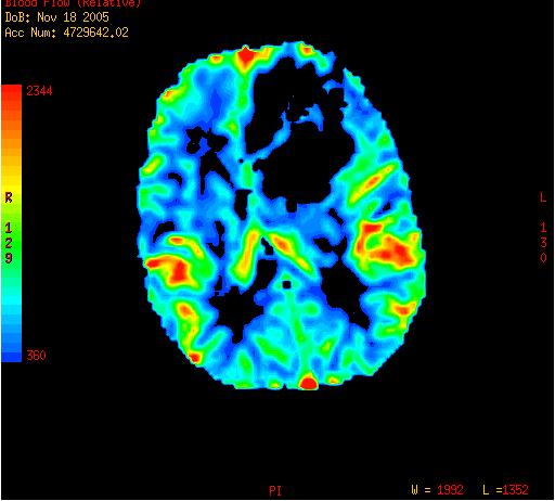
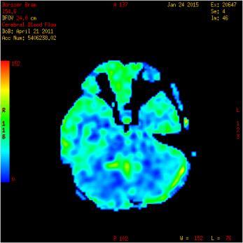
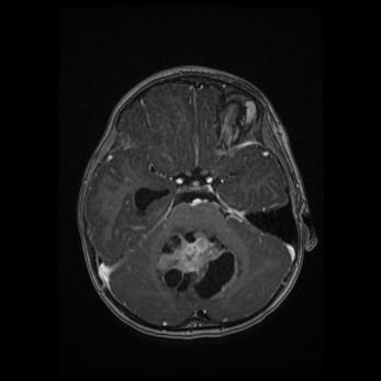
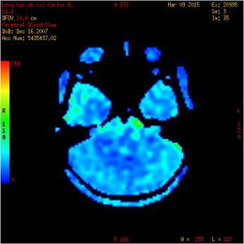
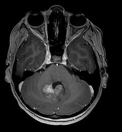
15 New Imaging Concepts in Central Nervous System Neoplasms What could be potential radiological biomarkers besides age and location? T1, T2, T1 after Gd, DWI, MRS? Or should we go for newer imaging modalities? ASL, APT, DKI?
16
17
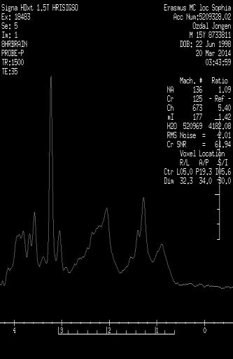
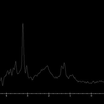
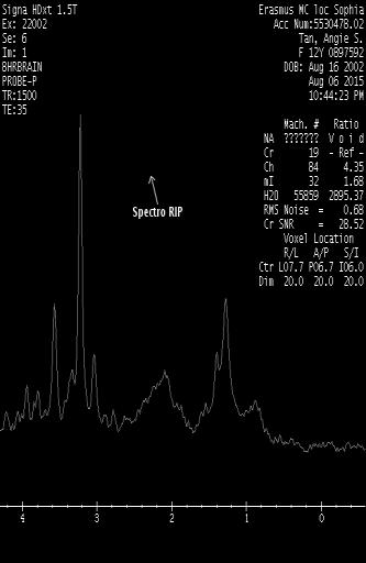
18 Medulloblastoma subgroups and MR Spectroscopy
19 Medulloblastoma subgroups and MR Spectroscopy

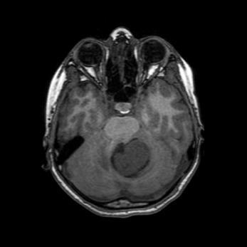
20 4y old male. Diagnosis?

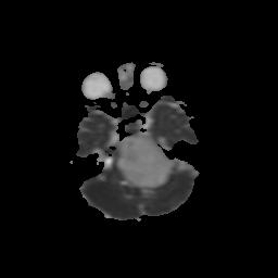
21 3y old girl Diagnosis?

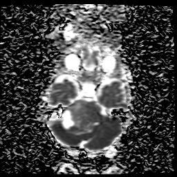
22 16m old boy Diagnosis?

23 1y old boy Diagnosis?
24 New Imaging Concepts in Central Nervous System Neoplasms When to use? Conventional and additional advanced MR sequences
25 New Imaging Concepts in Central Nervous System Neoplasms Best differential diagnosis
26 New Imaging Concepts in Central Nervous System Neoplasms Best differential diagnosis Location, age and imaging sequences
27 New Imaging Concepts in Central Nervous System Neoplasms Best differential diagnosis Location, age and imaging sequences Posterior fossa DWI for differentiation
28 New Imaging Concepts in Central Nervous System Neoplasms Best differential diagnosis Location, age and imaging sequences Posterior fossa DWI for differentiation ASL in combination with Gd enhancement (ratio) characterise brain tumors infra- and supratentorial
29 New Imaging Concepts in Central Nervous System Neoplasms Best differential diagnosis Location, age and imaging sequences Posterior fossa DWI for differentiation ASL in combination with Gd enhancement (ratio) characterise brain tumors infra- and supratentorial Medulloblastoma, use MRS to differentiate between the subgroups

30 6y old boy Diagnosis?
31 New Imaging Concepts in Central Nervous System Neoplasms Supratentorial brain tumors are less easy to characterise using conventional and advanced sequences DWI and MRS are no good predictors for tumor grading in glial tumors Still a lot of research has to be done to look for new radiological biomarkers in other common brain tumors like glial tumors
32 Another possible New Imaging Concept in Central Nervous System Neoplasms To abandon the use of Gd in neuro-oncology imaging: Recently multiple papers focus on the cumulation of Gd in the brain and their possible side effects In the future we have to look for alternative sequences which may give the same information Follow-up imaging without T1 after Gd? Arterial spin labeling (ASL) Amide Proton Transfer (APT) imaging could be an alternative? Diffusion kurtosis imaging (DKI)?


33 New Imaging Concepts in Central Nervous System Neoplasms
34 New Non-contrast Imaging Concepts in Central Nervous System Neoplasms Arterial spin labeling (ASL) No Gd contrast, measures CBF Three methods, continues, pulsed and pseudocontinues Pediatric brain tumor imaging Ratio with contrast enhancement may predict tumor grading No tumor subtyping
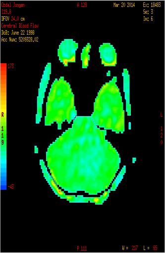
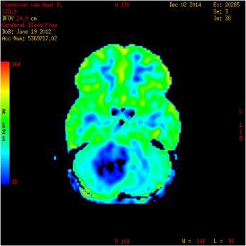
35
36 New Non-contrast Imaging Concepts in Central Nervous System Neoplasms Amide Proton Transfer (APT) imaging Type of chemical exchange dependent saturation transfer (CEST) magnetic resonance imaging (MRI), in which amide protons of endogenous mobile proteins and peptides in tissue are detected Especially useful in high grade brain tumors APT shows better diagnostic performance than diffusion- and perfusion-weighted imaging in nonenhancing gliomas Park KJ et al. Eur Radiol Feb 16. Added value of amide proton transfer imaging to conventional and perfusion MR imaging for evaluating the treatment response of newly diagnosed glioblastoma.
37 New Non-contrast Imaging Concepts in Central Nervous System Neoplasms Diffusion kurtosis imaging (DKI) Diffusion weighted method with 2 non-zero b-values More sensitive for brain tumor imaging May differentiate between low and high grade tumors


38 Courtesy to Susan Palasis Diffusion kurtosis imaging (DKI) in a 7 year old with resected large cell anaplastic medulloblastoma 2 nd post-op study 2 weeks later

39 Normalized kmean map to MNI space superimposed on T1 C+ kmean in right ROI is kmean in left ROI is Findings show definite tumor around 4 th ventricle and foramen of Luschka. Concern for tumor infiltration of right cerebellar hemisphere.
40 Summary Challenge: balancing desires and needs Stay connected Most accurate diagnosis Provide the best possible diagnosis Utilize the most accurate, cuttingedge techniques Fine-tune imaging protocol (D,FU) Incorporate the latest molecular signatures Do not disrupt current clinical diagnosis and patient management Weigh the availability and cost of novel diagnostic techniques Preserve the ability for longterm clinical, experimental and etiological correlations Courtesy of Dr. David Louis
41 New Imaging Concepts in Central Nervous System Neoplasms What are the new concepts? Molecular biology, Non Gd imaging How will it change our radiological thinking? Dedicated imaging protocols When to use? Depends on brain tumor subtyping
42 New Imaging Concepts in Central Nervous System Neoplasms Acknowledgements: Pieter Wesseling Zoltan Patay Susan Palasis Arthur Adams Jeroen Hendrikse Dik Rutgers
Amide Proton Transfer Imaging: A Novel MR Method for High-grade Brain Tumors.
 Amide Proton Transfer Imaging: A Novel MR Method for High-grade Brain Tumors. Poster No.: C-1732 Congress: ECR 2013 Type: Scientific Exhibit Authors: M. Ida, M. Ishizuka, T. Suzuki, Y. Kubo, K. Hino, S.
CNS pathology Third year medical students. Dr Heyam Awad 2018 Lecture 12: CNS tumours 2/3
 CNS pathology Third year medical students Dr Heyam Awad 2018 Lecture 12: CNS tumours 2/3 Pilocytic astrocytoma Relatively benign ( WHO grade 1) Occurs in children and young adults Mostly: in the cerebellum
Pediatric CNS Tumors. Disclosures. Acknowledgements. Introduction. Introduction. Posterior Fossa Tumors. Whitney Finke, MD
 Pediatric CNS Tumors Disclosures Whitney Finke, MD Neuroradiology Fellow PGY-6 University of Utah Health Sciences Center Salt Lake City, Utah None Acknowledgements Introduction Nicholas A. Koontz, MD Luke
Diffusion-weighted imaging and ADC mapping in the differentiation of intraventricular brain tumors
 Diffusion-weighted imaging and ADC mapping in the differentiation of intraventricular brain tumors Poster No.: C-2652 Congress: ECR 2010 Type: Educational Exhibit Topic: Neuro Authors: M. Gavrilov, T.
Case Report Atypical Presentation of Atypical Teratoid Rhabdoid Tumor in a Child
 Case Reports in Oncological Medicine Volume 2013, Article ID 815923, 4 pages http://dx.doi.org/10.1155/2013/815923 Case Report Atypical Presentation of Atypical Teratoid Rhabdoid Tumor in a Child Y. T.
Pediatric Brain Tumors: Updates in Treatment and Care
 Pediatric Brain Tumors: Updates in Treatment and Care Writer Classroom Rishi R. Lulla, MD MS Objectives Introduce the common pediatric brain tumors Discuss current treatment strategies for pediatric brain
Gliomas in the 2016 WHO Classification of CNS Tumors
 Gliomas in the 2016 WHO Classification of CNS Tumors Hindi N Al-Hindi, MD, FCAP Consultant Neuropathologist and Head Section of Anatomic Pathology Department of Pathology and Laboratory Medicine King Faisal
Emerging contrasts at ultrahigh fields" A. Dean Sherry
 Emerging contrasts at ultrahigh fields" A. Dean Sherry Advanced Imaging Research Center Department of Radiology UT Southwestern Medical Center Department of Chemistry & Biochemistry, UT Dallas ADVANCED
11/6/2013. Vanderbilt University Institute of Imaging Science (VUIIS) 7 Tesla Vascular Imaging. Evaluating stroke risk
 7 Tesla Vascular Imaging. Evaluating stroke risk") Vanderbilt University Institute of Imaging Science (VUIIS) New MI Techniques for Imaging Cerebrovascular Disease Manus J. Donahue Depts. of adiology, Physics, Neurology and Physics Vanderbilt University
Vanderbilt University Institute of Imaging Science (VUIIS) New MI Techniques for Imaging Cerebrovascular Disease Manus J. Donahue Depts. of adiology, Physics, Neurology and Physics Vanderbilt University
Role of Diffusion Mri In Differentiation Between The Common Pediatric Posterior Fossa Brain Tumors.
 The Egyptian Journal of Hospital Medicine (July 2018) Vol. 73 (2), Page 6090-6096 Role of Diffusion Mri In Differentiation Between The Common Pediatric Posterior Fossa Brain Tumors. HanaaAbdelkader Ahmed
Speed, Comfort and Quality with NeuroDrive
 Speed, Comfort and Quality with NeuroDrive Echelon Oval provides a broad range of capabilities supporting fast, accurate diagnosis of brain conditions and injuries. From anatomical depiction to vascular
Supratentorial brain tumors in the first year of life are challenging
 ORIGINAL RESEARCH PEDIATRICS Diffusion Imaging for Tumor Grading of Supratentorial Brain Tumors in the First Year of Life S.F. Kralik, A. Taha, A.P. Kamer, J.S. Cardinal, T.A. Seltman, and C.Y. Ho ABSTRACT
Role of functional MRI in evaluating intraaxial brain tumors Advances and pitfalls.
 Role of functional MRI in evaluating intraaxial brain tumors Advances and pitfalls. Poster No.: C-1685 Congress: ECR 2014 Type: Educational Exhibit Authors: A. R. Udare, A. Mahajan, S. Juvekar, P. Shetty,
Diffusion Restriction Precedes Contrast Enhancement in Glioblastoma Multiforme
 Diffusion Restriction Precedes Contrast Enhancement in Glioblastoma Multiforme Adil Bata 1, Jai Shankar 2 1 Faculty of Medicine, Class of 2017 2 Department of Diagnostic Radiology, Division of Neuroradiology,
Detection of Leptomeningeal CNS Metastases in Children
 Detection of Leptomeningeal CNS Metastases in Children Noah D. Sabin, M.D. Julie H. Harreld M.D. Kathleen J. Helton M.D. Zoltan Patay M.D., Ph.D. St. Jude Children s Research Hospital Memphis, TN Leptomeningeal
Outline. Neuroradiology. Diffusion Imaging in. Clinical Applications of. Basics of Diffusion Imaging. Basics of Diffusion Imaging
 Clinical Applications of Diffusion Imaging in Neuroradiology No disclosures Stephen F. Kralik Assistant Professor of Radiology Indiana University School of Medicine Department of Radiology and Imaging
Case Report Intracranial Capillary Hemangioma in the Posterior Fossa of an Adult Male
 Case Reports in Radiology Volume 2016, Article ID 6434623, 4 pages http://dx.doi.org/10.1155/2016/6434623 Case Report Intracranial Capillary Hemangioma in the Posterior Fossa of an Adult Male Jordan Nepute,
Role of proton magnetic resonance spectroscopy in diagnosis of pilocytic astrocytoma in children
 Alexandria Journal of Medicine (2012) 48, 131 137 Alexandria University Faculty of Medicine Alexandria Journal of Medicine www.sciencedirect.com ORIGINAL ARTICLE Role of proton magnetic resonance spectroscopy
Value of Subtraction Images in the Detection of Hemorrhagic Brain Lesions on Contrast-Enhanced MR Images
 681 Value of Subtraction Images in the Detection of Hemorrhagic Brain Lesions on Contrast-Enhanced MR Images Soheil L. Hanna 1 James W. Langston Suzanne A. Gronemeyer Contrast-enhanced T1-weighted MR images
WHO 2016 CNS Tumor Classification Update. DISCLOSURES (Arie Perry, MD) PATTERN RECOGNITION. Arie Perry, M.D. Director, Neuropathology
 PATTERN RECOGNITION. Arie Perry, M.D. Director, Neuropathology") WHO 2016 CNS Tumor Classification Update Arie Perry, M.D. Director, Neuropathology DISCLOSURES (Arie Perry, MD) I have no financial relationships to disclose. - and - I will not discuss off label use or
WHO 2016 CNS Tumor Classification Update Arie Perry, M.D. Director, Neuropathology DISCLOSURES (Arie Perry, MD) I have no financial relationships to disclose. - and - I will not discuss off label use or
Pleomorphic xanthoastrocytomas (PXAs) are rare neoplasms. Pleomorphic Xanthoastrocytoma of Childhood: MR Imaging and Diffusion MR Imaging Features
 are rare neoplasms. Pleomorphic Xanthoastrocytoma of Childhood: MR Imaging and Diffusion MR Imaging Features") Published July 3, 2014 as 10.3174/ajnr.A4011 ORIGINAL RESEARCH PEDIATRICS Pleomorphic Xanthoastrocytoma of Childhood: MR Imaging and Diffusion MR Imaging Features W. Moore, D. Mathis, L. Gargan, D.C. Bowers,
Published July 3, 2014 as 10.3174/ajnr.A4011 ORIGINAL RESEARCH PEDIATRICS Pleomorphic Xanthoastrocytoma of Childhood: MR Imaging and Diffusion MR Imaging Features W. Moore, D. Mathis, L. Gargan, D.C. Bowers,
Advancement in molecular tumor diagnosis
 3D APT whitepaper MR clinical applications Amide Proton Transfer weighted imaging: Advancement in molecular tumor diagnosis Kim van de Ven, PhD ; Jochen Keupp, PhD 2 Senior Clinical Scientist, Philips
Basics of Quantification of ASL
 Basics of Matthias van Osch Contents Thoughts about quantification Some quantification models Bloch equations Buxton model Parameters that need to be estimated Labeling efficiency M Bolus arrival time
Structural and functional imaging for the characterization of CNS lymphomas
 Structural and functional imaging for the characterization of CNS lymphomas Cristina Besada Introduction A few decades ago, Primary Central Nervous System Lymphoma (PCNSL) was considered as an extremely
Primary Central Nervous System Lymphoma with Lateral Ventricle Involvement
 The Open Medical Imaging Journal, 2012, 6, 103-107 103 Open Access Primary Central Nervous System Lymphoma with Lateral Ventricle Involvement Yumi Oie 1,*, Kazuhiro Murayama 1, Shinya Nagahisa 2, Masato
Astroblastoma: Radiologic-Pathologic Correlation and Distinction from Ependymoma
 AJNR Am J Neuroradiol 23:243 247, February 2002 Case Report Astroblastoma: Radiologic-Pathologic Correlation and Distinction from Ependymoma John D. Port, Daniel J. Brat, Peter C. Burger, and Martin G.
Childhood Brain and Spinal Cord Tumors Treatment Overview (PDQ )
") 1 di 8 04/03/2017 07.31 NCBI Bookshelf. A service of the National Library of Medicine, National Institutes of Health. PDQ Cancer Information Summaries [Internet]. Bethesda (MD): National Cancer Institute
1 di 8 04/03/2017 07.31 NCBI Bookshelf. A service of the National Library of Medicine, National Institutes of Health. PDQ Cancer Information Summaries [Internet]. Bethesda (MD): National Cancer Institute
Perfusion CT and perfusion MRI combined study in patients treated for glioblastoma multiforme: a pilot study
 Perfusion CT and perfusion MRI combined study in patients treated for glioblastoma multiforme: a pilot study Poster No.: C-0789 Congress: ECR 2012 Type: Scientific Paper Authors: P. AMATUZZO, S. Zizzari,
An analysis of MRI findings in patients referred with fits
 An analysis of MRI findings in patients referred with fits Pallewatte AS 1, Alahakoon S 1, Senanayake G 1, Bulathsinghela BC 1 1 National Hospital of Sri Lanka, Colombo, Sri Lanka Abstract Introduction:
MRI perfusion of brain tumors: any differences between supratentorial and infratentorial?
 MRI perfusion of brain tumors: any differences between supratentorial and infratentorial? Poster No.: C-2034 Congress: ECR 2012 Type: Scientific Paper Authors: M. Martucci, S. Gaudino, C. Schiarelli, R.
CT & MRI Evaluation of Brain Tumour & Tumour like Conditions
 CT & MRI Evaluation of Brain Tumour & Tumour like Conditions Dr. Anjana Trivedi 1, Dr. Jay Thakkar 2, Dr. Maulik Jethva 3, Dr. Ishita Virda 4 1 M.D. Radiology, Professor and Head, P.D.U. Medical College
PRINCESS MARGARET CANCER CENTRE CLINICAL PRACTICE GUIDELINES
 PRINCESS MARGARET CANCER CENTRE CLINICAL PRACTICE GUIDELINES CENTRAL NERVOUS SYSTEM MEDULLOBLASTOMA AND PNET CNS Site Group Medulloblastoma and PNET Author: Dr. Norm Laperriere 1. INTRODUCTION 3 2. PREVENTION
Diffusion Weighted Imaging in IBD: An Update Ethan A. Smith, MD
 Diffusion Weighted Imaging in IBD: An Update Ethan A. Smith, MD Section of Pediatric Radiology C.S. Mott Children s Hospital University of Michigan ethans@med.umich.edu Disclosures Royalties from Elsevier
Joana Ramalho, MD C. Ryan Miller, MD, PhD
 Joana Ramalho, MD C. Ryan Miller, MD, PhD Case 1 56 year old female Presented with: 3-4 weeks of visual symptoms (asymmetric vision loss, blurry & dark vision, photosensitivity & decreased peripheral vision)
PRINCESS MARGARET CANCER CENTRE CLINICAL PRACTICE GUIDELINES
 PRINCESS MARGARET CANCER CENTRE CLINICAL PRACTICE GUIDELINES CENTRAL NERVOUS SYSTEM ANAPLASTIC GLIOMAS CNS Site Group Anaplastic Gliomas Author: Dr. Norm Laperriere Date: February 20, 2018 1. INTRODUCTION
Diffusion-Weighted and Conventional MR Imaging Findings of Neuroaxonal Dystrophy
 AJNR Am J Neuroradiol 25:1269 1273, August 2004 Diffusion-Weighted and Conventional MR Imaging Findings of Neuroaxonal Dystrophy R. Nuri Sener BACKGROUND AND PURPOSE: Neuroaxonal dystrophy is a rare progressive
Magnetic Resonance Imaging. Basics of MRI in practice. Generation of MR signal. Generation of MR signal. Spin echo imaging. Generation of MR signal
 Magnetic Resonance Imaging Protons aligned with B0 magnetic filed Longitudinal magnetization - T1 relaxation Transverse magnetization - T2 relaxation Signal measured in the transverse plane Basics of MRI
FUNCTIONAL MAGNETIC RESONANCE IMAGING IN FOLLOW-UP OF CEREBRAL GLIAL TUMORS
 Anvita Bieza FUNCTIONAL MAGNETIC RESONANCE IMAGING IN FOLLOW-UP OF CEREBRAL GLIAL TUMORS Summary of Doctoral Thesis to obtain PhD degree in medicine Specialty Diagnostic Radiology Riga, 2013 Doctoral thesis
Joana Ramalho, MD C. Ryan Miller, MD, PhD
 Joana Ramalho, MD C. Ryan Miller, MD, PhD Case 1 3 month old baby girl Presented with new onset of seizures Newborn. Questionable blurring of the gray-white junction within the right occipital lobe. Findings
Lara A. Brandão, MD a,b, *, Mark S. Shiroishi, MD c, Meng Law, MD c. mri.theclinics.com KEYWORDS KEY POINTS
 Brain Tumors A Multimodality Approach with Diffusion- Weighted Imaging, Diffusion Tensor Imaging, Magnetic Resonance Spectroscopy, Dynamic Susceptibility Contrast and Dynamic Contrast-Enhanced Magnetic
Comparative Study of Pseudo-Continuous Arterial Spin Labeling and Dynamic Susceptibility Contrast Imaging at 3.0 Tesla in Brain Tumors
 Research Article imedpub Journals http://www.imedpub.com/ DOI: 10.21767/2572-0376.100018 Neuro-Oncology: Open Access Comparative Study of Pseudo-Continuous Arterial Spin Labeling and Dynamic Susceptibility
CNS TUMORS. D r. Ali Eltayb ( U. of Omdurman. I ). M. Path (U. of Alexandria)
. M. Path (U. of Alexandria)") CNS TUMORS D r. Ali Eltayb ( U. of Omdurman. I ). M. Path (U. of Alexandria) CNS TUMORS The annual incidence of intracranial tumors of the CNS ISmore than intraspinal tumors May be Primary or Secondary
CNS TUMORS D r. Ali Eltayb ( U. of Omdurman. I ). M. Path (U. of Alexandria) CNS TUMORS The annual incidence of intracranial tumors of the CNS ISmore than intraspinal tumors May be Primary or Secondary
Abdominal applications of DWI
 Postgraduate course, SPR San Antonio (Texas), May 14-15, 2013 Abdominal applications of DWI Rutger A.J. Nievelstein Wilhelmina Children s s Hospital, Utrecht (NL) Outline What is DWI? How to perform? Challenges
Functional Chest MRI in Children Hyun Woo Goo
 Functional Chest MRI in Children Hyun Woo Goo Department of Radiology and Research Institute of Radiology Asan Medical Center, University of Ulsan College of Medicine, Seoul, Korea No ionizing radiation
Site Specific Coding Rules MALIGNANT CENTRAL NERVOUS SYSTEM TUMORS
 Multiple Primary and Histology Site Specific Coding Rules MALIGNANT CENTRAL NERVOUS SYSTEM TUMORS 1 Prerequisites 2 Completion of Multiple Primary and Histology General Coding Rules 3 There are many ways
Musculoskeletal Sarcomas
 Musculoskeletal Sarcomas Robert C. Orth, M.D., Ph.D. Edward B. Singleton Department of Pediatric Radiology Texas Children s Hospital Page 0 xxx00.#####.ppt 9/23/2012 9:01:18 AM No disclosures Page 1 xxx00.#####.ppt
Histopathological Study and Categorisation of Brain Tumors
 Histopathological Study and Categorisation of Brain Tumors Ruchira Wadhwa 1*, Purvi Patel 2, Hansa Goswami 3 1 Third Year Resident, 2 Assistant Professor, 3 Professor and Head, Department of Pathology,
/13/$ IEEE
 Multivariate Discriminant Analysis of Multiparametric Brain MRI to Differentiate High Grade and Low Grade Gliomas - A Computer- Aided Diagnosis Development Study *, Zeynep Firat, Ilhami Kovanlikaya, Ugur
The New WHO Classification and the Role of Integrated Molecular Profiling in the Diagnosis of Malignant Gliomas
 The New WHO Classification and the Role of Integrated Molecular Profiling in the Diagnosis of Malignant Gliomas Stefan Prokop, MD Neuropathology Fellow Hospital of the University of Pennsylvania Background
Assessment of longer post-labeling delays to measure CBF using pseudo-continuous ASL with background suppression
 Assessment of longer post-labeling delays to measure CBF using pseudo-continuous ASL with background suppression Poster No.: C-2189 Congress: ECR 2013 Type: Authors: Keywords: DOI: Scientific Exhibit Y.
Diffusion-weighted magnetic resonance imaging (MRI) allows for tissue
 allows for tissue") MAGNETIC RESONANCE IMAGING / IMAGERIE PAR RÉSONANCE MAGNÉTIQUE Nonischemic causes of hyperintense signals on diffusion-weighted magnetic resonance images: a pictorial essay Jeffrey M. Hinman, MD; James
MAGNETIC RESONANCE IMAGING / IMAGERIE PAR RÉSONANCE MAGNÉTIQUE Nonischemic causes of hyperintense signals on diffusion-weighted magnetic resonance images: a pictorial essay Jeffrey M. Hinman, MD; James
Anaplastic Pilocytic Astrocytoma: The fusion of good and bad
 Anaplastic Pilocytic Astrocytoma: The fusion of good and bad Alexandrina Nikova 1, Charalampos-Chrysovalantis Chytoudis-Peroudis 2, Penelope Korkolopoulou 3 and Dimitrios Kanakis 4 Abstract 5 Pilocytic
Radiation therapy is the most effective adjuvant therapy in children
 CLINICAL REPORT PEDIATRICS Imaging Changes in Very Young Children with Brain Tumors Treated with Proton Therapy and Chemotherapy N.D. Sabin, T.E. Merchant, J.H. Harreld, Z. Patay, P. Klimo, Jr, I. Qaddoumi,
SPECIAL SLIDE SEMINAR CASE 3
 SPECIAL SLIDE SEMINAR CASE 3 Tihana Džombeta, MD Leo Pažanin, MD, PhD Department of Pathology, School of Medicine, University of Zagreb Department of Pathology, Clinical Hospital Centre Sestre milosrdnice
SCIENTIFIC PROGRAMME SNOLA THE STATE OF THE ART ON NEURO-ONCOLOGY th March
 SCIENTIFIC PROGRAMME SNOLA THE STATE OF THE ART ON NEURO-ONCOLOGY 2018 15th March 13h 13h45 ROOM 1 ROOM 2 ROOM 3 Imaging and pathology case discussion Lymphomas case discussion- Meningeomas Moderator:
Publication for the Philips MRI Community Issue 39 December 2009
 FieldStrength Publication for the Philips MRI Community Issue 39 December 2009 32-channel coil boosts 3.0T neuro imaging at Kennedy Krieger Kennedy Krieger Institute sees significantly better fmri, DTI,
Role of Diffusion Weighted Imaging in Intracranial Tumors with Pathological Correlation
 ORIGINAL RESEARCH www.ijcmr.com with Pathological Correlation J. S. Aswini Jyothi, P. Sree Hari 2, V. Karuna 3 ABSTRACT Introduction: Intracranial tumors can occur in all age groups and can be grossly
Prostate MRI: Not So Difficult. Neil M. Rofsky, MD, FACR, FSCBTMR, FISMRM Dallas, TX
 Prostate MRI: Not So Difficult Neil M. Rofsky, MD, FACR, FSCBTMR, FISMRM Dallas, TX What is the biggest barrier to your practice incorporating prostate MRI? 1) I don t know how to read the cases 2) I don
International Journal of Research and Review E-ISSN: ; P-ISSN:
 International Journal of Research and Review www.ijrrjournal.com E-ISSN: 2349-9788; P-ISSN: 2454-2237 Original Research Article Grading of Intracranial Neoplasms with MR Perfusion and MR Spectroscopy Niharika
Clinical Trials for Adult Brain Tumors - the Imaging Perspective
 Clinical Trials for Adult Brain Tumors - the Imaging Perspective Whitney B. Pope, M.D., Ph.D. Department of Radiology David Geffen School of Medicine at UCLA August 22, 2015 1 Disclosure of Financial Relationships
MALIGNANT GLIOMAS: TREATMENT AND CHALLENGES
 MALIGNANT GLIOMAS: TREATMENT AND CHALLENGES DISCLOSURE No conflicts of interest to disclose Patricia Bruns APRN, CNS Givens Brain Tumor Center Abbott Northwestern Hospital October 12, 2018 OBJECTIVES THEN
FOR CMS (MEDICARE) MEMBERS ONLY NATIONAL COVERAGE DETERMINATION (NCD) FOR MAGNETIC RESONANCE IMAGING:
 MEMBERS ONLY NATIONAL COVERAGE DETERMINATION (NCD) FOR MAGNETIC RESONANCE IMAGING:") National Imaging Associates, Inc. Clinical guidelines BONE MARROW MRI Original Date: July 2008 Page 1 of 5 CPT Codes: 77084 Last Review Date: September 2014 NCD 220.2 MRI Last Effective Date: July 2011
National Imaging Associates, Inc. Clinical guidelines BONE MARROW MRI Original Date: July 2008 Page 1 of 5 CPT Codes: 77084 Last Review Date: September 2014 NCD 220.2 MRI Last Effective Date: July 2011
General: Brain tumors are lesions that have mass effect distorting the normal tissue and often result in increased intracranial pressure.
 1 Lecture Objectives Know the histologic features of the most common tumors of the CNS. Know the differences in behavior of the different tumor types. Be aware of the treatment modalities in the various
Update on Pediatric Brain Tumors
 Update on Pediatric Brain Tumors David I. Sandberg, M.D. Director of Pediatric Neurosurgery & Associate Professor Dr. Marnie Rose Professorship in Pediatric Neurosurgery Pre-talk Questions for Audience
Refractory focal epilepsy: findings by MRI.
 Refractory focal epilepsy: findings by MRI. Doctors Nicolás Sgarbi, Osmar Telis Clinical Radiology Department Hospital de Clínicas Montevideo- Uruguay ABSTRACT Epilepsy is one of the most frequent neurological
To analyse whether ADC values have a correlation with survival or EGFR amplification status in glioblastoma
 To analyse whether ADC values have a correlation with survival or EGFR amplification status in glioblastoma R. Zalazar, M. Páramo, M. Hernández, P. Domínguez, J.Etxano, P.García Barquín, H.Quiceno Arias,
Whole-tumor apparent diffusion coefficient measurements in nephroblastoma: Can it identify blastemal predominance? Abstract Purpose To explore the
 Whole-tumor apparent diffusion coefficient measurements in nephroblastoma: Can it identify blastemal predominance? Abstract Purpose To explore the potential relation between whole-tumor apparent diffusion
Phase II Pediatric Study With Dabrafenib in Combination With Trametinib in Patients With HGG and LGG
 Find Studies About Studies Submit Studies Resources About Site Phase II Pediatric Study With Dabrafenib in Combination With Trametinib in Patients With HGG and LGG The safety and scientific validity of
Case SCIWORA in patient with congenital block vertebra
 Case 15428 SCIWORA in patient with congenital block vertebra Lucas Walgrave 1, Charlotte Vanhoenacker 1-2, Thomas Golinvaux 3, Filip Vanhoenacker3-5 1: Leuven University Hospital, Department of Radiology,
Anatomical and Functional MRI of the Pancreas
 Anatomical and Functional MRI of the Pancreas MA Bali, MD, T Metens, PhD Erasme Hospital Free University of Brussels Belgium mbali@ulb.ac.be Introduction The use of MRI to investigate the pancreas has
A New Trend in Vascular Imaging: the Arterial Spin Labeling (ASL) Sequence
 Sequence") A New Trend in Vascular Imaging: the Arterial Spin Labeling (ASL) Sequence Poster No.: C-1347 Congress: ECR 2013 Type: Educational Exhibit Authors: J. Hodel, A. GUILLONNET, M. Rodallec, S. GERBER, R. 1
A New Trend in Vascular Imaging: the Arterial Spin Labeling (ASL) Sequence Poster No.: C-1347 Congress: ECR 2013 Type: Educational Exhibit Authors: J. Hodel, A. GUILLONNET, M. Rodallec, S. GERBER, R. 1
Field Strength. Regional Perfusion Imaging (RPI) matches cerebral arteries to flow territories
 matches cerebral arteries to flow territories") Field Strength Changing how the world looks at MR. Regional Perfusion Imaging (RPI) matches cerebral arteries to flow territories Research groups in Utrecht, Baltimore and Singapore collaborate on this
Field Strength Changing how the world looks at MR. Regional Perfusion Imaging (RPI) matches cerebral arteries to flow territories Research groups in Utrecht, Baltimore and Singapore collaborate on this
JMSCR Vol 05 Issue 08 Page August 2017
 www.jmscr.igmpublication.org Impact Factor 5.84 Index Copernicus Value: 83.27 ISSN (e)-2347-176x ISSN (p) 2455-0450 DOI: https://dx.doi.org/10.18535/jmscr/v5i8.19 Magnetic Resonance Imaging in Evaluation
MRI/MRS Biomarkers. Robert E. Lenkinski, Ph.D.
 MRI/MRS Biomarkers Robert E. Lenkinski, Ph.D. Disclosure GE Healthcare-Research Grant Aspect MR-Scientific Advisor Aposense-Scientific Advisor Brainwatch-Scientific Advisor I will be discussing off-label
Childhood Brain and Spinal Cord Tumors Treatment Overview (PDQ )
") 1 di 14 27/11/2016 17.42 NCBI Bookshelf. A service of the National Library of Medicine, National Institutes of Health. PDQ Cancer Information Summaries [Internet]. Bethesda (MD): National Cancer Institute
1 di 14 27/11/2016 17.42 NCBI Bookshelf. A service of the National Library of Medicine, National Institutes of Health. PDQ Cancer Information Summaries [Internet]. Bethesda (MD): National Cancer Institute
NEURORADIOLOGY DIL part 5
 NEURORADIOLOGY DIL part 5 Masses and tumors K. Agyem MD, G. Hall MD, D. Palathinkal MD, Alexandre Menard March/April 2015 OVERVIEW Introduction to Neuroimaging - DIL part 1 Basic Brain Anatomy - DIL part
SUPPLEMENTARY INFORMATION
 SUPPLEMENTARY INFORMATION Differentiation between glioma and radiation necrosis using molecular magnetic resonance imaging of endogenous proteins and peptides Jinyuan Zhou 1,2*, Erik Tryggestad 3, Zhibo
Arterial Spin Labeling in Body MR
 Arterial Spin Labeling in Body MR Neil M. Rofsky, MD FACR, FISMRM, FSCBTMR Department of Radiology and Advanced Imaging Research Center None Disclosures Acknowledgements Ananth J. Madhuranthakam, Ph.D.
Supra- and infratentorial brain tumors from childhood to maternity
 Supra- and infratentorial brain tumors from childhood to maternity What to expect? I am going to show you the characteristic imaging findings of following tumors: Thierry A.G.M. Huisman, MD, FICIS, EQNR
Clinical approach of perfusionweighted
 Clinical approach of perfusionweighted imaging Perfusion is one of the most important parameters affected on the diagnosis, treatment planning, and disease response to therapy. There are some discriminative
Functional Magnetic Resonance Imaging with Arterial Spin Labeling: Techniques and Potential Clinical and Research Applications
 pissn 2384-1095 eissn 2384-1109 imri 2017;21:91-96 https://doi.org/10.13104/imri.2017.21.2.91 Functional Magnetic Resonance Imaging with Arterial Spin Labeling: Techniques and Potential Clinical and Research
Posterior fossa tumors: clues to differential diagnosis with case-based review
 Posterior fossa tumors: clues to differential diagnosis with case-based review Poster No.: C-0323 Congress: ECR 2017 Type: Educational Exhibit Authors: H. A. Aboughalia, M. Abdelhady; Doha/QA Keywords:
Diagnostic improvement from average image in acute ischemic stroke
 Diagnostic improvement from average image in acute ischemic stroke N. Magne (1), E.Tollard (1), O. Ozkul- Wermester (2), V. Macaigne (1), J.-N. Dacher (1), E. Gerardin (1) (1) Department of Radiology,
The Use of Early Postoperative MR in Detecting Residual Juvenile Cerebellar Pilocytic Astrocytoma
 AJNR Am J Neuroradiol 19:151 156, January 1998 The Use of Early Postoperative MR in Detecting Residual Juvenile Cerebellar Pilocytic Astrocytoma Nancy K. Rollins, Perry Nisen, and Kenneth N. Shapiro PURPOSE:
Imaging Findings of CNS Atypical Teratoid/Rhabdoid Tumors
 AJNR Am J Neuroradiol 25:476 480, March 2004 Case Report Imaging Findings of CNS Atypical Teratoid/Rhabdoid Tumors Atilla Arslanoglu, Nafi Aygun, Deapak Tekhtani, Leslie Aronson, Ken Cohen, Peter C. Burger,
Perfusion MRI. Youngkyoo Jung, PhD Associate Professor Radiology, Biomedical Engineering, and Clinical & Translational Science Institute
 Perfusion MRI Youngkyoo Jung, PhD Associate Professor Radiology, Biomedical Engineering, and Clinical & Translational Science Institute Perfusion The delivery of blood to a capillary bed in tissue Perfusion
Recommendations for cross-sectional imaging in cancer management, Second edition
 www.rcr.ac.uk Recommendations for cross-sectional imaging in cancer management, Second edition Tumours of the spinal cord Faculty of Clinical Radiology www.rcr.ac.uk Contents Primary spinal cord tumours
Pediatric Oncology. Vlad Radulescu, MD
 Pediatric Oncology Vlad Radulescu, MD Objectives Review the epidemiology of childhood cancer Discuss the presenting signs and symptoms, general treatment principles and overall prognosis of the most common
Perforating arteries originating from the posterior communicating artery: a 7.0-Tesla MRI study
 Eur Radiol (2009) 19: 2986 2992 DOI 10.1007/s00330-009-1485-4 MAGNETIC RESONANCE Mandy M. A. Conijn Jeroen Hendrikse Jaco J. M. Zwanenburg Taro Takahara Mirjam I. Geerlings Willem P. Th. M. Mali Peter
T2, T2*, ute. Yeo Ju Kim. Radiology, Inha University Hospital, Incheon, Korea
 SY28-1 T2, T2*, ute Yeo Ju Kim Radiology, Inha University Hospital, Incheon, Korea T2 relaxation times relate to the rate of transverse magnetization decay, caused by the loss of phase coherence induced
BRAIN STEM AND CEREBELLUM..
 Lecture Title: BRAIN STEM AND CEREBELLUM.. (CNS Block, Radiology) Dr. Hamdy Hassan Ass.Prof. Consultant Radiology Department KKHU King Saud University Lecture Objectives.. Students at the end of the lecture
MRI Assessment of the Right Ventricle and Pulmonary Blood Flow, Perfusion and Ventilation
 MRI Assessment of the Right Ventricle and Pulmonary Blood Flow, Perfusion and Ventilation Dr. Richard Thompson Department of Biomedical Engineering University of Alberta Heart and Lung Imaging Many Constantly
SCIENTIFIC PROGRAMME SNOLA UPDATE ON NEURO- ONCOLOGY th March
 SCIENTIFIC PROGRAMME SNOLA UPDATE ON NEURO- ONCOLOGY 2016 24th March 13h 13h45 pathology case case parasellar meningeoma case : posterior fossa pediatric tumor 13h45 16h Imaging for CNS lymphomas Parasellar
PERFUSION MRI CONTRAST BASED TECHNIQUES
 PERFUSION MRI CONTRAST BASED TECHNIQUES by Kenny K Israni Mar 28, 2006 PERFUSION - MRI Dynamic Susceptibility contrast Dynamic Relaxivity contrast STEADY-STATE STATE TECHNIQUES Steady-state Susceptibility
Goals for this Lecture. Case 1. Key Points MRI TECHNIQUES FOR DIFFERENTIAL DIAGNOSIS OF RECURRENT BRAIN LESIONS
 MRI TECHNIQUES FOR DIFFERENTIAL DIAGNOSIS OF RECURRENT BRAIN LESIONS Goals for this Lecture 1. Review common appearances for recurrent tumor and treatment effects on conventional MRI 2. Discuss current
The choroid plexus of the fourth ventricle and its arteries
 O R I G I N A L A R T I C L E Folia Morphol. Vol. 64, No. 3, pp. 194 198 Copyright 2005 Via Medica ISSN 0015 5659 www.fm.viamedica.pl The choroid plexus of the fourth ventricle and its arteries Mansoor
The Natural History of Cerebellar Hemangioblastomas in von Hippel-Lindau Disease
 AJNR Am J Neuroradiol 24:1570 1574, September 2003 The Natural History of Cerebellar Hemangioblastomas in von Hippel-Lindau Disease Andrew Slater, Niall R. Moore, and Susan M. Huson BACKGROUND AND PURPOSE:
Oligodendroglioma: imaging findings, radio-pathological correlation and evolution
 Oligodendroglioma: imaging findings, radio-pathological correlation and evolution Poster No.: C-2104 Congress: ECR 2013 Type: Authors: Keywords: DOI: Scientific Exhibit A. Hernandez Castro, M. D. Monedero
Translating MRS into clinical benefit for children with brain tumours
 Translating MRS into clinical benefit for children with brain tumours Andrew Peet NIHR Research Professor Childhood Cancer The Facts Cancer is the most common cause of death from disease in childhood Brain