Osmotic Demyelination Syndrome Case Report POPOVA RD, KALCHEV EB, VALCHEV GN, KALOYANOVA DV, TENEVA TG, BALEV BD
|
|
|
- Dwain Chandler
- 5 years ago
- Views:
Transcription


1 Osmotic Demyelination Syndrome Case Report POPOVA RD, KALCHEV EB, VALCHEV GN, KALOYANOVA DV, TENEVA TG, BALEV BD
2 Once upon a time on a Saturday shift u An emergency head CT scan was ordered from the ICU for a 39- year-old woman in coma, transferred from the Regional Hospital "St. Anna", Varna; u The only information we had was that after an abdominal operation the patient developed progressive impairment of consciousness, necessitating admission to the ICU. u In our hospital information system we found a brain MR study of the patient, performed 7 months ago. The report of the examination was Normal brain MRI.

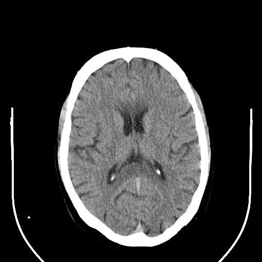
3 A non-contrast CT scan was performed:


4 The abnormality that we found first: Hypodense splenium of corpus callosum

5 We compared the findings with a CT scan, performed in the other hospital 6 days ago: Very slight hypodensity of the splenium


6 Our considerations: Transient lesion of the splenium? Marchiafava-Bignami?

7 Marchiafava-Bignami disease:


8 Our effort to gain some extra information:?? Radiology??? ICU

9 The complete patient history: A week before the patient was admitted to Regional Hospital "St. Anna", Varna after sudden development of abdominal pain and vomiting. She was diagnosed with incarcerated paraumbilical hernia; An emergency laparotomy was performed with resection of approximately 1.5 m partially necrotic intestine; The patient had previous history of 2 years with polydipsia and polyuria post partum and was diagnosed with partial diabetes insipidus. For these complains she received medical therapy, ceased in the early postoperative period; After the operation the patient developed progressive impairment of consciousness and a rise of Na+ levels, necessitating admission to the ICU. Rapid correction of the serum Na+ was performed.

10 Hmmm Electrolyte imbalance? Transient lesion of the splenium? Hyperosmolar state? Demyelination?
11 Osmotic demyelination syndrome Osmotic demyelination syndrome is a state of acute demyelination due to rapid shifts in serum osmolality. The entity was formerly called central pontine myelinolysis, for the demyelination process most typically affects the central pontine white matter with sparing of the periphery. It can also affect the cerebral and cerebellar white matter, which was formerly called extrapontine myelinolysis. Predisposing conditions include alcoholism, rapid correction of hyponatremia, hypernatremia, liver failure, organ transplantation, extensive burns, malnutrition and hyperosmolar hyperglycemia.

12 Osmotic demyelination syndrome Radiographic features CT CT may demonstrate low attenuation crossing the midline in the lower pons, although streak artifact frequently degrades this region and beam hardening phenomenon. MRI The earliest change is seen on DWI with restriction in the lower pons. This same region demonstrates eventual high T2 signal and later low T1 signal. The T1 and T2 changes may take up to two weeks to develop. Occasionally gadolinium enhancement is also demonstrated, just as in the acute phase of a multiple sclerosis (MS) plaque. The peripheral), as well as the periventricular and subpial regions, are typically spared. Similar appearances are seen in other parts of the brain: basal ganglia, midbrain and subcortical white matter. Signal characteristics of affected region include: T1: mildly or moderately hypointense T2: hyperintense, sparing the periphery and corticospinal tracts PD: hyperintense FLAIR: hyperintense DWI: hyperintense ADC: signal low or signal loss T1 C+ (Gd): usually there is no enhancement, but some authors reported that it may occur


13 CT findings revised:


14 The fowoll-up CT scans: 10 days later: A month later:


15 Thank you for your attention!
A Case of Osmotic Demyelination Presenting with Severe Hypernatremia
 Case Report ISSN 1738-5997 (Print) ISSN 2092-9935 (Online) Electrolyte Blood Press 13:30-36, 2015 http://dx.doi.org/10.5049/ebp.2015.13.1.30 A Case of Osmotic Demyelination Presenting with Severe Hypernatremia
A case of central pontine myelinolysis in a patient after liver transplantation
 46 A case of central pontine myelinolysis in a patient after liver transplantation Mouloudi E 1 MD, PhD, Papadopoulos S 1, MD, Massa E 1, MD, Giasnetsova T 1, MD, Theodoridou Th 1, MD, Anastasiou A 2,
A RARE CASE OF: MARCHIAFAVA-BIGNAMI DISEASE Manisha Panchal 1, Maulik Jethva 2, Anjana Trivedi 3, Pinkal Patel 4, Manish Yadav 5
 A RARE CASE OF: MARCHIAFAVA-BIGNAMI DISEASE Manisha Panchal 1, Maulik Jethva 2, Anjana Trivedi 3, Pinkal Patel 4, Manish Yadav 5 HOW TO CITE THIS ARTICLE: Manisha Panchal, Maulik Jethva, Anjana Trivedi,
Marchiafava Bignami Disease (MBD) and Diffusion Tensor Image (DTI) Tractography. Priscilla Chukwueke, MD, MPH
 and Diffusion Tensor Image (DTI) Tractography. Priscilla Chukwueke, MD, MPH") Marchiafava Bignami Disease (MBD) and Diffusion Tensor Image (DTI) Tractography Priscilla Chukwueke, MD, MPH INTRODUCTION Definition: A rare CNS disease characterized by demyelination of the Corpus Callosum.
Marchiafava Bignami Disease (MBD) and Diffusion Tensor Image (DTI) Tractography Priscilla Chukwueke, MD, MPH INTRODUCTION Definition: A rare CNS disease characterized by demyelination of the Corpus Callosum.
MRI OF THE THALAMUS. Mohammed J. Zafar, MD, FAAN Kalamazoo, MI
 1 MRI OF THE THALAMUS Mohammed J. Zafar, MD, FAAN Kalamazoo, MI Objectives: The thalamic nuclei can be involved in a wide variety of conditions. A systematic imaging approach would be useful for narrowing
Case 9511 Hypertensive microangiopathy
 Case 9511 Hypertensive microangiopathy Schepers S, Barthels C Section: Neuroradiology Published: 2011, Nov. 3 Patient: 67 year(s), male Authors' Institution Department of Radiology, Jessa ziekenhuis campus
Demyelinating Diseases of the Brain
 Department of Radiology University of California San Diego Demyelinating Diseases of the Brain John R. Hesselink, M.D. T1-Weighted Images Normal White Matter Contents Axons with envelope of myelin Neuroglia
Dean Shibata, M.D. Department of Radiology, University of Washington. White Matter, Metabolic, and Degenerative Disease.
 White Matter, Metabolic, and Degenerative Disease UW Radiology Review 2015 Dean Shibata, M.D Neuroradiology Department of Radiology, University of Washington MS (Multiple Sclerosis) ADEM (Acute Disseminated
1 MS Lesions in T2-Weighted Images
 1 MS Lesions in T2-Weighted Images M.A. Sahraian, E.-W. Radue 1.1 Introduction Multiple hyperintense lesions on T2- and PDweighted sequences are the characteristic magnetic resonance imaging (MRI) appearance
White matter diseases affecting the corpus callosum; demyelinating and metabolic diseases
 White matter diseases affecting the corpus callosum; demyelinating and metabolic diseases Poster No.: C-0199 Congress: ECR 2011 Type: Educational Exhibit Authors: J. H. Yoo; Seoul/KR Keywords: Neuroradiology
Patologie infiammatorie encefaliche e midollari
 Patologie infiammatorie encefaliche e midollari Maria Laura Stromillo Department of Medicine, Surgery and Neuroscience Inflammatory disorders of the CNS NMOSD ADEM Multiple Sclerosis Neuro-Myelitis Optica
International Journal of Health Sciences and Research ISSN:
 International Journal of Health Sciences and Research www.ijhsr.org ISSN: 2249-9571 Case Report Central Variant Posterior Reversible Encephalopathy Syndrome: A Masquerader with Brainstem and Basal Ganglia
Cerebral malaria: MR imaging spectrum
 Cerebral malaria: MR imaging spectrum Poster No.: C-2705 Congress: ECR 2010 Type: Educational Exhibit Topic: Neuro Authors: P. S. Naphade, M. D. Agrawal, S. S. Sankhe, K. M. Siva, B. K. Jain; Mumbai/IN
brain MRI for neuropsychiatrists: what do you need to know
 brain MRI for neuropsychiatrists: what do you need to know Christoforos Stoupis, MD, PhD Department of Radiology, Spital Maennedorf, Zurich & Inselspital, University of Bern, Switzerland c.stoupis@spitalmaennedorf.ch
Early Diffusion MR Imaging Findings and Short-Term Outcome in Comatose Patients with Hypoglycemia
 ORIGINAL RESEARCH K. Johkura Y. Nakae Y. Kudo T.N. Yoshida Y. Kuroiwa Early Diffusion MR Imaging Findings and Short-Term Outcome in Comatose Patients with Hypoglycemia BACKGROUND AND PURPOSE: The relationship
Marchiafava-Bignami Disease
 Bahrain Medical Bulletin, Vol. 36, No. 4, December 2014 Marchiafava-Bignami Disease Fahd Al-Khamis, MBBS, UODFN* Fozaih Al-Shamrani, MBBS, UODFN** Ibrahim Al- Ghanimi, MBBS, UODFN*** Sarah Abdulhafiz,
General Identification. Name: 江 X X Age: 29 y/o Gender: Male Height:172cm, Weight: 65kg Date of admission:95/09/27
 General Identification Name: 江 X X Age: 29 y/o Gender: Male Height:172cm, Weight: 65kg Date of admission:95/09/27 Chief Complaint Sudden onset of seizure for several minutes Present illness This 29-year
Clinical. Toxic Encephalopathies. Toxic encephalopathy. Imaging. Exogenous Toxins
 Clinical Toxic Encephalopathies Sumeet Kumar National Neuroscience Institute, Duke-NUS Medical School Singapore Decreased consciousness, confusion Excitability, convulsions Motor/sensory disturbances Extrapyramidal
Masses of the Corpus Callosum
 Masses of the Corpus Callosum Kesav Raghavan, HMS Year III Dr. Agenda Corpus Callosum Development and Anatomy Our Patient: Clinical Presentation Differential Diagnosis of Masses in the Corpus Callosum
Case Report Marchiafava-Bignami Disease in a Nonalcoholic Diabetic Patient
 Case Reports in Neurological Medicine Volume 2013, Article ID 979383, 4 pages http://dx.doi.org/10.1155/2013/979383 Case Report Marchiafava-Bignami Disease in a Nonalcoholic Diabetic Patient Sisira Yadala
NEURO IMAGING 2. Dr. Said Huwaijah Chairman of radiology Dep, Damascus Univercity
 NEURO IMAGING 2 Dr. Said Huwaijah Chairman of radiology Dep, Damascus Univercity I. EPIDURAL HEMATOMA (EDH) LOCATION Seventy to seventy-five percent occur in temporoparietal region. CAUSE Most likely caused
NEURORADIOLOGY Angela Lignelli, MD
 Neuroradiology NEURORADIOLOGY Angela Lignelli, MD Plain radiographs CT MRI Cerebral Angiogram Myelograms Neuroradiology Computerized Axial Tomography (CT) CT without and with contrast CTA CT angiogram
NEURORADIOLOGY Angela Lignelli, MD
 NEURORADIOLOGY Angela Lignelli, MD Neuroradiology Plain radiographs CT MRI Cerebral Angiogram Myelograms 1 Neuroradiology Computerized Axial Tomography (CT) CT without and with contrast CTA CT angiogram
Neuroimaging and Psychiatry
 Neuroimaging and Psychiatry Chris Gale Otago Regional Psychiatry Training Programme Feb 12, 2011 In psychosis. Decrease volume frontal, parietal, hippocampus, corpus collosum. Increase CSF volume Functional
Index. Note: Page numbers of article titles are in boldface type.
 Index Note: Page numbers of article titles are in boldface type. A Abdominal compartment syndrome, as complication of fluid resuscitation, 331 338 abdominal perfusion pressure, 332 fluid restriction practice
1 Maiser. 5-Fluorouracil (5-FU) Induced Acute Toxic Leukoencephalopathy. Samuel Maiser, MD. Department of Neurology, University of Minnesota
 Induced Acute Toxic Leukoencephalopathy. Samuel Maiser, MD. Department of Neurology, University of Minnesota") 1 Maiser 5-Fluorouracil (5-FU) Induced Acute Toxic Leukoencephalopathy Samuel Maiser, MD Department of Neurology, University of Minnesota Case This is a 57-year-old right-handed male with a history of
1 Maiser 5-Fluorouracil (5-FU) Induced Acute Toxic Leukoencephalopathy Samuel Maiser, MD Department of Neurology, University of Minnesota Case This is a 57-year-old right-handed male with a history of
Imaging PRES syndrome: typical, atypical and follow up findings.
 Imaging PRES syndrome: typical, atypical and follow up findings. Poster No.: C-2361 Congress: ECR 2013 Type: Educational Exhibit Authors: P. Borrego Jimenez, A. Villalba Gutiérrez, M. Á. López Pino, P.
Probable etiology of mild encephalopathy with reversible isolated lesions in the corpus callosum in children: A review of 20 cases from northern China
 Neurology Asia 2018; 23(2) : 153 158 Probable etiology of mild encephalopathy with reversible isolated lesions in the corpus callosum in children: A review of 20 cases from northern China Chao-Yang Li,
The central nervous system
 Sectc.qxd 29/06/99 09:42 Page 81 Section C The central nervous system CNS haemorrhage Subarachnoid haemorrhage Cerebral infarction Brain atrophy Ring enhancing lesions MRI of the pituitary Multiple sclerosis
Attenuation value in HU From -500 To HU From -10 To HU From 60 To 90 HU. From 200 HU and above
 Brain Imaging Common CT attenuation values Structure Air Fat Water Brain tissue Recent hematoma Calcifications Bone Brain edema and infarction Normal liver parenchyma Attenuation value in HU From -500
A Case of Central Pontine Myelinolysis in a Type 2 Diabetic Patient without Electrolyte Changes
 Endocrinol Metab 26(3):263-267, September 2011 CASE REPORT A Case of Central Pontine Myelinolysis in a Type 2 Diabetic Patient without Electrolyte Changes A Ra Jo, Ji Hye Suk, Jong Kun Ha, Chan Woo Jung,
Joana Ramalho, MD C. Ryan Miller, MD, PhD
 Joana Ramalho, MD C. Ryan Miller, MD, PhD Case 1 3 month old baby girl Presented with new onset of seizures Newborn. Questionable blurring of the gray-white junction within the right occipital lobe. Findings
Table 1: Baseline characteristics of 108 isolated vertigo patients Clinical or laboratory variable n (%) Female 67 (62%)
 Female 67 (62%)") 4. Results The 108 patients who fulfilled the inclusion and exclusion criteria were analyzed. Baseline demographic and epidemiological characteristics of the patients are given in Table 1. Table 1: Baseline
4. Results The 108 patients who fulfilled the inclusion and exclusion criteria were analyzed. Baseline demographic and epidemiological characteristics of the patients are given in Table 1. Table 1: Baseline
Toxic and Metabolic Disease of Nervous System
 Toxic and Metabolic Disease of Nervous System Reid R. Heffner, MD Distinguished Teaching Professor Emeritus Department of Pathology and Anatomy January 14, 2019 1 I HAVE NO CONFLICTS OF INTEREST OR DISCLOSURES
Stroke mimics. Case 1. Acute cases. History. 43 year old healthy male Shortly after awakening developed:
 Stroke mimics Acute cases Gothenburg 21. may 2007 History Case 1 43 year old healthy male Shortly after awakening developed: Left-sided lower facial weakness Left-sided arm paralysis and weakness in leg
Altered Mental Status After Liver Transplant
 REVIEW Altered Mental Status After Liver Transplant Helen S. Te, M.D. Mental status alteration occurs in up to 70% of liver transplant (LT) recipients in the early postoperative period and leads to longer
Magnetic Resonance Imaging for Neurological Conditions. Lawrance Yip Department of Radiology Queen Mary Hospital
 Magnetic Resonance Imaging for Neurological Conditions Lawrance Yip Department of Radiology Queen Mary Hospital Outline Strength and limitations of MRI for neurological conditions MR Imaging techniques
Toxins in Brain! Magnetic Resonance (MR) Imaging of Toxic Leukoencephalopathy A Pictorial Essay
 Imaging of Toxic Leukoencephalopathy A Pictorial Essay") Signature: Pol J Radiol, 2017; 82: 311-319 DOI: 10.12659/PJR.901791 REVIEW ARTICLE Received: 2016.10.02 Accepted: 2016.10.11 Published: 2017.06.13 Authors Contribution: A Study Design B Data Collection
Signature: Pol J Radiol, 2017; 82: 311-319 DOI: 10.12659/PJR.901791 REVIEW ARTICLE Received: 2016.10.02 Accepted: 2016.10.11 Published: 2017.06.13 Authors Contribution: A Study Design B Data Collection
Keep Imaging Simple: An Introduction To Neuroimaging
 Keep Imaging Simple: An Introduction To Neuroimaging Meghan Elkins, OD, FAAO Please silence all mobile devices and remove items from chairs so others can sit. Unauthorized recording of this session is
Role of MRI in acute disseminated encephalomyelitis
 Original Research Article Role of MRI in acute disseminated encephalomyelitis Shashvat Modiya 1*, Jayesh Shah 2, C. Raychaudhuri 3 1 1 st year resident, 2 Associate Professor, 3 HOD and Professor Department
Automated Identification of Neoplasia in Diagnostic Imaging text reports
 Automated Identification of Neoplasia in Diagnostic Imaging text reports "This work has been funded in whole or in part with Federal funds from the National Cancer Institute, National Institutes of Health,
Diffusion-Weighted and Conventional MR Imaging Findings of Neuroaxonal Dystrophy
 AJNR Am J Neuroradiol 25:1269 1273, August 2004 Diffusion-Weighted and Conventional MR Imaging Findings of Neuroaxonal Dystrophy R. Nuri Sener BACKGROUND AND PURPOSE: Neuroaxonal dystrophy is a rare progressive
Hypoglycemic encephalopathy mimicking acute ischemic stroke in clinical presentation and magnetic resonance imaging: a case report
 Chuang et al. BMC Medical Imaging (2019) 19:11 https://doi.org/10.1186/s12880-019-0310-z CASE REPORT Hypoglycemic encephalopathy mimicking acute ischemic stroke in clinical presentation and magnetic resonance
Disorders of water and sodium homeostasis. Prof A. Pomeranz 2017
 Disorders of water and sodium homeostasis Prof A. Pomeranz 2017 Pediatric (Nephrology) Tool Box Disorders of water and sodium homeostasis Pediatric Nephrology Tool Box Hyponatremiaand and Hypernatremia
Supplementary Appendix
 Supplementary Appendix This appendix has been provided by the authors to give readers additional information about their work. Supplement to: Carrera J-P, Forrester N, Wang E, et al. Eastern equine encephalitis
The Nervous Liver: Neurological complications associated with Cirrhosis & Liver Transplantation
 The Nervous Liver: Neurological complications associated with Cirrhosis & Liver Transplantation Poster No.: C-1397 Congress: ECR 2014 Type: Educational Exhibit Authors: A. Arora, A. Mukund, S. Rajesh,
Helpful Information for evaluation of new neurological symptoms in patients receiving TYSABRI
 Helpful Information for evaluation of new neurological symptoms in patients receiving TYSABRI This information is provided as an educational resource for healthcare providers and should be considered current
Year 2003 Paper two: Questions supplied by Tricia
 question 43 A 42-year-old man presents with a two-year history of increasing right facial numbness. He has a history of intermittent unsteadiness, mild hearing loss and vertigo but has otherwise been well.
CDE Exam Preparation Presented by Wendy Graham RD CDE May 4, 2017
 CDE Exam Preparation Presented by Wendy Graham RD CDE May 4, 2017 DKA at organ level 3 Diabetic Ketoacidosis Characteristics Ketones positive Anion Gap > 12 (High) Blood Sugar > 14 (High) Bicarbonate
MRI diagnosis of infantile Alexander disease in a 14 month old African boy
 MRI diagnosis of infantile Alexander disease in a 14 month old African boy Nondumiso Dlamini 1*, Vicci du Plessis 2 1. Department of Radiology, Greys' Hospital, Pietermaritzburg Metropolitan Complex, South
Metronidazole-induced encephalopathy (MIE) is a rare
 is a rare") ORIGINAL RESEARCH E. Kim D.G. Na E.Y. Kim J.H. Kim K.R. Son K.H. Chang MR Imaging of Metronidazole-Induced Encephalopathy: Lesion Distribution and Diffusion-Weighted Imaging Findings BACKGROUND AND PURPOSE:
ORIGINAL RESEARCH E. Kim D.G. Na E.Y. Kim J.H. Kim K.R. Son K.H. Chang MR Imaging of Metronidazole-Induced Encephalopathy: Lesion Distribution and Diffusion-Weighted Imaging Findings BACKGROUND AND PURPOSE:
IV Fluids. I.V. Fluid Osmolarity Composition 0.9% NaCL (Normal Saline Solution, NSS) Uses/Clinical Considerations
 Uses/Clinical Considerations") IV Fluids When administering IV fluids, the type and amount of fluid may influence patient outcomes. Make sure to understand the differences between fluid products and their effects. Crystalloids Crystalloid
IV Fluids When administering IV fluids, the type and amount of fluid may influence patient outcomes. Make sure to understand the differences between fluid products and their effects. Crystalloids Crystalloid
NEURO IMAGING OF ACUTE STROKE
 1 1 NEURO IMAGING OF ACUTE STROKE ALICIA RICHARDSON, MSN, RN, ACCNS-AG, ANVP-BC WENDY SMITH, MA, RN, MBA, SCRN, FAHA LYNN HUNDLEY, APRN, CNRN, CCNS, ANVP-BC 2 2 1 DISCLOSURES Alicia Richardson: Stryker
Tips and tricks for detecting diffuse axonal injury on CT and MR neuroimaging
 Tips and tricks for detecting diffuse axonal injury on CT and MR neuroimaging Poster No.: C-3080 Congress: ECR 2018 Type: Educational Exhibit Authors: M. Marinkic, D. Zadravec ; Zagreb/HR, Zageb/HR Keywords:
MR Imaging for diagnostic of metabolic encephalopathy
 MR Imaging for diagnostic of metabolic encephalopathy Poster No.: C-2142 Congress: ECR 2014 Type: Educational Exhibit Authors: A. Zeriouel, O. Addou, I. Kamaoui, M. Maaroufi, N. Sqalli, S. Tizniti; Fes/MA
MRI of the Brain: A Primer on What, How, Why, and When. September Amit Malhotra, Harvard Medical School, Year- IV. Gillian Lieberman, MD
 September 2000 MRI of the Brain: A Primer on What, How, Why, and When Hornak, J.P. The Basics of MRI. 1996-2000 Amit Malhotra, Harvard Medical School, Year- IV Magnetic Resonance Imaging A Brief History
Index. aneurysm, 92 carotid occlusion, 94 ICA stenosis, 95 intracranial, 92 MCA, 94
 A ADC. See Apparent diffusion coefficient (ADC) Aneurysm cerebral artery aneurysm, 93 CT scan, 93 gadolinium, 93 Angiography, 13 Anoxic brain injury, 25 Apparent diffusion coefficient (ADC), 7 Arachnoid
Pediatric CNS Tumors. Disclosures. Acknowledgements. Introduction. Introduction. Posterior Fossa Tumors. Whitney Finke, MD
 Pediatric CNS Tumors Disclosures Whitney Finke, MD Neuroradiology Fellow PGY-6 University of Utah Health Sciences Center Salt Lake City, Utah None Acknowledgements Introduction Nicholas A. Koontz, MD Luke
Radiologic-Pathologic Correlation of White Matter Disease
 The Neuroradiology Journal 22 (Suppl. 1): 26-32, 2009 www. centauro. it Radiologic-Pathologic Correlation of White Matter Disease J.G. SMIRNIOTOPOULOS 1,2,3,.. SMITH 1,2, E. RUSHING 2,4, F.M. MURPHY 2,
diabetes in adults Metabolic complications of
 Metabolic complications of diabetes in adults Dimitri MARGETIS MD ICU St ANTOINE PARIS Definition Diabetic acidoketosis Serious complication in type I diabetes : Hyperglycemia Metabolic acidosis Acidic
Rapid Regression of White Matter Changes in Hypoglycemic Encephalopathy
 www.ksmrm.org JKSMRM 18(4) : 357-361, 2014 pissn 1226-9751 / eissn 2288-3800 Case Report Rapid Regression of White Matter Changes in Hypoglycemic Encephalopathy Sang-wook Son, Kye-ho Lee, Dong-soo Yoo
MRI and differential diagnosis in patients suspected of having MS
 Andrea Falini Italy MRI and differential diagnosis in patients suspected of having MS IMPROVING THE PATIENT S LIFE THROUGH MEDICAL EDUCATION www.excemed.org Outline of presentation - Diagnostic criteria
Diabetic Emergencies: Ketoacidosis and the Hyperglycemic Hyperosmolar State. Adam Bursua, Pharm.D., BCPS
 Diabetic Emergencies: Ketoacidosis and the Hyperglycemic Hyperosmolar State Adam Bursua, Pharm.D., BCPS Objectives Describe the epidemiology of diabetic ketoacidosis (DKA) and the hyperglycemic hyperosmolar
Delayed Encephalopathy of Acute Carbon Monoxide Intoxication: Diffusivity of Cerebral White Matter Lesions
 AJNR Am J Neuroradiol 24:1592 1597, September 2003 Delayed Encephalopathy of Acute Carbon Monoxide Intoxication: Diffusivity of Cerebral White Matter Lesions Ji-hoon Kim, Kee-Hyun Chang, In Chan Song,
Case Rep Neurol 2010;2: DOI: /
 157 This is an Open Access article licensed under the terms of the Creative Commons Attribution- NonCommercial-NoDerivs 3.0 License (www.karger.com/oa-license), applicable to the online version of the
CASE OF THE WEEK PROFESSOR YASSER METWALLY
 CLINICAL PICTURE CLINICAL PICTURE A 40 Years old female patient, known to be suffering from multiple sclerosis, was presented clinically with painful diminution of vision in the right aye and mild weakness
Complete Recovery of Perfusion Abnormalities in a Cardiac Arrest Patient Treated with Hypothermia: Results of Cerebral Perfusion MR Imaging
 pissn 2384-1095 eissn 2384-1109 imri 2018;22:56-60 https://doi.org/10.13104/imri.2018.22.1.56 Complete Recovery of Perfusion Abnormalities in a Cardiac Arrest Patient Treated with Hypothermia: Results
MRI in MS. MRI in multiple sclerosis. MS T2 Lesions: Pathology. Brain lesions Morphology Matters. Sagittal FLAIR Morphology Matters
 MRI in multiple sclerosis MRI in MS Rohit Bakshi, MD, MA Breakstone Professor of Neurology & Radiology Director, Laboratory for Neuroimaging Research Senior Neurologist, MS Center Brigham & Women s Hospital
Marchiafava-Bignami Disease: Longitudinal MR Imaging and MR Spectroscopy Study
 AJNR Am J Neuroradiol 24:249 253, February 2003 Case Report Marchiafava-Bignami Disease: Longitudinal MR Imaging and MR Spectroscopy Study Anna Gambini, Andrea Falini, Lucia Moiola, Giancarlo Comi, and
Persistent lithium-induced neurotoxicity: direct effect of lithium and/or hypernatremia?
 Jefferson Journal of Psychiatry Volume 22 Issue 1 Article 3 July 2009 Persistent lithium-induced neurotoxicity: direct effect of lithium and/or hypernatremia? Ioana-Mihaela Popescu University of California
Fig.1: A, Sagittal 110x110 mm subimage close to the midline, passing through the cingulum. Note that the fibers of the corpus callosum run at a
 Fig.1 E Fig.1:, Sagittal 110x110 mm subimage close to the midline, passing through the cingulum. Note that the fibers of the corpus callosum run at a slight angle are through the plane (blue dots with
Caudal and Lentiform nuclei Myelinolysis following Endoscopical surgery for pediatric Craniopharyngioma: two cases report and literature review
 Wang et al. Chinese Neurosurgical Journal (2018) 4:21 https://doi.org/10.1186/s41016-018-0125-8 CHINESE MEDICAL ASSOCIATION CHINESE NEUROSURGICAL SOCIETY CASE REPORT Open Access Caudal and Lentiform nuclei
Diffusion-weighted magnetic resonance imaging (MRI) allows for tissue
 allows for tissue") MAGNETIC RESONANCE IMAGING / IMAGERIE PAR RÉSONANCE MAGNÉTIQUE Nonischemic causes of hyperintense signals on diffusion-weighted magnetic resonance images: a pictorial essay Jeffrey M. Hinman, MD; James
MAGNETIC RESONANCE IMAGING / IMAGERIE PAR RÉSONANCE MAGNÉTIQUE Nonischemic causes of hyperintense signals on diffusion-weighted magnetic resonance images: a pictorial essay Jeffrey M. Hinman, MD; James
Disclosure. + Outline. Case-based approach to neurological emergencies that might present to the ED
 Kathleen R. Fink, MD University of Washington 5 th Nordic Emergency Radiology Course May 21, 2015 Disclosure My spouse receives research salary support from: Bracco BayerHealthcare Guerbet Outline Case-based
Clinics in diagnostic imaging (175)
") Singapore Med J 2017; 58(3): 121-125 doi: 10.11622/smedj.2017017 CMEArticle Clinics in diagnostic imaging (175) Vijay Krishnan 1, MD, FRCR, Tze Chwan Lim 1, MBBS, FRCR, Francis Cho Hao Ho 2, MBBS, FRANZCR,
Singapore Med J 2017; 58(3): 121-125 doi: 10.11622/smedj.2017017 CMEArticle Clinics in diagnostic imaging (175) Vijay Krishnan 1, MD, FRCR, Tze Chwan Lim 1, MBBS, FRCR, Francis Cho Hao Ho 2, MBBS, FRANZCR,
MR imaging spectrum of bilateral symmetric hippocampal lesions
 MR imaging spectrum of bilateral symmetric hippocampal lesions Poster No.: C-2510 Congress: ECR 2010 Type: Educational Exhibit Topic: Neuro Authors: P. S. Naphade, M. D. Agrawal, S. S. Sankhe, K. M. Siva,
Review Article Thalamic Lesions: A Radiological Review
 Behavioural Neurology, Article ID 154631, 17 pages http://dx.doi.org/10.1155/2014/154631 Review Article Thalamic Lesions: A Radiological Review Dimitri Renard, Giovanni Castelnovo, Chantal Campello, Stephane
Structural and functional imaging for the characterization of CNS lymphomas
 Structural and functional imaging for the characterization of CNS lymphomas Cristina Besada Introduction A few decades ago, Primary Central Nervous System Lymphoma (PCNSL) was considered as an extremely
Brain AVM with Accompanying Venous Aneurysm with Intracerebral and Intraventricular Hemorrhage
 Cronicon OPEN ACCESS EC PAEDIATRICS Case Report Brain AVM with Accompanying Venous Aneurysm with Intracerebral and Intraventricular Hemorrhage Dimitrios Panagopoulos* Neurosurgical Department, University
Imaging Neurologic Emergencies: When and Where Radiology Makes a Difference
 Imaging Neurologic Emergencies: When and Where Radiology Makes a Difference James G. Smirniotopoulos, M.D. Radiology and Radiological Sciences Uniformed Services University Bethesda, MD Learning Objectives
NEURORADIOLOGY-NEUROPATHOLOGY CONFERENCE
 THE UNIVERSITY OF NORTH CAROLINA at CHAPEL HILL SEPTEMBER 2013 NEURORADIOLOGY-NEUROPATHOLOGY CONFERENCE Claudia da Costa Leite, MD, PhD Thomas Bouldin, MD CASE 1 6 y-o female with headaches and vomiting
Essentials of Clinical MR, 2 nd edition. 14. Ischemia and Infarction II
 14. Ischemia and Infarction II Lacunar infarcts are small deep parenchymal lesions involving the basal ganglia, internal capsule, thalamus, and brainstem. The vascular supply of these areas includes the
Pictorial Essay. Primary Lymphoma of the Central Nervous System: Typical and Atypical CT and MR Imaging Appearances
 Downloaded from www.ajronline.org by 37.44.197.38 on 02/11/18 from IP address 37.44.197.38. opyright RRS. For personal use only; all rights reserved Primary Lymphoma of the entral Nervous System: Typical
Case Report Paroxysmal Amnesia Attacks due to Hashimoto s Encephalopathy
 Case Reports in Medicine Volume 2016, Article ID 1267192, 4 pages http://dx.doi.org/10.1155/2016/1267192 Case Report Paroxysmal Amnesia Attacks due to Hashimoto s Encephalopathy Pelin Nar Senol, Aylin
Pediatric MS MRI Study Methodology
 General Pediatric MS MRI Study Methodology SCAN PREPARATION axial T2-weighted scans and/or axial FLAIR scans were obtained for all subjects when available, both T2 and FLAIR scans were scored. In order
MRI Atlas of MS Lesions
 MRI Atlas of MS Lesions M. A. Sahraian, E.-W. Radue MRI Atlas of MS Lesions With the collaboration of A. Gass, S. Haller, L. Kappos, J. Kesselring, J.-I. Kira, K. Weier 123 Prof. Dr. med. Mohammad Ali
Atypical Unilateral Posterior Reversible Encephalopathy Syndrome Mimicking a Middle Cerebral Artery Infarction
 Case Report Neuroimaging and Head & Neck http://dx.doi.org/10.3348/kjr.2015.16.5.1104 pissn 1229-6929 eissn 2005-8330 Korean J Radiol 2015;16(5):1104-1108 Atypical Unilateral Posterior Reversible Encephalopathy
11 th Annual Cerebrovascular Symposium 5/11-12/2017. Hypertonic Use D E R E K C L A R K
 Hypertonic Use D E R E K C L A R K 1 Outline Types of hyperosmolar therapy Review Cerebral Na Physiology Differences between periphery and BBB Acute phase Subacute phase Chronic changes Hypertonic Saline
Hyperglycaemic Emergencies GRI EDUCATION
 Hyperglycaemic Emergencies GRI EDUCATION LEARNING OUTCOMES Develop and describe your system of blood gas interpretation and recognise common patterns of acid-base abnormality. Describe the pathophysiology
Case Report. Herpes simplex virus encephalitis presenting as frontal lobe hemorrhage
 1 Case Report Herpes simplex virus encephalitis presenting as frontal lobe hemorrhage Authors: Shila, MD, *Jessica Erfan, MPAS, PA-C, Ray Bogitch, MD, Jefferson T. Miley, MD Department of Neurology, Dell
Pulmonary Arteriovenous Malformations Complicated with Paradoxical Embolic Stroke
 Archives of Clinical and Medical Case Reports doi: 10.26502/acmcr.96550032 Volume 2, Issue 4 Case Report Pulmonary Arteriovenous Malformations Complicated with Paradoxical Embolic Stroke Cheah Wai Hun
IMAGING OF HYPOXIC ISCHEMIC INJURY IN A NEONATE FN3 STATE MEETING NEMOURS CHILDREN'S HOSPITAL ORLANDO,FL 08/04/18
 IMAGING OF HYPOXIC ISCHEMIC INJURY IN A NEONATE FN3 STATE MEETING NEMOURS CHILDREN'S HOSPITAL ORLANDO,FL 08/04/18 Dhanashree Rajderkar,MD Assistant Professor Department of Radiology University of Florida
Original Article INTRODUCTION. Abstract
 Original Article Print ISSN: 2321-6379 Online ISSN: 2321-595X DOI: 10.17354/ijss/2018/19 Patterns of Restricted Diffusion within Corpus Callosum in Neonatal Hypoxic-Ischemic Encephalopathy and its Significance
Imaging Acute Stroke and Cerebral Ischemia
 Department of Radiology University of California San Diego Imaging Acute Stroke and Cerebral Ischemia John R. Hesselink, M.D. Causes of Stroke Arterial stenosis Thrombosis Embolism Dissection Hypotension
The Role of MRI in Multiple Sclerosis
 September 2006 The Role of MRI in Multiple Sclerosis David Cochran, Harvard Medical School Year IV Meet our patient WM 49yoF presents to Multiple Sclerosis Clinic for follow-up regarding worsening symptoms
Anoxic brain injury CT and MRI patterns - quick pictoral quide for junior radiologists.
 Anoxic brain injury CT and MRI patterns - quick pictoral quide for junior radiologists. Poster No.: C-1844 Congress: ECR 2017 Type: Educational Exhibit Authors: A. Kecler - Pietrzyk, W. Torreggiani ; Dublin/IE,
Diffusion weighted magnetic resonance imaging features of intracranial lesions
 INTERNATIONAL JOURNAL OF HEALTH RESEARCH IN MODERN INTEGRATED MEDICAL SCIENCES, ISSN 2394-8612 (P), ISSN 2394-8620 (O), Vol-2, Issue-4, Oct-Dec 2015 35 Original Article Diffusion weighted magnetic resonance
A Case of Anterograde Amnesia with Bilateral Hippocampus Involvement After Acute Glufosinate Ammonium Intoxication
 www.ksmrm.org JKSMRM 18(4) : 352-356, 2014 pissn 1226-9751 / eissn 2288-3800 Case Report A Case of Anterograde Amnesia with Bilateral Hippocampus Involvement After Acute Glufosinate Ammonium Intoxication
Brainstem. Steven McLoon Department of Neuroscience University of Minnesota
 Brainstem Steven McLoon Department of Neuroscience University of Minnesota 1 Course News Change in Lab Sequence Week of Oct 2 Lab 5 Week of Oct 9 Lab 4 2 Goal Today Know the regions of the brainstem. Know
A pictorial review of neurological complications of systemic lupus erythematosus and antiphospholipid syndrome
 A pictorial review of neurological complications of systemic lupus erythematosus and antiphospholipid syndrome Poster No.: C-2780 Congress: ECR 2010 Type: Educational Exhibit Topic: Neuro Authors: E. Tavernaraki,