such as lung cancer or breast cancer
|
|
|
- Gerard Harvey
- 6 years ago
- Views:
Transcription
1 Assessing Dosimetric Accuracy with Lucy 3D QA Phantom Victoria Honetschlager, Department of Physics, Minnesota State University Moorhead, Moorhead, MN ABSTRACT Stereotactic radiosurgery (SRS) is a radiation therapy method which treats a very small and well defined volume with a high dose of radiation. New technologies such as the Novalis Tx allow this treatment on a wide-scale clinical level. In 2011, Roger Maris Cancer Center acquired the Novalis Tx and the clinic is now able to treat far more patients than before. With this new linear accelerator comes a need to quantify the accuracy of it during a SRS treatment. This study utilizes a Lucite medical imaging phantom, LUCY 3D QA, and iplan software to image, plan, and treat a pseudo patient and quantify machine accuracy in absorbed dose delivered (dosimetric accuracy). INTRODUCTION More than 180,000 brain tumors are diagnosed each year [U.S News]. While this number sounds staggering, it is only about 2-3% of diagnosed adult cancer cases in the United States. Of these diagnoses, the large majority are metastatic 1. In fact, nearly one in four patients diagnosed with cancer will develop a metastatic brain tumor. Current clinical treatment techniques include: Surgery to remove or, in some cases, debulk 2 the tumor Chemotherapy, though it is largely inefficient at providing a comprehensive treatment for cranial cancers Radiation therapy, including whole brain radiation and stereotactic radiosurgery Many of these cases include a combination of treatments and the patient s interventional care is specifically tailored by a team of health care professionals such as radiation oncologists, 1 Secondary tumors spread from an initial cancer such as lung cancer or breast cancer 2 To remove a large portion of the tumor s mass to reduce its sixe medical physicists, dosimetrists, and radiation therapists. Radiation therapy is a broad category designation for a treatment that uses radiation to damage the DNA of cells in order to halt the reproduction of invasive cells. The tumor will shrink over time, often taking six months to several years to fully clear. Unlike surgery, radiation therapy does not physically remove the tumor. Within radiation therapy are many specific treatments, summarized in Appendix A. Unfortunately, radiation can also damage healthy cells surrounding the tumor, which can lead to side effects. Thus, much research has been dedicated to treatment methods that minimize damage to surrounding structures. WHY STEREOTACTIC RADIOSURGERY? Unlike whole brain radiation therapy- in which a large volume of the brain is given radiation to treat both the visible lumps of a tumor and a preemptive dose to developing tumors not seen on an MRI scan- stereotactic radiosurgery (SRS) utilizes a high dose of radiation to a very narrow target. SRS differs from more conventional radiation therapy in several aspects: Single large dose of radiation is used rather than multiple daily fractions [Loeffler et al.] Sharp dose gradients are produced so radiation outside the target volume in minimized [Suh] SRS allows for accuracy of treatment within a few millimeters of the target [Loeffler et al.] With this in mind, SRS can be very useful in cases where the tumor is hard to reach, such as intracranial growths. For tumors outside the brain, SBRT 3 can be of huge benefit for tumors located close to vital organs, or those subject to movement within the body. Table 1 summarizes 3 Clinically, SRS is used when discussing cranial lesions; the same technique used on locations other than the head is referred to as SBRT, or stereotactic body radiotherapy.
2 characteristics that make a patient ideal for SRS or SBRT treatment. 1. Well defined borders on either MRI and/or CT 2. Spherical in shape (most metastic cancer satisfies this) 3. Small (usually under 4 cm in diameter) 4. Located near critical structures such as spinal cord 5. Surgery not an option due to tumor location or patient health Table 1: Consideration factors for SRS treatment Since no surgery is actually performed, risks such as infection, hemorrhaging and spinal fluid leakage are greatly reduced. While patients may be sedated during treatment, general anesthesia is not required 4, meaning they are able to communicate throughout the entirety of the procedure. Since SRS is non-invasive, there is no scarring, unlike open skull surgery. Unfortunately, stereotactic radiosurgery treatment is that it is far costlier that whole-brain radiation therapy. Estimated Medicare costs for SRS are $10,000 to $27,000 per procedure, compared to $2,300 to $7,650 for whole brain radiation therapy. Most insurance plans now cover SRS treatment in cases where patients satisfy several of the criteria described in Table 1. MATERIALS AND METHODS This study takes a quality assurance phantom through the basic treatment process that patients follow, with some modifications. Here, however, rather than seeing through to a final radiation treatment, the process is halted at a final quality assurance check as there is no human patient to treat. Data collected while treating the phantom was analyzed to assess the accuracy of the plan and machine in delivering therapy. It is beneficial to familiarize one with the materials used, such as the linear accelerator, phantom and ion chamber, as well as gain an understanding of the treatment and quality assurance process before discussing this study s examination of the cranial SRS phantom. 4 This discussion is limited to adult patients. Often in pediatric cases, children and toddlers must be under general anesthesia, and require more intensive treatment and care. NOVALIS TX In 2007, Varian Medical Systems partnered with BrainLAB AG to launch the Novalis Tx. This clinical linear accelerator has the ability to be used as a stereotactic radiosurgery platform, allowing local cancer centers the ability to more cost effectively provide SRS treatment options to patients [Chang 2007]. All linear accelerators, including the Novalis Tx, accelerate electrons and then allow then to collide with a target made of heavy metal. From these collisions, high energy x-rays are produced. The x-rays are shaped with wedges 5, cones 6, or multileaf collimators 7 (MLC). The patient lies on a couch, while the gantry, which houses the beam, rotates around the patient. By rotating both the gantry and the couch, the ability to treat at any angle is maintained. With all linear accelerators operating on the same basis, one may be tempted to ask what sets the Novalis Tx apart or makes it worth the $4 million dollar price tag. New technologies such as dual energy modes allow deeper penetration, beneficial for SBRT applications. 120 leaves reside in the collimator, allowing for better beam shaping. Imaging technologies such as ExacTrac and OnBoard Imager (OBI) allow for precise patient alignment and respiratory gating 8, and the maximum dose rate is 1,000 monitor units per second- higher than many older and stand-alone linacs (Agazaryan & Schultz, 2011). Figure 1 shows an illustrated view of the image guided techniques featured on the Novalis Tx. For more specific feature information, please see Appendix B. 5 Collimator insert used to tailor the beam attenuation 6 Fitting over the end of the collimator that narrows the beam or field size; when conformal arcs are used with a cone beam, a circular or ovular shape is created 7 Small metal pieces that move one-dimensionally to create shapes in the head of the collimator. This allows a dosimetrist to tailor the shape of the radiation beam to the tumor shape. 8 Used when treating tumors susceptible to respiratory induced breathing, such as lung cancer; measures patient breathing to treat at optimal time intervals

3 Figure 1(right): ExacTrac x-ray configuration. X-ray tubes are mounted in the floor and flat panel imagers are mounted to the ceiling In summer of 2011, installation of the Novalis Tx began at Roger Maris Cancer Center in Fargo, North Dakota. The machine was commissioned in late fall, and patient treatment began in winter As such, very few SRS treatments have been performed at this location, and questions such as the machine accuracy were important to ascertain. This is the motivation on which this study relies. QUALITY ASSURANCE Due to both the high radiation dose and dose rate, quality assurance is of utmost importance for medical professionals when dealing with SRS treatment. The large majority of the quality assurance falls under the duties of the medical physicist. The American Association of Physicists in Medicine (AAPM) has published a series of guidelines derived from task groups. These standards are widely accepted in the field and generally dictate quality assurance protocol on the clinical level. Every morning, radiation therapists follow a machine warm-up protocol which ensures that the machine is working as expected. Daily, monthly and annual checks are performed by the medical physicists. These checks assure the quality of the linear accelerator itself, rather than the patient plan. Table 2 shows in more detail the guidelines AAPM has established in task group 142[Klein et al.]. Patient specific quality assurance is integrated within the treatment process. Figure 2 shows a visual representation of the treatment process based off a similar proposed model by Dr. Fang Yin at the Duke University Medical Center. Daily Checks Audiovisual monitoring Door interlock (door should not open when machine beam is on) Check that OBI and ExacTrac work properly Weekly Checks Quantitative checks on imaging systems Monthly Checks Energy check for photons Respiratory gating system check Annual Checks Subset of tests executed during machine commissioning Table 2: Summary of TG-142 quality assurance policies for medical accelerators This visual representation highlights the importance of quality assurance within the entirety of treatment. While planning software such as iplan do give dose rates expected within the treatment volume, it is important that medical physicists take a quantitative measurement using a phantom- or acrylic form similar to the density of tissue that is used to assess and analyze the delivery of treatment plans- for a cross-reference. In doing so, irregularities with the machine or patient alignment may be detected. This study relies on a slightly modified patient model, as seen in Figure 3. This assumes that a patient has come into the hospital and received a diagnostic MRI scan and an oncologist has already prescribed a stereotactic radiosurgery treatment. From there, the phantom is imaged, planned for and treated. One benefit to representing this process visually is that it becomes easy to recognize that an error in an early-stage of planning or imaging may be piggy-backed or carried along until treatment. For this reason the phantom is used from the earliest stages throughout the entirety of the treatment process.
4 Case Selection Initial diagnostic imaging (MRI) Patient consultation with oncologist SRS Treatment determination 3-D Simulation CT scan for treatment planning Scans sent to dosimetry along with oncologist prescription Assessment Patient returns for imaging to determine success of treatment Consult with oncologist to determine if further treatment is necessary Retreatment Treatment Radiation therapists deliver treatment to patient Correction needed to position? NO Quality Assurance Check #3 Image patient using OBI and ExacTrac Treatment Planning Dosimetrist develops treatment plan based on oncologists specifications Brain LAB iplan software NO YES Patient Set-Up Immobilize patient on table, align them with a fusion of CT and MRI images Quality Assurance Check Medical physicist checks dosimetrists plan, submits it to oncologist for approval Planning software calculates monitor units (MU) to be given Approved? YES Quality Assurance Check #2 Exact patient plan is executed on a Lucite phantom for quantitative analysis Figure 2: Treatment process from diagnostic imaging to success assessment. The process highlights three key aspects of treatment execution: planning (pink), quality assurance (blue), and treatment delivery (green).
5 Case Selection Assumed Initial diagnostic imaging (MRI) assumed to have occurred Patient consultation with oncologist SRS Treatment determination 3-D Simulation on Phantom CT scan phantom for treatment planning Scans sent to dosimetry, a trivial dose of radiation is assigned to later compare quantitatively Treatment Planning for Phantom Medial physicist develops treatment plan based on oncologists specifications Brain LAB iplan software Planning software generates TaPos to be used to align phantom NO Assessment Ion chamber sends information to electrometer Readings are recorded and analyzed Total dose measured at isocenter by phantom is compared to expected dose at isocenter from planning software YES Treatment Medical physicist delivers treatment to phantom Correction needed to position? NO Quality Assurance Check #3 Image phantom using OBI and ExacTrac Phantom Set-Up Immobilize phantom with invasive head ring on table, align them with TaPos and CT Image Quality Assurance Check Medical physicist checks plan Planning software calculates monitor units (MU) to be given Approved? YES Quality Assurance Check #2 Exact patient plan is executed on a Lucite phantom for quantitative analysis Figure 3: Treatment process from diagnostic imaging to data analysis for this study. This process is similar to the one highlighted in Figure 2.


6 LUCY 3D QA PHANTOM This study utilizes the LUCY 3D QA Phantom. The phantom is produced by Standard Imaging, Inc. for specific use in perform[ing] comprehensive quality assurance tests for an entire stereotactic radiosurgery procedure (Standard Imaging, Inc.) and costs approximately $33,000 when purchased with all accessory packages. Lucy is a highly precise phantom with tolerances of 0.1 mm (Standard Imaging). The phantom can be mounted onto commercial SRS frames, such as those produced by Varian Medical Systems and BrainLAB AG for use with the Novalis Tx linac, as shown in Figure 4. This unique ability to interface without respect to branding allows hospitals and clinics to image and treat the phantom in the exact manner and position as the patient on the table, assuring a valid and accurate quality assurance protocol. This dose verification study uses the Exradin A16 Microchamber Ion Chamber. Figure 5 also shows the A16 Ion Chamber inserted into the Lucy Phantom. Different inserts must be used with different ion chambers, as each is milled slightly different. The A16 functions in the same manner as all ion chambers. It consists of a gas filled enclosure between two conducting electrodes. When the gas between the electrodes is ionized by photons generated by the linear accelerator, a current is created and measured by an electrometer. The charge is reported in nanocouloumbs and a correction factor is applied to convert the charge into an absorbed dose. EXRADIN A-16 ION CHAMBER Drilled into the Lucy phantom is a dosimetry insert compatible with several different ion chambers. The insert is used to verify a prescribed dose at the isocenter, or volumetric center. The insert is milled so that the center of the Lucy phantom. Figure 5 shows this ion chamber quite clearly on the right side of the photograph. Figure 4(left): Spherical Lucy phantom fitted into an invasive BrainLAB frame for SRS treatment and fixed onto Novalis Tx treatment couch. One can clearly see the ion chamber insert on the right side of the Lucy phantom. Figure 5 (above): Exradin A16 Mirochamber Ion Chamber fitted into Lucy phantom. Readings taken by the ion chamber appear on an electrometer and allow the medical physicist to calculate a total dosage received by the phantom

7 SRS SPECIFIC QUALITY ASSURANCE Having discussed the overall quality assurance process and method for assessing accuracy in SRS plans, this section describes in greater detail the exact methods used to quantify this end-to-end test. CT SCANS FOR PLANNING CT Scans for treatment planning were performed on a Philips scanner at Roger Maris Cancer Center in Fargo, North Dakota. Two scans were taken with the Lucy phantom and A16 ion chamber in a head frame localizer. The localizer box contains metal rods that appear during imaging. These markings allow alignment of the phantom during treatment. Figure 6 shows a series of CT images from several orientations. After scans are taken, they are imported into the treatment planning software, in this case, BrainLAB AG s iplan software. TREATMENT PLANNING With the images imported into iplan, a treatment plan was developed. The plan uses conformal circular arcs, a method whereby the dose is delivered by several arcs that intersect around the isocenter. Each arc is called a field when assigned specific amount of radiation to deliver. Together they create a spherical dose distribution which is very effective for most cranial (especially metastatic) tumors. To do this, circular cones ranging in diameter for 0.5 mm to 30 mm are fit into the linear accelerator collimator. Cones give the best dose uniformity for circular lesions, but irregular shapes can be treated by overlaying several spherical dose distributions. In an effort to simplify the procedure, it was assumed that the phantom had a single, spherical lesion located at the isocenter. For visual comparison, Figure 7 shows the collimator head with and without a cone attachment. Figure 6: Series of CT images of Lucy phantom with A16 ion chamber. Illustrates axial, sagittal and coronal images. The lower right corner shows the orientation of the phantom during scan in a clear, easy to comprehend manner.

![This six-field 9 plan has an assigned, or prescribed, dose of 28.00 Gray 10 [Gy];after planning, however, the dose expected to be detected at the isocenter is 28.21 Gy. This difference is 0.](/docs-images/75/72445020/images/8-1.jpg "75% which is acceptable and within tolerances, as defined by AAPM.")
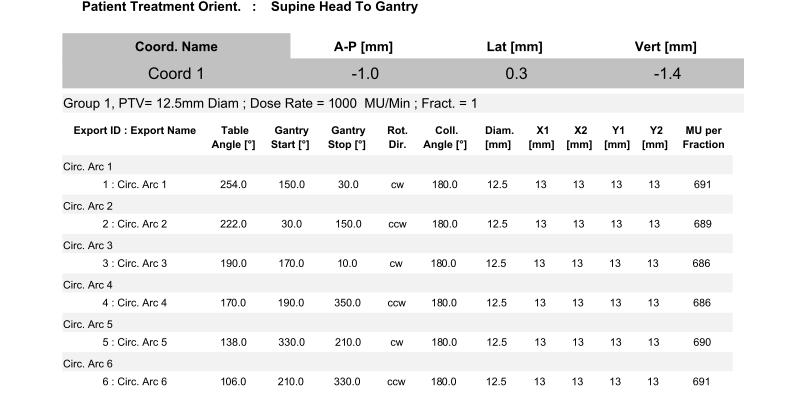
8 Figure 7: Collimator without any cone attachment (left) mm cone attachment (right). Specifically, this plan uses a 12.5mm cone to deliver treatment. As typical with most plans delivered at Roger Maris Cancer Center, the linear accelerator energy was planned at 6 MV at set for a dose rate of 1,000 monitor units/minute. This six-field 9 plan has an assigned, or prescribed, dose of Gray 10 [Gy];after planning, however, the dose expected to be detected at the isocenter is Gy. This difference is 0.75% which is acceptable and within tolerances, as defined by AAPM. Because the gantry travels dynamically through a starting and ending angle, collisions between the treatment couch and linac may occur. In order to reduce the likelihood of such 9 Common dosimetry planning uses either six or nine fields. 10 It may be a bit confusing that monitor units and Gray both seem to be getting tossed around. Unfortunately, the linac is not equipped to handle delivering dose in Gray and so the machine uses its own unit, a monitor unit, which is measure of the energy expended, while the medical physicist finds their calculations far easier when using units of Gray. Linacs are calibrated to deliver 1 cgy/mu for a 10cm x 10 cm field at 10 cm depth. an event and treat as closely to the phantom as possible, the plan features couch kicks, which is simply a vertical axis rotation of the treatment couch. By rotating the couch, a more even dose distribution can be given and hot spots that could occur from beam divergence are minimized. Figure 8 shows delivery parameters generated by iplan, including gantry angle rotation arcs for each field, treatment couch angles, and monitor units to be delivered by the linac for each field. The delivery parameters are in effect a plan summary showing the major details of the final plan. For more detailed plan parameters, please see Appendix C. iplan also created a Target Positioner Overlay (TaPo) which affixes to the head localizer box and allows for positioning based off the guided in-room lasers.


9 Figure 8: Delivery Parameters for SRS quality assurance plan with Lucy phantom. 6-field, 6 MV plan with 12.5 mm cone beam conformal circular arcs. HEAD FRAME LOCALIZATION With a finalized treatment plan in place, the next part of the process was to localize and position the phantom. During this phase, it is critical that the phantom isocenter matches as closely as possible with the machine isocenter so that treatment is spatially accurate. In order to do so, the Lucy phantom was placed in the invasive head ring, and mounted to the treatment couch with a specialized mount from Varian Medical Systems, Inc. and BrainLAB. Figure 9 (at left) shows the phantom mounted to the table. Knobs on the mount help correct the alignment of the phantom with the in-room lasers. In order to know what to align the phantom to, Lucy phantom is placed in the same head localizer that was used during the CT scan. The TaPo printouts are then attached to the Figure 9: Varian Medical Systems, Inc. and BrainLAB couch attachment for Lucy phantom in invasive head ring. Knobs that adjust the alignment (pitch, roll, and yaw) are boxed in yellow.


10 localizer frame. Figure 10 shows positioning of the Lucy phantom with the head localizer frame and TaPo overlay. Once proper positioning is achieved with the TaPo, the localizer frame can be removed and the ExacTrac system generates a kv x-ray image. This image is overlaid from the initial CT scan; it then enables the user to drag the two images together to align them as closely as possible. Once alignment is completed, then a suggested shift to table parameters is given based on the amount of realignment needed is made. Figure 11 shows the image fusion of the original CT image (red) and the OBI (blue). The images then get overlapped as shown in the set of images on the right and a suggested couch shift is given by the computer based on the amount of image adjustment. Suggested couch shifts based on x ray localization were 1mm. Figure 10: Positioning Lucy phantom for treatment using localizer frame (left) and TaPo overlay (right). In room lasers are used to align the target coordinate shown above. In order to this, alignment knobs may be adjusted as well as the couch position.


11 Figure 11: On-Board Imaging portal allows medical physicists the ability to image phantom on the couch and shift the table so current placement aligns with pre-treatment CT scans. Top image shows an initial in-room x-ray taken prior to any couch movement. From this, the two images were matched up to align and the couch was moved. Bottom image shows in-room x-ray post couch movement.
12 TREATMENT AND DATA COLLECTION With final positioning made and a properly positioned phantom, the machined delivered treatment through the treatment plan previously explained. Electrometer readings were recorded and later analyzed to compare the expected absorbed dose with the experimentally measured absorbed dose. RESULTS AND DISCUSSION ELECTROMETER READINGS Table 3 shows all six of the electrometer readings after the completion of each field s treatment. The chamber was calibrated by comparison with a PTW Farmer brand chamber model, a well regarded model in the field. The electrometer used was a Standard Imaging, Inc. SI Max 4000 model. Both the chamber and the electrometer are sent for calibration to an Accredited Dosimetry Calibration Laboratory (ADCL) approved by the American Association of Physicists in Medicine (AAPM). These labs run the equipment through a series of tests and then provide the clinic with a set of calibration factors to be used when taking measurements. Field Cumulative reading [pc] ± ± ± ± ± ± 5 Table 3: Electrometer readings for each field. Final electrometer reading for the entirety of treatment was pc. The raw electrometer readings do not provide a helpful reference for analysis. Thus, the charge readings must be converted into an absorbed dose 11 reading. Theoretically, the absorbed dose can be calculated by: (Eqn. 1) where D is the absorbed dose to water, M is the fully corrected chamber reading and N is the calibration coefficient. The calibration coefficient can be put in terms of clinically measured variables: (Eqn. 2) where: Ctp: temperature and pressure correction factor CF: overall correction factor based on ion chamber and electrometer correction factors measured at an ACDL ROF: relative output factor, which is measured on each linac for a particular field size; in this case pre-determined as The following quantities may be found from easily measured and recorded values: (Eqn. 3) (Eqn. 4) where CF chamber is the calibration factor of the chamber as determined by an AAPM ACDL and CF elect is the calibration factor of the electrometer, also determined by an AAPM ACDL. Table 4 shows these calibration factors. Chamber Electrometer A16 Calibration Factors cgy/c C/Rdg CF (CF chamber CF elect ) cgy/rdg Table 4: Calibration factors for PTW Farmer chamber and SI Max 4000 electrometer. Calibrated at an ACDL. Then the final absorbed dose can be found as: Q tot : final electrometer charge reading in nc (Eqn.5) 11 Absorbed dose is measured in Gray [Gy], which is the commonly used radiation measurement for medical physicists. iplan gives expected dose in Gy.
13 Figure 12: Microsoft Excel document used for data analysis. The data was then placed in a tabulated spreadsheet. Figure 12 shows a portion of the Microsoft Excel document which was used. Appendix D contains the entire spreadsheet. The expected dose at isocenter was Gy, or 2799 cgy. The absorbed dose as measured by the electrometer was cgy at isocenter, or 1.6 ± 0.1% difference. This falls within tolerances as defined by TG-142 and the AAPM 12. Overall, these results show excellent agreement between iplan dose calculation and measurement with the Lucy phantom. This demonstrates that an end-to-end test is appropriate in determining an end-to-end measurement of dosimetric accuracy for stereotactic radiosurgery processes. Further study of the Lucy 3D QA Phantom could include utilizing radiographic film during treatment, which is beneficial for small conformal fields such as those used in an SRS procedure. Film dosimetry allows spatial accuracy to be assessed [Robar 1999], which is to be pursued at a later date. 12 AAPM TG-40 published in 1994 recommended that dose delivered to patient be within ±5% of the prescribed dose. This is still the current recommendation, although many clinics such as Sanford run off smaller tolerances. Currently, Sanford Health aims for ±2% in most of their radiotherapy treatments
14 Appendix A: Breakdown of radiation therapy treatments Radiation Therapy Goal: irradiate and damage DNA of tumor cells to inhibit reproduction Internal (Brachytherapy) Radiation source placed inside or next to area requiring treatment External Beam Radiation delivered externally to the tumor location Permanent (Low dose rate) Radioactive source permanently implanted inside patient Temporary (Low or high dose rate) Radioactive source placed next to area; different sources allow for high or low dose rates Proton Therapy Cyclotron produces high energy protons, which do little harm to non-tumor tissue; only 9 centers in the U.S. IMRT Intensity Modulated Radiation Therapy (IMRT) uses a linear accelerator to vary the angle, shape and intensity of the radiation beams SRS Stereotactic Radiosurgery (SRS) uses precise, intense radiation dose to a target area; can treat when surgery is not possible. SRS traditionally refers t the head and sometimes neck SBRT Stereotactic Body Radiosurgery (SBRT) uses the same techniques as SRS, but on locations other than the head and neck. IGRT Image Guided Radiation Therapy (IGRT) uses medical imaging techniques while delivering treatment for accurate spatial placement
15 Appendix B: Novalis Tx Comprehensive Image Guided Radiosurgery (IGRS) ExacTrac X-Ray 6D system uses x-ray tubes and ceiling-mounted panels On-Board Imager (OBI) for soft-tissue targeting iplan Net BrainLAB AG s powerful planning software 13 Frameless SRS No longer must attach a frame to patient s skull Rapid Arc Fast and precise MLC controls mean dose delivery can occur up to 1,000 MU/second, shortening treatment times greatly Adaptive Gating Treat targets subject to respiration related movement, such as lung cancer patients 6-D Robotic Couch Easy patient positioning with both translational and rotational set-up 2.5-mmHD 120 MLC New micro-mlc leaves mean tighter beam shaping Dual Energy With both 6 and 20 MV energy, the ability to penetrate deeper is given Chart information is based off of product suite information provided by Varian. For more information about the Novalis Tx, please visit the Novalis Tx homepage at: 13 For product specific information, please visit:
16 Appendix C: iplan Treatment Planning Parameters
17
18 Appendix D: Electrometer Reading Analysis
19
20 Works Cited "Brain Tumors." U.S. News : n. page. Web. 16 Feb < Chang, B.K., Timmerman, R.D.. "Stereotactic body radiation therapy: A comprehensive review." ONS Connect. 6. (2007): Print. Klein, Eric, Joseph Hanley, et al. "Task Group 142 report: Quality assurance of medical accelerators." Medical Physics (2009): Print. Loeffler, Jay S., Eugene Rossitch, Robert Siddon, et al. "Role of Sterotactic Radiosurgery With a Linear Accelerator in Treatment of Intracranial Arteriovenous Malformations and Tumors in Children." Journal of Pediatrics (1990): Print. Robar, J.L., Clark, B.G.. "The use f radiographic film for linear accelerator stereotactic radiosurgical dosimetry." Medical Physics (1999): Print. Suh, John. "Stereotactic Radiosurgery for the Management of Brain Metases." New England Journal of Medicine (2010): Print. Yin, Fang. "Treatment quality assurance for linac based SRS/SBRT." SEAPPM Annual Meeting. Myrtle Beach Keynote.
EORTC Member Facility Questionnaire
 Page 1 of 9 EORTC Member Facility Questionnaire I. Administrative Data Name of person submitting this questionnaire Email address Function Phone Institution Address City Post code Country EORTC No Enter
Unrivaled, End-to-End
 PHANTOMS Unrivaled, End-to-End Stereotactic QA Industry-leading 0.1mm accuracy minimizes errors at each link in the stereotactic quality assurance chain. Stereotactic radiosurgery (SRS) is governed by
Implementing New Technologies for Stereotactic Radiosurgery and Stereotactic Body Radiation Therapy
 Implementing New Technologies for Stereotactic Radiosurgery and Stereotactic Body Radiation Therapy Implementation of radiosurgery and SBRT requires a fundamentally sound approach Errors don t blur out
I. Equipments for external beam radiotherapy
 I. Equipments for external beam radiotherapy 5 linear accelerators (LINACs): Varian TrueBeam 6, 10 & 18 MV photons, 6-18 MeV electrons, image-guided (IGRT) and intensity modulated radiotherapy (IMRT),
Can we hit the target? Can we put the dose where we want it? Quality Assurance in Stereotactic Radiosurgery and Fractionated Stereotactic Radiotherapy
 Quality Assurance in Stereotactic Radiosurgery and Fractionated Stereotactic Radiotherapy David Shepard, Ph.D. Swedish Cancer Institute Seattle, WA Timothy D. Solberg, Ph.D. University of Texas Southwestern
ph fax
 This product is available through: JRT Associates www.standardimaging.com 800-261-4446. ph 608-831-0025. fax 608-831-2202 5 Nepperhan Avenue, Suite 2B 3120 Deming Way Middleton WIElmsford, 53562-1461 NY
7/10/2015. Acknowledgments. Institution-specific TG-142? AAPM:Task Group-142. Failure-Mode & Effects Analysis
 Acknowledgments Thanks to Saiful Huq for an illuminating conversation about the application of TG-100 Jennifer O Daniel, Ph.D. & Fang-Fang Yin, Ph.D. Duke University Medical Center Annual AAPM Meeting,
Linac or Non-Linac Demystifying And Decoding The Physics Of SBRT/SABR
 Linac or Non-Linac Demystifying And Decoding The Physics Of SBRT/SABR PhD, FAAPM, FACR, FASTRO Department of Radiation Oncology Indiana University School of Medicine Indianapolis, IN, USA Indra J. Das,
Eric E. Klein, Ph.D. Chair of TG-142
 Eric E. Klein, Ph.D. Chair of TG-142 Professor of Radiation Oncology Washington University St. Louis, MO 2010 AAPM Annual Meeting Med. Phys. 21(4) 1994 Performance-based, comprehensive guidelines for preventing
STEREOTACTIC DOSE VERIFICATION PHANTOM VERSATILE STEREOTACTIC QA PHANTOMS
 PHANTOMS VERSATILE STEREOTACTIC QA For fast and accurate commissioning of Accuray CyberKnife treatment systems and patient specific dose verification plans STEREOTACTIC DOSE VERIFICATION PHANTOM Stereotactic
Radiosurgery. Most Important! 8/2/2012. Stereotactic Radiosurgery: State of the Art Technology and Implementation Linear Accelerator Radiosurgery
 Therapy SAM Symposium: WE-A-BRCD-1 Stereotactic Radiosurgery: State of the Art Technology and Implementation Linear Accelerator Radiosurgery Kamil M. Yenice, PhD Associate Professor Chief of Clinical Physics
Stereotactic Radiosurgery. Extracranial Stereotactic Radiosurgery. Linear accelerators. Basic technique. Indications of SRS
 Stereotactic Radiosurgery Extracranial Stereotactic Radiosurgery Annette Quinn, MSN, RN Program Manager, University of Pittsburgh Medical Center Using stereotactic techniques, give a lethal dose of ionizing
IROC Liver Phantom. Guidelines for Planning and Irradiating the IROC Liver Phantom. Revised July 2015
 IROC Liver Phantom Guidelines for Planning and Irradiating the IROC Liver Phantom. Revised July 2015 The study groups are requests that each institution keep the phantom for no more than 2 weeks. During
SBRT fundamentals. Outline 8/2/2012. Stereotactic Body Radiation Therapy Quality Assurance Educational Session
 Stereotactic Body Radiation Therapy Quality Assurance Educational Session J Perks PhD, UC Davis Medical Center, Sacramento CA SBRT fundamentals Extra-cranial treatments Single or small number (2-5) of
Normal tissue doses from MV image-guided radiation therapy (IGRT) using orthogonal MV and MV-CBCT
 using orthogonal MV and MV-CBCT") Received: 28 September 2017 Revised: 17 November 2017 Accepted: 28 December 2017 DOI: 10.1002/acm2.12276 RADIATION ONCOLOGY PHYSICS Normal tissue doses from MV image-guided radiation therapy (IGRT) using
Received: 28 September 2017 Revised: 17 November 2017 Accepted: 28 December 2017 DOI: 10.1002/acm2.12276 RADIATION ONCOLOGY PHYSICS Normal tissue doses from MV image-guided radiation therapy (IGRT) using
SHIELDING TECHNIQUES FOR CURRENT RADIATION THERAPY MODALITIES
 SHIELDING TECHNIQUES FOR CURRENT RADIATION THERAPY MODALITIES MELISSA C. MARTIN, M.S., FACR, FAAPM PRESIDENT AAPM - 2017 PRESIDENT - THERAPY PHYSICS INC., GARDENA, CA MELISSA@THERAPYPHYSICS.COM AAPM Spring
Quality assurance in external radiotherapy
 Quality assurance in external radiotherapy dr. Marius Laurikaitis medical physicist Oncological Hospital of Kaunas Medical University Hospital Kaunas, 2010-10-14 Acceptance and Commissioning Acceptance
S. Derreumaux (IRSN) Accidents in radiation therapy in France: causes, consequences and lessons learned
 Accidents in radiation therapy in France: causes, consequences and lessons learned") S. Derreumaux (IRSN) Accidents in radiation therapy in France: causes, consequences and lessons learned MEDICAL LINEAR ACCELERATORS Electron beam (MeV) Photon beam (MV) PRECISION REQUIRED IN RADIOTHERAPY
S. Derreumaux (IRSN) Accidents in radiation therapy in France: causes, consequences and lessons learned MEDICAL LINEAR ACCELERATORS Electron beam (MeV) Photon beam (MV) PRECISION REQUIRED IN RADIOTHERAPY
CyberKnife Technology in Ablative Radiation Therapy. Jun Yang PhD Cyberknife Center of Philadelphia Drexel University Jan 2017
 CyberKnife Technology in Ablative Radiation Therapy Jun Yang PhD Cyberknife Center of Philadelphia Drexel University Jan 2017 Objectives Components and work flow of CyberKnife Motion management of CyberKnife
Disclosure. Outline. Machine Overview. I have received honoraria from Accuray in the past. I have had travel expenses paid by Accuray in the past.
 Clinical Implementation of the CyberKnife Disclosure I have received honoraria from Accuray in the past. I have had travel expenses paid by Accuray in the past. Mary Ellen Masterson-McGary McGary CyberKnife
NIA MAGELLAN HEALTH RADIATION ONCOLOGY CODING STANDARD. Dosimetry Planning
 NIA MAGELLAN HEALTH RADIATION ONCOLOGY CODING STANDARD Dosimetry Planning CPT Codes: 77295, 77300, 77301, 77306, 77307, 77321, 77316, 77317, 77318, 77331, 77399 Original Date: April, 2011 Last Reviewed
Understanding Radiation Therapy. For Patients and the Public
 Understanding Radiation Therapy For Patients and the Public Introduction to Radiation Oncology Radiation has been an effective tool for treating cancer for more than 100 years. Radiation oncologists are
In-Room Radiographic Imaging for Localization
 In-Room Radiographic Imaging for Localization Fang-Fang Yin, Zhiheng Wang, Sua Yoo, Devon Godfrey, Q.-R. Jackie Wu Department of Radiation Oncology Duke University Medical Center Durham, North Carolina
Credentialing for the Use of IGRT in Clinical Trials
 Credentialing for the Use of IGRT in Clinical Trials James M. Galvin, DSc Thomas Jefferson University Hospital Jefferson Medical College Philadelphia, PA and The Radiation Therapy Oncology Group RADIATION
RADIATION ONCOLOGY RESIDENCY PROGRAM Competency Evaluation of Resident
 Resident s Name: RADIATION ONCOLOGY RESIDENCY PROGRAM Competency Evaluation of Resident Rotation: PHYS 703: Clinical Rotation 2 Inclusive dates of rotation: Feb. 26, 2016 Aug. 25, 2016 Director or Associate
Lung Spine Phantom. Guidelines for Planning and Irradiating the IROC Spine Phantom. MARCH 2014
 Lung Spine Phantom Guidelines for Planning and Irradiating the IROC Spine Phantom. MARCH 2014 The study groups are requesting that each institution keep the phantom for no more than 2 week. During this
Indiana University Health Proton Therapy Center. Chee-Wai Cheng, Ph.D.
 Indiana University Health Proton Therapy Center Chee-Wai Cheng, Ph.D. Machine configuration and layout Fixed beam line, double scattering and propeller Uniform scanning nozzle and snout Gantry room Range
SRS Uncertainty: Linac and CyberKnife Uncertainties
 SRS Uncertainty: Linac and CyberKnife Uncertainties Sonja Dieterich, PhD Linac/CyberKnife Technological Uncertainties 1 Linac Mechanical/Radiation Isocenters Depuydt, Tom, et al. "Computer aided analysis
Spatially Fractionated Radiation Therapy: GRID Sponsored by.decimal Friday, August 22, Pamela Myers, Ph.D.
 Spatially Fractionated Radiation Therapy: GRID Sponsored by.decimal Friday, August 22, 2014 Pamela Myers, Ph.D. Introduction o o o o o Outline GRID compensator Purpose of SFRT/GRID therapy Fractionation
MAX-HD SRS PHANTOM THE COMPREHENSIVE END-TO-END SRS PHANTOM SCAN PLAN LOCALIZE TREAT. distributed by:
 SRS PHANTOM SCAN PLAN LOCALIZE TREAT THE COMPREHENSIVE END-TO-END SRS PHANTOM distributed by: Tel: +33 (0) 42 88 68 41 info@orion-france.com www.orion-france.com 2, Avenue du General Balfourier 75016 Paris,
Amendment No. 2. Item No. 2 (Rfx/ Event number )
") Amendment 2 Sub: Amendment to the Document 06.12.2018 Ref.: Notice Inviting Bid ref. HITES/PCD/NCI-AIIMS/36/18-19 dated 26.09.2018 read with its Amendment no. 1 dated 19.11.18 The following changes have
Amendment 2 Sub: Amendment to the Document 06.12.2018 Ref.: Notice Inviting Bid ref. HITES/PCD/NCI-AIIMS/36/18-19 dated 26.09.2018 read with its Amendment no. 1 dated 19.11.18 The following changes have
SUPERIORITY OF A REAL TIME PLANNING TECHNIQUE OVER IMAGE GUIDED RADIATION THERAPY FOR THE TREATMENT OF PRIMARY PROSTATE CANCERS
 SUPERIORITY OF A REAL TIME PLANNING TECHNIQUE OVER IMAGE GUIDED RADIATION THERAPY FOR THE TREATMENT OF PRIMARY PROSTATE CANCERS Authors: Scott Merrick James Wong MD, Mona Karim MD, Yana Goldberg MD DISCLOSURE
Who Should Know Radiation Oncology Coding?
 Why Should We Learn Radiation Oncology Coding? Terry Wu, Ph.D. Chief Physicist Radiation Oncology Department Willis-Knighton Cancer Center Who Should Know Radiation Oncology Coding? Radiation Oncologist
Work partially supported by VisionRT
 Work partially supported by VisionRT Background of frameless intracranial stereotactic radiosurgery UCSD SRS/SRT procedure Clinical Results Summary Total prescribed doses : order of 10 50 Gy Planning targets
IMRT QUESTIONNAIRE. Address: Physicist: Research Associate: Dosimetrist: Responsible Radiation Oncologist(s)
") IMRT QUESTIONNAIRE Institution: Date: / / Address: Physicist: e-mail: Telephone: Fax: Research Associate: email: Telephone: Fax: Dosimetrist: email: Telephone: Fax: Responsible Radiation Oncologist(s)
IMRT QUESTIONNAIRE Institution: Date: / / Address: Physicist: e-mail: Telephone: Fax: Research Associate: email: Telephone: Fax: Dosimetrist: email: Telephone: Fax: Responsible Radiation Oncologist(s)
Verification of Relative Output Factor (ROF) Measurement for Radiosurgery Small Photon Beams
 Measurement for Radiosurgery Small Photon Beams") Verification of Relative Output Factor (ROF) Measurement for Radiosurgery Small Photon Beams Reduan A a, Mazurawati M b, Nur Iziana M a, Nik Ruzman NI b, Ahmad Z a and Ahmad Lutfi Y b a School of Health
Verification of Relative Output Factor (ROF) Measurement for Radiosurgery Small Photon Beams Reduan A a, Mazurawati M b, Nur Iziana M a, Nik Ruzman NI b, Ahmad Z a and Ahmad Lutfi Y b a School of Health
In-Room Radiographic Imaging for Localization
 In-Room Radiographic Imaging for Localization Fang-Fang Yin, Zhiheng Wang, Sua Yoo, Devon Godfrey, Q.-R. Jackie Wu Department of Radiation Oncology Duke University Medical Center Durham, North Carolina
PGY-1. Resident Review Session Schedule
 1. August Simulation & Treatment 1.1. Sim Setup 1.2. Sim Techniques 1.3. 4DCT 1.4. Breath Hold / Gating 1.5. Treatment Setup 1.6. Treatment Delivery 1.7. Filming 1.7.1. Port film 1.7.2. kv 1.7.3. CBCT
Radiotherapy physics & Equipments
 Radiotherapy physics & Equipments RAD 481 Lecture s Title: An Overview of Radiation Therapy for Health Care Professionals Dr. Mohammed Emam Vision :IMC aspires to be a leader in applied medical sciences,
RPC Liver Phantom Highly Conformal Stereotactic Body Radiation Therapy
 RPC Liver Phantom Highly Conformal Stereotactic Body Radiation Therapy Guidelines for Planning and Irradiating the RPC Liver Phantom. Revised Dec 2005 Credentialing for this protocol requires four steps:
Treatment Planning Evaluation of Volumetric Modulated Arc Therapy (VMAT) for Craniospinal Irradiation (CSI)
 for Craniospinal Irradiation (CSI)") Treatment Planning Evaluation of Volumetric Modulated Arc Therapy (VMAT) for Craniospinal Irradiation (CSI) Tagreed AL-ALAWI Medical Physicist King Abdullah Medical City- Jeddah Aim 1. Simplify and standardize
Treatment Planning Evaluation of Volumetric Modulated Arc Therapy (VMAT) for Craniospinal Irradiation (CSI) Tagreed AL-ALAWI Medical Physicist King Abdullah Medical City- Jeddah Aim 1. Simplify and standardize
IROC Lung Phantom 3D CRT / IMRT. Guidelines for Planning and Irradiating the IROC Lung Phantom. Revised Dec 2015
 IROC Lung Phantom 3D CRT / IMRT Guidelines for Planning and Irradiating the IROC Lung Phantom. Revised Dec 2015 The IROC requests that each institution keep the phantom for no more than 2 weeks. During
PHYS 383: Applications of physics in medicine (offered at the University of Waterloo from Jan 2015)
") PHYS 383: Applications of physics in medicine (offered at the University of Waterloo from Jan 2015) Course Description: This course is an introduction to physics in medicine and is intended to introduce
PHYS 383: Applications of physics in medicine (offered at the University of Waterloo from Jan 2015) Course Description: This course is an introduction to physics in medicine and is intended to introduce
Original Date: April 2016 Page 1 of 7 FOR CMS (MEDICARE) MEMBERS ONLY
 MEMBERS ONLY") National Imaging Associates, Inc. Clinical guidelines STEREOTACTIC RADIATION THERAPY: STEREO RADIOSURGERY (SRS) AND STEREOTACTIC BODY RADIATION THERAPY (SBRT) CPT4 Codes: Please refer to pages 5-6 LCD
National Imaging Associates, Inc. Clinical guidelines STEREOTACTIC RADIATION THERAPY: STEREO RADIOSURGERY (SRS) AND STEREOTACTIC BODY RADIATION THERAPY (SBRT) CPT4 Codes: Please refer to pages 5-6 LCD
IMRT/IGRT Patient Treatment: A Community Hospital Experience. Charles M. Able, Assistant Professor
 IMRT/IGRT Patient Treatment: A Community Hospital Experience Charles M. Able, Assistant Professor Disclosures I have no research support or financial interest to disclose. Learning Objectives 1. Review
Managing the imaging dose during image-guided radiation therapy
 Managing the imaging dose during image-guided radiation therapy Martin J Murphy PhD Department of Radiation Oncology Virginia Commonwealth University Richmond VA Imaging during radiotherapy Radiographic
RTOG DOSIMETRY DATA SUBMISSION
 Radiation Therapy Oncology Group American College of Radiology 1818 Market Street, Suite 1600 Philadelphia, PA 19103-3604 (215) 574-3189 (800) 227-5463 Ext. 4189 (215) 928-0153 Fax Phoenix, Arizona RTOG
FROM ICARO1 TO ICARO2: THE MEDICAL PHYSICS PERSPECTIVE. Geoffrey S. Ibbott, Ph.D. June 20, 2017
 FROM ICARO1 TO ICARO2: THE MEDICAL PHYSICS PERSPECTIVE Geoffrey S. Ibbott, Ph.D. June 20, 2017 1 DISCLOSURES My institution holds Strategic Partnership Research Agreements with Varian, Elekta, and Philips
SBRT REQUIRES: STEREOTACTIC BODY RADIOTHERAPY STEREOTACTIC BODY RADIOTHERAPY (SBRT) (SBRT) What s s in a name? Stereotactic Body Radiotherapy
 (SBRT) What s s in a name? Stereotactic Body Radiotherapy") INTRODUCTION TO STEREOTACTIC BODY RADIOTHERAPY: (I) Physics and Technology (II) Clinical Experience & (III) Radiobiological Considerations and Future Directions Stanley H. Benedict, Ph.D., Danny Song,
INTRODUCTION TO STEREOTACTIC BODY RADIOTHERAPY: (I) Physics and Technology (II) Clinical Experience & (III) Radiobiological Considerations and Future Directions Stanley H. Benedict, Ph.D., Danny Song,
EXACTRAC HIGHLY ACCURATE PATIENT MONITORING
 EXACTRAC HIGHLY ACCURATE PATIENT MONITORING PATIENT POSITION MONITORING ExacTrac is an in-room based monitoring system that detects intrafractional motion during treatment delivery. Two kv X-Ray units
A Patient s Guide to SRS
 A Patient s Guide to SRS Stereotactic Radiosurgery 230 Nebraska St. Sioux City, IA 51101 NOTES 230 Nebraska St. Sioux City, IA 51101 Contents page Introduction 1 SRS and how it works 2 The technology involved
CURRICULUM OUTLINE FOR TRANSITIONING FROM 2-D RT TO 3-D CRT AND IMRT
 CURRICULUM OUTLINE FOR TRANSITIONING FROM 2-D RT TO 3-D CRT AND IMRT Purpose The purpose of this curriculum outline is to provide a framework for multidisciplinary training for radiation oncologists, medical
Small field diode dosimetry
 Small field diode dosimetry Parham Alaei, Ph.D. Department of Radiation Oncology University of Minnesota NCCAAPM Symposium-October 10, 2013 1 Diodes as beam data collection detectors Diodes as in vivo
Additional Questions for Review 2D & 3D
 Additional Questions for Review 2D & 3D 1. For a 4-field box technique, which of the following will deliver the lowest dose to the femoral heads? a. 100 SSD, equal dmax dose to all fields b. 100 SSD, equal
Varian Treatment. Streamlined Treatment Delivery Management Application. Specifications
 Varian Treatment Streamlined Treatment Delivery Management Application Specifications Specifications Varian Treatment 1 Introduction Streamlined Treatment Delivery Management Varian Treatment verifies
The Journey of Cyberknife Commissioning
 The Journey of Cyberknife Commissioning Jun Yang Ph.D 1, Alan Cohen M.S. 2 1) Adjunct Associate Professor Drexel University Alliance Oncology 2) Chief Medical Physicist Accuray Incorporated X-ray Sources
Canadian Partnership for Quality Radiotherapy. Technical Quality Control Guidelines for Gamma Knife Radiosurgery. A guidance document on behalf of:
 Canadian Partnership for Quality Radiotherapy Technical Quality Control Guidelines for Gamma Knife Radiosurgery A guidance document on behalf of: Canadian Association of Radiation Oncology Canadian Organization
IGRT1 technologies. Paweł Kukołowicz Warsaw, Poland
 IGRT1 technologies Paweł Kukołowicz Warsaw, Poland Minimal prerequisite for good, efficient radiotherapy ICTP 2015 Paweł Kukołowicz 2/29 Minimal prerequisite for good, efficient radiotherapy Well trained
Special Procedures Rotation I/II SBRT, SRS, TBI, and TSET
 University of Michigan Department of Radiation Oncology Division of Radiation Physics Special Procedures Rotation I/II SBRT, SRS, TBI, and TSET Resident: Rotation staff mentor/ advisor: _Scott Hadley,
Protura Robotic Patient Positioning System. for efficiency + performance
 Protura Robotic Patient Positioning System for efficiency + performance Protura Robotic Patient Positioning System The Protura Robotic Patient Positioning System is the ultimate in robotic patient motion
MEDICAL MANAGEMENT POLICY
 PAGE: 1 of 8 This medical policy is not a guarantee of benefits or coverage, nor should it be deemed as medical advice. In the event of any conflict concerning benefit coverage, the employer/member summary
SBRT of Lung & Liver lesions using Novalis IGRT System. Patrick Silgen, M.S., DABR Park Nicollet Methodist Hospital
 SBRT of Lung & Liver lesions using Novalis IGRT System Patrick Silgen, M.S., DABR Park Nicollet Methodist Hospital It could be worse!!! Acknowledgements Michael Weber, M.S., DABR Brenden Garrity, M.S.,
IROC Head and Neck Phantom. Guidelines for Planning and Irradiating the IROC IMRT Phantom. Revised MARCH 2014
 IROC Head and Neck Phantom Guidelines for Planning and Irradiating the IROC IMRT Phantom. Revised MARCH 2014 The study groups are requesting that each institution keep the phantom for a period of time
Limits of Precision and Accuracy of Radiation Delivery Systems
 Limits of Precision and Accuracy of Radiation Delivery Systems Jean M. Moran, Ph.D. 1 and Timothy Ritter, Ph.D. 2 1 University of Michigan, Ann Arbor, Michigan 2 Ann Arbor Veterans Affairs Hospital, Ann
Herlev radiation oncology team explains what MRI can bring
 Publication for the Philips MRI Community Issue 46 2012/2 Herlev radiation oncology team explains what MRI can bring The radiotherapy unit at Herlev University Hospital investigates use of MRI for radiotherapy
Varian Edge Experience. Jinkoo Kim, Ph.D Henry Ford Health System
 Varian Edge Experience Jinkoo Kim, Ph.D Henry Ford Health System Disclosures I participate in research funded by Varian Medical Systems. Outline of Presentation Review advanced imaging in Varian Edge Linear
X-Ray Guided Robotic Radiosurgery for Solid Tumors
 X-Ray Guided Robotic Radiosurgery for Solid Tumors Mohan Bodduluri Accuray Incorporated 570 Del Rey Avenue Sunnyvale, CA 94085 USA and J. M. McCarthy Department of Mechanical and Aerospace Engineering
Accuracy Requirements and Uncertainty Considerations in Radiation Therapy
 Departments of Oncology and Medical Biophysics Accuracy Requirements and Uncertainty Considerations in Radiation Therapy Introduction and Overview 6 August 2013 Jacob (Jake) Van Dyk Conformality 18 16
Quality Assurance of Ultrasound Imaging in Radiation Therapy. Zuofeng Li, D.Sc. Murty S. Goddu, Ph.D. Washington University St.
 Quality Assurance of Ultrasound Imaging in Radiation Therapy Zuofeng Li, D.Sc. Murty S. Goddu, Ph.D. Washington University St. Louis, Missouri Typical Applications of Ultrasound Imaging in Radiation Therapy
IMRT Planning Basics AAMD Student Webinar
 IMRT Planning Basics AAMD Student Webinar March 12, 2014 Karen Chin Snyder, MS Senior Associate Physicist Department of Radiation Oncology Disclosures The presenter has received speaker honoraria from
Nuclear Associates
 Nuclear Associates 37-013 GARD Users Manual March 2005 Manual No. 37-013-1 Rev. 2 2004, 2005 Fluke Corporation, All rights reserved. Printed in U.S.A. All product names are trademarks of their respective
Image Guided in Radiation Therapy (IGRT) Chumpot Kakanaporn Med Phys Radiation Oncology Siriraj Hospital
 Chumpot Kakanaporn Med Phys Radiation Oncology Siriraj Hospital") Image Guided in Radiation Therapy (IGRT) Chumpot Kakanaporn Med Phys Radiation Oncology Siriraj Hospital EBT Process Diagnosis Simulation Tx Planning Tx Verification Tx Delivery X-ray CT MRI NM Patho Process
Image Guided in Radiation Therapy (IGRT) Chumpot Kakanaporn Med Phys Radiation Oncology Siriraj Hospital EBT Process Diagnosis Simulation Tx Planning Tx Verification Tx Delivery X-ray CT MRI NM Patho Process
Dosisverifikation mit Delta 4 Discover während der Behandlung
 Dosisverifikation mit Delta 4 Discover während der Behandlung Anders Adolfson, Regional Sales Manager at Scandidos AK IMRT 2015 in Erlangen, March 19 th 2015 Agenda Which problem does Delta 4 Discover
Medical Dosimetry Graduate Certificate Program IU Graduate School & The Department of Radiation Oncology IU Simon Cancer Center
 Medical Dosimetry Graduate Certificate Program IU Graduate School & The Department of Radiation Oncology IU Simon Cancer Center All students accepted into the Medical Dosimetry Graduate Certificate Program
Learning Objectives. Clinically operating proton therapy facilities. Overview of Quality Assurance in Proton Therapy. Omar Zeidan
 Overview of Quality Assurance in Proton Therapy Omar Zeidan AAPM 2012 Charlotte, NC July 30 st, 2012 Learning Objectives Understand proton beam dosimetry characteristics and compare them to photon beams
Elekta Synergy Digital accelerator for advanced IGRT
 Elekta Synergy Digital accelerator for advanced IGRT Setting the standard for confident care The Field of Radiation Therapy is Constantly Changing Being able to take full advantage of the latest clinical
Treatment Efficiency and Optimization of Patient Care with IBA ProteusOne
 Treatment Efficiency and Optimization of Patient Care with IBA ProteusOne Terry Wu, Ph.D. Chief Physicist, Radiation Oncology Department Willis-Knighton Cancer Center/Proton Therapy Center Shreveport,
To Reduce Hot Dose Spots in Craniospinal Irradiation: An IMRT Approach with Matching Beam Divergence
 SCIENCE & TECHNOLOGY To Reduce Hot Dose Spots in Craniospinal Irradiation: An IMRT Approach with Matching Beam Divergence Alburuj R. Rahman*, Jian Z. Wang, Dr. Z. Huang, Dr. J. Montebello Department of
Quality Assurance of TPS: comparison of dose calculation for stereotactic patients in Eclipse and iplan RT Dose
 Petrovic B Comparison of dose calculation algorithms for stereotaxy Quality Assurance of TPS: comparison of dose calculation for stereotactic patients in and RT Dose Borislava Petrovic 1, Aleksandra Grządziel
Intensity Modulated Radiation Therapy: Dosimetric Aspects & Commissioning Strategies
 Intensity Modulated Radiation Therapy: Dosimetric Aspects & Commissioning Strategies ICPT School on Medical Physics for Radiation Therapy Justus Adamson PhD Assistant Professor Department of Radiation
7/31/2012. Volumetric modulated arc therapy. UAB Department of Radiation Oncology. Richard Popple, Ph.D.
 UAB Department of Radiation Oncology Volumetric modulated arc therapy Richard Popple, Ph.D. Disclosures UAB has research agreements with Varian Medical Systems Speaking honoraria from Varian Medical Systems
Efficient SIB-IMRT planning of head & neck patients with Pinnacle 3 -DMPO
 Investigations and research Efficient SIB-IMRT planning of head & neck patients with Pinnacle 3 -DMPO M. Kunze-Busch P. van Kollenburg Department of Radiation Oncology, Radboud University Nijmegen Medical
IBA Dosimetry Company Presentation. Patient Safety & Quality Control
 IBA Dosimetry Company Presentation Patient Safety & Quality Control IBA Mission Protect, enhance and save lives Protect, Enhance, and Save Lives - 2 - The History of IBA Dosimetry A story of innovation
Verification of treatment planning system parameters in tomotherapy using EBT Radiochromic Film
 Verification of treatment planning system parameters in tomotherapy using EBT Radiochromic Film E.B.Rajmohan¹, Pratik Kumar¹, Bhudatt Paliwal,² David Westerly², N.Gopishankar³, R.K.Bisht³, D.Tewatia²,
Technical Study. Institution University of Texas Health San Antonio. Location San Antonio, Texas. Medical Staff. Daniel Saenz. Niko Papanikolaou.
 Technical Study Stereotactic Radiosurgery with Elekta Versa HD and Monaco Accuracy of a single isocenter, multiple brain metastases VMAT plan delivered to a pseudo-patient dosimetric gel phantom Institution
IMRT QA: Point Dose Measurements or 2D Array?
 QA for IMRT IMRT QA: Point Dose Measurements or D Array? General Linac QA for IMRT MLC checks Dose linearity at low MU Patient-specific measurements Performed using various methods Purpose is to verify
Quality Assurance in Stereotactic. Radiotherapy. Swedish Cancer Institute Seattle, WA
 Quality Assurance in Stereotactic iosurgery and Fractionated Stereotactic Radiotherapy David Shepard, Ph.D. Swedish Cancer Institute Seattle, WA Timothy D. Solberg, Ph.D. University of Texas Southwestern
A Comparison of IMRT and VMAT Technique for the Treatment of Rectal Cancer
 A Comparison of IMRT and VMAT Technique for the Treatment of Rectal Cancer Tony Kin Ming Lam Radiation Planner Dr Patricia Lindsay, Radiation Physicist Dr John Kim, Radiation Oncologist Dr Kim Ann Ung,
Intrafractional Junction Shifts Utilizing Multileaf Collimation: A Novel CSI Planning Technique. Rodney Hood RT(R)(T)CMD
(T)CMD") Intrafractional Junction Shifts Utilizing Multileaf Collimation: A Novel CSI Planning Technique Rodney Hood RT(R)(T)CMD Happy Father s Day! Quiet Room Beam me up Scotty! What is CSI? CSI-DURHAM! CSI Craniospinal
Intrafractional Junction Shifts Utilizing Multileaf Collimation: A Novel CSI Planning Technique Rodney Hood RT(R)(T)CMD Happy Father s Day! Quiet Room Beam me up Scotty! What is CSI? CSI-DURHAM! CSI Craniospinal
Practical Reference Dosimetry Course April 2015 PRDC Program, at a glance. Version 1.0. Day 1 Day 2 Day 3 Day 4
 Practical Reference Dosimetry Course 21-24 April 2015 PRDC 2015 Program, at a glance Version 1.0 Day 1 Day 2 Day 3 Day 4 Quantities and Units Free air chambers Uncertainties Brachytherapy traceability
Technique For Plan Quality and Efficiency Using VMAT Radiosurgery For Patients with Multiple Brain Metastases
 Technique For Plan Quality and Efficiency Using VMAT Radiosurgery For Patients with Multiple Brain Metastases Kimberly Dempsey, BS, CMD, RT(T) Heather Smith, MS, CMD, RT(R)(T) The University of Alabama
Commissioning and Radiobiology of the INTRABEAM System
 Commissioning and Radiobiology of the INTRABEAM System Susha Pillai and Junan Zhang Scheme INTRABEAM System. Physics Commissioning, QA, and Radiation Protection Radiobiology 1 Disclosure OHSU is an INTRABEAM
Image Guided Stereotactic Radiotherapy of the Lung
 Image Guided Stereotactic Radiotherapy of the Lung Jamie Marie Harris, MS DABR Avera McKennan Radiation Oncology September 25, 2015 Stereotactic Body Radiotherapy - Clinical Dose/Fractionation - Normal
Imaging of Scattered Radiation for Real Time Tracking of Tumor Motion During Lung SBRT
 Imaging of Scattered Radiation for Real Time Tracking of Tumor Motion During Lung SBRT April 25 nd, 2015 Lung Cancer Lung cancer is the most lethal cancer: Over 224,000 new diagnoses in the U.S. predicted
WHOLE-BRAIN RADIOTHERAPY WITH SIMULTANEOUS INTEGRATED BOOST TO MULTIPLE BRAIN METASTASES USING VOLUMETRIC MODULATED ARC THERAPY
 doi:10.1016/j.ijrobp.2009.03.029 Int. J. Radiation Oncology Biol. Phys., Vol. 75, No. 1, pp. 253 259, 2009 Copyright Ó 2009 Elsevier Inc. Printed in the USA. All rights reserved 0360-3016/09/$ see front
Patient-specific quality assurance for intracranial cases in robotic radiosurgery system
 JBUON 2018; 23(1): 179-184 ISSN: 1107-0625, online ISSN: 2241-6293 www.jbuon.com E-mail: editorial_office@jbuon.com ORIGINAL ARTICLE Patient-specific quality assurance for intracranial cases in robotic
IROC Prostate Phantom. Guidelines for Planning and Treating the IROC IMRT Prostate Phantom. Revised March 2014
 IROC Prostate Phantom Guidelines for Planning and Treating the IROC IMRT Prostate Phantom. Revised March 2014 The study groups are requesting that each institution keep the phantom for a period of time
M. J. Maryanski, Three Dimensional BANG Polymer Gel Dosimeters AAPM'99, CE Course
 Three Dimensional BANG Polymer Gel Dosimeters Marek J. Maryanski MGS Research, Inc. Guilford, CT Educational objectives: Describe the need for high-resolution 3D dosimetry in 3D CRT. Explain the physics
4 Essentials of CK Physics 8/2/2012. SRS using the CyberKnife. Disclaimer/Conflict of Interest
 SRS using the CyberKnife Sonja Dieterich, PhD, DABR Associate Professor University of California Davis Disclaimer/Conflict of Interest Consulting agreements with Broncus Medical and CyberHeart, Inc. Scientific
Managing the imaging dose during Image-guided Radiotherapy. Martin J Murphy PhD Department of Radiation Oncology Virginia Commonwealth University
 Managing the imaging dose during Image-guided Radiotherapy Martin J Murphy PhD Department of Radiation Oncology Virginia Commonwealth University Radiographic image guidance has emerged as the new paradigm