10/2/17. A 45-year-old female presents with a lesion on the cheek, rule out basal cell carcinoma. Overview
|
|
|
- Justina Rodgers
- 6 years ago
- Views:
Transcription

 Multiple trichoepitheliomas, cylindromas,")

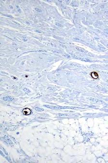
1 10/2/17 Overview Desmoplastic trichoepithelioma vs. morpheaform BCC vs. microcystic adnexal carcinoma Trichilemmomas & Cowden s syndrome Cutaneous Adnexal Tumours with Clinical Impact Rajiv M. Patel, M.D. RCPA NZ ASM 2017 (9:00-9:45am, Saturday, ) Multiple trichoepitheliomas, cylindromas, spiradenomas & Brooke-Spiegler Digital papillary adenocarcinoma Sebaceous tumors & Muir-Torre syndrome Trichodiscoma fibrofolliculomas/acrochordons & Birt-Hogg-Dube syndrome Proliferating pilar tumor vs. SCC 2 A 45-year-old female presents with a lesion on the cheek, rule out basal cell carcinoma 1

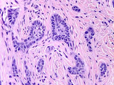
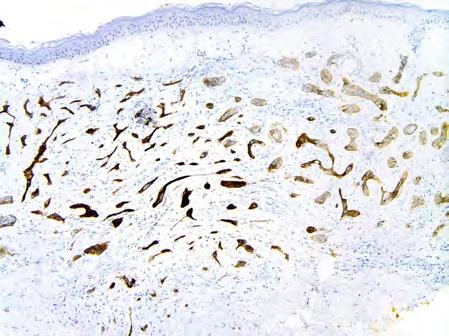
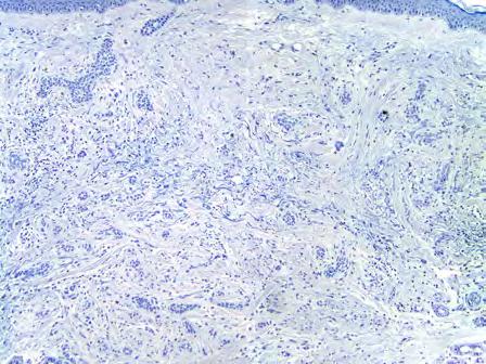
2 BerEP4 CK20 Diagnosis? Morpheaform Basal Cell Carcinoma (MBCC) 2


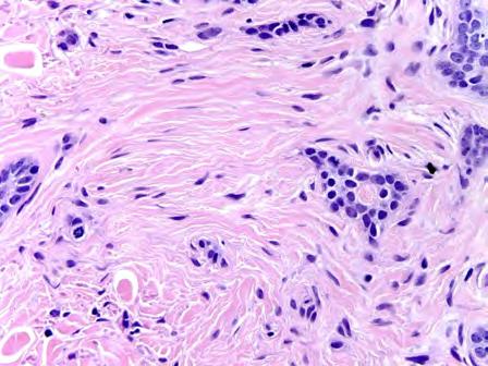
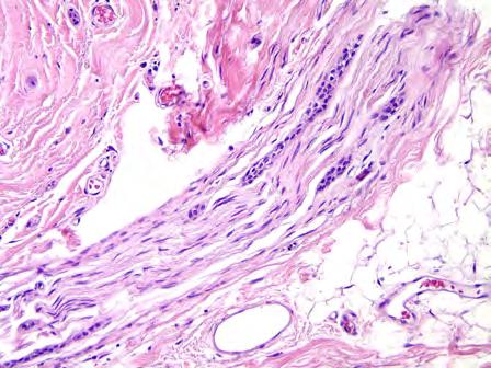
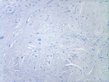
3 MBCC: Clinical 5% of all BCCs Head and neck Ill-defined, tethered, pale and shiny plaque that resembles morphea Lacks translucent appearance MBCC: Histology Ill-defined borders Infiltrative narrow strands of basaloid cells +/- Epidermal connection Blue, densely fibrotic /desmoplastic stroma Patchy lymphoid aggregates Inconspicuous myxoid change and clefting between tumor strands and stroma Perineural invasion present in 3% of morpheaform and infiltrative BCCs MBCC MBCC Deep infiltrative growth MBCC MBCC Cellular desmoplastic stroma Mild pleomorphism Frequent apoptoses 3


")
 Microcystic adnexal")


4 MBCC MBCC: BerEP4 Coarse chromatin Mitoses MBCC: CK20 CK20 stain highlighting retained Merkel cells in DTE Absence of intratumoral Merkel cells Differential Diagnosis Sclerosing basaloid neoplasms: Morpheaform BCC (MBCC) Desmoplastic trichoepithelioma (DTE) Microcystic adnexal carcinoma (MAC) H&E histopathology is the key IHC helpful in superficial biopsies Desmoplastic trichoepithelioma: Flesh colored annular plaque with central dell 4


")


5 10/2/17 DTE DTE Sclerosing variant of trichoepithelioma Usually presents on central face of adults often with central umbilication Cords of basaloid tumor cells in desmoplastic stroma Keratocysts and calcification (key distinguishing features) Intrastromal clefts Papillary mesenchymal bodies usually not present Well circumscribed DTE DTE Intrastromal clefts Intrastromal clefts DTE: CK20 DTE: CK20 Intratumoral Merkel cells Intratumoral Merkel cells 5

 MAC Clinical Features Adult patients (rare cases in children) Presents as firm plaque with central depression on upper lip, around mouth")

6 MBCC DTE Circumscription Poor Good Epid. connection +/- +/- Keratinous cysts +/- + Stroma Desmoplastic, cellular (blue) Sclerotic (pink) Lymph. aggregates + +/- Clefting Between tumor and stroma Intrastromal Nuclear atypia + - Mitosis/Apoptosis + -/Rare Perineural invasion +/- -/+ MBCC DTE CK20 (Merkel cells) -/Rare + Androgen Receptor + - CD34 (stromal cells) - + BerEP4 Diffuse + Focal + Bcl-2 Diffuse + Peripheral + Microcystic adnexal carcinoma (MAC) MAC Clinical Features Adult patients (rare cases in children) Presents as firm plaque with central depression on upper lip, around mouth or eyelids Microscopic features Infiltrative growth pattern Basaloid strands of tumor cells Superficially keratocysts Duct lumen formation often seen in deeper aspects of tumor Perineural invasion Dense sclerotic pink stroma Diffuse dermal involvement MAC MAC Syringomatous keratinous cysts Infiltration of deep tissue 6


7 MAC MAC Sclerotic (pink) stroma Subtle infiltration by small nests MAC MAC: CEA Eccrine ductular differentiation Highlights ductal lumina MAC MAC: CK20 Perineural invasion Absence of intratumoral Merkel cells 7

8 MAC: BerEP4 MAC Behavior Locally destructive Recurrence rate 30-40%; lower with Mohs surgery Rarely metastasizes Practical tip Up to 85% of MACs misdiagnosed as DTE or syringoma on small biopsies If you are uncertain of diagnosis on shave biopsy, ask for a repeat punch biopsy to assess for infiltrative growth and perineural invasion MBCC MAC Circumscription Poor Poor Epidermal connection +/- +/- Keratinous cysts +/- + Ductules - + Stroma Desmoplastic (blue) Sclerotic (pink) Lymph. aggregates + Usually - Clefting + - Nuclear atypia + Minimal Mitosis/Apoptosis + Infrequent Perineural invasion +/- Frequently + MBCC MAC EMA - Ductules + CEA - Ductules + BerEP4 + - Bcl-2 Diffuse + Focal + Androgen Receptor + - CK20 (Merkel cells) - - CD34 (stromal cells) - - MBCC: Prognosis & Treatment Infiltrative, difficult to obtain clear margins More aggressive and higher recurrence rate compared to other BCC subtypes Nodal metastases extremely rare Mohs surgery for facial lesions +/- Radiation for perineural invasion MBCC: Key Points Look for the desmoplastic and inflamed stroma, nuclear atypia, mitoses and apoptoses Use a panel of immunostains in superficial biopsies or difficult cases If a definitive diagnosis cannot be established on a partial biopsy, recommend excision 8

 hamartoma tumor syndrome (PHTS)")

9 A 33-year-old male with multiple, flesh colored papules around the nose Diagnosis? Multiple trichilemmomas Trichilemmoma Clinical features Usually presents on nose or lip of adults Multiple lesions major criterion of Cowden syndrome Part of the phosphatase & tensin homolog deleted on chromosome 10 (PTEN) hamartoma tumor syndrome (PHTS) Differential diagnosis Basal cell carcinoma with trichilemmal differentiation Mucinous change, clefting, apoptoses Inverted follicular keratosis Squamous eddies Verruca vulgaris No hair follicle involvement 9


 (Gangliocytoma) Meningioma GI")
 Acral keratoses Mucocutaneous")
 Intel")
 Thyroid structural lesions (e.g.")
: 1.")
10 Trichilemmoma Clear to pale cells Microscopic features Connects to epidermis Superficially coarse keratohyaline granules resembling koilocytes Endophytic growth Lobular architecture Clear to pale cells Peripheral palisading Eosinophilic basement membrane Peripheral palisading Cuticle Cowden s syndrome: Multiple trichilemmomas, acral keratoses, and oral mucosal cobblestoning. Increased risk of breast, thyroid, endometrial carcinomas Cowden (PTEN hamartoma) syndrome Sclerotic fibromas Major Criteria: Breast cancer Endometrial cancer Follicular thyroid cancer Lhermitte Duclos disease (LDD) (Gangliocytoma) Meningioma GI hamartomas or ganglioneuromas Macrocephaly ( 97 percentile: 58 cm for females, 60 cm for males) Macular pigmentation of the glans penis Multiple mucocutaneous lesions: Trichilemmomas ( 3, at least one biopsy proven) Acral keratoses Mucocutaneous neuromas ( 3) Oral papillomas Minor Criteria: Autism spectrum disorder Colon cancer Esophageal glycogenic acanthosis ( 3) Lipomas ( 3) Intellectual disability (i.e., IQ 75) Renal cell carcinoma Testicular lipomatosis Papillary thyroid cancer (papillary or follicular variant) Thyroid structural lesions (e.g., adenoma, nodule(s), goiter) Vascular anomalies (including multiple intracranial developmental venous anomalies) Operational diagnosis in an individual (either of the following): 1. Three or more major criteria, but one must include macrocephaly, LDD, or GI hamartomas; or 2. Two major and three minor criteria. Operational diagnosis in a family where one individual is diagnostic for Cowden syndrome: 1. Any two major criteria with or without minor criteria; or 2. One major and two minor criteria; or 3. Three minor criteria Multiple lesions associated with Cowden syndrome 10

 Multiple lesions associated with Cowden syndrome")


 A 38-year-old female presents with multiple, non-descript")
11 Sclerotic fibroma: CD 34 Desmoplastic trichilemmoma Similar to conventional TL except areas with infiltrative appearance and desmoplastic stroma Key to diagnosis: areas resembling TL, bland cytologic features & peritumoral BM (PAS, laminin, COL IV+) Multiple lesions associated with Cowden syndrome Trichilemmoma: Key Points Desmoplastic stroma (peritumoral BM) Have a high index of suspicion for this lesion on the lips & nose Multiple lesions are associated with Cowden syndrome BCC & SCC (DTL) most important DDx More atypical, look for other histologic features of these tumors Recognition of peritumoral basement membrane is the key to recognizing desmoplastic TL (PAS, laminin, ColIV+) A 38-year-old female presents with multiple, non-descript flesh colored papules on the central forehead 11


12 Diagnosis? Multiple Trichoepitheliomas Trichoepithelioma AKA cribiform trichoblastoma Clinical features Presents as skin colored papule Usually central face but location variable Solitary or multiple Multiple lesions associated with Brooke-Spiegler syndrome Trichoepithelioma Microscopic features Centered in mid dermis Sometimes has attachment to overlying epidermis Basaloid tumor cells arranged in nodules, nests or cords Tumor nodules often have branching pattern Keratocyst formation Papillary mesenchymal bodies Fibroblastic stroma Papillary mesenchymal body 12


 Multiple & ruptured keratocysts a clue to Dx")
13 Ruptured keratocyst Keratocyst Thrichoepithelioma vs. Basal cell carcinoma Dermal mucin in stroma Trichoepithelioma Younger adults Fibroblastic stroma No atypia, low mitotic rate No tumor retraction present Retained Merkel cells on CK20 stain Basal cell carcinoma Older adults Mucinous stroma May have atypia, more mitotic figures Tumor retraction often present No retained Merkel cells on CK20 stain Trichoepithelioma: Key points Head & neck esp. the face Multiple TE associated with multiple cylindromas, spiradenomas, salivary gland tumors, trichilemmomas, or BCC (Brook-Spiegler) Multiple & ruptured keratocysts a clue to Dx Generally lacks apoptotic bodies seen in BCC Demonstrates retained CK20 + Merkel cells, unlike BCC Tumor-stroma retraction space 13


 and")
14 22-year-old female with multiple smooth red papules & nodules on the scalp Diagnosis? Tumor with features of spiradenoma (right) and cylindroma (left) Spiradenocylindroma 14


15 Cylindroma Clinical features Adult F:M ratio 9:1 Smooth red papules, nodules (turban tumors) 90% head and neck, especially scalp Associated with Brooke- Spiegler syndrome Autosomal dominant Mutation in CYLD Cylindroma Microscopic features Nests of basaloid tumor cells surrounded by dense basement membrane material Tumor lobules have complex pattern Jigsaw puzzle pattern Small basaloid cells and larger pale cells Small basaloid cells Basement membrane Larger pale cells Spiradenoma Clinical features Most common in young adults Presents on upper half of body 75% on ventral surface Dermal mass, often painful Multiple lesions associated with Brooke-Spiegler syndrome Spiradenoma Microscopic features Basophilic nodules in dermis Fibrous capsule sometimes present Less complex pattern than cylindroma Two cell types: small basaloid cells and larger polygonal cells Focal duct lumen formation Eosinophilic globules within tumor lobules often present May have thick basement membrane May have areas indistinguishable from cylindroma 15


 Spiradenoma less complex growth pattern Often features of both")
 Basal cell carcinoma most practical DDx Search")
16 Ducts Polygonal cells Hyaline globules Basaloid cells Brooke-Spiegler syndrome Inherited in an autosomal-dominant fashion Consists of multiple spiradenomas associated with multiple trichoepitheliomas, cylindromas, BCC & benign & malignant tumours of the parotid gland Responsible gene is CYLD located at 16q12-q13 shows sequence mutations or loss of heterozygosity at 16q typical of a tumour suppressor gene Spiradenoma/cylindroma: Key points Similar cellular composition (basaloid & larger polygonal cells) Spiradenoma less complex growth pattern Often features of both in the same tumor Multiple lesions a marker of Brooke-Spiegler syndrome Rare malignant variants described (carcinoma ex cylindroma & spiradenoma) Basal cell carcinoma most practical DDx Search for additional features of BCC 16


17 62-year-old male with firm nodule of right index finger Diagnosis? Digital papillary adenocarcinoma Digital Papillary Adenocarcinoma Formerly divided in digital papillary adenoma and digital papillary adenocarcinoma Now recognized that there is no difference in behavior based on histology Both considered low-grade adenocarcinomas 17


 Squamous metaplasia Cystic spaces Clear cell change Papillary")
18 Digital Papillary Adenocarcinoma Clinical features Middle aged adults M>F (7:1) Fingers, toes, palms, and soles Often painful Behavior: Frequent recurrence and metastasis Have a high index of suspicion for any adnexal tumor at acral site Digital Papillary Adenocarcinoma Microscopic features Deep dermis to subcutis, relatively circumscribed Cystic spaces Back to back glands Papillary structures Mitoses & necrosis often encountered Squamous metaplasia and clear cell change common (but not predominant) Immunophenotype: Positive for keratin, S100, CEA (not diagnostically useful) Squamous metaplasia Cystic spaces Clear cell change Papillary projections Back to back glands 18


19 Differential: Digital papillary adenocarcinoma Hidradenoma Both have solid and cystic areas Clear cell change common in both Not papillary Lacks back to back glands Uncommon on digits Key points: Digital papillary adenocarcinoma Deceptively low-grade adenocarcinoma with recurrence in 50% and metastasis in 14% of cases, respectively Have a high index of suspicion for the Dx for any adnexal tumor arising at an acral site Complete excision with free margins, sometimes requires amputation Long term follow-up Usefulness of adjuvant therapy and SLNBx have not been defined 63-year-old male with a lesion on the neck


 & central mature sebocytes No or little cytologic atypia No or")
20 MSH2 Diagnosis? Low-grade cystic sebaceous neoplasm 115 Sebaceous Hyperplasia Middle aged and older patients Usually presents on central face Clinically resembles BCC Hyperplasia of normal sebaceous glands Increased number of glands Lobules of sebaceous glands grouped around follicle DDx: Sebaceous adenoma, nevus sebaceus Sebaceous Adenoma Clinical features Middle-aged and older patients Usually on head and neck Clinically resembles basal cell carcinoma Usually sporadic but may be hereditary and multiple Most common tumor associated with Muir-Torre syndrome Sebaceous neoplasms, SCC & visceral carcinomas (laryngeal & GI) Sebaceous Adenoma Abnormal sebaceous gland that connects to epidermis Not associated with follicle Predominantly composed of mature sebocytes Lobules of peripheral basaloid cells ( 50%) & central mature sebocytes No or little cytologic atypia No or rare mitotic figures DDx: Sebaceous hyperplasia, BCC with sebaceous differentiation Superficial biopsy may be difficult to distinguish from sebaceous carcinoma Epidermal connection 20


21 Sebaceoma Basaloid cells 50% Sebaceous adenoma variant Clinically similar to sebaceous adenoma Most common on head and neck Microscopic features Predominantly composed of basaloid cells (>50%) rather than mature sebocytes Basaloid cells intermingled with mature sebocytes No to mild atypia Mitotic figures often present but no atypical mitotic figures Basaloid cells admixed with sebocytes >50% basaloid cells 21

 Non-ocular associated with Muir-Torre syndrome Behavior Local recurrence 30-40% Metastasis")

 and EMA usually positive Infiltrative")
22 Differential: Sebaceoma Basal cell carcinoma with sebaceous differentiation BCC has peripheral palisading Retraction spaces Positive for Ber-EP4, unlike sebaceoma Sebaceous carcinoma Clinical: Sebaceous carcinoma Middle aged to older patients Two clinical locations: ocular (75%) and non-ocular (25%) Non-ocular associated with Muir-Torre syndrome Behavior Local recurrence 30-40% Metastasis 20-25% Mortality 10-20% Pathologic features: Sebaceous carcinoma Basaloid cells > sebocytes Microscopic features Nodular or infiltrative growth pattern Basaloid cells > sebocytes Nuclear atypia and mitotic figures Atypical mitotic figures common Squamous metaplasia and pagetoid spread common Especially ocular tumors Immunophenotype: Androgen receptor (AR) and EMA usually positive Infiltrative Nuclear atypia Atypical mitoses 22

 Heritable cancer syndrome associated with hereditary nonpolyposis colorectal cancer (HNPCC) MTS definition: heritable cancer syndrome with at least one sebaceous")
 ~ 40% of patients with sebaceous tumors have MTS Mean age sebaceous neoplasm in MTS: 63 yrs Range: 37-85 yrs.")
23 Sebaceous carcinoma of eyelid with pagetoid growth pattern High-grade sebaceous carcinoma Sebocytic differentiation difficult to appreciate 133 Note: cytoplasmic vacuoles indenting nuclei Differential: Sebaceous carcinoma Basal cell carcinoma with sebaceous differentiation Retraction spaces and peripheral palisading Androgen receptor positivity less prominent Clear cell squamous cell carcinoma Clear cells result of glycogen not lipid (PAS+) Negative for androgen receptor, negative or weak positivity for EMA Melanoma Melanin often present Lacks cytoplasmic vacuoles that indent nuclei Muir-Torre Syndrome (MTS) Heritable cancer syndrome associated with hereditary nonpolyposis colorectal cancer (HNPCC) MTS definition: heritable cancer syndrome with at least one sebaceous neoplasm and one underlying visceral malignancy Associated visceral malignancies: GI (47%), GU (21%), and breast (12%), most common Skin tumors in MTS: sebaceous adenoma (most common), sebaceous carcinoma and keratoacanthoma Muir-Torre Syndrome (MTS) ~ 40% of patients with sebaceous tumors have MTS Mean age sebaceous neoplasm in MTS: 63 yrs Range: yrs. Cutaneous tumors may be concurrent with diagnosis of visceral malignancy (9-22%) subsequent to visceral malignancy (56-59%) precede diagnosis of visceral malignancy (>6%) Muir-Torre Syndrome (MTS) Due to defects in DNA mismatch repair enzymes, usually MSH2 (90%) or MLH1 If sebaceous neoplasm encountered, consider screening for MTS Can screen with immunostains Loss of nuclear staining suggests MTS Not 100% specific If immunostains not available, correlate with family history. If there is family history of colon cancer or other cancer, or the patient is relatively young (<60), consider early screening for colon cancer 23


24 MSH2 PPV of lack of mismatch repair gene by IHC for diagnosis of Muir-Torre syndrome Gene that is lacking by IHC Positive predictive value (PPV) MSH2 55% MLH1 88% MSH6 67% Loss of staining in tumor cells MLH1 & MSH6 100% MSH2, MLH1 & MSH6 100% MLH, MutL Homolog; MSH, MutS Homolog. V. Chhibber, K. Dresser, M. MahalingamMSH-6: extending the reliability of immunohistochemistry as a screening tool in Muir-Torre syndrome. Mod Pathol, 21 (2008), pp Canned comments for sebaceous neoplasms 1. Initial biopsy with sebaceous neoplasm: Sebaceous neoplasms may arise sporadically or in association with the Muir-Torre syndrome. Immunohistochemical stains for mismatch repair proteins (MSH2, MSH6, MLH1 and PMS2) are available and can be performed upon the clinician s request. 2. Reporting of positive MMR results: Immunohistochemical stains against the mismatch repair proteins show loss of expression for and retained expression for within the sebaceous neoplasm. These results are not diagnostic but may be seen in Muir-Torre syndrome. Correlation with clinical findings and family history is required. Molecular testing (microsatellite instability and/or gene mutation analysis) may be performed to confirm germline mutation if clinically indicated. 3. Reporting of negative MMR results: Immunohistochemical stains against the mismatch repair proteins show intact expression for MSH2, MSH6, MLH1 and PMS2 within the sebaceous neoplasm. These results speak against but do not entirely exclude Muir-Torre syndrome. Correlation with clinical findings and family history is required. Molecular testing (microsatellite instability and/or gene mutation analysis) may be considered if clinical concern for Muir-Torre syndrome persists Key points: Sebaceous neoplasms & MT Precise classifications of sebaceous neoplasms may not be possible in limited biopsies In this case a dx of low-grade or atypical sebaceous neoplasm with a recommendation for complete excision appropriate May be sporadic or associated with MT Screening for MSI by IHC is reliable & cost effective Results suggesting MSI should be followed up with definitive genetic testing and counseling year-old male with multiple papules on the nose 24


25 Diagnosis? Fibrofolliculoma 145 Clinical: Fibrofolliculoma/trichodiscoma Flesh-colored papules on face, neck Indistinguishable clinically Favored to be follicular hamartomas Multiple in Birt-Hogg-Dube syndrome Pathology: Fibrofolliculoma/trichodiscoma Fibrofolliculoma Central distorted hair follicle Thin anastomosing strands of follicular epithelium centered around infundibular portion of follicle Pale fibrotic stroma Trichodiscoma Mainly fibrous Often only with surrounding collarette of epithelium Birt-Hogg-Dube: Trichodiscoma Fibrofolliculoma/trichodiscoma: Central distorted follicle with surrounding fibrosis and a surrounding network of thin anastomosing strands of follicular epithelium. Predominantly fibrous stroma surrounded by epidermal collarette 25


26 Birt-Hogg- Dube: Trichodiscomas Birt-Hogg-Dube Autosomal Dominant Follicular hamartomas (Fibrofolliculomas, Perifollicular fibromas, trichodiscomas) and acrochordons Renal tumors Oncocytoma, chromophobe RCC and clear cell RCC Pulmonary symptoms Emphysema, cysts, spontaneous pneumothoraces Less frequently thyroid CA Predominantly fibrous stroma surrounded by epidermal collarette Birt-Hogg-Dube Mutation in folliculin (FCLN) on 17p 11.2 Thought to be a tumor suppressor acting as a regulator of mtor pathway Encodes folliculin which interacts with FNIP1 and FNIP2 Differential: Fibrofolliculoma/trichodiscoma BCC Retraction artifact, mucinous stroma, palisading nuclei, atypia Neurofolliular hamartoma Spindle cell rich trichodiscoma S100 & CD34+ Perifollicular fibroma Form of fibrofolliculoma Perifollicular fibrosis surrounds normal follicles rather than the epithelial strands seen in fibrofolliculoma Differential: Fibrofolliculoma/trichodiscoma Fibrofolliculoma Perifollicular fibroma Neurofollicular hamartoma Spindle cell trichodiscoma with atypia and fatty metaplasia


27 Key points: Fibrofolliculoma/trichodiscoma Fibrofolliculoma & trichodiscoma represent the opposite ends of a spectrum of the same lesion Multiple lesions are a marker for Birt-Hogg-Dube syndrome, which is associated with renal tumors, pulmonary symptoms & rarely thyroid carcinoma Neurofollicular hamartoma, perfollicular fibroma and spindle cell predominant trichodiscoma are histologic variants These lesions are benign 62-year-old female with a solitary exophytic lesion on the scalp, present for many years Diagnosis? Proliferating pilar tumor
 Often large exophytic")


 abrupt keratinization (lacks")


28 Clinical: Proliferating pilar tumor (PPT) Usually older individuals Females > males Scalp most common site (90%) Often large exophytic tumors measuring 10cm in diameter Recurrence after excision infrequent 2% of pilar tumors will develop into PPT Rare instances of transformation into malignant PPT Metastasis to regional lymph nodes may occur Complete excision with margin of normal tissue Pathology: Proliferating pilar tumor (PPT) Lobular proliferation of squamous epithelium with trichilemmal (pilar) abrupt keratinization (lacks granular layer), hylanized membrane at periphery, peripheral palisading Well-demarcated peripheral margin Remnant pilar cyst usually present Epithelium often demonstrates: Variable clear cell change Disordered proliferation Nuclear pleomorphism Prominent nucleoli Mitoses DDx: SCC= More atypical, infiltrative boarders, rapid evolution Well-demarcated Abrupt keratinization Disordered growth Central cystic area SCC Atypia Pilar cyst Mitosis

29 Key features distinguishing PPT from SCC Preexisting simple pilar cyst Good circumscription Smooth peripheral margins Trichilemmal (abrupt) keratinization Lack of significant cytologic atypia & minimal mitotic activity Presence of peripheral palisading Presence of hyaline basement membrane Acknowledgements Thank you to my colleagues: May Chan & Steve Billings for sharing PPT slides used in this talk
Appendageal skin tumors
 Appendageal skin tumors Ibrahim Khalifeh, M.D. Associate Professor Department of Pathology American University of Beirut Medical Center Beirut, Lebanon Appendageal tumors Neoplasms whose differentiation
Basal cell carcinoma 5/28/2011
 Goal of this Presentation A practical approach to the diagnosis of cutaneous carcinomas and their mimics Thaddeus Mully, MD University of California San Francisco To review common non-melanoma skin cancers
SEBACEOUS NEOPLASMS. Dr. Prachi Saraogi Clinical Fellow in Dermatology
 SEBACEOUS NEOPLASMS Dr. Prachi Saraogi Clinical Fellow in Dermatology Sebaceous neoplasms Sebaceous adenoma (Benign) Sebaceous carcinoma (Malignant) SEBACEOUS ADENOMA Benign tumours composed of incompletely
- Selected Tumors of the Skin Appendages - Primary vs. Metastasis
 - Selected Tumors of the Skin Appendages - Primary vs. Metastasis Napa Valley 2018 Victor G. Prieto, MD, PhD Chair of Pathology UT MD Anderson Cancer Center vprieto@mdanderson.org Napa Valley in May Introduction
Cowden Syndrome PTEN Hamartoma Tumor Syndrome. ACCME/Disclosure. 1. Background. Outline
 MASSACHUSETTS GENERAL HOSPITAL HARVARD MEDICAL SCHOOL PATHOLOGY Cowden Syndrome PTEN Hamartoma Tumor Syndrome ACCME/Disclosure Vania Nosé, MD, PhD Professor of Pathology Director of Anatomic Pathology
Keratinocyte tumors. Actinic Keratosis. Squamous cell carcinoma in situ. Squamous Cell Carcinoma. (aka Bowen s disease)
") Actinic Keratosis Keratinocyte tumors Prepared by Kurt Schaberg Precancerous, risk of malignancy ~8-20% per year (progresses to SCC); Due to chronic sun exposure Rough scaly plaque; typically due to sun
Actinic Keratosis Keratinocyte tumors Prepared by Kurt Schaberg Precancerous, risk of malignancy ~8-20% per year (progresses to SCC); Due to chronic sun exposure Rough scaly plaque; typically due to sun
Whitney A. High, MD, JD, MEng
 ADS Dermatopathology Meeting 2014 Selected Adnexal Tumors Whitney A. High, MD, JD, MEng Associate Professor, Dermatology & Pathology Director of Dermatopathology (Dermatology) University of Colorado School
Apocrine and eccrine adnexal tumors
 Apocrine and eccrine adnexal tumors Timothy McCalmont, MD University of California San Francisco, CA ECCRINE TUMORS Generally uncommon, because the sweat apparatus has little proliferative potential (hyperproliferation
Apocrine and eccrine adnexal tumors
 Apocrine and eccrine adnexal tumors Timothy McCalmont, MD University of California San Francisco, CA Past misguided notions Syringoma is eccrine Eccrine poroma rather than eccrine or apocrine poroma Spiradenoma
Desmoplastic Melanoma R/O BCC. Clinical Information. 74 y.o. man with lesion on left side of neck r/o BCC
 R/O BCC Sabine Kohler, M.D. Professor of Pathology and Dermatology Dermatopathology Service Stanford University School of Medicine Clinical Information 74 y.o. man with lesion on left side of neck r/o
Simplified approach to cutaneous adnexal tumors
 Simplified approach to cutaneous adnexal tumors Chandra Smart, MD Associate Clinical Professor UCLA Department of Pathology Introduction: Cutaneous adnexal neoplasms (CANs) are a diverse group of tumors
Samer Ghosn, MD Associate professor, Derpartment of Dermatology American University of Beirut Medical Center. Follicular lesions
 Samer Ghosn, MD Associate professor, Derpartment of Dermatology American University of Beirut Medical Center Follicular lesions Introduction Follicular lesions are important to recognize: For proper management
Prior Authorization. Additional Information:
 Genetic Testing for Cowden Syndrome - PTEN Gene MP9488 Covered Service: Prior Authorization Required: Additional Information: Yes when meets criteria below Yes as shown below Pre and post-test genetic
Salivary Glands 3/7/2017
 Salivary Glands 3/7/2017 Goals and objectives Focus on the entities unique to H&N Common board type facts Information for your future practice Salivary Glands Salivary Glands Major gland. Paratid. Submandibular.
BSD 2015 Case 19. Female 21. Nodule on forehead. The best diagnosis is:
 BSD 2015 Case 19 Female 21. Nodule on forehead. The best diagnosis is: A. mixed tumour of skin B. porocarcinoma C. nodular hidradenoma D. metastatic adenocarcinoma BSD 2015 Case 19 Female 21 Nodule on
Cutaneous Adnexal Tumors
 Cutaneous Adnexal Tumors Lesions with Predominant Follicular Differentiation Special Emphasis on Basal Cell Carcinoma 2014-04-01 Prof. Dr. med. Katharina Glatz Pathologie Cutaneous Adnexal Tumors Hair
Pathology of the skin. 2nd Department of Pathology, Semmelweis University
 Pathology of the skin 2nd Department of Pathology, Semmelweis University Histology of the skin Epidermis: Stratum corneum Stratum granulosum Stratum spinosum Stratum basale Dermis: papillary and reticular
Journal of International Academy of Forensic Science & Pathology (JIAFP)
") Journal of International Academy of Forensic Science & Pathology (JIAFP) ISSN 2395-0722 MICROCYSTIC ADNEXAL CARCINOMA-A CASE REPORT WITH REVIEW OF LITERATURE Case Report Sulakshana M S 1,Natarajan M 2
Journal of International Academy of Forensic Science & Pathology (JIAFP) ISSN 2395-0722 MICROCYSTIC ADNEXAL CARCINOMA-A CASE REPORT WITH REVIEW OF LITERATURE Case Report Sulakshana M S 1,Natarajan M 2
American College of Mohs Surgery. Diagnostic Quality Control Program (Review of Answers)
") American College of Mohs Surgery Diagnostic Quality Control Program 2010 (Review of Answers) Question 1 A flesh-colored plaque on the right ala of an otherwise healthy 57 year-old male is referred for
American College of Mohs Surgery Diagnostic Quality Control Program 2010 (Review of Answers) Question 1 A flesh-colored plaque on the right ala of an otherwise healthy 57 year-old male is referred for
USCAP Neuropathology night panel CASE 2
 USCAP Neuropathology night panel CASE 2 B.K. Kleinschmidt-DeMasters MD University of Colorado at Denver and Health Sciences Center Denver, Colorado The Chinese Wall, Flat Tops Wilderness, Colorado Clinical
Benign versus Cancerous Lesions How to tell the difference FMF 2014 Christie Freeman MD, CCFP, DipPDerm, MSc
 1 Benign versus Cancerous Lesions How to tell the difference FMF 2014 Christie Freeman MD, CCFP, DipPDerm, MSc Benign lesions Seborrheic Keratoses: Warty, stuck-on Genetics and birthdays Can start in late
Myxo-inflammatory Fibroblastic sarcoma
 AKA Myxo-inflammatory Fibroblastic sarcoma Acral Myxoinflammatory fibroblastic sarcomaam.j.surg.path1998; 22; 911-924 Inflammatory myxoid tumour of soft parts with bizarre giant cells [Pathol.Res.Pract.
Benign and malignant epithelial lesions: Seborrheic keratosis: A common benign pigmented epidermal tumor occur in middle-aged or older persons more
 Benign and malignant epithelial lesions: Seborrheic keratosis: A common benign pigmented epidermal tumor occur in middle-aged or older persons more common on the trunk; but extremities, head and neck are
Lesions Mimicking Adenoid Cystic Carcinoma. Diagnostic Problems in Salivary Gland Pathology An Update 5/29/2009
 Diagnostic Problems in Salivary Gland Pathology An Update Lesions Mimicking Adenoid Cystic Carcinoma Stacey E. Mills, M.D. W.S. Royster Professor of Pathology Director of Surgical and Cytopathology University
3. Histopathology. 1. Introduction. 2. Case History. Volume 6 Issue 4, April Licensed Under Creative Commons Attribution CC BY
 Spiradenocylindroma with Trichoepithelioma A Collision Tumor with Multiple Differentiation R. Lavanya 1, S. K. Sridevi 2, P. Viswanathan 3, P. V. S.Prasad 4 1 II nd Year Post Graduate, Department of Pathology,
Skin Adnexal Tumors - A Histopathological Spectrum at a Tertiary Care Hospital
 Original Article GCSMC J Med Sci Vol (VI) No (I) January-June 2017 Skin Adnexal Tumors - A Histopathological Spectrum at a Tertiary Care Hospital Neeraja Barve*, Hansa Goswami**, Urvi Parikh *** Abstract
أملس عضلي غرن = Leiomyosarcoma. Leiomyosarcoma 1 / 5
 Leiomyosarcoma 1 / 5 EPIDEMIOLOGY Exact incidence is unknown, but older studies suggest that leiomyosarcomas comprise approximately 3 percent of soft-tissue sarcomas. Superficial leiomyosarcoma occurs
Salivary Gland Cytology
 Salivary Gland Cytology Diagnostic challenges and potential pitfalls Tarik M. Elsheikh, MD Professor and Medical Director Anatomic Pathology Cleveland Clinic FNA Salivary Gland Lesions Indications Distinguish
Case Report A Rare Cutaneous Adnexal Tumor: Malignant Proliferating Trichilemmal Tumor
 Case Reports in Medicine Volume 2015, Article ID 742920, 4 pages http://dx.doi.org/10.1155/2015/742920 Case Report A Rare Cutaneous Adnexal Tumor: Malignant Proliferating Trichilemmal Tumor Omer Alici,
Slide seminar. Asist. Prof. Jože Pižem, MD, PhD Institute of Pathology Medical Faculty, University of Ljubljana
 Slide seminar Asist. Prof. Jože Pižem, MD, PhD Institute of Pathology Medical Faculty, University of Ljubljana Case 5 A 57-year-old man with a dermal/subcutaneous lesion on the scalp, which was interpreted
What is new on adnexal neoplasms. Omar P. Sangueza, MD. Professor and Director of Dermatopathology. Wake Forest University School of Medicine
 What is new on adnexal neoplasms Omar P. Sangueza, MD Professor and Director of Dermatopathology Wake Forest University School of Medicine Winston Salem, North Carolina Carney complex is an autosomal dominant
Papillary Lesions of the breast
 Papillary Lesions of the breast Emad Rakha Professor of Breast Pathology The University of Nottingham Papillary lesions of the breast are a heterogeneous group of disease, which are characterised by neoplastic
BASAL CELL CARCINOMA WITH ECCRINE DIFFERENTIATION: A RARE ENTITY Divvya B 1, Rehana Tippoo 2, P. Viswanathan 3, B. Krishnaswamy 4, A.
 BASAL CELL CARCINOMA WITH ECCRINE DIFFERENTIATION: A RARE ENTITY Divvya B 1, Rehana Tippoo 2, P. Viswanathan 3, B. Krishnaswamy 4, A. Anvar Ali 5 HOW TO CITE THIS ARTICLE: Divvya B, Rehana Tippoo, P. Viswanathan,
Papillary Lesions of the Breast A Practical Approach to Diagnosis. (Arch Pathol Lab Med. 2016;140: ; doi: /arpa.
 Papillary Lesions of the Breast A Practical Approach to Diagnosis (Arch Pathol Lab Med. 2016;140:1052 1059; doi: 10.5858/arpa.2016-0219-RA) Papillary lesions of the breast Span the spectrum of benign,
Case Report Clear Cell Basal Cell Carcinoma
 SAGE-Hindawi Access to Research Volume 2011, Article ID 386921, 4 pages doi:10.4061/2011/386921 Case Report Clear Cell Basal Cell Carcinoma Deba P. Sarma, 1 Daniel Olson, 1 Jennifer Olivella, 1 Tracey
Cutaneous metastases. Thaddeus Mully. University of California, San Francisco Professor, Departments of Pathology and Dermatology
 Cutaneous metastases Thaddeus Mully University of California, San Francisco Professor, Departments of Pathology and Dermatology DISCLOSURE OF RELATIONSHIPS WITH INDUSTRY Thaddeus Mully Course C005 Essential
confusing, especially in small, limited biopsies. One such case of digital papillary adnexal adenocarcinoma with unusual histologic features will be
 1 Cutaneous Adnexal Lesions: The Must-Know Stuff 2011 USCAP Annual Meeting Dermatopathology Companion Meeting Doina Ivan, M.D. University of Texas MD Anderson Medical Center, Houston, TX The diagnosis
MECHANISMS OF HUMAN DISEASE: LABORATORY SESSION PATHOLOGY OF THE SKIN LAB. Friday, February 12, :30 am 11:00 am
 MECHANISMS OF HUMAN DISEASE: LABORATORY SESSION PATHOLOGY OF THE SKIN LAB Friday, February 12, 2012 9:30 am 11:00 am FACULTY COPY GOALS: Describe the basic clinical and morphologic features of various
Synonyms. Nephrogenic metaplasia Mesonephric adenoma
 Nephrogenic Adenoma Synonyms Nephrogenic metaplasia Mesonephric adenoma Definition Benign epithelial lesion of urinary tract with tubular, glandular, papillary growth pattern Most frequently in the urinary
Diseases of the breast (1 of 2)
") Diseases of the breast (1 of 2) Introduction A histology introduction Normal ducts and lobules of the breast are lined by two layers of cells a layer of luminal cells overlying a second layer of myoepithelial
Diseases of the breast (1 of 2) Introduction A histology introduction Normal ducts and lobules of the breast are lined by two layers of cells a layer of luminal cells overlying a second layer of myoepithelial
Histopathology: skin pathology
 Histopathology: skin pathology These presentations are to help you identify, and to test yourself on identifying, basic histopathological features. They do not contain the additional factual information
Update in Salivary Gland Pathology. Benjamin L. Witt University of Utah/ARUP Laboratories February 9, 2016
 Update in Salivary Gland Pathology Benjamin L. Witt University of Utah/ARUP Laboratories February 9, 2016 Objectives Review the different appearances of a selection of salivary gland tumor types Establish
ARIZONA SOCIETY OF PATHOLOGISTS 13 TH APRIL 2013 HEAD AND NECK CYTOPATHOLOGY. F ZAHRA ALY, MD, PhD
 ARIZONA SOCIETY OF PATHOLOGISTS 13 TH APRIL 2013 HEAD AND NECK CYTOPATHOLOGY F ZAHRA ALY, MD, PhD The main areas sites amenable for cytopathology include lymph nodes, thyroid, major salivary glands especially
04/10/2018. Intraductal Papillary Neoplasms Of Breast INTRADUCTAL PAPILLOMA
 Intraductal Papillary Neoplasms Of Breast Savitri Krishnamurthy MD Professor of Pathology Deputy Division Head The University of Texas MD Anderson Cancer Center 25 th Annual Seminar in Pathology Pittsburgh,
Objectives. Salivary Gland FNA: The Milan System. Role of Salivary Gland FNA 04/26/2018
 Salivary Gland FNA: The Milan System Dr. Jennifer Brainard Section Head Cytopathology Cleveland Clinic Objectives Introduce the Milan System for reporting salivary gland cytopathology Define cytologic
Disclosure. Relevant Financial Relationship(s) None. Off Label Usage None MFMER slide-1
 None. Off Label Usage None MFMER slide-1") Disclosure Relevant Financial Relationship(s) None Off Label Usage None 2013 MFMER slide-1 Case Presentation A 43 year old male, with partial nephrectomy for a right kidney mass 2013 MFMER slide-2 2013
Disclosure Relevant Financial Relationship(s) None Off Label Usage None 2013 MFMER slide-1 Case Presentation A 43 year old male, with partial nephrectomy for a right kidney mass 2013 MFMER slide-2 2013
Disclosures. Parathyroid Pathology. Objectives. The normal parathyroid 11/10/2012
 Disclosures Parathyroid Pathology I have nothing to disclose Annemieke van Zante MD/PhD Assistant Professor of Clinical Pathology Associate Chief of Cytopathology Objectives 1. Review the pathologic features
Oncocytic-Appearing Salivary Gland Tumors. Oncocytic, Cystic, Mucinous, and High Grade Salivary Gland Tumors SALIVARY GLAND FNA: PART II
 William C. Faquin, MD, PhD Professor of Pathology Harvard Medical School Director of Head and Neck Pathology Massachusetts Eye and Ear Massachusetts General Hospital SALIVARY GLAND FNA: PART II Oncocytic,
Mody. AIS vs. Invasive Adenocarcinoma of the Cervix
 Common Problems in Gynecologic Pathology Michael T. Deavers, M.D. Houston Methodist Hospital, Houston, Texas Common Problems in Gynecologic Pathology Adenocarcinoma in-situ (AIS) of the Cervix vs. Invasive
Normal thyroid tissue
 Thyroid Pathology Overview Normal thyroid tissue Normal thyroid tissue with follicles filled with colloid. Thyroid cells form follicles, spheres of epithelial cells (always single layered in health, usually
Diagnostic Cytology of Cancer Cases
 Diagnostic Cytology of Cancer Cases Somporn Techangamsuwan Companion Animal Cancer Research Unit (CAC-RU) Department of Pathology, Faculty of Veterinary Science, Chulalongkorn University 1 Tumor or Non-tumor
MECHANISMS OF HUMAN DISEASE: LABORATORY SESSION PATHOLOGY OF THE SKIN LAB. Friday, February 13, :30 am 11:00 am
 MECHANISMS OF HUMAN DISEASE: LABORATORY SESSION PATHOLOGY OF THE SKIN LAB Friday, February 13, 2009 9:30 am 11:00 am FACULTY COPY GOALS: Describe the basic clinical and morphologic features of various
Dermatopathology: The tumor is composed of keratinocytes which show atypia, increase mitoses and abnormal mitoses.
 Squamous cell carcinoma (SCC): A common malignant tumor of keratinocytes arising in the epidermis, usually from a precancerous condition: 1- UV induced actinic keratosis, usually of low grade malignancy.
Gross appearance of nodular hyperplasia in material obtained from suprapubic prostatectomy. Note the multinodular appearance and the admixture of
 Tiền liệt tuyến Tiền liệt tuyến Gross appearance of nodular hyperplasia in material obtained from suprapubic prostatectomy. Note the multinodular appearance and the admixture of solid and microcystic areas.
Introduction. Results. Discussion. Histopathologic and immunohistochemical findings. Results. conclusions,
 1/5 2/5 Carcinoma distinctive carcinoma. form erysipeloides (CE), metastasis. which clinically Itfrom has resembles been termed erysipelas, is an uncommon, but may extend It164 toclassically back, presents
A 5 Year Histopathological Study of Skin Adnexal Tumors at a Tertiary Care Hospital
 IOSR Journal of Dental and Medical Sciences (IOSR-JDMS) e-issn: 2279-0853, p-issn: 2279-0861.Volume 14, Issue 4 Ver. VII (Apr. 2015), PP 01-05 www.iosrjournals.org A 5 Year Histopathological Study of Skin
Microcystic Squamous Cell Carcinoma of the Lung A Clinicopathologic Study of Three Cases
 Anatomic Pathology / Microcystic SCC of the Lung Microcystic Squamous Cell Carcinoma of the Lung A Clinicopathologic Study of Three Cases Annikka Weissferdt, MD, and Cesar A. Moran, MD Key Words: Squamous
المركب النموذج--- سبيتز وحمة = Type Spitz's Nevus, Compound SPITZ NEVUS 1 / 7
 SPITZ NEVUS 1 / 7 Epidemiology An annual incidence rate of 1.4 cases of Spitz nevus per 100,000 individuals has been estimated in Australia, compared with 25.4 per 100,000 individuals for cutaneous melanoma
Note: The cause of testicular neoplasms remains unknown
 - In the 15- to 34-year-old age group, they are the most common tumors of men. - Tumors of the testis are a heterogeneous group of neoplasms that include: I. Germ cell tumors : 95%; all are malignant.
Corporate Medical Policy
 Corporate Medical Policy Genetic Testing for PTEN Hamartoma Tumor Syndrome File Name: Origination: Last CAP Review: Next CAP Review: Last Review: genetic_testing_for_pten_hamartoma_tumor_syndrome 4/2013
My Journey into the World of Salivary Gland Sebaceous Neoplasms
 My Journey into the World of Salivary Gland Sebaceous Neoplasms Douglas R. Gnepp Warren Alpert Medical School at Brown University Rhode Island Hospital Pathology Department Providence RI Asked to present
Differential Diagnosis of Oral Masses. Palatal Lesions
 Differential Diagnosis of Oral Masses Palatal Lesions Palatal Masses Periapical Abscess Torus Palatinus Mucocele Lymphoid Hyperplasia Adenomatous Hyperplasia Benign Salivary Neoplasms Malignant Salivary
Disorders of Cell Growth & Neoplasia. Histopathology Lab
 Disorders of Cell Growth & Neoplasia Histopathology Lab Paul Hanna April 2010 Case #84 Clinical History: 5 yr-old, West Highland White terrier. skin mass from axillary region. has been present for the
FNA OF SALIVARY GLANDS: A PRACTICAL APPROACH
 FNA OF SALIVARY GLANDS: A PRACTICAL APPROACH FNA of Salivary Glands: Challenges Wide range of neoplastic and non-neoplastic lesions Cytological overlap between the different benign and malignant tumors
5/21/2018. Prostate Adenocarcinoma vs. Urothelial Carcinoma. Common Differential Diagnoses in Urological Pathology. Jonathan I.
 Common Differential Diagnoses in Urological Pathology Jonathan I. Epstein Prostate Adenocarcinoma vs. Urothelial Carcinoma 1 2 NKX3.1 NKX3.1 3 4 5 6 Proposed ISUP Recommendations Option to use PSA as a
MALIGNANT POROMA SYNONYM: POROCARCINOMA ECCRINE POROMA MALIGNANT Divvya B 1, M. Valluvan 2, Rehana Tippoo 3, P. Viswanathan 4, R.
 MALIGNANT POROMA SYNONYM: POROCARCINOMA ECCRINE POROMA MALIGNANT Divvya B 1, M. Valluvan 2, Rehana Tippoo 3, P. Viswanathan 4, R. Ramesh 5 HOW TO CITE THIS ARTICLE: Divvya B, M. Valluvan, Rehana Tippoo,
Enterprise Interest None
 Enterprise Interest None B3 lesions of the breast What are they at surgery? Case 4 Edi Brogi MD PhD Attending Pathologist - Director of Breast Pathology Memorial Sloan Kettering Cancer Center New York
Various hereditary, acquired and neoplastic conditions can lead to cyst formation in the kidney.
 Dr. Fatima AlAl-Hashimi Hashimi,, MD, FRCPath Salmaniya Medical Complex, Bahrain Various hereditary, acquired and neoplastic conditions can lead to cyst formation in the kidney. The most frequently encountered
Desmoplastic trichoepithelioma; a new case and review of the literature with emphasis on its differential diagnosis
 Hong Kong J. Dermatol. Venereol. (2014) 22, 135-139 Case Report Desmoplastic trichoepithelioma; a new case and review of the literature with emphasis on its differential diagnosis M Concha-Rogazy, S Alvarez-Veliz,
PLEOMORPHIC ADENOMA ( BENIGN MIXED TUMOR )
") ( BENIGN MIXED TUMOR ) Grossly, the tumor is freely movable, solid, sometimes lobulated and occasionally cystic. If recurrent, multinodular masses are common. Histologically, within a fibrous capsule,
( BENIGN MIXED TUMOR ) Grossly, the tumor is freely movable, solid, sometimes lobulated and occasionally cystic. If recurrent, multinodular masses are common. Histologically, within a fibrous capsule,
Cluster designation 5 staining of normal and non-lymphoid neoplastic skin*
 J Cutan Pathol 2005: 32: 50 54 Copyright # Blackwell Munksgaard 2005 Blackwell Munksgaard. Printed in Denmark Journal of Cutaneous Pathology Cluster designation 5 staining of normal and non-lymphoid neoplastic
Los Angeles Society Of Pathologists Dr. Shobha Castelino Prabhu
 Los Angeles Society Of Pathologists Dr. Shobha Castelino Prabhu Loma Linda University Medical Center June 12, 2007 CASE 1 76 year-old gentleman Status post right parotidectomy 1 year ago for a rare tumor
Case Report Endocrine Mucin-Producing Sweat Gland Carcinoma, a Histological Challenge
 Hindawi Volume 2017, Article ID 6343709, 4 pages https://doi.org/10.1155/2017/6343709 Case Report Endocrine Mucin-Producing Sweat Gland Carcinoma, a Histological Challenge Mary Anne Brett, Samih Salama,
1 NORMAL HISTOLOGY AND METAPLASIAS
 1 NORMAL HISTOLOGY AND METAPLASIAS, MD Anatomy and Histology 1 Metaplasias 2 ANATOMY AND HISTOLOGY The female breast is composed of a branching duct system, which begins at the nipple with the major lactiferous
A clinicopathologic study of skin appendageal tumors
 Net Study A clinicopathologic study of skin appendageal tumors Pradeep S. Nair Department of Dermatology and Venereology, Medical College Hospital, Trivandrum- 695 011, Kerala, India Address for correspondence:
Epithelial tumors. Dr. F.F. Khuzin, PhD Dr. M.O. Mavlikeev
 Epithelial tumors Dr. F.F. Khuzin, PhD Dr. M.O. Mavlikeev Epithelial tumors Tumors from the epithelium are the most frequent among tumors. There are 2 group features of these tumors: The presence in most
PSA. HMCK, p63, Racemase. HMCK, p63, Racemase
 Case 1 67 year old male presented with gross hematuria H/o acute prostatitis & BPH Urethroscopy: small, polypoid growth with a broad base emanating from the left side of the verumontanum Serum PSA :7 ng/ml
CASE year old male with a PET avid nodule in the left adrenal gland
 CASE 1 55 year old male with a PET avid nodule in the left adrenal gland Case 1 Adrenal gland parenchyma partly replaced by a spindle cell tumour with mild nuclear pleomorphism Atypical mitoses present
Problem diagnoses. Current issues in Anatomic pathology. Problem Diagnoses in Tumors of the Oral Cavity 5/29/2009
 Current issues in Anatomic pathology Problem Diagnoses in Tumors of the Oral Cavity Richard Jordan DDS PhD FRCPath Professor of Oral Pathology & Pathology Director, UCSF Oral Pathology Diagnostic Laboratory
21/07/2017. Hobnail endothelial cells are not the same as epithelioid endothelial cells
 UPDATE IN CUTANEOUS VASCULAR S DERMATOPATHOLOGY SESSION BELFAST PATHOLOGY JUNE 21/2017 Dr E Calonje St John s Institute of Dermatology, London, United Kingdom THE FAMILY OF VASCULAR S WITH EPITHELIOID
Educational Cases EQA November T.J. Palmer Raigmore Hospital Inverness
 Educational Cases EQA November 2013 T.J. Palmer Raigmore Hospital Inverness Case 2 Clinical Details Dob 11 February 1951 PMH: 1964 Extraction of 45 aet 13 yr 1966 Cyst between 44 and 46 enucleated 1973
Special slide seminar
 Special slide seminar Tomáš Rozkoš The Fingerland Department of Pathology Charles University Medical Faculty and Faculty Hospital in Hradec Králové Czech Republic Case history, 33 years old resistance
Submission of samples. Cytology of Lumps and Bumps. Evaluation of samples. Use caution interpreting. Criteria of malignancy.
 Submission of samples Cytology of Lumps and Bumps Paul Avery VMD, PhD, DACVP paul.avery@colostate.edu Air dry only No wet fixation using formalin or ethanol Stain 1-2 on-site to evaluate quality Send all
Case 18. M75. Excision of mass on scalp. Clinically SCC. The best diagnosis is:
 Case 18 M75. Excision of mass on scalp. Clinically SCC. The best diagnosis is: A. Pilomatrical carcinoma B. Adnexal carcinoma NOS C. Metastatic squamous cell carcinoma D.Primary squamous cell carcinoma
Spectrum of Preneoplastic and Neoplastic Cystic Lesions of the Kidney in Adult. by dr. Banan Burhan Mohammed Lecturer in Pathology Department
 Spectrum of Preneoplastic and Neoplastic Cystic Lesions of the Kidney in Adult by dr. Banan Burhan Mohammed Lecturer in Pathology Department Various hereditary, acquired, and neoplastic conditions can
Nasreen A. Syed, MD F.C. Blodi Eye Pathology Laboratory University of Iowa
 Nasreen A. Syed, MD F.C. Blodi Eye Pathology Laboratory University of Iowa No financial disclosures No discussion of off-label use of medications or unapproved devices 67 year old male referred to Oculoplastics
LUMPS AND BUMPS: AN ORGANIZED APPROACH TO DIAGNOSIS AND MANAGEMENT
 LUMPS AND BUMPS: AN ORGANIZED APPROACH TO DIAGNOSIS AND MANAGEMENT Tammy P. Than, M.S., O.D., F.A.A.O. The University of Alabama at Birmingham / School of Optometry 1716 University Blvd. Birmingham, AL
Histopathological Study of Tumours of Epidermis and Epidermal Appendages
 Original 460 Article Indian Journal of Pathology: Research and Practice Volume 6 Number 2, April - June 2017 (Part 2) DOI: http://dx.doi.org/10.21088/ijprp.2278.148x.6217.22 Histopathological Study of
The Relevance of Cytologic Atypia in Cutaneous Neural Tumors
 The Relevance of Cytologic Atypia in Cutaneous Neural Tumors Recent Findings - New Developments New Problems Zsolt B. Argenyi, M.D. Professor of Pathology & Dermatology Director of Dermatopathology Department
Spindle Cell Lesions Of The Breast. Emad Rakha Professor of Breast Pathology and Consultant Pathologist
 Spindle Cell Lesions Of The Breast Emad Rakha Professor of Breast Pathology and Consultant Pathologist * SCLs comprise a wide spectrum of diseases, ranging from reactive processes to aggressive malignant
Pitfalls in thyroid tumor pathology. Prof.Valdi Pešutić-Pisac MD, PhD
 Pitfalls in thyroid tumor pathology Prof.Valdi Pešutić-Pisac MD, PhD Too many or... Tumour herniation through a torn capsule simulating capsular invasion fibrous capsule with a sharp discontinuity, suggestive
BAP-oma & BEYOND MICHAEL A NOWAK, MD
 BAP-oma & BEYOND MICHAEL A NOWAK, MD CONFLICTS No conflicts with the content of this lecture BAP-oma Wiesner 2011: Families with multiple tan dome-shaped papules of head, neck, trunk, and extremities.
Neoplasia literally means "new growth.
 NEOPLASIA Neoplasia literally means "new growth. A neoplasm, defined as "an abnormal mass of tissue the growth of which exceeds and is uncoordinated with that of the normal tissues and persists in the
CASE REPORT SOLITARY SEBACEOUS NEVUS OF JADASSOHN COMPLICATED BY SQUAMOUS CELL CARCINOMA AND BASAL CELL CARCINOMA
 CASE REPORT Dennis H. Kraus, MD, Section Editor SOLITARY SEBACEOUS NEVUS OF JADASSOHN COMPLICATED BY SQUAMOUS CELL CARCINOMA AND BASAL CELL CARCINOMA Ahmad Ridzwan Arshad, FRCS, 1 Wan S. Azman, MS, 1 Ayadurai
Disclosure of Relevant Financial Relationships
 Squamous entities of the thyroid: Reactive to Neoplastic Michelle D. Williams Associate Professor Dept of Pathology, Head & Neck Section University of Texas MD Anderson Cancer Center Disclosure of Relevant
Salivary gland tumor cytologic and histologic correlation: Algorithmic and risk stratification based approaches
 Salivary gland tumor cytologic and histologic correlation: Algorithmic and risk stratification based approaches Christopher C. Griffith, MD, PhD Raja R. Seethala, MD 1. Salivary gland tumor cytology: A
2 to 3% of All New Visceral Cancers Peak Incidence is 6th Decade M:F = 2:1 Grossly is a Bright Yellow, Necrotic Mass with a Pseudocapsule
 GENITOURINARY PATHOLOGY Kathleen M. O Toole, M.D. Renal Cell Carcinoma 2 to 3% of All New Visceral Cancers Peak Incidence is 6th Decade M:F = 2:1 Grossly is a Bright Yellow Necrotic Mass Grossly is a Bright
Neoplasia 2018 Lecture 2. Dr Heyam Awad MD, FRCPath
 Neoplasia 2018 Lecture 2 Dr Heyam Awad MD, FRCPath ILOS 1. List the differences between benign and malignant tumors. 2. Recognize the histological features of malignancy. 3. Define dysplasia and understand
Malignant tumors of melanocytes: Part 1. Deba P Sarma, MD., Omaha
 Malignant tumors of melanocytes: Part 1 Deba P Sarma, MD., Omaha The melanocytic tumor is one of the most difficult and confusing areas in Dematopathology. It is true that most (95%) of such lesions are
When Immunostains Can Get You in Trouble: Gynecologic Pathology p16: Panacea or Pandora s Box?
 When Immunostains Can Get You in Trouble: Gynecologic Pathology p16: Panacea or Pandora s Box? Teri A. Longacre, MD Stanford Medicine Stanford California pi6 in Gynecologic Pathology: Panacea or Pandora