What really matters When and Why. Pathology of Uterine Mesenchymal Lesions. Nafisa Wilkinson London
|
|
|
- Marilyn Banks
- 5 years ago
- Views:
Transcription
1 What really matters When and Why Pathology of Uterine Mesenchymal Lesions Nafisa Wilkinson London


2 Patient centred approach immunohistochemistry Histological diagnosis Next generation sequencing Genetic mutations The patient! Gross examination 2
3 Mesenchymal lesions what matters Is it smooth muscle or endometrial stromal? Smooth Muscle Benign/ Uncertain Malignant Potential/ Malignant Endometrial Stromal Benign/ LGESS/ HGESS/ Undifferentiated U. Sarcoma 3
4 Clinical relevance: management options If radiologically malignant MDT review CT chest, abdo, pelvis TAH+BSO+omentectomy ( ESS) TAH (min) Leiomyosarcoma Referral to sarcoma team (registration) Adjuvant treatment (if indicated) LMS (no good evidence for hormonal mnx) ESS (Anti oestrogen or progesterone) Recurrences managed by Sarcoma team 4
5 Aims Clinical History what matters? Gross examination sampling- what matters? Histological examination when should you worry? Diagnosing a leiomyosarcoma when should you worry? Diagnosing an ESS- What is important? Immunohistochemistry How to use it? Molecular markers How and when to use them? Prognostication? Any markers that are reliable? 5
6 Clinical History what matters? Age Young patient with multiple fibroids? HLRCC syndrome Hormonal status- peri or post menopausal (concern for malignancy) Drug history GnRH agonist treatment, Tranexamic acid, Ulipristal Acetate (Esmya) Previous procedures embolisation, laser Rx, rapid growth following embolization (concerning feature) 6
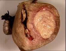
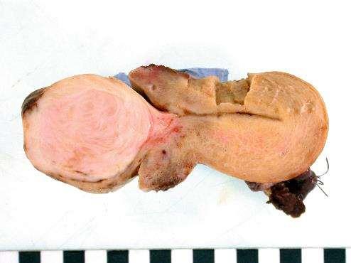
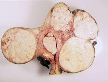
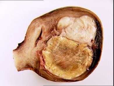
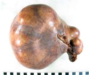
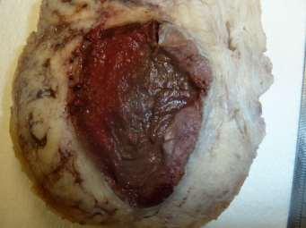
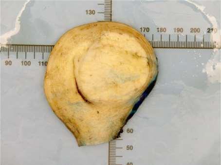
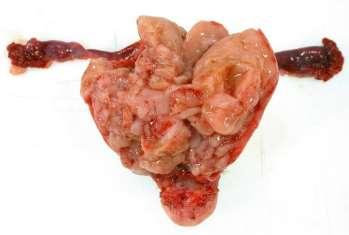
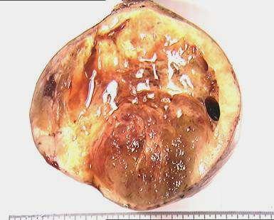
7 Gross examination Size (largest more likely to be malignant) Numerous- grossly examine all Hysterectomy: Sample largest 3 or 4 Myomectomy: rep sections of each if no worrisome features Colour Uniform, white whorled appearance bulging at you Yellow - sample Heterogenous- sample, esp Tumour/ myometrial border Polypoid with myometrial component- sample 7

8 Leiomyoma typical
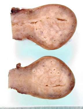
9 Appearances that do not matter. 9

10 Not typical gross appearance 10
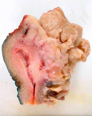
11 Concerning gross appearance
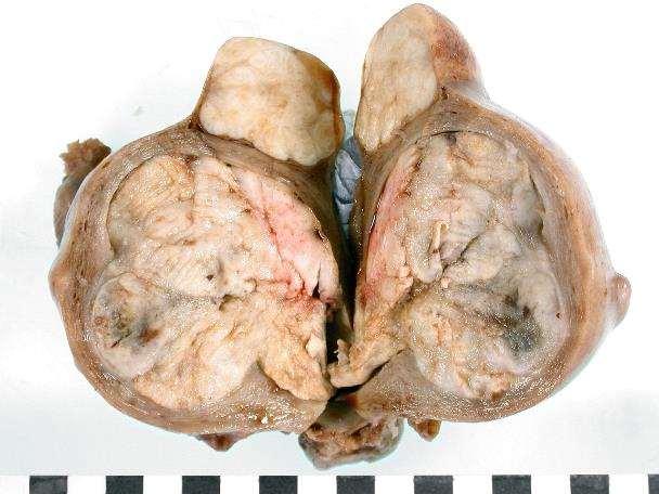
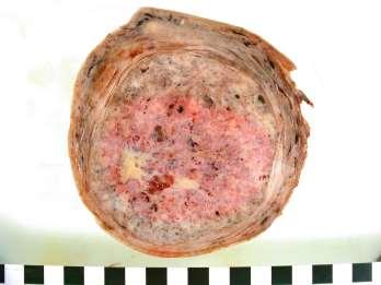
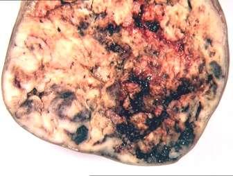
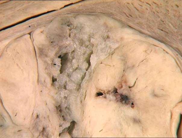
12 Heterogenous gross appearance 12


13 Fixation important to assess mesenchymal lesions Following fixation Fresh 13
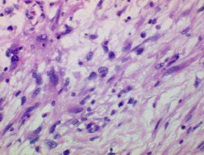
14 Leiomyosarcoma Most important diagnosis for the patient Account for 1% of all uterine malignancies Most patients are post menopausal Usually confined to the uterus when diagnosed Recurrent disease- lung and pelvis (commonest site of metastasis) Bone, cranial/intracranial, skin and soft tissue 5 year survival rate stage I and II is 40-70% 15-25% for all stages. STAGE MOST IMPORTANT PROGNOSTIC INDICATOR 14

15 STAGING FOR UTERINE SARCOMAS Int J Obstet Gynecol 2009, 104, 179 Measure maximum tumour dimension Block adnexa in entirety 15
16 Grading in LMS No robust grading system that relates to prognosis Soft tissue sarcoma grading not relevant to uterine LMS All are high grade at present 16
17 Problems with dx of leiomyosarcoma Leiomyoma variants that have some but not all features malignancy STUMP (Smooth Muscle Tumours of Uncertain Malignant Potential) Is it a Leiomyosarcoma? 17
18 Diagnosis of leiomyosarcoma - 2 of 3 main criteria required for usual type Mitotic Activity (increased > 10MF/10HPF) Coagulative Tumour cell necrosis Cytologic atypia (diffuse) Vascular invasion (10-20%) Infiltrative border often seen (if searched for!) Bell, Kempson and Hendrickson: Am J Surg Pathol 1994:18;


19 Diffuse Cytologic Atypia Mitotically active, atypical forms, marked DIFFUSE atypia Low power very cellular neoplasm 19


20 Coagulative necrosis Overtly apparent coagulative necrosis


21 Leiomyosarcoma: haemorrhage and necrosis Highly cellular tumour 21


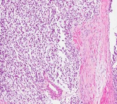
22 Remember hyaline necrosis is seen in LMS Zone of granulation tissue between viable and non-viable tumour 22


23 Sample extensively at tumour/myometrial border Vascular invasion at advancing edge of tumour
24 LMS-immunohistochemistry Desmin * p53, P16 and Ki 67 H-caldesmon * Smooth muscle actin * ER, PR and AR positive (30-40%) C-kit (CD117) and DOG 1 may be positive (no c-kit mutation found) CD10 can be positive * Cytokeratins, EMA may be positive * Diagnostically useful markers

25 Mitotically active leiomyoma check where you count mitoses beware submucosal leiomyoma under ulcerated surface! Ki-67 very helpful Identifies zone of proliferation No atypia, no coagulative necrosis, smooth border with myometrium. 25


26 Cellular leiomyomata No Atypia No increase in mitotic activity Irregular peripheral border Not infiltrative border


27 Leiomyoma with bizarre nuclei Typically, no mitoses or 1-2/ 10 HPF, no coagulative tumour cell necrosis 27

28 Leiomyoma with bizarre nuclei 28


29 FH deficient leiomyoma (HLRCC) Younger patients <40 years, multiple leiomyomata, staghorn vessels, eosinophilic globules, per-nuclear halos 29


30 With thanks to Dr Sakinah Thiryayi Intravenous leiomyomatosis 30


31 Intravenous leiomyomatosis No cytologic atypia or brisk mitotic activity, note IVL can show All the same changes that are seen within intramyometrial leiomyomata IVL in Uterus With thanks to Dr Sakinah Thiryayi 31 Tumour within right ventricle
32 Myxoid leiomyosarcoma 32


33 Myxoid Leiomyosarcoma Mitotic activity Alcian blue +ve 33
34 Myxoid Leiomyosarcoma Severe cytologic atypia + / - tumour cell necrosis Any Mitotic index Mitotic Index > 2MF/10 HPF No cytologic atypia No necrosis 34
35 EPITHELIOID LEIOMYOSARCOMA Very rare especially pure epithelioid tumours Few cases in literature Diffuse moderate to severe cytologic atypia > 3 MF/ 10 HPF Necrosis - microscopic 35

36 EPITHELIOID LEIOMYOSARCOMA 36


37 PEComa Epithelioid morphology Clear /eosinophilic cytoplasm Pink granular cytoplasm Centrally located/round nuclei Nested growth pattern Epithelioid and spindled cells TSC1 or TSC 2 mutations Dysregulation of the mtor signalling pathway mtor inhibitors may be helpful in Rx Express melanocytic and muscle markers 37
 Endometrial stromal sarcoma (high grade) specific t(10:17) Undifferentiated Uterine")
38 WHO Updated grading for EST Endometrial stromal nodule Endometrial stromal sarcoma (low grade) Endometrial stromal sarcoma (high grade) specific t(10:17) Undifferentiated Uterine Sarcoma 38


39 Cellular leiomyoma Vs ESN Cellular leiomyoma Endometrial stromal nodule 39
40 LG ESS Clinical features Age usually < 50 years Pelvic or abdominal pain/abnormal vaginal bleeding Variable sized neoplasm (polypoid / bulky) 1/3 rd extrauterine pelvic extension at diagnosis May present with metastasis (Ovary common site) Indolent and protracted course (characterised by recurrences) May be associated with prolonged oestrogenic stimulation, tamoxifen Rx or prior pelvic irradiation 40


41 Endometrial Stromal sarcoma - LG Stromal cells proliferate around small calibre arterioles Resembles the stroma seen in the proliferative phase 41


42 LGESS pushing, tongue-like growth Extensive lymphovascular space permeation Note no stromal response 42


43 ESS vs Leiomyosarcoma ESS- Proliferation around arterioles Leiomyosarcoma- proliferation in fascicles 43


44 Endometrial stromal tumour Endometrial stromal proliferation in the absence of glands consider ESS as possible dx Note if base not identified cannot exclude an ESS on curettage material 44
45 LGESS -Immunohistochemistry CD10 strong diffuse positive (usually) * ER/ PR/ WT1 : typically positive * SMA - often positive Desmin- occasionally positive * H-caldesmon negative (+ ve smooth muscle * differentiation) C-Kit (CD117) may be positive but no c KIT mutations Aromatase Androgen receptor may be positive (sex cord like areas) AE1/AE3 epithelial differentiation Inhibin/ calretinin/melan-a and CD99- may be positive * = Diagnostically useful markers 45
46 ESS LG Molecular genetics t(7;17) -80% JAZF1-SUZ12 t(6;7)- 6% PHF1-JAZF1 Am J Surg Pathol 2011; 35: Chiang S et al Frequency gene rearrangements endometrial stromal tumours t(6;10) -4% EPC1-PHF1 46
47 ESS Low Grade Molecular genetics useful Pelvic tumour ER, PR positive Not given history of previous ESS (low grade). Establishes dx in most cases 47

48 HGESS- Dual Cell population 48

49 Necrosis seen Round cell component 49

50 Extensive LVSI 50
51 HG ESS Immunohistochemistry High grade component CD10 ve ER ve PR ve Cyclin D1 (>70%) strong, diffuse, nuclear +ve C KIT (cytoplasmic strong) DOG 1( -ve) in high grade and low grade areas Beta-catenin (cytoplasmic) no nuclear positivity Negative for: EMA, SMA, desmin, caldesmon, HMB-45, Melan A and cytokeratin 51

52 Cyclin D 1 diffuse positive Courtesy Dr Oliva Courtesy Dr Esther Oliva

53 FISH t(10;17)(q22;p13) Courtesy of Drs Lee and Oliva, YWHAE-NUTM2 ESS 53
54 Undifferentiated uterine sarcoma Definition A tumour arising in the endometrium or myometrium, lacking resemblance to proliferative phase endometrial stroma with high-grade cytological features and no specific type of differentiation Rare tumour, patients post menopausal, mean age 60 years Prognosis: Poor. Patients present with high stage disease (>60%). Even patients with stage I disease DOD within 2 years Adjuvant therapy no therapeutic benefit 54
55 Undifferentiated Uterine sarcoma Heterogeneous group of sarcomas with high mitotic activity and necrosis lacking diagnostic criteria for: ESS (high grade) Leiomyosarcoma Rhabdomyosarcoma Adenosarcoma with sarcomatous overgrowth Carcinosarcoma (esp when sarcoma has overgrown carcinoma) Undifferentiated or dedifferentiated endometrial carcinoma Complex Karyotype (many structural and numerical aberrations) 55

56 Endometrial Stromal Sarcoma: prognosis Am J Surg Pathol, 2012: 36,

57 Smooth muscle tumour of uncertain malignant potential WHO
58 Leiomyoma with bizarre nuclei Downes and Hart MIB-1 BZL <10% Suggests Leiomyosarcoma >15% Some consider any SMT with diffuse moderate to severe atypia No tumour cell necrosis >5 to < 9 MFs /10HPF leiomyosarcoma others STUMP Small number have recurred. 58
59 Leiomyoma with bizarre nuclei Croce, Young and Oliva 2014 Am J Surg Pathol cases Mitotic counts 0 to 7/10 HPF (average 1-2/10HPF) 37 (63%) had <2 MF/10 HPF 19 (32%) had 2-5 MF/10 HPF 3 ( 5%) had 6,7,7 MF/10 HPF 2 with focal and 1 diffuse BN 2.9,5.7 and 5.5. years FU respectively None recurred (follow-up 1 to 13 years) 59
60 STUMP sample carefully and generously SMT with coagulative necrosis but < 10 MF/ 10 HPF SMT with diffuse cytologic atypia, <10MF/10HPF and no necrosis or unsure about necrosis SMT with focal or multifocal moderate to severe atypia but <10 MF/ 10 HPF SMT with no necrosis or atypia but > 15 MF/ 10 HPF 60

61 Leiomyosarcoma No reliable prognostic markers Adjuvant treatment is used to variable effect Need to await specific markers before significant impact on Rx NGS of tumours and precision medicine may be the answer NCI and the precision medicine gov 61
62 Mesenchymal tumours Thorough sampling of tumours that look unusual especially at the tumour/ myometrial border. Use immunohistochemistry as a panel Investigate carefully before labelling a stromal neoplasm an undifferentiated uterine sarcoma (much worse prognosis). Use molecular markers for low grade and HG ESS to support diagnosis especially of pelvic tumours which are recurrent stromal sarcomas. Have a low threshold for referral of these tumours as they are rare. 62
ESS: Pathologic Insights
 GEIS XVI INTERNATIONAL SYMPOSIUM Seville 4th October 2018 ESS: Pathologic Insights Sílvia Bagué The Royal Marsden Hospital London (United Kingdom) I have no conflicts of interest Endometrial stromal sarcoma
Endometrial Stromal Tumors
 Endometrial Stromal Tumors WHO Categories: Endometrial Stromal Nodule (ESN) Endometrial Stromal Sarcoma, low grade (LGESS) Endometrial Stromal Sarcoma, high grade (HGESS) Undifferentiated Uterine Sarcoma
Mody. AIS vs. Invasive Adenocarcinoma of the Cervix
 Common Problems in Gynecologic Pathology Michael T. Deavers, M.D. Houston Methodist Hospital, Houston, Texas Common Problems in Gynecologic Pathology Adenocarcinoma in-situ (AIS) of the Cervix vs. Invasive
05/07/2018. Types of challenges. Challenging cases in uterine pathology. Case 1 ` 65 year old female Post menopausal bleeding Uterine Polyp
 Types of challenges Challenging cases in uterine pathology Nafisa Wilkinson Gynaecological Pathologist UCLH London Lack of complete history often, NO clinical history at all! Cases from other centres often
Diagnostic problems in uterine smooth muscle tumors
 Diagnostic problems in uterine smooth muscle tumors Marina Kos Ljudevit Jurak Clinical Department of Pathology, Clinical Hospital Center Sestre milosrdnice, Zagreb Institute of Pathology, University of
64 YO lady THBSO for prolapse At gross : A 3 cm endometrial polyp in the fundus
 Case 6 64 YO lady THBSO for prolapse At gross : A 3 cm endometrial polyp in the fundus Numerous irregular, large glands with leaf-like pattern Large glands with broad-based papillary infolding into the
CyclinD1 Positive High-Grade Endometrial Stromal Sarcoma: A Fascinating Entity!
 Case Report DOI: 10.21276/APALM.1530 CyclinD1 Positive High-Grade Endometrial Stromal Sarcoma: A Fascinating Entity! Divya Shelly*, Imtiaz Ahmed, Sampath K. Srinivasagowda and Reena Bharadwaj Department
Gynecologic Evening Specialty Conference. Karuna Garg, MD University of California San Francisco
 Gynecologic Evening Specialty Conference Karuna Garg, MD University of California San Francisco Disclosure of Relevant Financial Relationships The USCAP requires that anyone in a position to influence
Uterine Mesenchymal Tumors from a Gynaecological Point of View: A Mini-Review
 EC Gynaecology Special Issue - 2017 Uterine Mesenchymal Tumors from a Gynaecological Point of View: A Mini-Review Mini Review Dr. Huseyin Aydogmus, Dr. Servet Gencdal, Dr. Nihan Gencdal and Dr. Serpil
Original Article Endometrial stromal sarcoma: a clinicopathological analysis of 14 cases
 Int J Clin Exp Pathol 2018;11(5):2799-2804 www.ijcep.com /ISSN:1936-2625/IJCEP0073760 Original Article Endometrial stromal sarcoma: a clinicopathological analysis of 14 cases Fuqiang Wang *, Ruixue Lei
International Journal of Case Reports in Medicine
 International Journal of Case Reports in Medicine Vol. 2013 (2013), Article ID 665097, 28 minipages. DOI:10.5171/2013.665097 www.ibimapublishing.com Copyright 2013 Hemalatha A. L., Varna I, Deepthi B.
Normal endometrium: A, proliferative. B, secretory.
 Normal endometrium: A, proliferative. B, secretory. Nội mạc tử cung Nội mạc tử cung Cyclic changes in endometrium.. Approximate relationship of useful microscopic changes. Arias-Stella reaction in endometrial
Disclosure. Case. Mixed Tumors of the Uterine Corpus and Cervix. I have nothing to disclose
 Mixed Tumors of the Uterine Corpus and Cervix Marisa R. Nucci, M.D. Division of Women s and Perinatal Pathology Department of Pathology Brigham and Women s Hospital Boston, MA UCSF Current Issues in Anatomic
SMOOTH MUSCLE TUMOURS
 SMOOTH MUSCLE TUMOURS NORMAL SMOOTH MUSCLE Cytology Immunohistochemistry Ultrastructure Masson Trichrome Smooth Muscle Ultrastructure Many myofilaments running parallel to the long axis of the smooth
Department of Pathology, Royal Group of Hospitals Trust, Belfast, Northern Ireland.
 UTERINE ADENOSARCOMA W Glenn McCluggage Department of Pathology, Royal Group of Hospitals Trust, Belfast, Northern Ireland. Definition of Adenosarcoma: A mixed tumor composed of benign neoplastic glandular
Clinicopathologic correlation of endometrial stromal sarcomas: a retrospective study of 42 cases
 Original Article Clinicopathologic correlation of endometrial stromal sarcomas: a retrospective study of 42 cases Fengjie Wang 1,2, Yongfen Yi 1, Yi Luo 3, Jing Chen 1 1 Department of Pathology, Molecular
STUMPed for a Diagnosis Contemporary Management of Uterine Sarcomas
 UCSF Helen Diller Family Comprehensive Cancer Center Disclosures I have no financial disclosures STUMPed for a Diagnosis Contemporary Management of Uterine Sarcomas Lee-may Chen, MD Department of Obstetrics,
Endometrial Stromal Sarcoma
 May 26, 2011 By Sushila Ladumor, MD [1] Endometrial stromal sarcoma (ESS) is a rare malignant tumor of the endometrium, occurring in the age group of 40-50 years. History The 50-year-old, female patient
UTERINE SARCOMAS CURRENT THERAPEUTIC OPTIONS
 Review Journal of Translational Medicine and Research, volume 19, no. 1-2, 2014 UTERINE SARCOMAS CURRENT THERAPEUTIC OPTIONS N. Bacalbaæa 1, A. Traistaru 2, I. Bãlescu 3 1 Carol Davila University of Medicine
The Good Uterine Sarcomas: What Do You Need to Know
 The Good Uterine Sarcomas: What Do You Need to Know Anais Malpica, M.D. Departments of Pathology and Gynecologic Oncology The University of Texas M.D. Anderson Cancer Center Matthew Powell, M.D. Division
UTERINE SARCOMA EXAMPLE OF A UTERINE SARCOMA USING PROPOSED TEMPLATE
 UTERINE SARCOMA EXAMPLE OF A UTERINE SARCOMA USING PROPOSED TEMPLATE Case: Adenosarcoma with heterologous elements and stromal overgrowth o TAH, BSO, omentectomy, staging biopsies of cul-de-sac, bladder
Dr Sanjiv Manek Oxford. Oxford Pathology Course 2010 for FRCPath Illustration-Cellular Pathology. Oxford Radcliffe NHS Trust
 Dr Sanjiv Manek Oxford Oxford Pathology Course 2010 for FRCPath Illustration-Cellular Pathology. Oxford Radcliffe NHS Trust Ovarian Endometrial Vulvo-vaginal Cervical Illustration-Cellular Pathology. Oxford
Standards and datasets for reporting cancers Dataset for histopathological reporting of uterine sarcomas. September 2018
 Standards and datasets for reporting cancers Dataset for histopathological reporting of uterine sarcomas September 2018 Authors: Professor W Glenn McCluggage, Royal Group of Hospitals, Belfast Professor
59 yo male with past medical history of prostate carcinoma, presented with upper abdominal pain
 December 2016 59 yo male with past medical history of prostate carcinoma, presented with upper abdominal pain Contributed by: Divya Sharma, MD. Fellow, Gastrointestinal Pathology, Department of Pathology
The role of immunohistochemistry in surgical pathology of the uterine corpus and cervix
 The role of immunohistochemistry in surgical pathology of the uterine corpus and cervix Prof. Ben Davidson, MD PhD Department of Pathology, Norwegian Radium Hospital, Oslo University Hospital, Oslo, Norway
Atypical uterine leiomyoma: a case report and review of the literature
 DOI 10.1186/s13256-016-0800-3 CASE REPORT Open Access Atypical uterine leiomyoma: a case report and review of the literature Suzana Manxhuka-Kerliu 1*, Irma Kerliu-Saliu 2, Vjollca Sahatciu-Meka 3, Lloreta
Classification (1) Classification (3) Classification (2) Spindle cell lesions. Spindle cell lesions of bladder (Mills et al.
 Classification (3) Classification (2) Spindle cell lesions. Spindle cell lesions of bladder (Mills et al.") Non-epithelial tumours and nonepithelial tumour-like lesions of the bladder Dr Jonathan H Shanks The Christie NHS Foundation Trust, Manchester, UK Classification (1) Myofibroblastic proliferations and
Non-epithelial tumours and nonepithelial tumour-like lesions of the bladder Dr Jonathan H Shanks The Christie NHS Foundation Trust, Manchester, UK Classification (1) Myofibroblastic proliferations and
Female Genital Tract Lab. Dr. Nisreen Abu Shahin Assistant Professor of Pathology University of Jordan
 Female Genital Tract Lab Dr. Nisreen Abu Shahin Assistant Professor of Pathology University of Jordan Ovarian Pathology A 20-year-old female presented with vague left pelvic pain. Pelvic exam revealed
أملس عضلي غرن = Leiomyosarcoma. Leiomyosarcoma 1 / 5
 Leiomyosarcoma 1 / 5 EPIDEMIOLOGY Exact incidence is unknown, but older studies suggest that leiomyosarcomas comprise approximately 3 percent of soft-tissue sarcomas. Superficial leiomyosarcoma occurs
Diagnostically Challenging Cases in Gynecologic Pathology
 Diagnostically Challenging Cases in Gynecologic Pathology Eric C. Huang, M.D., Ph.D. Department of Pathology and Laboratory Medicine University of California, Davis Medical Center Case 1 Presentation 38
Scotland and Northern Ireland EQA Scheme. Circulation 46
 Scotland and Northern Ireland EQA Scheme Circulation 46 Special Educational Cases E1 and E2 Presented by Dr K Robertson Case E1 Female 42 year old with heavy menstrual and intermenstrual bleeding. IUS
Case Report. Low Grade Endometrial Stromal Sarcoma: Report of a Rare Uterine Malignancy
 Iranian 64 Journal of Pathology (2013) 8 (1), 64-70 Case Report Low Grade Endometrial Stromal Sarcoma: Report of a Rare Uterine Malignancy Pinki Pandey 1, Alok Dixit 2, Aparna Tanwar 1, Nanak Chand Mahajan
Staging and Treatment Update for Gynecologic Malignancies
 Staging and Treatment Update for Gynecologic Malignancies Bunja Rungruang, MD Medical College of Georgia No disclosures 4 th most common new cases of cancer in women 5 th and 6 th leading cancer deaths
57th Annual HSCP Spring Symposium 4/16/2016
 An Unusual Malignant Spindle Cell Lesion to Involve the Breast Erinn Downs-Kelly, D.O. Associate Professor of Pathology University of Utah & ARUP Laboratories No disclosures Case 39 y/o female with no
Newer soft tissue entities
 Newer soft tissue entities Examples among fibroblastic tumors Turku, May 6, 2010 Markku Miettinen, M.D. AFIP, Washington, DC Fibroblastic neoplasms Solitary fibrous tumor /Hemangiopericytoma Low-grade
3/25/2019. Rare uterine cancers ~3% Leiomyosarcoma Carcinosarcoma (MMMT) Endometrial Stromal Sarcomas Aggressive tumors High Mortality Rates
 Endometrial Stromal Sarcomas Aggressive tumors High Mortality Rates") J. Anthony Rakowski D.O., F.A.C.O.O.G. MSU SCS Board Review Coarse Rare uterine cancers ~3% Leiomyosarcoma Carcinosarcoma (MMMT) Endometrial Stromal Sarcomas Aggressive tumors High Mortality Rates Signs
J. Anthony Rakowski D.O., F.A.C.O.O.G. MSU SCS Board Review Coarse Rare uterine cancers ~3% Leiomyosarcoma Carcinosarcoma (MMMT) Endometrial Stromal Sarcomas Aggressive tumors High Mortality Rates Signs
Case: The patient is a 24 year- old female who was found to have multiple mural nodules within the antrum. Solid and cystic components were noted on
 Case: The patient is a 24 year- old female who was found to have multiple mural nodules within the antrum. Solid and cystic components were noted on imaging. There is no significant past medical history.
A 25 year old female with a palpable mass in the right lower quadrant of her abdomen
 May 2016 A 25 year old female with a palpable mass in the right lower quadrant of her abdomen Contributed by: Paul Ndekwe, MD, Resident Physician, Indiana University School of Department of Pathology and
I have nothing to disclose
 A 47 year old female with multiple lung nodules Disclosure of Relevant Financial Relationships Tamar Giorgadze, MD, PhD Professor of Pathology Medical College of Wisconsin Milwaukee, Wisconsin USCAP requires
Trophoblastic tumors
 Trophoblastic tumors Uterus tumor course Oslo, 21-22/1/16 Prof. Ben Davidson, MD PhD Department of Pathology, Norwegian Radium Hospital, Oslo University Hospital, Oslo, Norway Cases 45 38 39 4 Case 45
Icd 10 uterine leiomyosarcoma
 Icd 10 uterine leiomyosarcoma Search Aetna considers myomectomy or hysterectomy using power morcellation experimental and investigational for the removal of uterine fibroids because its safety and. Free,
An Overview of Genital Stromal Tumors
 An Overview of Genital Stromal Tumors By Konstantinos Linos MD, FCAP, FASDP Bone, Soft Tissue and Dermatopathology Assistant Professor of Pathology Dartmouth-Hitchcock Medical Center Geisel School of Medicine
RETROPERITONEAL RECURRENCE OF UTERINE SMOOTH MUSCLE TUMOR OF UNCERTAIN MALIGNANT POTENTIAL AS LEIOMYOSARCOMA
 CASE REPORT Korean J Obstet Gynecol 2012;55(12):996-1000 http://dx.doi.org/10.5468/kjog.2012.55.12.996 pissn 2233-5188 eissn 2233-5196 RETROPERITONEAL RECURRENCE OF UTERINE SMOOTH MUSCLE TUMOR OF UNCERTAIN
CASO 1. Xavier Matias-Guiu Hospital Universitari Arnau de Vilanova, Universitat de Lleida, IRBLLEIDA.
 CASO 1 Xavier Matias-Guiu Hospital Universitari Arnau de Vilanova, Universitat de Lleida, IRBLLEIDA. ASSESSMENT OF PRIMARY ORIGIN IN PATIENTS WITH A PREVIOUS HISTORY OF CANCER Primary Tumor T1 T5 T6 T13
21/07/2017. Hobnail endothelial cells are not the same as epithelioid endothelial cells
 UPDATE IN CUTANEOUS VASCULAR S DERMATOPATHOLOGY SESSION BELFAST PATHOLOGY JUNE 21/2017 Dr E Calonje St John s Institute of Dermatology, London, United Kingdom THE FAMILY OF VASCULAR S WITH EPITHELIOID
Financial disclosures
 Mesenchymal Neoplasms with Melanocytic Differentiation By Konstantinos Linos MD, FCAP, FASDP Bone, Soft Tissue and Dermatopathology Assistant Professor of Pathology Dartmouth-Hitchcock Medical Center Geisel
Icd 10 uterine leiomyosarcoma
 Icd 10 uterine leiomyosarcoma Free, official coding info for 2018 ICD - 10 -CM C55 - includes detailed rules, notes, synonyms, ICD -9-CM conversion, index and annotation. Histologically confirmed nonresectable
CME/SAM. Diagnosis of Endometrial Stromal Tumors. A Clinicopathologic Study of 25 Biopsy Specimens With Identification of Problematic Areas
 AJCP / Original Article Diagnosis of Endometrial Stromal Tumors A Clinicopathologic Study of 25 Biopsy Specimens With Identification of Problematic Areas Sten Stemme, MD, PhD, 1 Mehran Ghaderi, PhD, 1
Enterprise Interest No disclosures.
 Enterprise Interest No disclosures. Secondary Tumours in Uropathology Case 2 PRESENTED AT: EUROPEAN CONGRESS OF PATHOLOGY 18 #ECP2018 Slides are the property of the author. Permission required for reuse.
Interesting Cases in Gynecologic Pathology. Michael Ward, MD Surgical Pathology Fellow University of Utah Health Sciences Center Salt Lake City, UT
 Interesting Cases in Gynecologic Pathology Michael Ward, MD Surgical Pathology Fellow University of Utah Health Sciences Center Salt Lake City, UT Case 1 History: 50 year old woman with a uterine mass
Endometrial Stromal Sarcoma Arising from Endometrial Polyp: A Case Report
 Kobe J. Med. Sci., Vol. 64, No. 2, pp. E36-E42, 2018 Endometrial Stromal Sarcoma Arising from Endometrial Polyp: A Case Report SAKI SATO 1, YOJIRO OJIMA 1, MASATOSHI KANDA 1, TOMOHIKO KIZAKI 2 and NORIYUKI
ACCME/Disclosures ALK FUSION-POSITIVE MESENCHYMAL TUMORS. Tumor types with ALK rearrangements. Anaplastic Lymphoma Kinase. Jason L.
 Companion Meeting of the International Society of Bone and Soft Tissue Pathology The Evolving Concept of Mesenchymal Tumors ALK FUSION-POSITIVE MESENCHYMAL TUMORS Jason L. Hornick, MD, PhD March 13, 2016
Ovarian mucinous borderline tumor accompanied by LGESS with myxoid change: a case report and literature review
 https://doi.org/10.1186/s40001-017-0295-4 European Journal of Medical Research CASE REPORT Open Access Ovarian mucinous borderline tumor accompanied by LGESS with myxoid change: a case report and literature
Disclosure. Relevant Financial Relationship(s) None. Off Label Usage None MFMER slide-1
 None. Off Label Usage None MFMER slide-1") Disclosure Relevant Financial Relationship(s) None Off Label Usage None 2013 MFMER slide-1 Case Presentation A 43 year old male, with partial nephrectomy for a right kidney mass 2013 MFMER slide-2 2013
Disclosure Relevant Financial Relationship(s) None Off Label Usage None 2013 MFMER slide-1 Case Presentation A 43 year old male, with partial nephrectomy for a right kidney mass 2013 MFMER slide-2 2013
3/27/2017. Disclosure of Relevant Financial Relationships
 Ophthalmic Pathology Evening Specialty Conference USCAP 2017 5 th March, 2017 Mukul K. Divatia, MD Assistant Professor Department of Pathology & Genomic Medicine Weill Cornell Medical College Houston Methodist
6/5/2010. Outline of Talk. Endometrial Alterations That Mimic Cancer & Vice Versa: Metaplastic / reactive changes. Problems in Biopsies/Curettages
 Outline of Talk Endometrial Alterations That Mimic Cancer & Vice Versa: Problems in Biopsies/Curettages Metaplastic / reactive changes Mucinous change Microglandular hyperplasia-like change Squamous metaplasia
1/10/2018. Soft Tissue Tumors Showing Melanocytic Differentiation. Overview. Desmoplastic/ Spindle Cell Melanoma
 2016 MFMER slide-1 2016 MFMER slide-2 2016 MFMER slide-3 Soft Tissue Tumors Showing Melanocytic Differentiation Andrew L. Folpe, M.D. Professor of Laboratory Medicine and Pathology Mayo Clinic, Rochester,
Enterprise Interest Nothing to declare
 Enterprise Interest Nothing to declare Diagnoses one would not like to miss in soft tissue pathology early in your career Marta Sbaraglia, MD Department of Pathology Hospital of Treviso University of Padua
5 Mousa Al-Abbadi. Ola Al-juneidi & Obada Zalat. Ahmad Al-Tarefe
 5 Mousa Al-Abbadi Ola Al-juneidi & Obada Zalat Ahmad Al-Tarefe Abnormal Uterine Bleeding (AUB) AUB is a very common scenario or symptom where women complain of menorrhagia (heavy and/or for long periods),
Unusual Osteoblastic Secondary Lesion as Predominant Metastatic Disease Spread in Two Cases of Uterine Leiomyosarcoma
 49 Unusual Osteoblastic Secondary Lesion as Predominant Metastatic Disease Spread in Two Cases of Uterine Leiomyosarcoma Loredana Miglietta a Maria Angela Parodi b Luciano Canobbio b Luca Anselmi c a Medical
Presenter: Yeh-Han Wang M.D.
 Korea-Taiwan-Japan Joint Meeting for Gynecological Pathology Mini-lecture Female Adnexal Tumor of Probable Wolffian Origin (FATWO) in Taiwan: A Small Case Series and Literature Review Presenter: Yeh-Han
Desmoplastic Melanoma R/O BCC. Clinical Information. 74 y.o. man with lesion on left side of neck r/o BCC
 R/O BCC Sabine Kohler, M.D. Professor of Pathology and Dermatology Dermatopathology Service Stanford University School of Medicine Clinical Information 74 y.o. man with lesion on left side of neck r/o
GUT-C 11/30/2017. Debasmita Das, M.D. PGY-1 Danbury Hospital
 GUT-C 11/30/2017 Debasmita Das, M.D. PGY-1 Danbury Hospital CLINICAL SUMMARY 8/2017 59 year old female Presented to the ED with 1 month history of general malaise, fever and weight loss PMH: Significant
SOFT TISSUE TUMOR PATHOLOGY: AN UPDATE
 SOFT TISSUE TUMOR PATHOLOGY: AN UPDATE Jason L. Hornick, MD, PhD July 18, 2013 Department of Pathology Brigham and Women s Hospital Harvard Medical School Boston, MA, USA I have no disclosures. New Soft
Endometrial Stromal Sarcoma of the Uterus with Arterial Tumor Embolus
 ISPUB.COM The Internet Journal of Gynecology and Obstetrics Volume 12 Number 1 Endometrial Stromal Sarcoma of the Uterus with Arterial Tumor Embolus D Feng, D Wolfson Citation D Feng, D Wolfson. Endometrial
Uterine sarcomas. Nomonde Mbatani 1,2 Alexander B. Olawaiye 3 Jaime Prat 4, * Abstract 1 INTRODUCTION FIGO CANCER REPORT 2018
 DOI: 10.1002/ijgo.12613 FIGO CANCER REPORT 2018 Uterine sarcomas Nomonde Mbatani 1,2 Alexander B. Olawaiye 3 Jaime Prat 4, * 1 Department of Obstetrics and Gynecology, Groote Schuur Hospital/ University
Case Presentation. Maha Akkawi, MD, Fatima Obeidat, MD, Tariq Aladily, MD. Department of Pathology Jordan University Hospital Amman, Jordan
 Case Presentation Maha Akkawi, MD, Fatima Obeidat, MD, Tariq Aladily, MD Department of Pathology Jordan University Hospital Amman, Jordan The 25th Annual Congress of the ADIAP The 8/11/2013 1 5th International
Update on Cutaneous Mesenchymal Tumors. Thomas Brenn
 Update on Cutaneous Mesenchymal Tumors Thomas Brenn Cutaneous Mesenchymal Tumours Wide morphological and biological spectrum Myofibroblastic, smooth muscle, neural, vascular, apidocytic, undifferentiated;
Special slide seminar
 Special slide seminar Tomáš Rozkoš The Fingerland Department of Pathology Charles University Medical Faculty and Faculty Hospital in Hradec Králové Czech Republic Case history, 33 years old resistance
Current Concept in Ovarian Carcinoma: Pathology Perspectives
 Current Concept in Ovarian Carcinoma: Pathology Perspectives Rouba Ali-Fehmi, MD Professor of Pathology The Karmanos Cancer Institute, Wayne State University School of Medicine Current Concept in Ovarian
Leiomyosarcoma of the inferior vena cava: 1 case. B. Bancel, A. Rode, C. Ducerf. Hôpital CROIX ROUSSE LYON. Case report
 Leiomyosarcoma of the inferior vena cava: 1 case B. Bancel, A. Rode, C. Ducerf Hôpital CROIX ROUSSE LYON Bucharest Nov 2011 Case report 34 yr-old woman, no antecedent Sept 2004: Abdominal upper right quadrant
Benign and malignant epithelial lesions: Seborrheic keratosis: A common benign pigmented epidermal tumor occur in middle-aged or older persons more
 Benign and malignant epithelial lesions: Seborrheic keratosis: A common benign pigmented epidermal tumor occur in middle-aged or older persons more common on the trunk; but extremities, head and neck are
Two cases of perivascular epithelioid cell tumor of the uterus: clinical, radiological and pathological diagnostic challenge
 DOI 10.1186/s40001-017-0248-y European Journal of Medical Research CASE REPORT Open Access Two cases of perivascular epithelioid cell tumor of the uterus: clinical, radiological and pathological diagnostic
Ovarian Clear Cell Carcinoma
 Ovarian Clear Cell Carcinoma Rouba Ali-Fehmi, MD Professor of Pathology The Karmanos Cancer Institute, Wayne State University School of Medicine 50 year old woman with chief complaint of shortness of breath
A 42-year-old woman with a liver mass
 April 2016 Case of the Month A 42-year-old woman with a liver mass Contributed by: Natalia I. Rush, MD, Resident Physician, Indiana University School of Medicine, Department of Pathology and Laboratory
4/12/2018. MUSC Pathology Symposium Kiawah Island April 18, Jesse K. McKenney, MD
 MUSC Pathology Symposium Kiawah Island April 18, 2018 Jesse K. McKenney, MD 1 Urothelial Carcinoma with Alternative Differentiation 2 Urothelial Carcinoma with Alternative Differentiation Recognition as
Diplomate of the American Board of Pathology in Anatomic and Clinical Pathology
 A 33-year-old male with a left lower leg mass. Contributed by Shaoxiong Chen, MD, PhD Assistant Professor Indiana University School of Medicine/ IU Health Partners Department of Pathology and Laboratory
Spindle Cell Lesions Of The Breast. Emad Rakha Professor of Breast Pathology and Consultant Pathologist
 Spindle Cell Lesions Of The Breast Emad Rakha Professor of Breast Pathology and Consultant Pathologist * SCLs comprise a wide spectrum of diseases, ranging from reactive processes to aggressive malignant
Brief History. Identification : Past History : HTN without regular treatment.
 Brief History Identification : Name : 陳 x - Admission : 94/10/06 Gender : male Age : 75 y/o Chief Complaint : Urinary difficulty for months. Past History : HTN without regular treatment. Brief History
Article begins on next page
 Leiomyoma of the Vulva Rutgers University has made this article freely available. Please share how this access benefits you. Your story matters. [https://rucore.libraries.rutgers.edu/rutgers-lib/50624/story/]
My personal experience at University of Toronto and recent updates of
 My personal experience at University of Toronto and recent updates of Endocrine Pathology Toshitetsu Hayashi M.D. Ph.D. ¹Department of Diagnostic Pathology, Takamatsu Red Cross Hospital, Japan ²Laboratory
Mu ath M.A. Rjoub Supervised by: Dr. Huda Zahawi, FRCPath. King Abdullah University Hospital )KAUH(
KAUH(") Mu ath M.A. Rjoub Supervised by: Dr. Huda Zahawi, FRCPath. King Abdullah University Hospital )KAUH( Clinical History A 56 year old single female, presented complaining of postmenopausal bleeding. She underwent
Mu ath M.A. Rjoub Supervised by: Dr. Huda Zahawi, FRCPath. King Abdullah University Hospital )KAUH( Clinical History A 56 year old single female, presented complaining of postmenopausal bleeding. She underwent
International Society of Gynecological Pathologists Symposium 2007
 International Society of Gynecological Pathologists Symposium 2007 Anais Malpica, M.D. Department of Pathology The University of Texas M.D. Anderson Cancer Center Grading of Ovarian Cancer Histologic grade
G3.02 The malignant potential of the neoplasm should be recorded. CG3.02a
 G3.02 The malignant potential of the neoplasm should be recorded. CG3.02a Conventional adrenocortical neoplasm. Each of the below parameters is scored 0 when absent and 1 when present. 3 or more of these
Salivary Glands 3/7/2017
 Salivary Glands 3/7/2017 Goals and objectives Focus on the entities unique to H&N Common board type facts Information for your future practice Salivary Glands Salivary Glands Major gland. Paratid. Submandibular.
Circulation: X Case number: 501 Number of responses: 84 Date: 4 MAY 12
 Circulation: X Case number: 500 Number of responses: 81 Date: 4 MAY 12 Female, aged 65 TAH and BSO for G1 endometrioid adenocarcinoma. Tumour positive with inhibin, vimentin, CD56 and SMA. Negative with
The Relevance of Cytologic Atypia in Cutaneous Neural Tumors
 The Relevance of Cytologic Atypia in Cutaneous Neural Tumors Recent Findings - New Developments New Problems Zsolt B. Argenyi, M.D. Professor of Pathology & Dermatology Director of Dermatopathology Department
CASE REPORT Primary uterine angiosarcoma with rhabdoid morphology : A case report
 Malaysian J Pathol 2018; 40(2) : 203 207 CASE REPORT Primary uterine angiosarcoma with rhabdoid morphology : A case report Jatin S GANDHI, Meenakshi KAMBOJ, Gurudutt GUPTA and Neha SETH Department of Histopathology
Low-grade endometrial stromal sarcoma is a rare uterine
 Review Article Unusual Presentations of Gynecologic Tumors Primary, Extrauterine, Low-Grade Endometrioid Stromal Sarcoma Ramya P. Masand, MD Context. Low-grade endometrial stromal sarcomas, when uterine
Histologic diagnosis of gestational trophoblastic diseases (GTD)
") 1 Histologic diagnosis of gestational trophoblastic diseases (GTD) Masaharu Fukunaga, M.D. Department of Pathology, the Jikei University Daisan Hospital, Tokyo, Japan Hydatidiform moles With the increased
1 Histologic diagnosis of gestational trophoblastic diseases (GTD) Masaharu Fukunaga, M.D. Department of Pathology, the Jikei University Daisan Hospital, Tokyo, Japan Hydatidiform moles With the increased
No financial or other disclosures
 Case 2014-5 Esther N. Bit-Ivan, DO Northwestern University Jason Wang, MD Jason Park, MD Korgun Koral, MD Children s Medical Center Charles Timmons, MD Veena Rajaram, MD No financial or other disclosures
Keywords solitary fibrous tumor, dedifferentiation, dedifferentiated solitary fibrous tumor, STAT6, GRIA2, cytokeratin, rhabdomyosarcomatous
 758452IJSXXX10.1177/1066896918758452International Journal of Surgical PathologyCreytens et al research-article2018 Pitfalls in Pathology Multifocal Cytokeratin Expression in a Dedifferentiated Solitary
The World Health Organization defines PEComas as mesenchymal
 ORIGINAL ARTICLE Perivascular Epithelioid Cell Neoplasms of Soft Tissue and Gynecologic Origin A Clinicopathologic Study of 26 Cases and Review of the Literature Andrew L. Folpe, MD,* Thomas Mentzel, MD,
number Done by Corrected by Doctor Maha Shomaf
 number 16 Done by Waseem Abo-Obeida Corrected by Zeina Assaf Doctor Maha Shomaf MALIGNANT NEOPLASMS The four fundamental features by which benign and malignant tumors can be distinguished are: 1- differentiation
Selected Pseudomalignant Soft Tissue Tumors of the Skin and Subcutis
 Selected Pseudomalignant Soft Tissue Tumors of the Skin and Subcutis Andrew L. Folpe, M.D. Professor of Laboratory Medicine and Pathology Mayo Clinic, Rochester, MN folpe.andrew@mayo.edu 2016 MFMER slide-1
Original Article Diagnostic value of progesterone receptor, p16, p53 and phh3 expression in uterine atypical leiomyoma
 Int J Clin Exp Pathol 2015;8(6):7196-7202 www.ijcep.com /ISSN:1936-2625/IJCEP0008719 Original Article Diagnostic value of progesterone receptor, p16, p53 and phh3 expression in uterine atypical leiomyoma
Women's Reproductive Health Research Center, Tabriz University of Medical Sciences, Tabriz, Iran. 2
 Advanced Pharmaceutical Bulletin Adv Pharm Bull, 2015, 5(Suppl 1), 683-687 doi: 10.15171/apb.2015.093 http://apb.tbzmed.ac.ir Short Communication Immunohistochemical Profile of Uterine Leiomyoma with Bizarre
All authors abide by the Association for Medical Ethics (AME) ethical rules of disclosure.
 ethical rules of disclosure.") Longo F, Musumeci G, Parenti R, Vecchio G, Magro G. Atypical cell leiomyoma of the uterus with amianthoid-like fibers: A case report. OA Case Reports 2013 Nov 15;2(14):137. Licensee OA Publishing London
Longo F, Musumeci G, Parenti R, Vecchio G, Magro G. Atypical cell leiomyoma of the uterus with amianthoid-like fibers: A case report. OA Case Reports 2013 Nov 15;2(14):137. Licensee OA Publishing London
The presence of myxoid stroma in malignant uterine
 ORIGINAL ARTICLE Myxoid Leiomyosarcoma of the Uterus A Clinicopathologic Analysis of 30 Cases and Review of the Literature With Reappraisal of Its Distinction From Other Uterine Myxoid Mesenchymal Neoplasms
Low-Grade Periductal Stromal of Breast: a case report
 Low-Grade Periductal Stromal of Breast: a case report Rosanna Nenna 1 Cosimo Damiano Inchingolo 1 Domenico Palmieri 2 Annalisa De Lucia 1 Giusy Elicio 1 Pina Miscioscia 1 ( 1 ) U.O.C. di Anatomia Patologica,