SOFT TISSUE TUMOR PATHOLOGY: AN UPDATE
|
|
|
- Austin Montgomery
- 5 years ago
- Views:
Transcription


1 SOFT TISSUE TUMOR PATHOLOGY: AN UPDATE Jason L. Hornick, MD, PhD July 18, 2013 Department of Pathology Brigham and Women s Hospital Harvard Medical School Boston, MA, USA
2 I have no disclosures.
3 New Soft Tissue Tumors Not really new tumors! Uncommon lesions that previously went unrecognized, or Were confused with more common tumor types Collected over years of observation and finally formally described
4 Value in Describing New Soft Tissue Tumors Provide guidelines for separating histologically similar lesions Significant differences in clinical behavior and/or prognosis Facilitate appropriate clinical management and patient follow-up
5 Discovery of New Molecular Genetic Findings in Soft Tissue Tumors Provides insights into pathogenesis Often validates histology-based classification Results in new tools for reproducible diagnosis: Antibodies for immunohistochemistry Probes for FISH / primers for RT-PCR
6 OUTLINE 1. Hemosiderotic fibrolipomatous tumor and myxoinflammatory fibroblastic sarcoma 2. Pseudomyogenic hemangioendothelioma 3. Atypical intradermal smooth muscle neoplasm and cutaneous leiomyosarcoma 4. Hybrid schwannoma/perineurioma 5. Myoepithelioma/myoepithelial carcinoma 6. Post-radiation atypical vascular lesions and cutaneous angiosarcoma
7 HEMOSIDEROTIC FIBROLIPOMATOUS TUMOR AND MYXOINFLAMMATORY FIBROBLASTIC SARCOMA
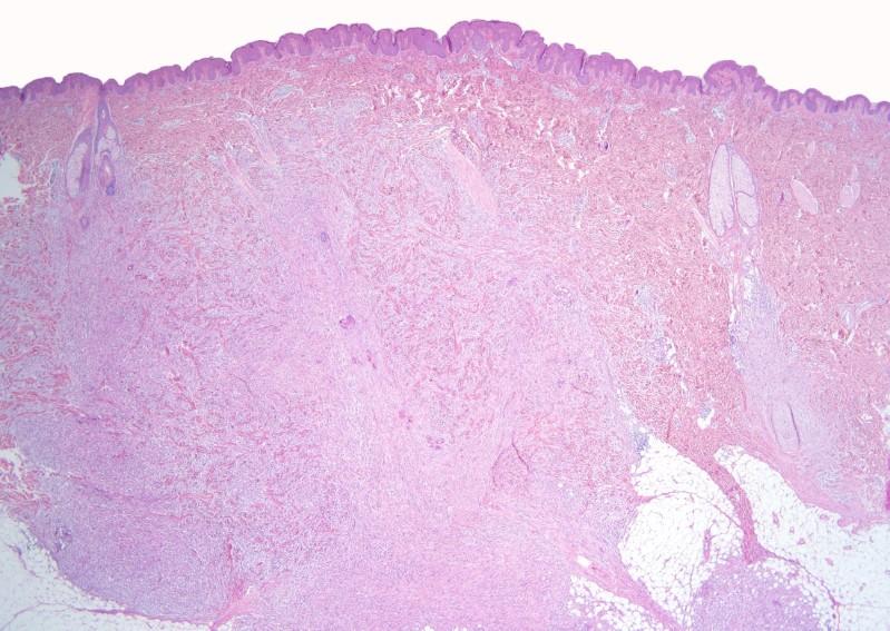
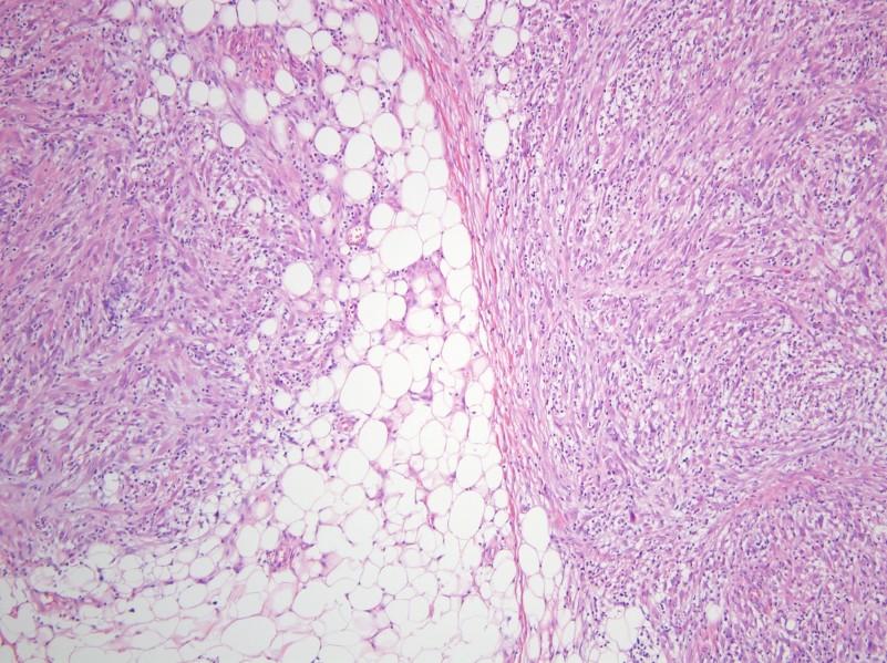
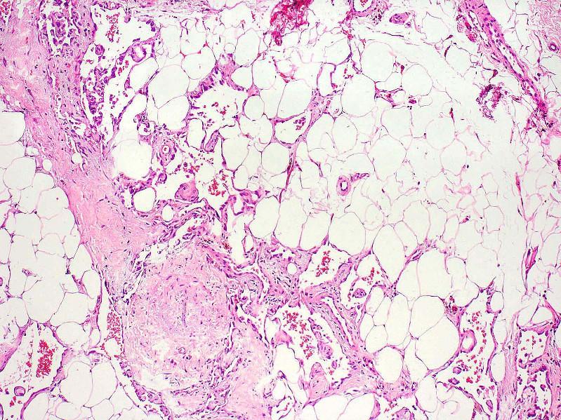
8 Hemosiderotic Fibrolipomatous Tumor First described as hemosiderotic fibrohistiocytic lipomatous lesion Believed to be a reactive process Now known to be neoplastic Uncommon subcutaneous tumor Ankle and foot of adults Ill-defined margins May recur locally (non-destructive)
9 Hemosiderotic Fibrolipomatous Tumor: Histology Variable admixture of mature adipose tissue and bland fibroblasts Short fascicles and whorls Honeycomb infiltration of fat Prominent hemosiderin deposition May show focal nuclear atypia and pleomorphism Some cases with focally prominent myxoid stroma

10 Hemosiderotic Fibrolipomatous Tumor

11 Hemosiderotic Fibrolipomatous Tumor
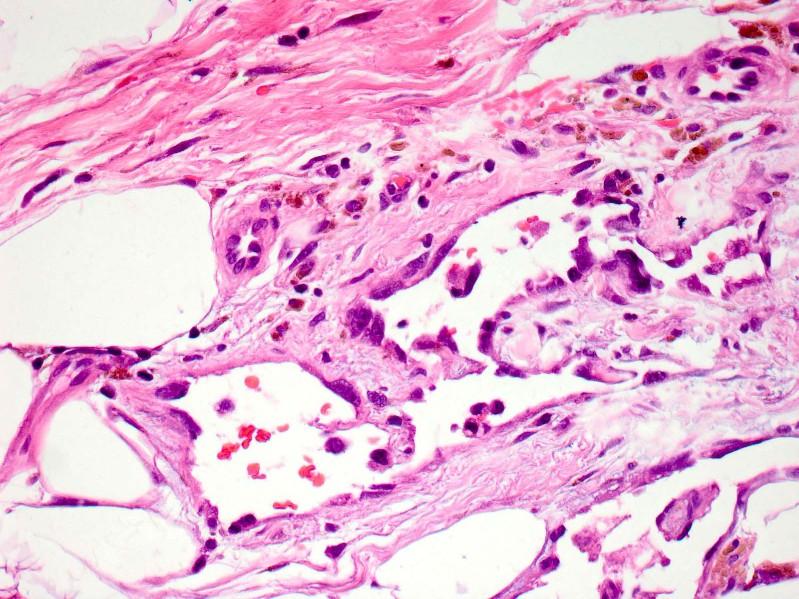
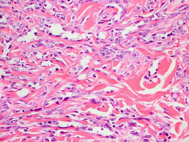
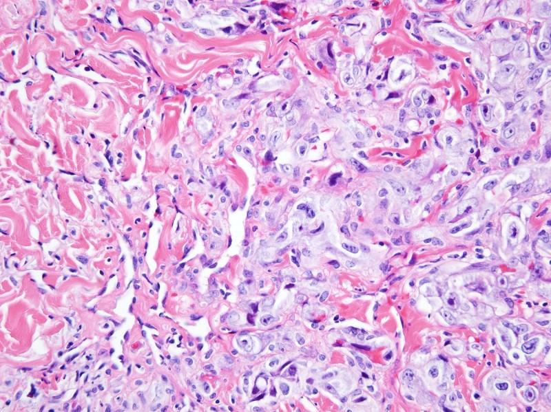
12 Myxoinflammatory Fibroblastic Sarcoma Originally also described as: Inflammatory myxohyaline tumor of distal extremities with virocyte or Reed-Sternberglike cells Inflammatory myxoid tumor of soft parts with bizarre giant cells Low grade sarcoma Hands/fingers/wrists >> ankles/feet Significant recurrent potential Rarely metastasize
13 Myxoinflammatory Fibroblastic Sarcoma: Histology Multinodular, infiltrative Hypocellular, myxoid stroma-rich areas with pseudolipoblasts Cellular areas with fibroblastic spindle cells and prominent chronic inflammation Highly variable proportions Large atypical mononuclear/multinucleate cells with inclusion-like nucleoli ( Reed- Sternberg-like or virocyte-like )

14 Myxoinflammatory Fibroblastic Sarcoma

15 Myxoinflammatory Fibroblastic Sarcoma

16 Myxoinflammatory Fibroblastic Sarcoma

17 Myxoinflammatory Fibroblastic Sarcoma

18 Myxoinflammatory Fibroblastic Sarcoma

19 Myxoinflammatory Fibroblastic Sarcoma

20 Myxoinflammatory Fibroblastic Sarcoma

21 Myxoinflammatory Fibroblastic Sarcoma

22 Myxoinflammatory Fibroblastic Sarcoma

23 Myxoinflammatory Fibroblastic Sarcoma Reed-Sternberg-like cells
24 Relationship between these tumor types? MIFS may contain focal areas indistinguishable from HFLT HFLT may contain focal areas with myxoid stroma and occasional atypical or pleomorphic cells HFLT may recur as MIFS

25 Hybrid HFLT/MIFS

26 Hybrid HFLT/MIFS
27 Shared Molecular Genetics! t(1;10)(p22;q24) TGFBR3-MGEA5 Ring chromosomes with 3p12 amplification Same findings in HFLT, MIFS, and hybrid tumors
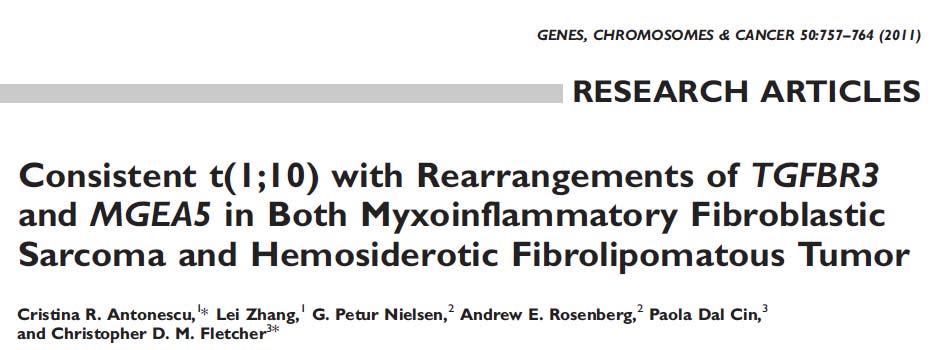
28
29 Elco CP et al. Am J Surg Pathol 2011
(p13;q24),del(3)(p11), 15[6]")

30 HFLT karyotype & FISH TGFBR3 45,XX, t(1;10)(p13;q24),del(3)(p11), 15[6] MGEA5 Centromeric Telomeric Courtesy of Paola Dal Cin, Ph.D. Genes Chromosomes Cancer 2011


31 HFLT MIFS MIFS
32 PSEUDOMYOGENIC HEMANGIOENDOTHELIOMA
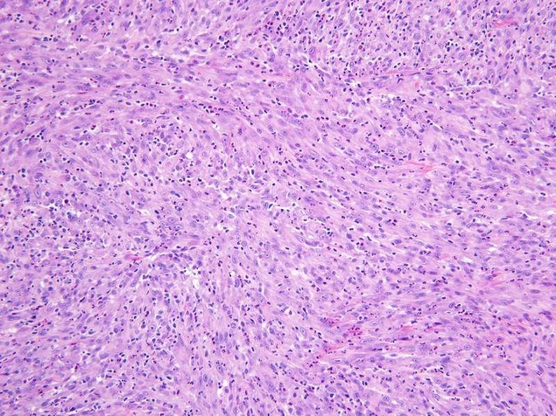
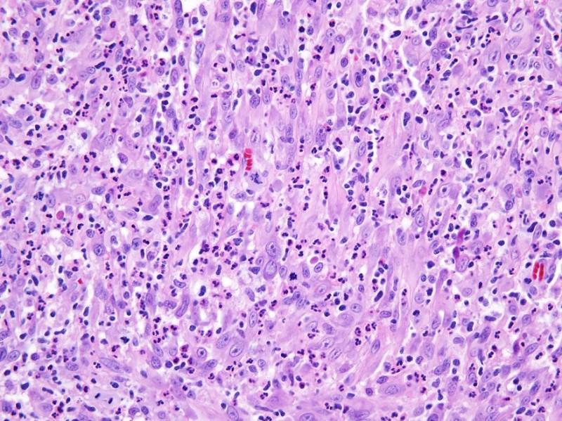
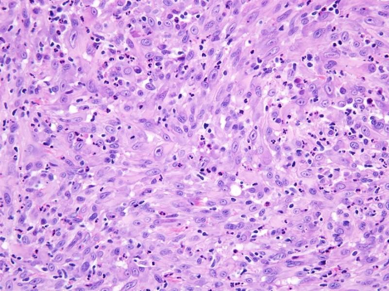
33 Pseudomyogenic Hemangioendothelioma Originally reported as fibroma-like variant of epithelioid sarcoma Also known as epithelioid sarcoma-like hemangioendothelioma Distinctive rarely metastasizing endothelial neoplasm Often presents with multiple discontiguous nodules in different tissue planes of a limb Histologically mimics a myoid tumor
34 Pseudomyogenic Hemangioendothelioma: Clinical Features Striking male predominance (5:1) Young adults (peak in 2 nd and 3 rd decades) Most patients (75%) present with cutaneous nodules 50% intramuscular; 20% intraosseous lesions 50% local recurrences or develop additional nodules in same anatomic area Usually within 1-2 years of first excision Despite ominous clinical presentation, distant metastasis rare
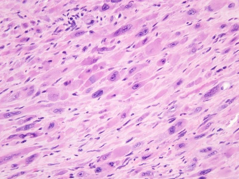
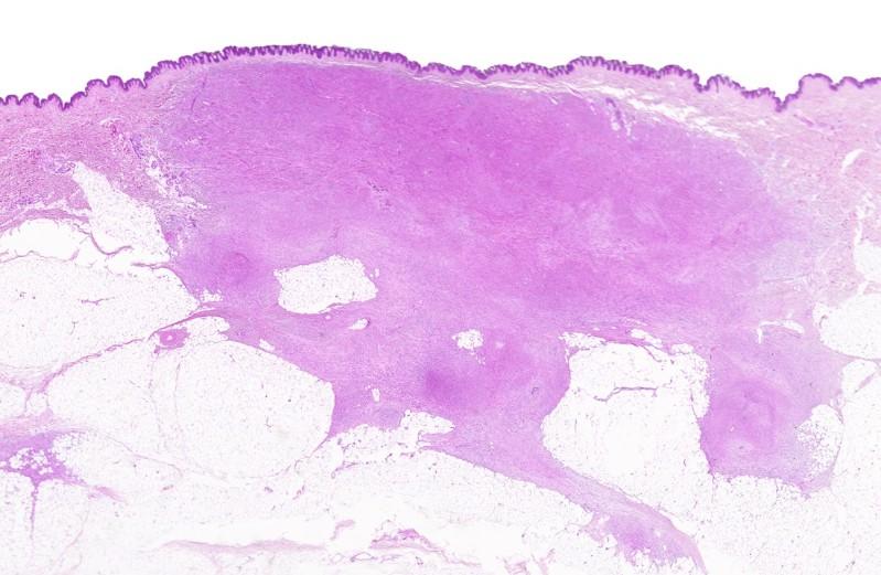
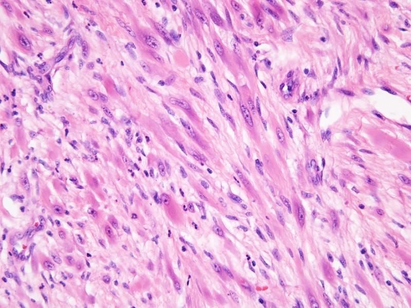
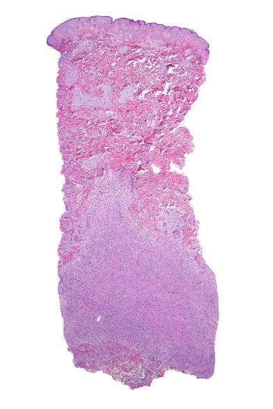
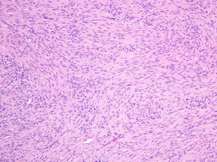
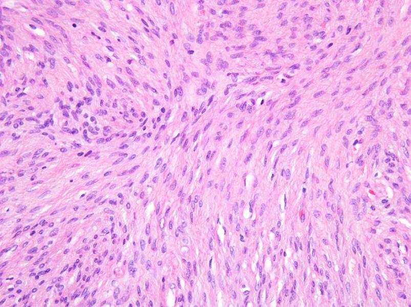
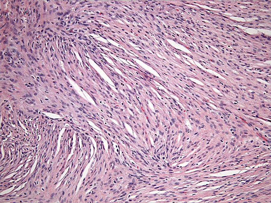
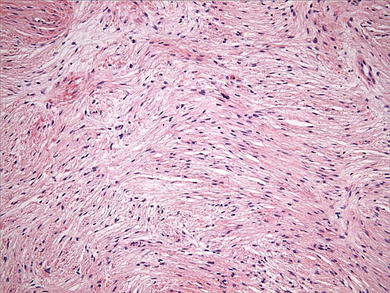
35 Pseudomyogenic Hemangioendothelioma: Histology Infiltrative margins Sheets, loose fascicles Relatively uniform plump spindle cells with abundant brightly eosinophilic cytoplasm May mimic rhabdomyoblasts 50% with prominent stromal neutrophils Cutaneous tumors often show overlying epidermal hyperplasia Often infiltrate into subcutaneous tissue
36 Pseudomyogenic Hemangioendothelioma
37 Pseudomyogenic Hemangioendothelioma
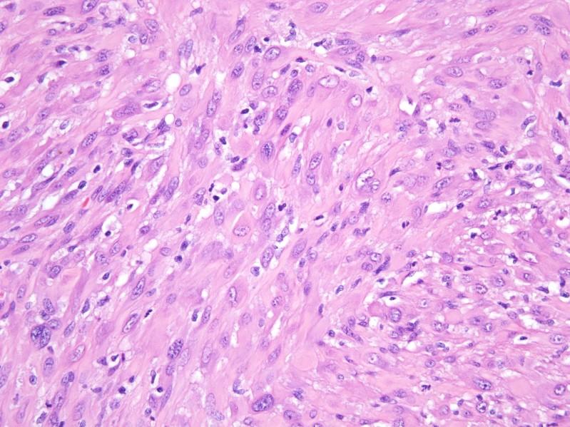
38 Pseudomyogenic Hemangioendothelioma
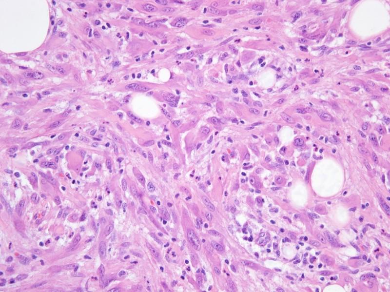
39 Pseudomyogenic Hemangioendothelioma
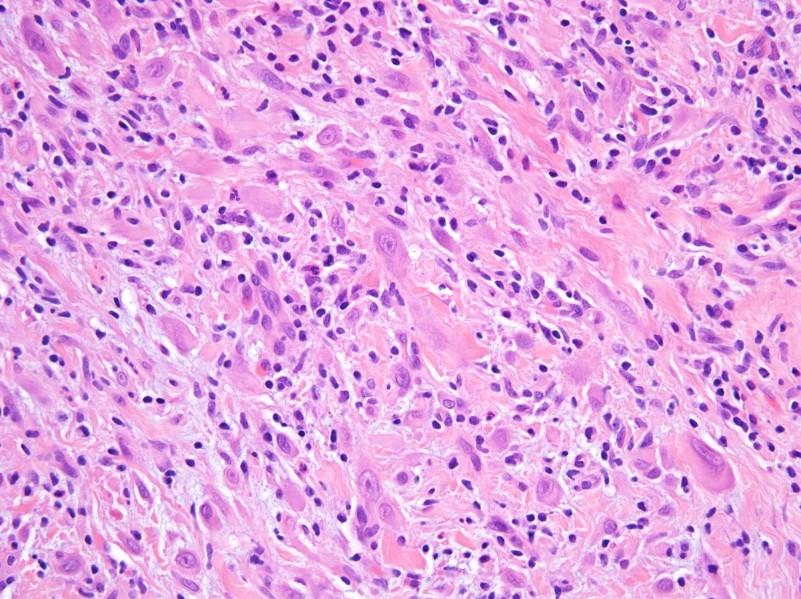
40 Pseudomyogenic Hemangioendothelioma
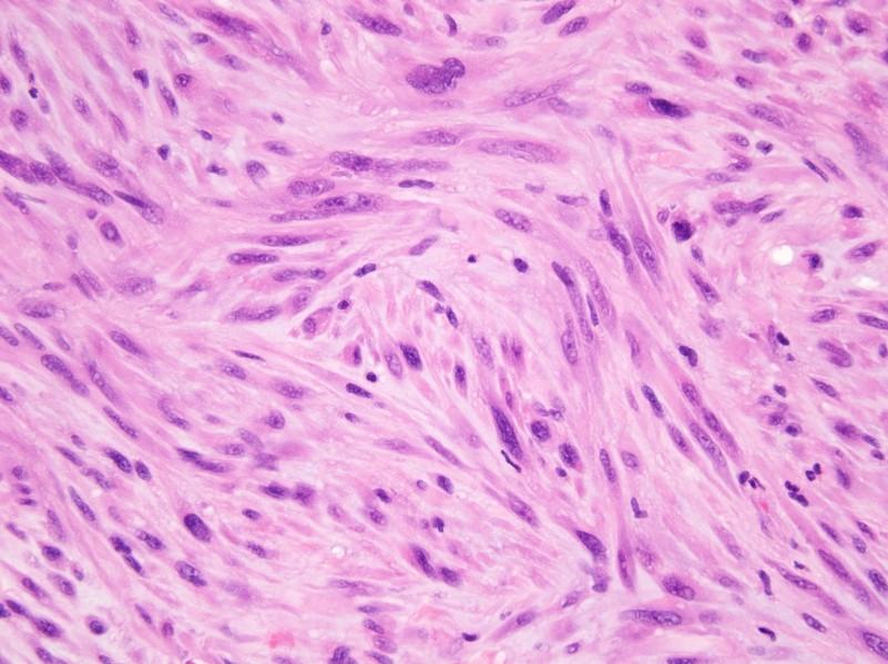
41 Pseudomyogenic Hemangioendothelioma
42 Pseudomyogenic Hemangioendothelioma
43 Pseudomyogenic Hemangioendothelioma
44 Pseudomyogenic Hemangioendothelioma
45
46 Pseudomyogenic Hemangioendothelioma
47 Pseudomyogenic Hemangioendothelioma
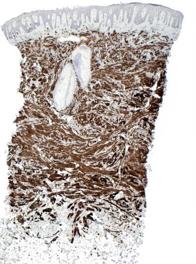
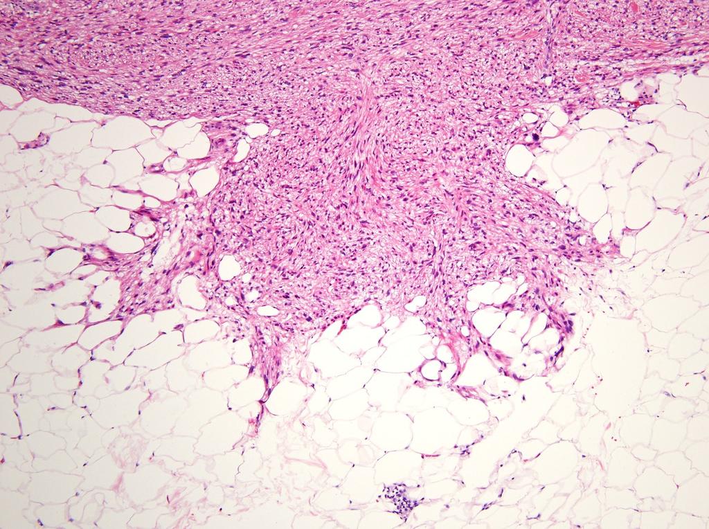
48 Pseudomyogenic Hemangioendothelioma
49 Pseudomyogenic Hemangioendothelioma: Immunohistochemistry Diffusely positive for keratin AE1/AE3 Nuclear staining for FLI1 and ERG 50% positive for CD31 Negative for EMA, keratin MNF116, CD34 Expression of INI1 retained
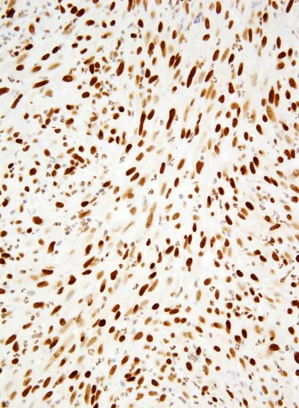
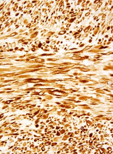
50 Pseudomyogenic Hemangioendothelioma AE1/AE3 ERG
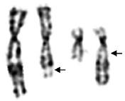
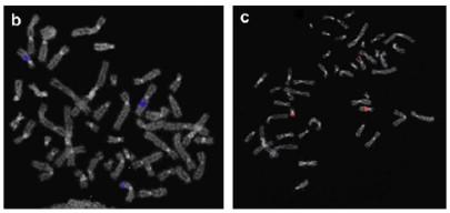
51 der(7)t(7;19) in 1 of 9 additional cases by FISH
52 ATYPICAL INTRADERMAL SMOOTH MUSCLE NEOPLASM AND CUTANEOUS LEIOMYOSARCOMA
53 Cutaneous Leiomyosarcoma Nomenclature widely used for cutaneous smooth muscle tumors with nuclear atypia and/or mitotic activity Predilection for middle-aged and elderly adult male patients Usually arise on trunk and lower extremities Older studies did not distinguish between purely dermal and subcutaneous tumors When confined to the dermis, no metastatic potential Sarcoma designation inappropriate!
54
55 Atypical Cutaneous Smooth Muscle Tumors: Outcome Massi et al. 36 cases Follow-up on 27 cases (16 limited to dermis; 11 minimal subcutaneous extension) 3 local recurrence 1 distant metastasis at 15 yrs (primary tumor with minimal subcutaneous extension) Kraft and Fletcher 84 cases Follow-up on 52 cases 18 local recurrence None metastasized
56 Atypical Intradermal Smooth Muscle Neoplasm: Histology Predominantly infiltrative growth pattern Nodular areas Fascicles ramifying through dermal collagen bundles Limited extension into superficial subcutaneous tissue with pushing border Spindle cells with broad nuclei, brightly eosinophilic cytoplasm Degree of nuclear atypia, pleomorphism, mitotic activity irrelevant
57 Atypical Intradermal Smooth Muscle Neoplasm: Immunohistochemistry Similar to smooth muscle tumors of other sites: SMA, desmin, caldesmon nearly always positive Broad-spectrum keratins expressed in 50%

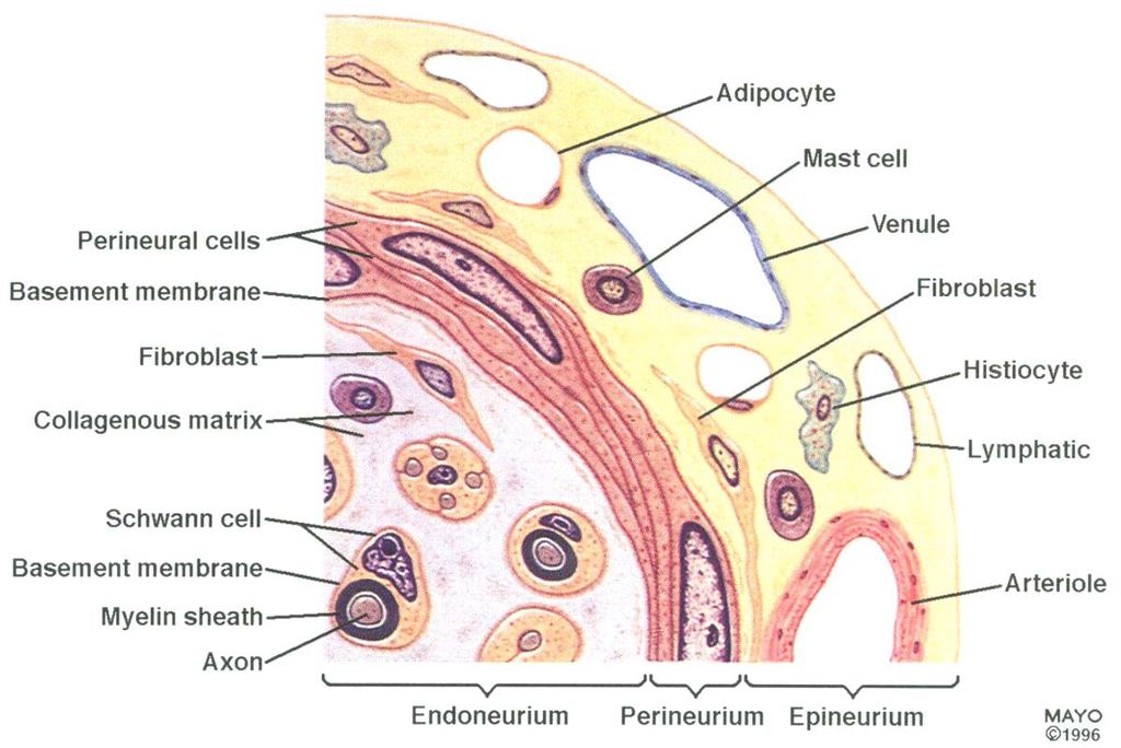
58 Pilar Leiomyoma desmin

59 Pilar Leiomyoma


60 Atypical Intradermal Smooth Muscle Neoplasm desmin

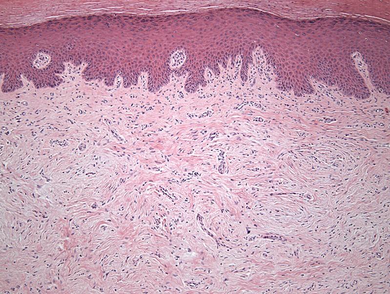
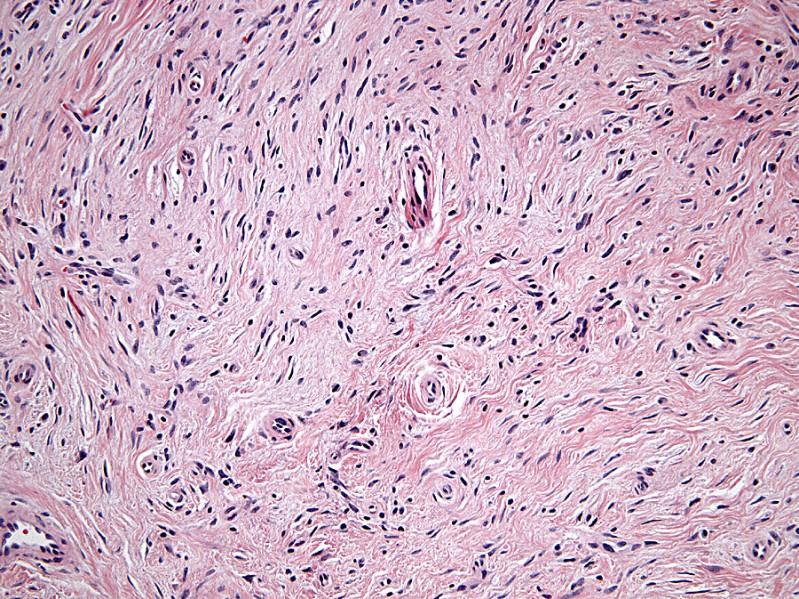
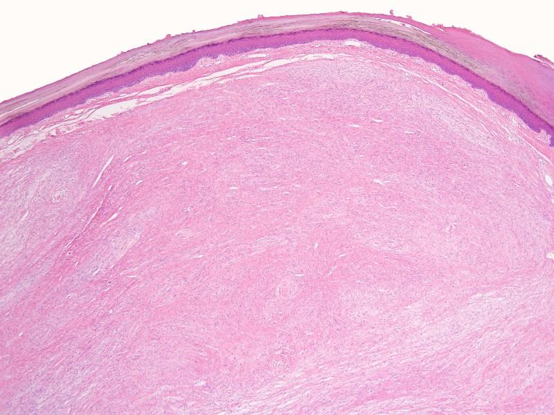
61 Atypical Intradermal Smooth Muscle Neoplasm

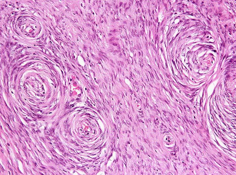
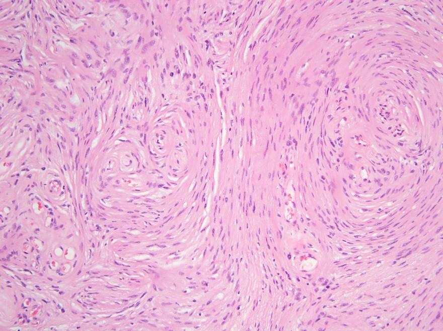
62 Atypical Intradermal Smooth Muscle Neoplasm
63 Cutaneous Leiomyosarcoma
64 Classification of Smooth Muscle Tumors of the Superficial Soft Tissues Confined to the dermis: Pilar leiomyoma Atypical intradermal smooth muscle neoplasm Involve dermis and infiltrate subcutis: Cutaneous leiomyosarcoma Involve subcutis: Angioleiomyoma Subcutaneous leiomyosarcoma
65 HYBRID SCHWANNOMA/PERINEURIOMA

66 THE NERVE SHEATH
67
68 Hybrid Nerve Sheath Tumors Benign nerve sheath tumors: Schwannoma Neurofibroma Perineurioma In recent years, tumors containing elements of >1 type of nerve sheath tumor recognized First: hybrid neurofibroma/schwannoma (schwannomatous nodules within otherwise typical neurofibroma) Recently: hybrid schwannoma/perineurioma
69 Soft Tissue Perineurioma: Clinical Features Equal gender distribution Wide age range; uncommon in children Most common in limbs, but can arise at essentially any anatomic site Painless subcutaneous mass ~25% deep soft tissue; 10% skin Benign, rarely recur
70 Soft Tissue Perineurioma: Histology Well circumscribed, unencapsulated Storiform, lamellar, whorled architecture Collagenous >> myxoid stroma Thin, elongated spindle cells with delicate bipolar cytoplasmic processes Some cases with ovoid cells EMA positive (may be weak) CD34 in 65%; claudin-1 in 30%
71 Soft Tissue Perineurioma
72 Soft Tissue Perineurioma
73 Soft Tissue Perineurioma
74 Soft Tissue Perineurioma
75 Soft Tissue Perineurioma
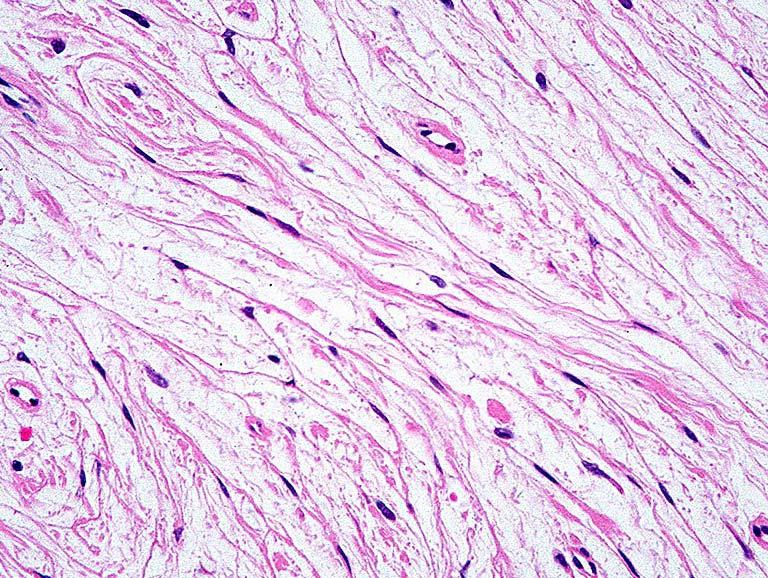
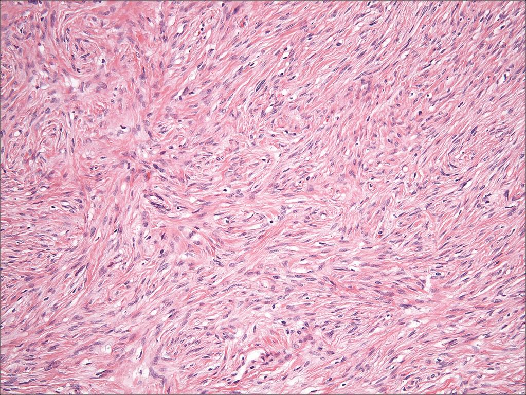
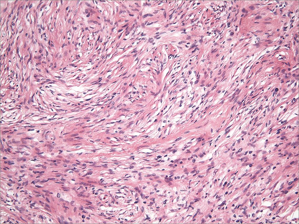
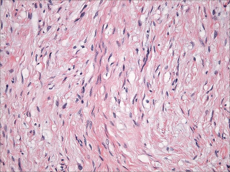
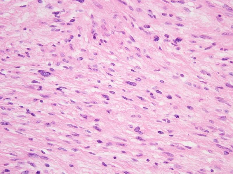
76 Soft Tissue Perineurioma
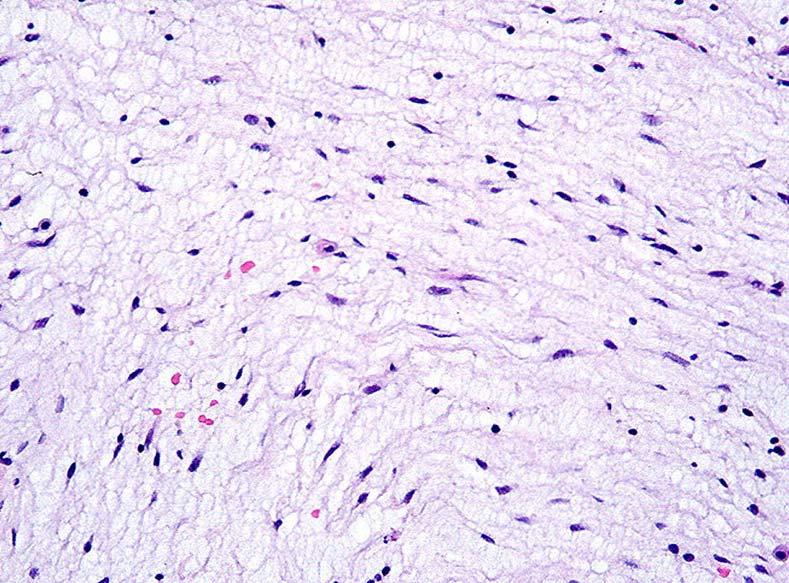
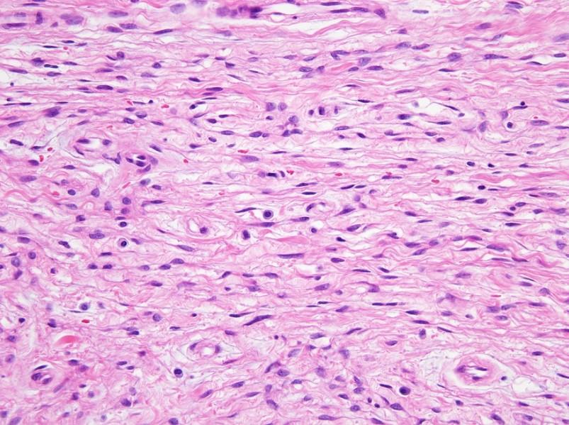
77 Soft Tissue Perineurioma
78 Cutaneous Perineurioma
79 Cutaneous Perineurioma
80 Soft Tissue Perineurioma

81 Soft Tissue Perineurioma EMA

82 Soft Tissue Perineurioma EMA

83 Soft Tissue Perineurioma CD34
84 Hybrid Schwannoma/Perineurioma: Clinical Features Usually involve skin and subcutaneous tissue More rarely deep soft tissue or visceral sites Wide age range and anatomic distribution No association with neurofibromatosis Rarely recur locally
85 Hybrid Schwannoma/Perineurioma: Histology Architecture like soft tissue perineurioma: Storiform and whorled growth pattern Predominantly Schwannian cytology: Plump spindle cells with tapering nuclei and eosinophilic cytoplasm Less conspicuous perineurial cell component: Slender nuclei and delicate elongated cytoplasmic processes Degenerative atypia relatively common
86 Hybrid Schwannoma/Perineurioma
87 Hybrid Schwannoma/Perineurioma
88 Hybrid Schwannoma/Perineurioma
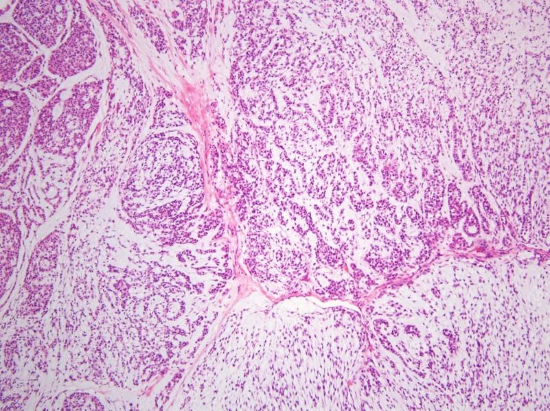
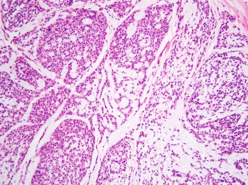
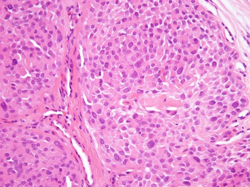
89 Hybrid Schwannoma/Perineurioma
90 Hybrid Schwannoma/Perineurioma
91 Hybrid Schwannoma/Perineurioma
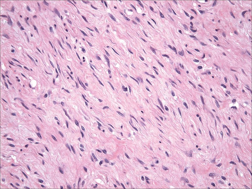
92 Hybrid Schwannoma/Perineurioma
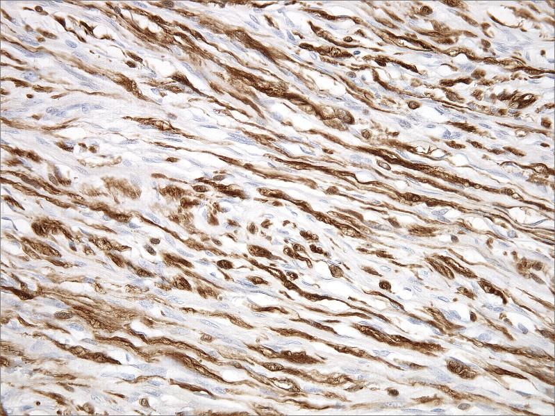
93 Hybrid Schwannoma/Perineurioma S100
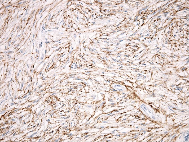
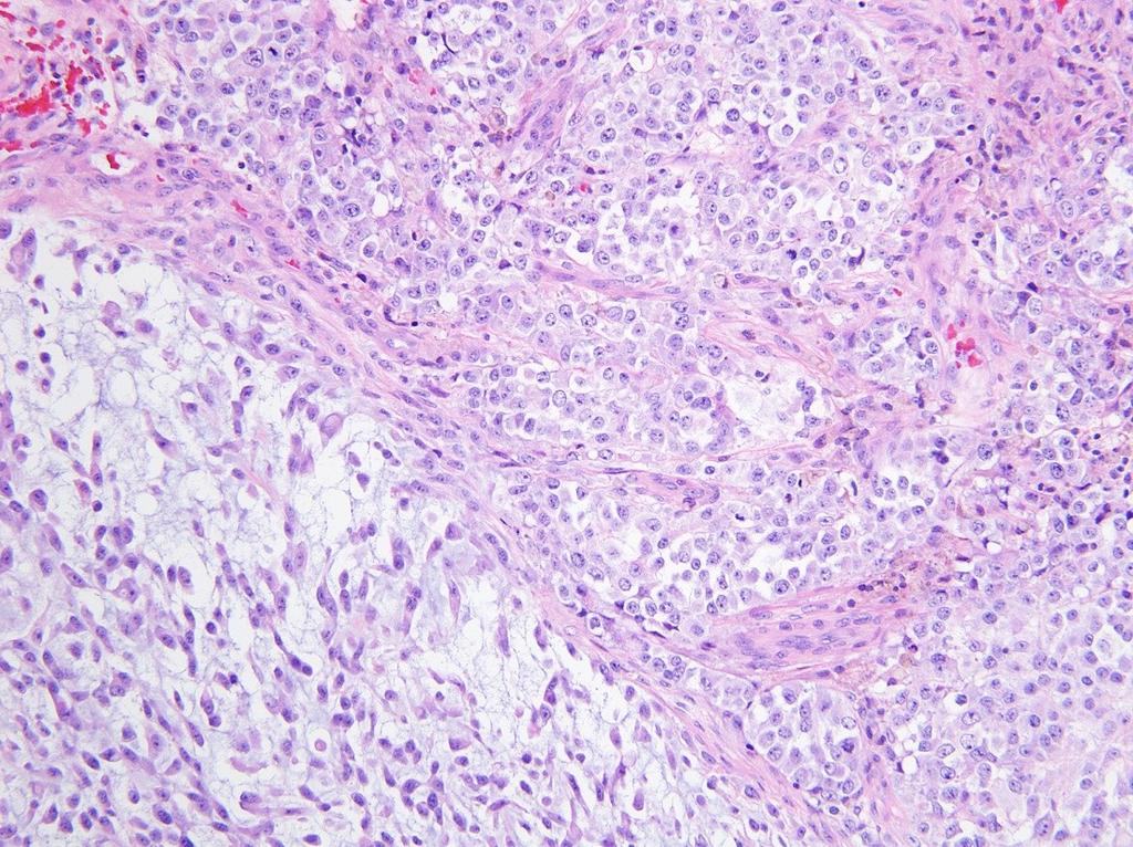
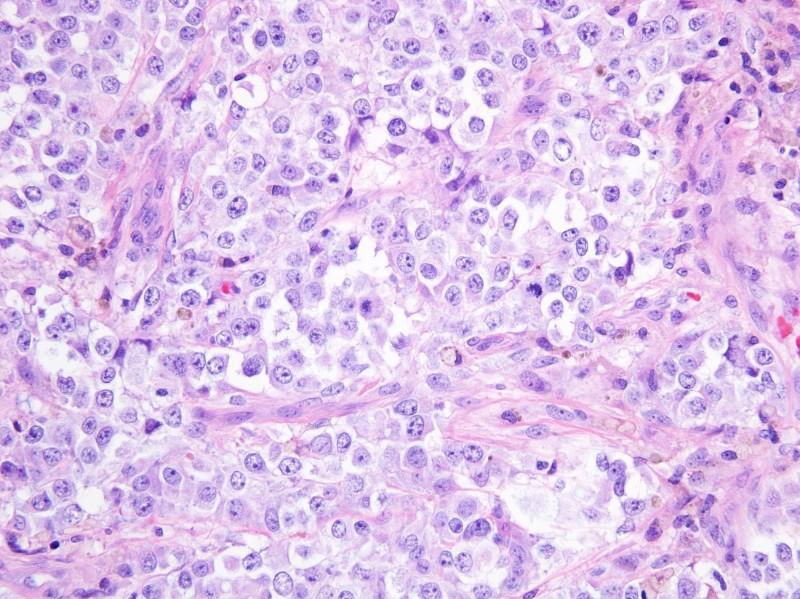
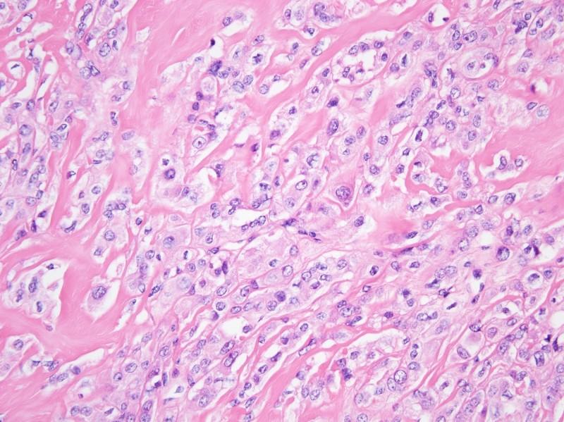
94 Hybrid Schwannoma/Perineurioma EMA

95 Hybrid Schwannoma/Perineurioma EMA (brown) S100 (red)
96 MYOEPITHELIOMA/ MYOEPITHELIAL CARCINOMA
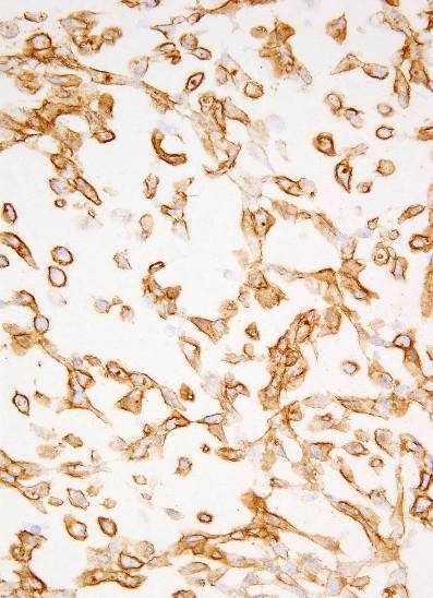
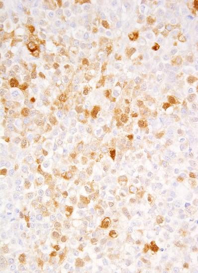
97 Myoepithelial Tumors of Skin and Soft Tissue Histologically similar to salivary gland counterparts Cutaneous myoepithelial tumors lie on a spectrum, ranging from mixed tumor ( chondroid syringoma ) to pure myoepithelioma, to malignant counterparts Malignant myoepithelial tumors known as myoepithelial carcinomas Cutaneous myoepithelioma: predilection for extremities, male predominance Usually present as painless nodule
98 Myoepithelial Tumors of Skin and Soft Tissue: Histology Lobulated architecture Reticular, trabecular, or nested growth pattern Variably prominent myxoid stroma Predominantly epithelioid cells with eosinophilic cytoplasm Wide range in cytology: spindle cell, clear cell, plasmacytoid (hyaline cell) components Intratumoral heterogeneity typical Myoepithelial carcinoma: moderate/severe nuclear atypia
99 Myoepithelioma
100 Myoepithelioma
101 Myoepithelioma
102 Myoepithelial Carcinoma
103 Myoepithelial Carcinoma
104 Myoepithelial Carcinoma
105 Myoepithelial Tumors of Skin and Soft Tissue: Immunohistochemistry Usually positive for keratins, EMA, S100 50% positive for GFAP Variable expression of muscle markers (esp. calponin, SMA) Variable expression of p63 Distinctive syncytial variant: negative for keratins
106 Myoepithelioma EMA S100
107 Cutaneous Syncytial Myoepithelioma Distinctive variant exclusive to the skin Sheets of ovoid to epithelioid cells Palely eosinophilic cytoplasm Indistinct cell borders Irregular tumor margins Benign, rarely recur

108 Cutaneous Syncytial Myoepithelioma

109 Cutaneous Syncytial Myoepithelioma

110 Cutaneous Syncytial Myoepithelioma

111 Cutaneous Syncytial Myoepithelioma

112 Cutaneous Syncytial Myoepithelioma

113 Cutaneous Syncytial Myoepithelioma
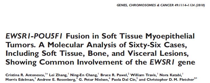
114 Myoepithelial Tumors of Skin and Soft Tissue: Molecular Genetics EWSR1 rearrangements in ~50% Novel fusion partners EWSR1-PBX1 EWSR1-POU5F1 EWSR1-ZNF444 These fusion partners only account for 1/3 of myoepithelial tumors with EWSR1 rearrangements Cutaneous syncytial myoepithelioma EWSR1-?
115


116 Antonescu et al. Genes Chromosomes Cancer 2010

117 Courtesy of Alex Lazar
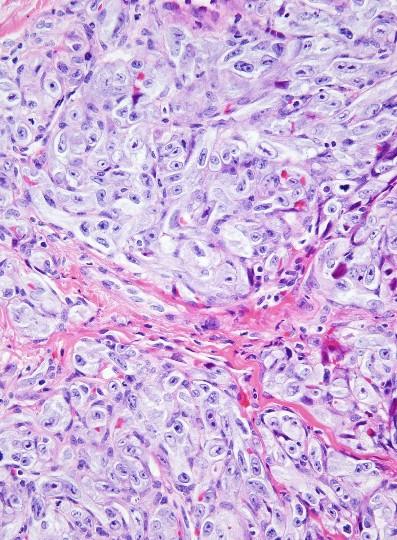
118 POST-RADIATION ATYPICAL VASCULAR LESIONS AND CUTANEOUS ANGIOSARCOMA
119 Post-radiation Atypical Vascular Lesions and Cutaneous Angiosarcoma Spectrum of cutaneous vascular proliferations after breast-conserving surgery and radiation therapy Atypical vascular lesions may be multifocal, usually benign course Angiosarcoma locally destructive and significant potential for metastasis Considerable clinical and histologic overlap
120 Atypical Post-radiation Vascular Proliferation: Histology Often relatively well-circumscribed Superficial to mid-dermis Dilated, dissecting vascular channels Endothelial cells with hobnail appearance common No multilayering, mitotic activity, or significant nuclear atypia No infiltration of subcutaneous tissue

121 Atypical Post-radiation Vascular Proliferation

122 Atypical Post-radiation Vascular Proliferation

123 Atypical Post-radiation Vascular Proliferation

124 Atypical Post-radiation Vascular Proliferation

125 Atypical Post-radiation Vascular Proliferation
126 Cutaneous Angiosarcoma
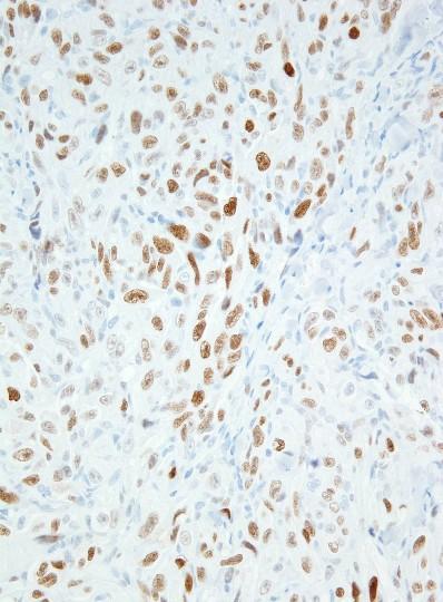
127 Cutaneous Angiosarcoma
128 Cutaneous Angiosarcoma
129 Epithelioid Angiosarcoma

130 Spindle Cell Angiosarcoma
131 Post-radiation Cutaneous Angiosarcoma: Molecular Genetics Consistent high-level amplification of MYC gene (8q24) Not observed in atypical vascular lesions, primary mammary angiosarcoma, or angiosarcoma of sun-damaged skin Subset also shows amplification of FLT4 gene (encoding VEGFR3)
132

133 Guo et al. Genes Chromosomes Cancer 2011 Manner et al. Am J Pathol 2010

134

135 Cutaneous Angiosarcoma MYC
136 Cutaneous Angiosarcoma MYC
137 Summary Classification of soft tissue tumors continues to evolve as new lesions are described Recognition of distinct clinical behavior helps guide management and follow-up Molecular genetic discoveries provide insights into pathogenesis and classification Lead to development of new tools to improve diagnostic accuracy
Myxo-inflammatory Fibroblastic sarcoma
 AKA Myxo-inflammatory Fibroblastic sarcoma Acral Myxoinflammatory fibroblastic sarcomaam.j.surg.path1998; 22; 911-924 Inflammatory myxoid tumour of soft parts with bizarre giant cells [Pathol.Res.Pract.
21/07/2017. Hobnail endothelial cells are not the same as epithelioid endothelial cells
 UPDATE IN CUTANEOUS VASCULAR S DERMATOPATHOLOGY SESSION BELFAST PATHOLOGY JUNE 21/2017 Dr E Calonje St John s Institute of Dermatology, London, United Kingdom THE FAMILY OF VASCULAR S WITH EPITHELIOID
ACCME/Disclosures ALK FUSION-POSITIVE MESENCHYMAL TUMORS. Tumor types with ALK rearrangements. Anaplastic Lymphoma Kinase. Jason L.
 Companion Meeting of the International Society of Bone and Soft Tissue Pathology The Evolving Concept of Mesenchymal Tumors ALK FUSION-POSITIVE MESENCHYMAL TUMORS Jason L. Hornick, MD, PhD March 13, 2016
Selected Pseudomalignant Soft Tissue Tumors of the Skin and Subcutis
 Selected Pseudomalignant Soft Tissue Tumors of the Skin and Subcutis Andrew L. Folpe, M.D. Professor of Laboratory Medicine and Pathology Mayo Clinic, Rochester, MN folpe.andrew@mayo.edu 2016 MFMER slide-1
Advances in the clinicopathological and molecular classification of cutaneous mesenchymal neoplasms
 Histopathology 2016 DOI: 10.1111/his.12930 REVIEW Advances in the clinicopathological and molecular classification of cutaneous mesenchymal neoplasms Danielle C Costigan & Leona A Doyle 1 Department of
The Genetics of Myoepithelial Tumors: salivary glands, soft tissue and bone
 The Genetics of Myoepithelial Tumors: salivary glands, soft tissue and bone Cristina Antonescu, MD Memorial Sloan-Kettering Cancer Center, New York Nothing to declare Disclosure Spectrum of Myoepithelial
Newer soft tissue entities
 Newer soft tissue entities Examples among fibroblastic tumors Turku, May 6, 2010 Markku Miettinen, M.D. AFIP, Washington, DC Fibroblastic neoplasms Solitary fibrous tumor /Hemangiopericytoma Low-grade
أملس عضلي غرن = Leiomyosarcoma. Leiomyosarcoma 1 / 5
 Leiomyosarcoma 1 / 5 EPIDEMIOLOGY Exact incidence is unknown, but older studies suggest that leiomyosarcomas comprise approximately 3 percent of soft-tissue sarcomas. Superficial leiomyosarcoma occurs
Malignant Peripheral Nerve Sheath Tumor
 C H A P T E R 120 Malignant Peripheral Nerve Sheath Tumor Currently, malignant peripheral nerve sheath tumor (MPNST) is the most commonly used generic name for the neoplasms known in the past as neurosarcoma,
The Relevance of Cytologic Atypia in Cutaneous Neural Tumors
 The Relevance of Cytologic Atypia in Cutaneous Neural Tumors Recent Findings - New Developments New Problems Zsolt B. Argenyi, M.D. Professor of Pathology & Dermatology Director of Dermatopathology Department
1/10/2018. Soft Tissue Tumors Showing Melanocytic Differentiation. Overview. Desmoplastic/ Spindle Cell Melanoma
 2016 MFMER slide-1 2016 MFMER slide-2 2016 MFMER slide-3 Soft Tissue Tumors Showing Melanocytic Differentiation Andrew L. Folpe, M.D. Professor of Laboratory Medicine and Pathology Mayo Clinic, Rochester,
Desmoplastic Melanoma R/O BCC. Clinical Information. 74 y.o. man with lesion on left side of neck r/o BCC
 R/O BCC Sabine Kohler, M.D. Professor of Pathology and Dermatology Dermatopathology Service Stanford University School of Medicine Clinical Information 74 y.o. man with lesion on left side of neck r/o
A 25 year old female with a palpable mass in the right lower quadrant of her abdomen
 May 2016 A 25 year old female with a palpable mass in the right lower quadrant of her abdomen Contributed by: Paul Ndekwe, MD, Resident Physician, Indiana University School of Department of Pathology and
Financial disclosures
 Mesenchymal Neoplasms with Melanocytic Differentiation By Konstantinos Linos MD, FCAP, FASDP Bone, Soft Tissue and Dermatopathology Assistant Professor of Pathology Dartmouth-Hitchcock Medical Center Geisel
Diplomate of the American Board of Pathology in Anatomic and Clinical Pathology
 A 33-year-old male with a left lower leg mass. Contributed by Shaoxiong Chen, MD, PhD Assistant Professor Indiana University School of Medicine/ IU Health Partners Department of Pathology and Laboratory
Financial disclosures
 Cutaneous Mesenchymal Neoplasms with EWSR1 Rearrangement By Konstantinos Linos MD, FCAP, FASDP Bone, Soft Tissue and Dermatopathology Assistant Professor of Pathology Dartmouth-Hitchc Geisel School of
Evening Specialty Conference Bone and Soft Tissue Pathology. Diagnostic pitfalls in bone and soft tissue pathology
 Evening Specialty Conference Bone and Soft Tissue Pathology. Case 1 Elizabeth G Demicco, MD, PhD Mount Sinai Hospital, New York Disclosure of Relevant Financial Relationships USCAP requires that all planners
Disclosure of Relevant Financial Relationships
 Surgical Pathology Companion Meeting Case 5: Locally Recurrent Chest wall Mass Cristina Antonescu, MD Department of Pathology, Memorial Sloan Kettering Cancer Center Disclosure of Relevant Financial Relationships
57th Annual HSCP Spring Symposium 4/16/2016
 An Unusual Malignant Spindle Cell Lesion to Involve the Breast Erinn Downs-Kelly, D.O. Associate Professor of Pathology University of Utah & ARUP Laboratories No disclosures Case 39 y/o female with no
Cutaneous Mesenchymal Neoplasms with EWSR1 Rearrangement
 Cutaneous Mesenchymal Neoplasms with EWSR1 Rearrangement By Konstantinos Linos MD, FCAP, FASDP Bone, Soft Tissue and Dermatopathology Assistant Professor of Pathology Dartmouth-Hitchcock Medical Center
Self assessment case. Dr Saleem Taibjee Dorset County Hospital, Dorchester
 Self assessment case Dr Saleem Taibjee saleemtaibjee@gmail.com Dorset County Hospital, Dorchester Clinical details 34-year-old man: Shave excision Skin tag / papilloma left thigh The best diagnosis is:
3/24/2017 DENDRITIC CELL NEOPLASMS: HISTOLOGY, IMMUNOHISTOCHEMISTRY, AND MOLECULAR GENETICS. Disclosure of Relevant Financial Relationships
 DENDRITIC CELL NEOPLASMS: HISTOLOGY, IMMUNOHISTOCHEMISTRY, AND MOLECULAR GENETICS Jason L. Hornick, M.D., Ph.D. Director of Surgical Pathology and Immunohistochemistry Brigham and Women s Hospital Professor
Spindle Cell Lesions Of The Breast. Emad Rakha Professor of Breast Pathology and Consultant Pathologist
 Spindle Cell Lesions Of The Breast Emad Rakha Professor of Breast Pathology and Consultant Pathologist * SCLs comprise a wide spectrum of diseases, ranging from reactive processes to aggressive malignant
Case 27 Male 42. Painless, static, well-circumscribed, subcutaneous nodule right lower leg,?lipoma. The best diagnosis is:
 Case 27 Male 42. Painless, static, well-circumscribed, subcutaneous nodule right lower leg,?lipoma. The best diagnosis is: A. Angiosarcoma B. Haemangiopericytoma C.Myopericytoma D.Myofibroma E. Angioleiomyoma
An Overview of Cutaneous Vascular Neoplasms
 An Overview of Cutaneous Vascular Neoplasms By Konstantinos Linos MD, FCAP, FASDP Bone, Soft Tissue and Dermatopathology Assistant Professor of Pathology Dartmouth-Hitchcock Medical Center Geisel School
Enterprise Interest Nothing to declare
 Enterprise Interest Nothing to declare Diagnoses one would not like to miss in soft tissue pathology early in your career Marta Sbaraglia, MD Department of Pathology Hospital of Treviso University of Padua
Update on Cutaneous Mesenchymal Tumors. Thomas Brenn
 Update on Cutaneous Mesenchymal Tumors Thomas Brenn Cutaneous Mesenchymal Tumours Wide morphological and biological spectrum Myofibroblastic, smooth muscle, neural, vascular, apidocytic, undifferentiated;
An Overview of Genital Stromal Tumors
 An Overview of Genital Stromal Tumors By Konstantinos Linos MD, FCAP, FASDP Bone, Soft Tissue and Dermatopathology Assistant Professor of Pathology Dartmouth-Hitchcock Medical Center Geisel School of Medicine
5/10. Pathology Soft tissue tumors. Farah Bhani. Mohammed Alorjani
 5/10 Pathology Soft tissue tumors Mohammed Alorjani Farah Bhani Slides are included in this sheet. Objectives: Soft tissue tumors 1. Describe soft tissue tumors. 2. Understand the classification of soft
Slide seminar. Asist. Prof. Jože Pižem, MD, PhD Institute of Pathology Medical Faculty, University of Ljubljana
 Slide seminar Asist. Prof. Jože Pižem, MD, PhD Institute of Pathology Medical Faculty, University of Ljubljana Case 5 A 57-year-old man with a dermal/subcutaneous lesion on the scalp, which was interpreted
Disclosures. An update on ancillary techniques in the diagnosis of soft tissue tumors. Ancillary techniques. Introduction
 Disclosures An update on ancillary techniques in the diagnosis of soft tissue tumors. I have nothing to disclose. Andrew Horvai, MD, PhD Clinical Professor, Pathology Introduction Ancillary techniques
Soft Tissue Perineurioma
 The Korean Journal of Pathology 2009; 43: 266-70 DOI: 10.4132/KoreanJPathol.2009.43.3.266 Soft Tissue Perineurioma - A Case Report - Jun Mo Kim Joon Hyuk Choi Department of Pathology, Yeungnam University
Dermatopathology. Dr. Rafael Botella Estrada. Hospital La Fe de Valencia
 Dermatopathology Dr. Rafael Botella Estrada. Hospital La Fe de Valencia DERMATOPATHOLOGY CASE CHALLENGE: RECOGNIZING MIMIS AND MASQUERADERS Rosalie Elenitsas. University of Pennsylvania Spectrum Lupus
Cellular Neurothekeoma
 Cellular Neurothekeoma Scott W Binder, MD Pritzker Professor of Pathology & Dermatology Sr. Vice Chair Director, Pathology Clinical Services Chief, Dermatopathology Geffen/UCLA School of Medicine Clinical
CASE REPORT Benign epithelioid peripheral nerve sheath tumour resembling schwannoma
 Malaysian J Pathol 2014; 36(3) : 217 221 CASE REPORT Benign epithelioid peripheral nerve sheath tumour resembling schwannoma Thejasvi KRISHNAMURTHY MD and SR NIVEDITHA MD, DNB Department of Pathology,
SMOOTH MUSCLE TUMOURS
 SMOOTH MUSCLE TUMOURS NORMAL SMOOTH MUSCLE Cytology Immunohistochemistry Ultrastructure Masson Trichrome Smooth Muscle Ultrastructure Many myofilaments running parallel to the long axis of the smooth
Update On Lipomatous Tumors: Old Standbys and New Concepts
 Update On Lipomatous Tumors: Old Standbys and New Concepts John R. Goldblum, M.D. Chairman, Department of Anatomic Pathology Cleveland Clinic Professor of Pathology Cleveland Clinic Lerner College of Medicine
USCAP 2011: ASDP companion meeting. Steven D. Billings 1
 USCAP 2011: ASDP companion meeting. Steven D. Billings (billins@ccf.org) 1 Spindle cell tumors that make you say, Oh $*&%! This lecture will focus on examples of cutaneous tumors that present particular
59 yo male with past medical history of prostate carcinoma, presented with upper abdominal pain
 December 2016 59 yo male with past medical history of prostate carcinoma, presented with upper abdominal pain Contributed by: Divya Sharma, MD. Fellow, Gastrointestinal Pathology, Department of Pathology
Special slide seminar
 Special slide seminar Tomáš Rozkoš The Fingerland Department of Pathology Charles University Medical Faculty and Faculty Hospital in Hradec Králové Czech Republic Case history, 33 years old resistance
Disclosures. An update on ancillary techniques in the diagnosis of soft tissue tumors. Ancillary techniques. Introduction
 Disclosures An update on ancillary techniques in the diagnosis of soft tissue tumors. I have nothing to disclose. Andrew Horvai, MD, PhD Clinical Professor, Pathology Introduction Ancillary techniques
Pancreas. Atrophy, acinar cell. Pathogenesis: Diagnostic key features:
 Pancreas Atrophy, acinar cell Pathogenesis: Decrease in number and/or size of acinar cells may be due to spontaneous or experimentally induced degenerative changes, apoptosis, or a sequel of chronic inflammation.
BSD 2015 Case 19. Female 21. Nodule on forehead. The best diagnosis is:
 BSD 2015 Case 19 Female 21. Nodule on forehead. The best diagnosis is: A. mixed tumour of skin B. porocarcinoma C. nodular hidradenoma D. metastatic adenocarcinoma BSD 2015 Case 19 Female 21 Nodule on
Fun with Fat. General Rules. Case
 Fun with Fat General Rules Imaging: location (deep vs. superficial) Superficial lesions are seldom liposarcomas Deep lesions may be benign or malignant Myxoid stroma is common in benign and malignant lesions
Classification (1) Classification (3) Classification (2) Spindle cell lesions. Spindle cell lesions of bladder (Mills et al.
 Classification (3) Classification (2) Spindle cell lesions. Spindle cell lesions of bladder (Mills et al.") Non-epithelial tumours and nonepithelial tumour-like lesions of the bladder Dr Jonathan H Shanks The Christie NHS Foundation Trust, Manchester, UK Classification (1) Myofibroblastic proliferations and
Non-epithelial tumours and nonepithelial tumour-like lesions of the bladder Dr Jonathan H Shanks The Christie NHS Foundation Trust, Manchester, UK Classification (1) Myofibroblastic proliferations and
Endometrial Stromal Tumors
 Endometrial Stromal Tumors WHO Categories: Endometrial Stromal Nodule (ESN) Endometrial Stromal Sarcoma, low grade (LGESS) Endometrial Stromal Sarcoma, high grade (HGESS) Undifferentiated Uterine Sarcoma
Case Presentation. Maha Akkawi, MD, Fatima Obeidat, MD, Tariq Aladily, MD. Department of Pathology Jordan University Hospital Amman, Jordan
 Case Presentation Maha Akkawi, MD, Fatima Obeidat, MD, Tariq Aladily, MD Department of Pathology Jordan University Hospital Amman, Jordan The 25th Annual Congress of the ADIAP The 8/11/2013 1 5th International
ESS: Pathologic Insights
 GEIS XVI INTERNATIONAL SYMPOSIUM Seville 4th October 2018 ESS: Pathologic Insights Sílvia Bagué The Royal Marsden Hospital London (United Kingdom) I have no conflicts of interest Endometrial stromal sarcoma
Diagnostic Approach to Soft Tissue Tumors
 SECTION 2 Diagnostic Approach to Soft Tissue Tumors Overview Biopsy and Resection of Soft Tissue Tumors 20 Clinical Approach Age- and Location-Based Approach to Diagnosis 24 Histologic Approach Pattern-Based
Other New entities in soft tissue tumors.
 Other New entities in soft tissue tumors. Angelo Paolo Dei Tos MD Departments of Pathology and Oncology General Hospital of Treviso, Italy Introduction During the past decade classification schemes have
Slide Seminar Spanish Society of Pathology
 Slide Seminar Spanish Society of Pathology John R. Goldblum, M.D. Chairman, Department of Anatomic Pathology Cleveland Clinic Professor of Pathology Cleveland Clinic Lerner College of Medicine 1921 Original
Primary Cutaneous CD30-Positive T-cell Lymphoproliferative Disorders
 Primary Cutaneous CD30-Positive T-cell Lymphoproliferative Disorders Definition A spectrum of related conditions originating from transformed or activated CD30-positive T-lymphocytes May coexist in individual
Mody. AIS vs. Invasive Adenocarcinoma of the Cervix
 Common Problems in Gynecologic Pathology Michael T. Deavers, M.D. Houston Methodist Hospital, Houston, Texas Common Problems in Gynecologic Pathology Adenocarcinoma in-situ (AIS) of the Cervix vs. Invasive
Salivary Glands 3/7/2017
 Salivary Glands 3/7/2017 Goals and objectives Focus on the entities unique to H&N Common board type facts Information for your future practice Salivary Glands Salivary Glands Major gland. Paratid. Submandibular.
IN THE NAME OF GOD Dr. Kheirandish Oral and maxillofacial pathology
 IN THE NAME OF GOD Dr. Kheirandish Oral and maxillofacial pathology ORAL FOCAL MUCINOSIS Uncommon Tumorlike Cutaneous myxoid cyst Overproduction of hyaluronic acid by firoblasts Young adults Female Gingiva
3/27/2017. Disclosure of Relevant Financial Relationships
 Ophthalmic Pathology Evening Specialty Conference USCAP 2017 5 th March, 2017 Mukul K. Divatia, MD Assistant Professor Department of Pathology & Genomic Medicine Weill Cornell Medical College Houston Methodist
Notice of Faculty Disclosure
 Mesenchymal Tumors of the Vulva: Old, New, Something(s) Different Napa Valley Conference Pathology Education Partners Inc May 15, 2018 Teri A. Longacre, M.D. longacre@stanford.edu Stanford University,
Case 1 10/2/17. Myxoid Soft Tissue Tumors & Tumor-like Lesions. Myxofibro- or Fibromyxo-?: Myxoid Soft Tissue Tumours We Are All Mixed Up About
 Myxoid Soft Tissue Tumors & Tumor-like Lesions Myxofibro- or Fibromyxo-?: Myxoid Soft Tissue Tumours We Are All Mixed Up About Rajiv M. Patel, M.D. RCPA NZ ASM 2017 (4:15-5:00pm, Saturday, 23-09-17) Heterogenous
Pathology of Sarcoma ELEANOR CHEN, MD, PHD, ASSISTANT PROFESSOR DEPARTMENT OF PATHOLOGY UNIVERSITY OF WASHINGTON
 Pathology of Sarcoma ELEANOR CHEN, MD, PHD, ASSISTANT PROFESSOR DEPARTMENT OF PATHOLOGY UNIVERSITY OF WASHINGTON Presentation outline Background and epidemiology of sarcomas Sarcoma classification Sarcoma
Mayo Medical Laboratories
 Mayo Medical Laboratories Virtual Lectures 2014 MFMER 2016 MFMER slide-1 Virtual Lectures Planning Committee Disclosure Summary As a provider accredited by ACCME, College of Medicine, Mayo Clinic (Mayo
Benign and malignant epithelial lesions: Seborrheic keratosis: A common benign pigmented epidermal tumor occur in middle-aged or older persons more
 Benign and malignant epithelial lesions: Seborrheic keratosis: A common benign pigmented epidermal tumor occur in middle-aged or older persons more common on the trunk; but extremities, head and neck are
Dermatopathology. Dr. Rafael Botella Estrada. Hospital La Fe de Valencia
 Dermatopathology Dr. Rafael Botella Estrada. Hospital La Fe de Valencia Melanoma and mimics Dr. Martin Mihm Malignant lesions result from the accumulation of mutations Class I lesions (benign) Class II
Immunohistochemical Staining for Claudin-1 Can Help Distinguish Meningiomas From Histologic Mimics
 Anatomic Pathology / CLAUDIN-1 IN MENINGIOMAS Immunohistochemical Staining for Claudin-1 Can Help Distinguish Meningiomas From Histologic Mimics Hejin P. Hahn, MD, PhD, Elizabeth A. Bundock, MD, PhD, and
No financial or other disclosures
 Case 2014-5 Esther N. Bit-Ivan, DO Northwestern University Jason Wang, MD Jason Park, MD Korgun Koral, MD Children s Medical Center Charles Timmons, MD Veena Rajaram, MD No financial or other disclosures
Rhabdomyomas and Rhabdomyosarcomas (RMS) David M. Parham, MD Chief of Anatomic Pathology
 David M. Parham, MD Chief of Anatomic Pathology") Rhabdomyomas and Rhabdomyosarcomas (RMS) David M. Parham, MD Chief of Anatomic Pathology Tumors of skeletal muscle: Rhabdomyomas and rhabdomyosarcomas Embryonal muscle 2 3 4 5 6 7 8 Rhabdomyoma Benign
Rhabdomyomas and Rhabdomyosarcomas (RMS) David M. Parham, MD Chief of Anatomic Pathology Tumors of skeletal muscle: Rhabdomyomas and rhabdomyosarcomas Embryonal muscle 2 3 4 5 6 7 8 Rhabdomyoma Benign
CHAPTER 4. Smooth Muscle Tumours
 CHAPTER 4 Smooth Muscle Tumours Smooth muscle tumours arising at non-cutaneous, non-uterine locations have been the focus of a considerable conceptual shift in recent years and this is ongoing. Specifically,
Part 1. Slides 1-38, Rita Alaggio Soft tissue tumors Trondheim 14. mars 2013
 Part 1 Slides 1-38, Rita Alaggio Soft tissue tumors Trondheim 14. mars 2013 Pediatric Pathology Soft Tissue Tumors AN UPDATE Rita Alaggio Azienda Ospedaliera Università di Padova Soft Tissue Tumors More
4/12/2018. MUSC Pathology Symposium Kiawah Island April 18, Jesse K. McKenney, MD
 MUSC Pathology Symposium Kiawah Island April 18, 2018 Jesse K. McKenney, MD 1 Urothelial Carcinoma with Alternative Differentiation 2 Urothelial Carcinoma with Alternative Differentiation Recognition as
Atypical Palisaded Myofibroblastoma of Lymph Node: Report of a rare case.
 ISPUB.COM The Internet Journal of Pathology Volume 10 Number 1 Atypical Palisaded Myofibroblastoma of Lymph Node: Report of a rare case. V Kinnera, R Nandyala, M Yootla, K Mandyam Citation V Kinnera, R
Hemangioendothelioma with a Prominent Lymphoid Infiltrate Mimicking Follicular Dendritic Cell Tumor: Report of a Case
 Journal of Cancer Research Updates, 2013, 2, 135-139 135 Hemangioendothelioma with a Prominent Lymphoid Infiltrate Mimicking Follicular Dendritic Cell Tumor: Report of a Case Justin Kerstetter 1, Mia Perez
SESSION 1: GENERAL (BASIC) PATHOLOGY CONCEPTS Thursday, October 16, :30am - 11:30am FACULTY COPY
 PATHOLOGY CONCEPTS Thursday, October 16, :30am - 11:30am FACULTY COPY") SESSION 1: GENERAL (BASIC) PATHOLOGY CONCEPTS Thursday, October 16, 2008 9:30am - 11:30am FACULTY COPY GOAL: Describe the basic morphologic (structural) changes which occur in various pathologic conditions.
SESSION 1: GENERAL (BASIC) PATHOLOGY CONCEPTS Thursday, October 16, 2008 9:30am - 11:30am FACULTY COPY GOAL: Describe the basic morphologic (structural) changes which occur in various pathologic conditions.
Case year female. Routine Pap smear
 Case 1 57 year female Routine Pap smear Diagnosis? 1. Atypical glandular cells of unknown significance (AGUS) 2. Endocervical AIS 3. Endocervical adenocarcinoma 4. Endometrial adenocarcinoma 5. Adenocarcinoma
Oncocytic-Appearing Salivary Gland Tumors. Oncocytic, Cystic, Mucinous, and High Grade Salivary Gland Tumors SALIVARY GLAND FNA: PART II
 William C. Faquin, MD, PhD Professor of Pathology Harvard Medical School Director of Head and Neck Pathology Massachusetts Eye and Ear Massachusetts General Hospital SALIVARY GLAND FNA: PART II Oncocytic,
Gross appearance of nodular hyperplasia in material obtained from suprapubic prostatectomy. Note the multinodular appearance and the admixture of
 Tiền liệt tuyến Tiền liệt tuyến Gross appearance of nodular hyperplasia in material obtained from suprapubic prostatectomy. Note the multinodular appearance and the admixture of solid and microcystic areas.
Case: The patient is a 24 year- old female who was found to have multiple mural nodules within the antrum. Solid and cystic components were noted on
 Case: The patient is a 24 year- old female who was found to have multiple mural nodules within the antrum. Solid and cystic components were noted on imaging. There is no significant past medical history.
Histopathology: skin pathology
 Histopathology: skin pathology These presentations are to help you identify, and to test yourself on identifying, basic histopathological features. They do not contain the additional factual information
Case: The patient is a 62 year old woman with a history of renal cell carcinoma that was removed years ago. A 2.4 cm liver mass was found on CT
 Case: The patient is a 62 year old woman with a history of renal cell carcinoma that was removed years ago. A 2.4 cm liver mass was found on CT during follow- up. ALT, AST, Alk Phos and bilirubin were
Disclosure. Relevant Financial Relationship(s) None. Off Label Usage None MFMER slide-1
 None. Off Label Usage None MFMER slide-1") Disclosure Relevant Financial Relationship(s) None Off Label Usage None 2013 MFMER slide-1 Case Presentation A 43 year old male, with partial nephrectomy for a right kidney mass 2013 MFMER slide-2 2013
Disclosure Relevant Financial Relationship(s) None Off Label Usage None 2013 MFMER slide-1 Case Presentation A 43 year old male, with partial nephrectomy for a right kidney mass 2013 MFMER slide-2 2013
Normal endometrium: A, proliferative. B, secretory.
 Normal endometrium: A, proliferative. B, secretory. Nội mạc tử cung Nội mạc tử cung Cyclic changes in endometrium.. Approximate relationship of useful microscopic changes. Arias-Stella reaction in endometrial
USCAP Pediatrics Evening Subspecialty Conference 2015
 USCAP Pediatrics Evening Subspecialty Conference 2015 Sunday 22 March 2015 Alexander Lazar MD/PhD Department of Pathology S Section of Bone Soft TIssue Pathology Sarcoma Research Center The Case Patient
Synonyms. Nephrogenic metaplasia Mesonephric adenoma
 Nephrogenic Adenoma Synonyms Nephrogenic metaplasia Mesonephric adenoma Definition Benign epithelial lesion of urinary tract with tubular, glandular, papillary growth pattern Most frequently in the urinary
Differential Diagnosis of Oral Masses. Palatal Lesions
 Differential Diagnosis of Oral Masses Palatal Lesions Palatal Masses Periapical Abscess Torus Palatinus Mucocele Lymphoid Hyperplasia Adenomatous Hyperplasia Benign Salivary Neoplasms Malignant Salivary
Diseases of the breast (1 of 2)
") Diseases of the breast (1 of 2) Introduction A histology introduction Normal ducts and lobules of the breast are lined by two layers of cells a layer of luminal cells overlying a second layer of myoepithelial
Diseases of the breast (1 of 2) Introduction A histology introduction Normal ducts and lobules of the breast are lined by two layers of cells a layer of luminal cells overlying a second layer of myoepithelial
Inflammatory pseudotumor
 Inflammatory pseudotumor Inflammatory pseudotumor (IPT) Heterogeneous group of lesions of obscure etiology On physical and radiographic examination often confused with malignancy Synonyms Plasma cell granuloma
What really matters When and Why. Pathology of Uterine Mesenchymal Lesions. Nafisa Wilkinson London
 What really matters When and Why Pathology of Uterine Mesenchymal Lesions Nafisa Wilkinson London Patient centred approach immunohistochemistry Histological diagnosis Next generation sequencing Genetic
Gross appearance of peritoneal cysts. They have a thin, translucent wall and contain a clear fluid.
 Gross appearance of peritoneal cysts. They have a thin, translucent wall and contain a clear fluid. So-called multicystic benign mesothelioma. A, Gross appearance. So-called multicystic benign mesothelioma.
Tumors of Adipose Tissue Tumors Epidemiology Clinical Features. Morphology. Mature Adipocytes Separated by delicate fibrous septa
 Tumors of Adipose Tissue Lipoma Liposarcoma Most commonly happens in female The most common soft tissue tumor o Originates from matured Adipocytes Most commonly happes at the 4 th and 5 th decade of life
GUT-C 11/30/2017. Debasmita Das, M.D. PGY-1 Danbury Hospital
 GUT-C 11/30/2017 Debasmita Das, M.D. PGY-1 Danbury Hospital CLINICAL SUMMARY 8/2017 59 year old female Presented to the ED with 1 month history of general malaise, fever and weight loss PMH: Significant
Gastrointestinal stromal tumor
 Gastrointestinal stromal tumor 영남의대병리학교실 최준혁 Classification of gastrointestinal mesenchymal tumor Gastrointestinal stromal tumor(gist) Smooth muscle tumors : leiomyoma, leiomyosarcoma Neurogenic tumors
ARIZONA SOCIETY OF PATHOLOGISTS 13 TH APRIL 2013 HEAD AND NECK CYTOPATHOLOGY. F ZAHRA ALY, MD, PhD
 ARIZONA SOCIETY OF PATHOLOGISTS 13 TH APRIL 2013 HEAD AND NECK CYTOPATHOLOGY F ZAHRA ALY, MD, PhD The main areas sites amenable for cytopathology include lymph nodes, thyroid, major salivary glands especially
What is New in the 2015 WHO Lung Cancer Classification? Zhaolin Xu, MD, FRCPC, FCAP
 What is New in the 2015 WHO Lung Cancer Classification? Zhaolin Xu, MD, FRCPC, FCAP Professor, Dept of Pathology, Dalhousie University, Canada Pulmonary Pathologist and Cytopathologist, QEII HSC Senior
A case of giant cell tumour of soft parts in a horse Francesco Cian 1, Sarah Whiteoak 2, Jennifer Stewart 1
 A case of giant cell tumour of soft parts in a horse Francesco Cian 1, Sarah Whiteoak 2, Jennifer Stewart 1 1 Animal Health Trust, Newmarket, UK 2 608 Equine and Farm Vets, Rowington, UK Signalment: Horse,
5/21/2018. Prostate Adenocarcinoma vs. Urothelial Carcinoma. Common Differential Diagnoses in Urological Pathology. Jonathan I.
 Common Differential Diagnoses in Urological Pathology Jonathan I. Epstein Prostate Adenocarcinoma vs. Urothelial Carcinoma 1 2 NKX3.1 NKX3.1 3 4 5 6 Proposed ISUP Recommendations Option to use PSA as a
Financial disclosures
 An update on immunohistochemical markers in mesenchymal neoplasms By Konstantinos Linos MD, FCAP, FASDP Assistant Professor of Pathology Geisel School of Medicine at Dartmouth Dartmouth-Hitchcock Medical
Case Report Intraneural malignant perineurioma: a case report and review of literature
 Int J Clin Exp Pathol 2014;7(7):4503-4507 www.ijcep.com /ISSN:1936-2625/IJCEP0000923 Case Report Intraneural malignant perineurioma: a case report and review of literature Yong Huang 1, Hongwei Li 1, Zhengwen
Papillary Lesions of the Breast A Practical Approach to Diagnosis. (Arch Pathol Lab Med. 2016;140: ; doi: /arpa.
 Papillary Lesions of the Breast A Practical Approach to Diagnosis (Arch Pathol Lab Med. 2016;140:1052 1059; doi: 10.5858/arpa.2016-0219-RA) Papillary lesions of the breast Span the spectrum of benign,
Pathological Classification of Hepatocellular Carcinoma
 3 rd APASL Single Topic Conference: HCC in 3D Pathological Classification of Hepatocellular Carcinoma Glenda Lyn Y. Pua, M.D. HCC Primary liver cancer is the 2 nd most common cancer in Asia HCC is the
Lung Tumor Cases: Common Problems and Helpful Hints
 Lung Tumor Cases: Common Problems and Helpful Hints Brandon T. Larsen, MD, PhD Senior Associate Consultant Department of Laboratory Medicine and Pathology Mayo Clinic Arizona Arizona Society of Pathologists
Primary Myoepithelioma of the Testis - A Case Report -
 The Korean Journal of Pathology 2011; 45(S1): S20-24 DOI: 10.4132/KoreanJPathol.2011.45.S1.S20 Primary Myoepithelioma of the Testis - A Case Report - Seong Muk Jeong 1 Jung Hee Lee 1 Won Young Park 1 Na
Supplementary Figure 1. Spitzoid Melanoma with PPFIBP1-MET fusion. (a) Histopathology (4x) shows a domed papule with melanocytes extending into the
 Histopathology (4x) shows a domed papule with melanocytes extending into the") Supplementary Figure 1. Spitzoid Melanoma with PPFIBP1-MET fusion. (a) Histopathology (4x) shows a domed papule with melanocytes extending into the deep dermis. (b) The melanocytes demonstrate abundant
Supplementary Figure 1. Spitzoid Melanoma with PPFIBP1-MET fusion. (a) Histopathology (4x) shows a domed papule with melanocytes extending into the deep dermis. (b) The melanocytes demonstrate abundant