The Incidental Renal lesion
|
|
|
- Cuthbert Wilson
- 5 years ago
- Views:
Transcription


1 The Incidental Renal lesion
2 BACKGROUND Increase in abdominal CT/US in last 15 years Resulted in detection of many (small) renal lesions 50% > 50yrs has at least 1 lesion majority simple cysts Renal lesions usually categorised as solid or cystic Some remain indeterminate
 Benign Bosniak 4 RCC TCC")

3 SPECTRUM Simple cyst Bosniak 2 AML (Oncocytoma) Benign Bosniak 4 RCC TCC Lymphoma Malignant Indeterminate Complex cyst 2F/3 Infection Haemorrhage Solid SRM s


4

5 Questions What would be the consequence of NOT characterising incidental lesions? What would be the consequence of NOT following up incidental lesions? By size? By age/ co-morbidities?

6 Solid masses Size Enhancement Fat content Shape Surrounds History

7 Size Cysts No relationship with malignancy Solid : Frank et al J Urol 2003; 170: Reviewed 2770 resected tumours; benign in: 25% < 3cm 30% < 2cm 44% < 1cm Jeon et al Urology 2010; 81/376 masses <4cm benign (21%)
 that do not enhance. 2F May contain multiple hairline-thin septa with perceived enhancement.")
8 Bosniak Classification 1 Simple cyst with a hairline-thin wall that does not enhance. Water attenuation. (-10 to + 20 HU) 2 May contain a few hairline-thin septa. Fine or slightly thickened calcification Sharply marginated uniformly high-attenuating lesions (<3 cm) that do not enhance. 2F May contain multiple hairline-thin septa with perceived enhancement. Minimal thickening of the wall or septa calcification may be thick or nodular No measurable contrast enhancement. 3 Cystic mass with thickened irregular or smooth walls or septa, in which measurable enhancement is present at CT, MRI or CEUS. 4 Clearly malignant cystic masses that can have all of the criteria of category III, but also contain distinct enhancing soft-tissue nodules
9 Bosniak Classification 2F May contain multiple hairline-thin septa with perceived enhancement. Minimal thickening of the wall or septa calcification may be thick or nodular No measurable contrast enhancement. OBSERVE 2-5 YEARS 3 Cystic mass with thickened irregular or smooth walls or septa, in which measurable enhancement is present at CT, MRI or CEUS. REMOVE /ABLATE

10 ULTRASOUND Minimally complex cysts (BOSNIAK 2) Few hairline septa Acoustic enhancement Calcification acoustic shadowing. Requires CT
 Thickened")

11 ULTRASOUND Complex cysts: (BOSNIAK 2F 4) Thickened septa Solid elements Flow on colour doppler require CT evaluation


12 CT Renal Cysts B2 Minimally complex cysts - Few hairline septa which may enhance (not measurably) - Fine calcification in wall or septum - Homogenously hyperdense cyst < 3 cm (40-90 HU)

13 CT Renal cysts B2F BOSNIAK 2F cannot be considered benign without a period of observation (2-5 years) Introduced 2003* Thickened irregular or multiple septa No measurable enhancement Dense calcification *Israel & Bosniak, Radiology 2005; 236: 44-50

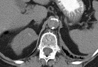
14 CT Renal cysts B4


15 CT Technique UNENHANCED 30 SECONDS 10 MINUTES 120 SECONDS


16
17 Solid masses Size Enhancement Fat content Shape Surrounds History


18 Measurement of Hounsfield Units >20 Enhancement, equivocal 6 HU 30 HU 12 HU 63 HU


19 Change = 6HU = no follow up 38 HU 44 HU
20 Solid masses Size Enhancement Fat content Shape Surrounds History
 Measure on the un-enhanced phase 4.")
21 CT - Angiomyolipoma? Macroscopic fat (- 20) Angiomyolipoma? Micro fat: -Pixel mapping line/square 4 pixels of fat attenuation (-10 HU) Measure on the un-enhanced phase 4.5% AML minimal fat Halpenny et al. Clin Rad 2010; 65:
22 Solid masses Size Enhancement Fat content Shape Surrounds History


23 Shape Tumour perinephric Infarct Treatment effect


24 Post partial nephrectomy
25 Solid masses Size Enhancement Fat content Shape Surrounds History


26 Post CT Cryotherapy
27 Indeterminate masses on CT Too small to characterise (< 5mm) Most renal lesions under 1cm will be benign cysts Psuedo-enhancement (> 10 HU) due to recon algorithm used to compensate for beam hardening. Seen in smaller and intra renal lesions. Especially on CT Urograms No precontrast scan


28 Does it go black on liver settings? WL 40 WW 400 WL 100 WW 150


29


30 CT KUB Mass > 70 HU = 99% hyperdense cyst* Mass < 20 HU = simple cyst Recall for post contrast - Calcification - Attenuation HU *Jonisch et al Radiology 2007: 243:
31 MRI Characterisation of solid renal masses In and out of phase techniques, loss of signal in fat containing masses on OP T2W Coronal Fat saturated pre and post contrast T1W imaging (ax & cor) Enhancement = increase in SI on ROI of > 15%* *Hecht et al, Radiology 2004; 232:
32 Kwon et al, Int Urol & Nephrol 2015; 47: indeterminate renal masses on CT had MRI Surgical correlation 47 benign (AML, Onco, cysts) CT Sens 27.7% Spec 94.5% MRI Sens 68.1 Spec 91.8% 15 patients could have avoided surgery MRI has incremental benefit to distinguish RCC from indeterminate renal masses on CT
 Improved contrast resolution Calcification not seen No")
33 MRI Characterisation of cystic masses Better depiction of septa on T2 weighted imaging Use of subtraction same parameters both aquisitions (post subtracted from pre-contrast) Improved contrast resolution Calcification not seen No pseudo-enhancement.


34 Bosniak III

35 Role of Biopsy Lesions indeterminate by imaging alone Prior to treatment Reliable diff between RCC and Oncocytoma Reliable histological subtype and Fuhrman grade


36 Technique < 3cm CT guided Platelets < 50, INR <1.5 Prone / decubitus Breath held insp or exp 17 Gauge Coaxial needle 18 Gauge biospy needle 2 cores
37 Hu et al, Human Pathology, 2015; 46: consecutive core biospies (0.5-24cm, 78% small ) 89% positive (of these 77% malignant, 23% benign) 93% accurate in histological subtype of RCC 11% non diagnostic


38 Complications metanalysis of 16,000* Haemorrhage / haematuria Infection Adjacent organ puncture Renal loss 0.1% Mortality 0.031% Track seeding - < 0.01 for RCC. Non diagnostic / false negative 10% *Upport AJR 2010; 194:
39 Questions What would be the consequence of NOT characterising incidental lesions? Acceptance of diagnostic uncertainty Avoidance of overdiagnosis and unnecessary surveillance What would be the consequence of NOT following up incidental lesions?

40 What % of incidental masses are significant? 3001 CT colonograms 433 (14.4%) had a mass >1cm (mean 2.5) 5 were complex/solid 4 RCC s detected with mean FU 3.3 yrs % chance of malignancy Heterogeneity on post contrast CT is a feature of RCC O Connor SD, Pickhardt PJ, Kim DH, Oliva MR, Silverman SG. Incidental finding of renal masses at unenhanced CT: preva- lence and analysis of features for guiding management. AJR Am J Roentgenol 2011; 197(1):
41 B2F how long to FU? 198 patients with B2F 86.5% Incidental findings Followed up 2 years 98% unchanged class, 66% unchanged size. 2% had partial nephrectomy, 0.5% malignant. Imaging for 2 years Savings if discharged at 2 (per year) Raslan et al, J Clinical Urology 2016; 9;
42 B2F how long to FU? In a fiscally constrained system such as the NHS, clinicians have a duty to use the allocated resources responsibly, accepting there is a small risk of an adverse outcome Kings Fund: Better value in the NHS: The role of changes in clinical practice.

43 Surveillance of small solid renal masses YES YES? NO NO
44 Summary -Ignore Lesions < 1cm (unless heterogenous) on CT or MRI Homogenous lesions < 20 or > 70 HU - unenhanced CT Lesions that look black on liver windows PV CT Macroscopic fat < 2cm Single thin septated cyst on US > 60 yrs.

45 Summary Solid masses Size Enhancement Fat content Shape Cystic masses Bosniak Classification 2F No enhancement 3 Measureable enhancement in septa. Surrounds History

46 S Silverman, G Israel, Q Trinh Radiology 2015; 275: 28-42

47
INTERDISCIPLINARY DISCUSSIONS IN LOCALISED RCC DIAGNOSIS AND SURGICAL STRATEGIES FOR ATYPICAL RENAL CYSTIC LESIONS. Maria Cova
 INTERDISCIPLINARY DISCUSSIONS IN LOCALISED RCC DIAGNOSIS AND SURGICAL STRATEGIES FOR ATYPICAL RENAL CYSTIC LESIONS Maria Cova Radiology Department University of Trieste (IT) Eleventh European International
Renal Mass Biopsy: Needed Now More than Ever
 Renal Mass Biopsy: Needed Now More than Ever Stuart G. Silverman, MD, FACR Professor of Radiology Harvard Medical School Director, Abdominal Imaging and Intervention Brigham and Women s Hospital Boston,
Role of imaging in RCC. Ultrasonography. Solid lesion. Cystic RCC. Solid RCC 31/08/60. From Diagnosis to Treatment: the Radiologist Perspective
 Role of imaging in RCC From Diagnosis to Treatment: the Radiologist Perspective Diagnosis Staging Follow up Imaging modalities Limitations and pitfalls Duangkamon Prapruttam, MD Department of Therapeutic
Renal Masses in Patients with Known Extrarenal Primary Primary Cancer Primary Primary n Met Mets s RCC Beni L mphoma Lung Breast Others
 The Importance of Stuart G. Silverman, MD, FACR Professor of Radiology Harvard ard Medical School Director, Abdominal Imaging and Intervention Brigham and Women s Hospital Boston, MA The Importance of
Management of the Incidental Renal Mass on CT: A White Paper of the ACR Incidental Findings Committee
 ORIGINAL ARTICLE Management of the Incidental Renal Mass on CT: A White Paper of the ACR Incidental Findings Committee Brian R. Herts, MD a, Stuart G. Silverman, MD b, Nicole M. Hindman, MD c, Robert G.
CME Article Clinics in diagnostic imaging (135)
") Medical Education Singapore Med J 2011; 52(5) : 384 CME Article Clinics in diagnostic imaging (135) Pojchamarnwiputh S, Muttarak M, Sriplakich S H 1a 1b 1c 1d Fig. 1 (a) Axial unenhanced; (b & c) delayed
Medical Education Singapore Med J 2011; 52(5) : 384 CME Article Clinics in diagnostic imaging (135) Pojchamarnwiputh S, Muttarak M, Sriplakich S H 1a 1b 1c 1d Fig. 1 (a) Axial unenhanced; (b & c) delayed
Renal masses - the role of diagnostic imaging
 Renal masses - the role of diagnostic imaging Poster No.: C-2471 Congress: ECR 2015 Type: Educational Exhibit Authors: V. Rai#; Bjelovar/HR Keywords: Cysts, Cancer, Structured reporting, Ultrasound, MR,
How To Approach Renal Masses? - Differential Diagnosis On Image
 How To Approach Renal Masses? - Differential Diagnosis On Image Poster No.: C-1646 Congress: ECR 2015 Type: Educational Exhibit Authors: A. E. A. G. Costa, A. Gomes, A. Duarte, I. Távora; Lisbon/PT Keywords:
(2/3 PRCC!) (2/3 PRCC!)
 (2/3 PRCC!)") Approach to the Incidental Solid Renal Mass Stuart G. Silverman, MD, FACR Professor of Radiology Harvard ard Medical School Director, Abdominal Imaging and Intervention Brigham and Women s Hospital Boston,
Approach to the Incidental Solid Renal Mass Stuart G. Silverman, MD, FACR Professor of Radiology Harvard ard Medical School Director, Abdominal Imaging and Intervention Brigham and Women s Hospital Boston,
Hyperechoic renal masses
 Hyperechoic renal masses Jean-Yves Meuwly, MD Department of Diagnostic and Interventional Radiology, University Hospital Lausanne, Switzerland Department of Diagnostic and Interventional Radiology Renal
Renal Mass Biopsy Should be Used for Most SRM - PRO
 Renal Mass Biopsy Should be Used for Most SRM - PRO Tony Finelli, MD, MSc, FRCSC Head, Division of Urology GU Site Lead, Princess Margaret Cancer Center GU Cancer Lead, Cancer Care Ontario Associate Professor,
Renal Cell Carcinoma: Attenuation Values on Unenhanced CT
 Genitourinary Imaging Original Research Pooler et al. Attenuation Values of Unenhanced CT of Renal Cell Carcinoma Genitourinary Imaging Original Research B. Dustin Pooler 1 Perry J. Pickhardt 1 Stacy D.
Caveat sonologist Mistakes to avoid in Kidney Ultrasound
 Caveat sonologist Mistakes to avoid in Kidney Ultrasound Simon Freeman Derriford Hospital, Plymouth simonfreeman@nhs.net Bear trap 1 Report: There is a 4cm solid mass arising from the left kidney likely
Small renal mass: differential diagnosis on image
 Small renal mass: differential diagnosis on image Poster No.: R-0166 Congress: RANZCR-AOCR 2012 Type: Educational Exhibit Authors: H. Lee, K. S. Lee, M. J. Kim; Anyang/KR Keywords: Cysts, Cancer, Staging,
Measure #405: Appropriate Follow-up Imaging for Incidental Abdominal Lesions National Quality Strategy Domain: Effective Clinical Care
 Measure #405: Appropriate Follow-up Imaging for Incidental Abdominal Lesions National Quality Strategy Domain: Effective Clinical Care 2016 PQRS OPTIONS FOR INDIVIDUAL MEASURES: CLAIMS, REGISTRY DESCRIPTION:
ESUR 2018, Sept. 13 th.-16 th., 2018 Barcelona, Spain
 ESUR 2018, Sept. 13 th.-16 th., 2018 Barcelona, Spain OUR APPROACH Incidental adrenal nodule/mass Isaac R Francis, M.B;B.S University of Michigan, Ann Arbor, Michigan Disclosures None (in memory) M Korobkin,
AB MR Interpretation Overview
 AB MR Interpretation Overview Goal of AB MR interpretation is to maintain high sensitivity and specificity In order to minimize false positives and short term follow ups, it is fundamental to focus only
Soft Tissue Tumour & Sarcoma Imaging Guidelines 2012
 Soft Tissue Tumour & Sarcoma Imaging Guidelines 2012 Version Control This is a controlled document please destroy all previous versions on receipt of a new version. Date Approved: March 2011 reissued April
Contrast-enhanced ultrasound (CEUS) in the evaluation and characterization of complex renal cysts
 in the evaluation and characterization of complex renal cysts") Contrast-enhanced ultrasound (CEUS) in the evaluation and characterization of complex renal cysts Poster No.: C-2812 Congress: ECR 2018 Type: Educational Exhibit Authors: J. A. Torres de Abreu Macedo,
Contrast-enhanced ultrasound (CEUS) in the evaluation and characterization of complex renal cysts Poster No.: C-2812 Congress: ECR 2018 Type: Educational Exhibit Authors: J. A. Torres de Abreu Macedo,
Imaging in breast cancer. Mammography and Ultrasound Donya Farrokh.MD Radiologist Mashhad University of Medical Since
 Imaging in breast cancer Mammography and Ultrasound Donya Farrokh.MD Radiologist Mashhad University of Medical Since A mammogram report is a key component of the breast cancer diagnostic process. A mammogram
The role of Bosniak classification in malignant tumor diagnosis: A single institution experience
 Original Article - Urological Oncology http://dx.doi.org/10.4111/icu.2016.57.2.100 pissn 2466-0493 eissn 2466-054X The role of Bosniak classification in malignant tumor diagnosis: A single institution
Is renal cryoablation becoming an effective alternative to partial nephrectomy?
 Is renal cryoablation becoming an effective alternative to partial nephrectomy? J GARNON 1, G TSOUMAKIDOU 1, H LANG 2, A GANGI 1 1 department of interventional radiology 2 department of urology University
Role Of Active Surveillance And Volume Monitoring In Patients With Small Renal Masses
 Yale University EliScholar A Digital Platform for Scholarly Publishing at Yale Yale Medicine Thesis Digital Library School of Medicine January 2012 Role Of Active Surveillance And Volume Monitoring In
The diagnostic criteria of multilocular renal cysts
 Case Report 772 Multilocular Renal Cysts with Renal Cell Carcinoma: Report of Four Cases Chia-Hsi Chen, MD; Cheng-Keng Chuang, MD, PhD; Chun-Te Wu, MD; Kwai-Fong Ng 1, MD; Shuen-Kuei Liao 2, PhD According
MANAGEMENT RECOMMENDATIONS
 1 MANAGEMENT RECOMMENDATIONS 1. Adrenal masses!!!!!!! page 2 2. Liver Masses!!!!!!! page 3 3. Obstetric US Soft Markers for Aneuploidy!! pages 4-6 4. Ovarian and Adnexal Cysts!!!!! pages 7-10 5. Pancreatic
11/1/2014. Radiologic incidentalomas Ordering pitfalls Newer technology and applications
 Bilal Tahir, MD Gitasree Borthakur, MD Indiana University School of Medicine Department of Radiology & Imaging Sciences October 31, 2014 ACP 2014 Dr. V. Aaron Nuclear (vaaron@iupui.edu) Dr. S. Westphal
Intracystic papillary carcinoma of the breast
 Intracystic papillary carcinoma of the breast Poster No.: C-1932 Congress: ECR 2011 Type: Educational Exhibit Authors: V. Dimarelos, F. TZIKOS, N. Kotziamani, G. Rodokalakis, 1 2 3 1 1 1 2 T. MALKOTSI
Renal biopsy is mandatory for every small renal mass
 Renal biopsy is mandatory for every small renal mass Ben Challacombe Consultant Urologist The Urology Centre Guy s and St. Thomas Hospital NHS Foundation Trust Oncocytoma High Risk Partial converted to
Evaluation of Thyroid Nodules
 Evaluation of Thyroid Nodules Stephan Kowalyk, MD January 25 28, 2018 1 Primary goal Exclude malignancy Incidental thyroid nodules If found on CT, MRI, PET scan, carotid Doppler ULTRASOUND!! January 25
Review Article Radiologic Evaluation of Small Renal Masses (I): Pretreatment Management
: Pretreatment Management") Hindawi Publishing Corporation Advances in Urology Volume 2008, Article ID 415848, 16 pages doi:10.1155/2008/415848 Review Article Radiologic Evaluation of Small Renal Masses (I): Pretreatment Management
Hindawi Publishing Corporation Advances in Urology Volume 2008, Article ID 415848, 16 pages doi:10.1155/2008/415848 Review Article Radiologic Evaluation of Small Renal Masses (I): Pretreatment Management
STANDARDIZED MANAGEMENT RECOMMENDATIONS FOR ADRENAL NODULES: EVIDENCE-BASED CONSENSUS POWERSCRIBE MACROS FROM AN ACADEMIC/PRIVATE PRACTICE
 STANDARDIZED MANAGEMENT RECOMMENDATIONS FOR ADRENAL NODULES: EVIDENCE-BASED CONSENSUS POWERSCRIBE MACROS FROM AN ACADEMIC/PRIVATE PRACTICE COLLABORATIVE Pamela Johnson 1, Darcy Wolfman 2, Upma Rawal 3,
Adnexal Masses and Problem Solving Pelvic MRI
 28th Congress of the Hungarian Society of Radiologists RCR Session Budapest June 2016 Adnexal Masses and Problem Solving Pelvic MRI DrSarah Swift St James s University Hospital Leeds, UK Objectives Characterisation
Bilateral Renal Angiomyolipomas with Invasion of the Renal Vein: A Case Report
 Case Study TheScientificWorldJOURNAL (2008) 8, 145 148 TSW Urology ISSN 1537-744X; DOI 10.1100/tsw.2008.29 Bilateral Renal Angiomyolipomas with Invasion of the Renal Vein: A Case Report C. Blick, N. Ravindranath,
Diagnostic accuracy of percutaneous renal tumor biopsy May 10 th 2018
 Diagnostic accuracy of percutaneous renal tumor biopsy May 10 th 2018 Dr. Tzahi Neuman Dep.Of Pathology Hadassah Medical Center Jerusalem, Israel, (tneuman@hadassah.org.il) Disclosure: 1 no conflicts of
Audit of split-bolus CT urography for the investigation of haematuria over a 12 month period at two district general hospitals
 Audit of split-bolus CT urography for the investigation of haematuria over a 12 month period at two district general hospitals Poster No.: C-1349 Congress: ECR 2010 Type: Educational Exhibit Topic: Genitourinary
IN THE NAME OF GOD POV: CYSTIC OVARIAN LESION
 IN THE NAME OF GOD POV: CYSTIC OVARIAN LESION CASE 1 20 years old girl with AUB and pelvic pain from 2 weeks ago Impression :Simple unilocular 6 cm ovarian cyst Next step? Almost certainly benign so FU
Is Ultrasound Useful for Further Evaluation of Homogeneously Hyperattenuating Renal Lesions Detected on CT?
 Genitourinary Imaging Original Research Genitourinary Imaging Original Research Mahadevaswamy Siddaiah 1 Satheesh Krishna Matthew D. F. McInnes Jeffrey S. Quon Wael M. Shabana Demetri Papadatos Nicola
The Incidental Renal Mass in the Primary Care Setting
 The Incidental Renal Mass in the Primary Care Setting Adele M. Caruso, MSN, CRNP Adult Nurse Practitioner The Perelman School of Medicine at the University of Pennsylvania Abstract There are approximately
Do Incidental Hyperechoic Renal Lesions Measuring Up to 1 cm Warrant Further Imaging? Outcomes of 161 Lesions
 Genitourinary Imaging Original Research Genitourinary Imaging Original Research Ankur M. Doshi 1 Abimbola Ayoola Andrew B. Rosenkrantz Doshi AM, Ayoola A, Rosenkrantz AB Keywords: angiomyolipoma, hyperechoic
ADRENAL MR: PEARLS AND PITFALLS
 ADRENAL MR: PEARLS AND PITFALLS Frank Miller, M.D. Lee F. Rogers MD Professor of Medical Education Chief, Body Imaging Section and Fellowship Medical Director, MR Imaging Professor of Radiology Northwestern
Thyroid in a Nutshell Dublin Catherine Kirkpatrick Consultant Sonographer ULHT
 Thyroid in a Nutshell Dublin 2017 Catherine Kirkpatrick Consultant Sonographer ULHT Acknowledgements Dr. Steve Colley Dr. Rhodri Evans Dr. Rhian Rhys Dr. Andrew McQueen Aims Anatomy & Physiology Incidence
REVIEW. Distinguishing benign from malignant adrenal masses
 Cancer Imaging (2003) 3, 102 110 DOI: 10.1102/1470-7330.2003.0006 CI REVIEW Distinguishing benign from malignant adrenal masses Isaac R Francis Professor of Radiology, Department of Radiology, University
LIVER IMAGING TIPS IN VARIOUS MODALITIES. M.Vlychou, MD, PhD Assoc. Professor of Radiology University of Thessaly
 LIVER IMAGING TIPS IN VARIOUS MODALITIES M.Vlychou, MD, PhD Assoc. Professor of Radiology University of Thessaly Hepatocellular carcinoma is a common malignancy for which prevention, screening, diagnosis,
Patient Selection for Ablative Therapies. Adrian D Joyce Leeds UK
 Patient Selection for Ablative Adrian D Joyce Leeds UK Therapy Renal Cell Ca USA: 30,000 new cases annually >12,000 deaths RCC accounts for 3% of all adult malignancy 40% of patients will die from their
Cystitis cystica is a rare chronic reactive inflammatory
 JOURNAL OF ENDOUROLOGY CASE REPORTS Volume 3.1, 2017 Mary Ann Liebert, Inc. Pp. 34 38 DOI: 10.1089/cren.2017.0010 Case Report Cystitis Cystica as a Large Solitary Bladder Cyst Stephanie Potts, MBChB and
ADRENAL LESIONS 10/09/2012. Adrenal + lesion. Introduction. Common causes. Anatomy. Financial disclosure. Dr. Boraiah Sreeharsha. Nothing to declare
 ADRENAL LESIONS Financial disclosure Nothing to declare Dr. Boraiah Sreeharsha MBBS;FRCR;FRCPSC Introduction Adrenal + lesion Adrenal lesions are common 9% of the population Increase in the detection rate
CURRENT METHODS IN IMAGE GUIDED BREAST BIOPSY
 CURRENT METHODS IN IMAGE GUIDED BREAST BIOPSY Stuart Silver April 24, 2004 OBJECTIVES Review development of current techniques Discuss stereotactic breast biopsy Discuss US guided breast biopsy 1 OBJECTIVES
GUIDELINES FOR PULMONARY NODULE MANAGEMENT : RECENT CHANGES AND UPDATES
 Venice 2017 GUIDELINES FOR PULMONARY NODULE MANAGEMENT : RECENT CHANGES AND UPDATES Heber MacMahon MB, BCh Department of Radiology The University of Chicago Disclosures Consultant for Riverain Medical
HEPATO-BILIARY IMAGING
 HEPATO-BILIARY IMAGING BY MAMDOUH MAHFOUZ MD PROF.OF RADIOLOGY CAIRO UNIVERSITY mamdouh.m5@gmail.com www.ssregypt.com CT ABDOMEN Indications Patient preparation Patient position Scanogram Fasting 4-6 hours
Case-based discussion:
 Case-based discussion: Pailin Kongmebhol, M.D. Department of Radiology Faculty of Medicine Chiang Mai University There are many guidelines for managing thyroid nodules Two important guidelines: 2015 American
ADRENAL MEDULLARY DISORDERS: PHAEOCHROMOCYTOMAS AND MORE
 ADRENAL MEDULLARY DISORDERS: PHAEOCHROMOCYTOMAS AND MORE DR ANJU SAHDEV READER AND CONSULTANT RADIOLOGIST QUEEN MARY UNIVERSITY AND ST BARTHOLOMEW S HOSPITAL BARTS HEALTH, LONDON, UK DISCLOSURE OF CONFLICT
Radiologic Findings of Mucocele-like Tumors of the breast: Can we differentiate pure benign from associated with high risk lesions?
 Radiologic Findings of Mucocele-like Tumors of the breast: Can we differentiate pure benign from associated with high risk lesions? Poster No.: C-0332 Congress: ECR 2014 Type: Educational Exhibit Authors:
Analysis of Changes in Attenuation of Proven Renal Cysts on Different Scanning Phases of Triphasic MDCT
 Eugene P. Chung 1 Brian R. Herts 1,2 Grant Linnell 1 Andrew C. Novick 2 Nancy Obuchowski 1,3 Deirdre M. Coll 1,4 Mark E. Baker 1 Received June 24, 2003; accepted after revision August 28, 2003. Presented
Management A Guideline Based Approach to the Incidental Pancreatic Cysts. Common Cystic Pancreatic Neoplasms.
 Management 2016 A Guideline Based Approach to the Incidental Pancreatic Cysts ISMRM 2016 Masoom Haider, MD, FRCP(C) Professor of Radiology, University of Toronto Clinician Scientist, Ontario Institute
Newcastle HPB MDM updated radiology imaging protocol recommendations. Author Dr John Scott. Consultant Radiologist Freeman Hospital
 Newcastle HPB MDM updated radiology imaging protocol recommendations Author Dr John Scott. Consultant Radiologist Freeman Hospital This document is intended as a guide to aid radiologists and clinicians
Cystic Renal Cell Carcinomas: Do They Grow, Metastasize, or Recur?
 Genitourinary Imaging Original Research Jhaveri et al. Growth Patterns of Cystic Renal Cell Carcinomas Genitourinary Imaging Original Research Kartik Jhaveri 1 Priya Gupta 1 Azadeh Elmi 2 Lior Flor 1 Hadas
THYROID NODULES: THE ROLE OF ULTRASOUND
 THYROID NODULES: THE ROLE OF ULTRASOUND NOVEMBER 2017 DR. DEAN DURANT DEFINITION Thyroid nodule: Focal area within the thyroid gland with echogenicity different from surrounding parenchyma. THYROID NODULES
4,3,2,1...How Many Phases are Needed? Balancing Diagnostic Efficacy and Radiation Modulation for MDCT Imaging of Renal Cell Carcinoma
 4,3,2,1...How Many Phases are Needed? Balancing Diagnostic Efficacy and Radiation Modulation for MDCT Imaging of Renal Cell Carcinoma Jeremy Hackworth, MD, MS Steven P Rowe, MD, PhD Satomi Kawamoto, MD
Ultrasound screening of soft tissue masses in the trunk and extremity - a BSG guide for ultrasonographers and primary care
 Ultrasound screening of soft tissue masses in the trunk and extremity - a BSG guide for ultrasonographers and primary care Introduction Soft tissue masses in the trunk and extremity are common and most
Thyroid Nodules: US Risk Stratification. Alex Tessnow, MD, FACE, ECNU University of Texas Southwestern Associate Professor of Medicine Dallas, Texas
 Thyroid Nodules: US Risk Stratification Alex Tessnow, MD, FACE, ECNU University of Texas Southwestern Associate Professor of Medicine Dallas, Texas Which of the following is true? A. All echogenic foci
ADRENAL INCIDENTALOMA. Jamii St. Julien
 ADRENAL INCIDENTALOMA Jamii St. Julien Outline Definition Differential Evaluation Treatment Follow up Questions Case Definition The phenomenon of detecting an otherwise unsuspected adrenal mass on radiologic
Small (< 4 cm) Renal Masses: Differentiation of Angiomyolipoma Without Visible Fat From Renal Cell Carcinoma Using Unenhanced and Contrast-Enhanced CT
 Renal Masses: Differentiation of Angiomyolipoma Without Visible Fat From Renal Cell Carcinoma Using Unenhanced and Contrast-Enhanced CT") Genitourinary Imaging Original Research Takahashi et al. CT of Small Renal Masses Genitourinary Imaging Original Research Naoki Takahashi 1 Shuai Leng 1 Kazuhiro Kitajima 1,2 Daniel Gomez-Cardona 1,3 Prabin
Genitourinary Imaging Original Research Takahashi et al. CT of Small Renal Masses Genitourinary Imaging Original Research Naoki Takahashi 1 Shuai Leng 1 Kazuhiro Kitajima 1,2 Daniel Gomez-Cardona 1,3 Prabin
Recommendations for cross-sectional imaging in cancer management, Second edition
 www.rcr.ac.uk Recommendations for cross-sectional imaging in cancer management, Second edition Renal and adrenal tumours Faculty of Clinical Radiology www.rcr.ac.uk Contents Renal cell carcinoma 3 Clinical
WHAT IS THE ROLE OF ACTIVE SURVEILLANCE
 WHAT IS THE ROLE OF ACTIVE SURVEILLANCE IN THE CONTEXT OF RENAL ABLATION AND PARTIAL NEPHRECTOMY? Alessandro Volpe University of Eastern Piedmont Novara, Italy RCC INCIDENCE SEER DATABASE (1975-2006) RCC
Case Report Papillary Renal Cell Carcinoma Seeding along a Percutaneous Biopsy Tract
 Case Reports in Urology Volume 2015, Article ID 925254, 4 pages http://dx.doi.org/10.1155/2015/925254 Case Report Renal Cell Carcinoma Seeding along a Percutaneous Biopsy Tract Deanne Soares, Nariman Ahmadi,
MRI Abdomen Protocol Pancreas/MRCP with Contrast
 MRI Abdomen Protocol Pancreas/MRCP with Contrast Reviewed By: Brett Mollard, MD; Anna Ellermeier, MD Last Reviewed: July 2018 Contact: (866) 761-4200 Standard uses: 1. Characterization of cystic and solid
Contrast Enhanced Ultrasound of Parenchymal Masses in Children
 Contrast Enhanced Ultrasound of Parenchymal Masses in Children Sue C Kaste, DO On behalf of Beth McCarville, MD St. Jude Children s Research Hospital Memphis, TN Overview Share St. Jude experience with
Pitfalls and Limitations of Breast MRI. Susan Orel Roth, MD Professor of Radiology University of Pennsylvania
 Pitfalls and Limitations of Breast MRI Susan Orel Roth, MD Professor of Radiology University of Pennsylvania Objectives Review the etiologies of false negative breast MRI examinations Discuss the limitations
Retroperitoneal Teratoma Heather Borders, MD
 Retroperitoneal Teratoma Heather Borders, MD 03/04/2012 History Newborn with congenitally diagnosed mass. No other clinical symptoms. Diagnosis Retroperitoneal Teratoma; Immature teratoma, grade 1, with
Imaging of Kidney Cancer
 119 Imaging of Kidney Cancer RADIOLOGIC CLINICS OF NORTH AMERICA Radiol Clin N Am 45 (2007) 119 147 Jingbo Zhang, MD*, Robert A. Lefkowitz, MD, Ariadne Bach, MD - Detection and diagnosis - CT scan Solid
Jesse Civan, M.D. Medical Director, Jefferson Liver Tumor Center
 Liver Tumors Jesse Civan, M.D. Medical Director, Jefferson Liver Tumor Center Differential Diagnosis Malignant Metastatic from non-hepatic primary Hepatocellular carcinoma Cholangiocarcinoma Biliary cystcarcinoma
Shadow because the air
 Thyroid Ultrasound Thyroid US examination needs: 1. high frequency transducer 2. extended patient's neck 3. check all the neck area because the swelling could be in areas other than the thyroid such as
James Cassuto, MS IV. Shekher Maddineni, MD Samuel McCabe, MD Vascular and Interventional Radiology
 Student: Attendings: Department: James Cassuto, MS IV Grigory Rozenblit, MD Shekher Maddineni, MD Samuel McCabe, MD Vascular and Interventional Radiology Chief Complaint & HPI 61 year old female who is
Principal Site Investigator ENHANCE (Evaluation of Thyroid FNA Genomic Signature) study: An IRB approved study with funding to Rochester Regional
 study: An IRB approved study with funding to Rochester Regional") October 20 th 2018 Principal Site Investigator ENHANCE (Evaluation of Thyroid FNA Genomic Signature) study: An IRB approved study with funding to Rochester Regional Health from Veracyte Review ultrasound
October 20 th 2018 Principal Site Investigator ENHANCE (Evaluation of Thyroid FNA Genomic Signature) study: An IRB approved study with funding to Rochester Regional Health from Veracyte Review ultrasound
The role of endoscopy in the diagnosis and treatment of cystic pancreatic neoplasms
 The role of endoscopy in the diagnosis and treatment of cystic pancreatic neoplasms CYSTIC LESIONS AND FLUID COLLECTIONS OF THE PANCREAS Their pathology ranges from pseudocysts and pancreatic necrosis
PULMONARY NODULES AND MASSES : DIAGNOSTIC APPROACH AND NEW MANAGEMENT GUIDELINES. https://tinyurl.com/hmpn2018
 PULMONARY NODULES AND MASSES : DIAGNOSTIC APPROACH AND NEW MANAGEMENT GUIDELINES Heber MacMahon MB, BCh Department of Radiology The University of Chicago https://tinyurl.com/hmpn2018 Disclosures Consultant
Contrast-Enhanced Spectral Mammography
 Contrast-Enhanced Spectral Mammography Illuminating Breast Cancer Detection SenoBright HD TM gehealthcare.com/senobright Mammography is the most reliable imaging technique for breasts, but limitations
AACE/ACE Advanced Endocrine Neck Ultrasound Training Course 2016
 AACE/ACE Advanced Endocrine Neck Ultrasound Training Course 2016 This 9mm left inferior nodule should remind us all why we re here! There is no absolute number of images required for documentation
Imaging Findings of Primary Angiomyolipoma of the Pancreas: A Case Report 췌장의원발성혈관근육지방종의영상소견 1 예 : 증례보고
 Case Report pissn 1738-2637 / eissn 2288-2928 https://doi.org/10.3348/jksr.2017.77.1.9 Imaging Findings of Primary Angiomyolipoma of the Pancreas: A Case Report 췌장의원발성혈관근육지방종의영상소견 1 예 : 증례보고 Hye Hee Kim,
of Thyroid Lesions Comet Tail Crystals
 2 Ultrasound Features of Thyroid Lesions There are many different features indicating a certain benign or malignant tumor type, but many of these are overlapping signs. Combining several features is considered
Cystic Pancreatic Lesions: Approach to Diagnosis
 Cystic Pancreatic Lesions: Approach to Diagnosis Poster No.: R-0130 Congress: RANZCR-AOCR 2012 Type: Educational Exhibit Authors: A. AGARWAL, R. M. Mendelson; Perth/AU Keywords: Cysts, Biopsy, Endoscopy,
Pathologic Characteristics of Solitary Small Renal Masses. Can They Be Predicted by Preoperative Clinical Parameters?
 Anatomic Pathology / Pathology of Small Renal Masses Pathologic Characteristics of Solitary Small Renal Masses Can They Be Predicted by Preoperative Clinical Parameters? Tom DeRoche, MD, 1 Esteban Walker,
Low-Dose CT: Clinical Studies & the Radiologist Perspective
 Low-Dose CT: Clinical Studies & the Radiologist Perspective RD-ASiR RD-MBIR SD-FBP RD=0.35 msv (80% dose reduction) Perry J. Pickhardt, MD UW School of Medicine & Public Health Low-Dose CT: Clinical Overview
CT & MRI of Benign Liver Neoplasms Srinivasa R Prasad
 CT & MRI of Benign Liver Neoplasms Srinivasa R Prasad No financial disclosures Acknowledgements Many thanks to Drs. Heiken, Narra & Menias (MIR) Dr. Sahani (MGH) for sharing images Benign Liver Tumors:
ISSN X (Print) Research Article. *Corresponding author Dr. Amlendu Nagar
 Research Article. *Corresponding author Dr. Amlendu Nagar") Scholars Journal of Applied Medical Sciences (SJAMS) Sch. J. App. Med. Sci., 2015; 3(3A):1069-1073 Scholars Academic and Scientific Publisher (An International Publisher for Academic and Scientific Resources)
Scholars Journal of Applied Medical Sciences (SJAMS) Sch. J. App. Med. Sci., 2015; 3(3A):1069-1073 Scholars Academic and Scientific Publisher (An International Publisher for Academic and Scientific Resources)
Kidney Case 1 SURGICAL PATHOLOGY REPORT
 Kidney Case 1 Surgical Pathology Report February 9, 2007 Clinical History: This 45 year old woman was found to have a left renal mass. CT urography with reconstruction revealed a 2 cm medial mass which
GUIDELINES ON RENAL CELL CANCER
 20 G. Mickisch (chairman), J. Carballido, S. Hellsten, H. Schulze, H. Mensink Eur Urol 2001;40(3):252-255 Introduction is characterised by a constant rise in incidence over the last 50 years, with a predominance
SCBT-MR 2015 Incidentaloma on Chest CT
 SCBT-MR 2015 Incidentaloma on Chest CT Reginald F. Munden MD, DMD, MBA I have no conflicts of interest to report Incidentaloma Pulmonary Nodule Mediastinal Lymph Node Coronary Artery Calcium Incidental
Policies, Standards, and Guidelines. Guidelines on Breast Ultrasound Examination and Reporting
 Policies, Standards, and Guidelines Guidelines on Breast Ultrasound Examination and Reporting Approved by Council June 2018 Approved: June 2018 Guidelines on Breast Ultrasound Examination and Reporting
objectives Pitfalls and Pearls in PET/CT imaging Kevin Robinson, DO Assistant Professor Department of Radiology Michigan State University
 objectives Pitfalls and Pearls in PET/CT imaging Kevin Robinson, DO Assistant Professor Department of Radiology Michigan State University To determine the regions of physiologic activity To understand
Category Term Definition Comments 1 Major Categories 1a
 Working Lexicon Categories, Terms & Definitions Category Term Definition Comments 1 Major Categories 1a Physiologic Category (consistent with normal ovarian physiology) Follicle Simple 3 cm in premenopausal
X-Ray Corner. Imaging Approach to Cystic Liver Lesions. Pantongrag-Brown L. Solitary cystic liver lesions. Hepatic simple cyst (Figure 1)
") THAI J 136 Imaging Approach to Cystic Liver Lesions GASTROENTEROL 2013 X-Ray Corner Imaging Approach to Cystic Liver Lesions Pantongrag-Brown L Cystic liver lesions are common findings in daily practice
THAI J 136 Imaging Approach to Cystic Liver Lesions GASTROENTEROL 2013 X-Ray Corner Imaging Approach to Cystic Liver Lesions Pantongrag-Brown L Cystic liver lesions are common findings in daily practice
LUNG NODULES: MODERN MANAGEMENT STRATEGIES
 Department of Radiology LUNG NODULES: MODERN MANAGEMENT STRATEGIES Christian J. Herold M.D. Department of Biomedical Imaging and Image-guided Therapy Medical University of Vienna Vienna, Austria Pulmonary
Histogram Analysis of Small Solid Renal Masses: Differentiating Minimal Fat Angiomyolipoma From Renal Cell Carcinoma
 Genitourinary Imaging Original Research Chaudhry et al. Histogram Analysis of Small Solid Renal Masses Genitourinary Imaging Original Research Humaira S. Chaudhry 1,2 Matthew S. Davenport 1,3 Christopher
Original Report. Mucocele-Like Tumors of the Breast: Mammographic and Sonographic Appearances. Katrina Glazebrook 1 Carol Reynolds 2
 Katrina Glazebrook 1 Carol Reynolds 2 Received January 2, 2002; accepted after revision August 28, 2002. 1 Department of Radiology, Mayo Clinic, 200 First St. S.W., Rochester, MN 55905. Address correspondence
Incidental Esophageal Findings on Chest CT. Amira Hussien, MD, Elliot Fishman, MD, Bouchra Younes, MD, Ahmed Hatw. Johns Hopkins Medical Institution
 Incidental Esophageal Findings on Chest CT Amira Hussien, MD, Elliot Fishman, MD, ouchra Younes, MD, Ahmed Hatw. Johns Hopkins Medical Institution I have nothing to disclose. DISCLOSURE INTRODUCTION Although
Sex: 女 Age: 51 Occupation: 無 Admission date:92/07/22
 Sex: 女 Age: 51 Occupation: 無 Admission date:92/07/22 Chief complaint Unknown fever for one month Hand tremor and left huge renal tumor was noted Present illness Suffered from fever for one month, hand
Contents. Basic Ultrasound Principles and Terminology. Ultrasound Nodule Characteristics
 Contents Basic Ultrasound Principles and Terminology Basic Ultrasound Principles... 1 Ultrasound System... 2 Linear Transducer for Superficial Images and Ultrasound-Guided FNA... 3 Scanning Planes... 4
Detailed Program of the second BREAST IMAGING AND INTERVENTIONS PROGRAM am am : Clinician s requirements from breast imaging
 Detailed Program of the second BREAST IMAGING AND INTERVENTIONS PROGRAM 2012 Day one, 2 nd November BREAST IMAGING AND INTERVENTIONS PROGRAM 2012 9.00 AM 9.10 am Introduction 9.10 am - 9.30 am : Clinician