(2/3 PRCC!) (2/3 PRCC!)
|
|
|
- Kristina Benson
- 5 years ago
- Views:
Transcription


1 Approach to the Incidental Solid Renal Mass Stuart G. Silverman, MD, FACR Professor of Radiology Harvard ard Medical School Director, Abdominal Imaging and Intervention Brigham and Women s Hospital Boston, MA Approach to the Incidental Solid Renal Mass Stuart G. Silverman, MD, FACR Disclosure of financial relationship with relevant commercial interest Lippincott, Williams, and Wilkins Philadelphia, PA Book Royalties The Problem Renal masses are ubiquitous Some benign masses cannot be differentiated from using imaging alone behavior varies and cannot be entirely predicted by imaging or pathology features. Management is hence controversial Outline Imaging vital in all aspects of management Biopsy now an accepted test The JACR white paper Differential Diagnosis Consider Pseudotumors Vascular abnormality Inflammatory Before Traumatic considering Cystic or solid neoplasms Page 1
2 Management Recommendations Large (> 3cm) Recommend/Comment Very small until 1 cm General Population Management Recommendations Large (> 3cm) Provided there is no detectable fat by CT or MRI Recommend/Comment Very small until 1 cm General Population Containing Fat Cells containing fat cells is rare! Most reported cases are of small amounts of fat associated w/ Ca 2+, and even rarer without Ca 2+. Fat Cells in Mechanisms Mature fat cells Lipid-laden macrophages (2/3 P!) Osseous metaplasia Cholesterol necrosis Angiomyolipoma The identification of fat cells in a noncalcified renal mass, in an adult, is virtually diagnostic of a benign renal angiomyolipoma Fat cells (FC) vs. Intracytoplasmic Lipid (ICL) Kidney FC ICL N * Y AML Y Y * Except case reports AML vs on MRI As renal cell carcinoma (clear cell type) may contain intracytoplasmic lipid, chemical shift MRI should not be used alone to discriminate renal angiomyolipoma from renal cell carcinoma. Page 2


3 Solid Masses may be benign Of 2,770 nephrectomies /NSS for solid renal masses, % benign Masses < 3 cm 25% benign Masses < 2 cm 30% benign Masses < 1 cm 44% benign Frank et al J Urol 2003 Solid Masses may be benign Benign Tumors resected % 73 Angiomyolipoma 18 Papillary adenoma* 4 Not otherwise specified 4 Metanephric adenoma 1 *Papillary <5mm Frank et al J Urol 2003 vs Oncocytic : Immunocytochemistry Onc Onc AE1/AE3 + + EMA Colloidal Fe - +* S100A1 + - *specific for Chromophobe Liu and Fanning Cancer Cytopath 2001 Li et al Histopathology 2007 Hyperdense + Enhancing typically papillary, or clear cell sub-type that has bled Angiomyolipoma with minimal fat Lymphoma Metanephric adenoma (rare) Leiomyoma (rare) Metastases (rare) Silverman et al RadioGraphics 2007 MRI Features of Renal Masses T1 dark T2 CE bright + (CC type) dark dark + (papillary) dark dark + AML with minimal fat Page 3
4 AML with Minimal Fat Approximately 4-5% AML contain little or no fat and are small,, and homogeneously enhancing masses Only 2% of are and homogeneously enhancing Jinzaki et al Radiology 1997 AML with Minimal Fat Biopsy can be used to diagnose AML, particularly with the aid of immunocytochemistry AML MART1 + - SMA + - HMB Granter et al Cancer 1999 AML Diagnostic Criteria CT - ROI < -10 HU MRI - fat suppression (not OOPS alone) Biopsy -+/ +/- fat cells; thick walled vessels, smooth muscle (SMA and HMB45) AML with Minimal fat AMLs demonstrating no fat have a characteristic appearance ( and enhancing) that is not common for. Biopsy can be used to biopsy them, and avoid unnecessary surgery Short (dark) T2 Masses Hemorrhagic cyst (papillary type or clear cell that bled) AML (minimal fat, rich in smooth ms spindle cells) Leiomyoma of capsule AML w/ min fat vs Sensitivity, specificity, and accuracy for combination of T2 SI ratio less than 0.9 and ([SII greater than 20% and T1 SI ratio greater than 1.2] or arterial-to to-delayed enhancement ratio greater than 1.5) were 73% (11 of 15), 99% (103 of 104), and 96% (114 of 119), respectively, for differentiating AML from (Sasiwimonphan et al Radiology 2012) AML (%SI changed = 350) enhance more than CC (% SI change = 230) in CMP (Vargas et al Radiology 2012) Problem is differentiating P from AML, not all, and a probable diagnosis is not adequate in all cases. Can MRI be used alone? Page 4
 CT or MRI at 3-6 mos, and 12 mos, then yearly Very small until 1 cm General Population and Growth")
5 Management Recommendations Recommend/Comment Large (> 3cm) or observe Consider biopsy or observe Very small until 1.5 cm Limited life expectancy/co-morbidity Management Recommendations Recommend/Comment Large (> 3cm) CT or MRI at 3-6 mos, and 12 mos, then yearly Very small until 1 cm General Population and Growth Solid The smaller the mass, the more likely it is benign. W/U lesions that grow to 1 cm Growth is concerning but not diagnostic of a malignancy. Lack of growth may be useful indicator of a benign neoplasm, or at least of benign behavior. Management flowchart Management flowchart Berland LL et al, JACR 2010 Berland LL et al, JACR 2010 Page 5
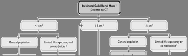

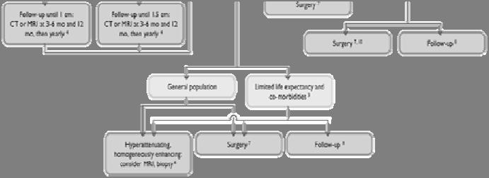
6 Management flowchart Berland LL et al, JACR 2010 Page 6
Renal Masses in Patients with Known Extrarenal Primary Primary Cancer Primary Primary n Met Mets s RCC Beni L mphoma Lung Breast Others
 The Importance of Stuart G. Silverman, MD, FACR Professor of Radiology Harvard ard Medical School Director, Abdominal Imaging and Intervention Brigham and Women s Hospital Boston, MA The Importance of
Renal Mass Biopsy: Needed Now More than Ever
 Renal Mass Biopsy: Needed Now More than Ever Stuart G. Silverman, MD, FACR Professor of Radiology Harvard Medical School Director, Abdominal Imaging and Intervention Brigham and Women s Hospital Boston,
The Incidental Renal lesion
 The Incidental Renal lesion BACKGROUND Increase in abdominal CT/US in last 15 years Resulted in detection of many (small) renal lesions 50% > 50yrs has at least 1 lesion majority simple cysts Renal lesions
ESUR 2018, Sept. 13 th.-16 th., 2018 Barcelona, Spain
 ESUR 2018, Sept. 13 th.-16 th., 2018 Barcelona, Spain OUR APPROACH Incidental adrenal nodule/mass Isaac R Francis, M.B;B.S University of Michigan, Ann Arbor, Michigan Disclosures None (in memory) M Korobkin,
Management of the Incidental Renal Mass on CT: A White Paper of the ACR Incidental Findings Committee
 ORIGINAL ARTICLE Management of the Incidental Renal Mass on CT: A White Paper of the ACR Incidental Findings Committee Brian R. Herts, MD a, Stuart G. Silverman, MD b, Nicole M. Hindman, MD c, Robert G.
Small renal mass: differential diagnosis on image
 Small renal mass: differential diagnosis on image Poster No.: R-0166 Congress: RANZCR-AOCR 2012 Type: Educational Exhibit Authors: H. Lee, K. S. Lee, M. J. Kim; Anyang/KR Keywords: Cysts, Cancer, Staging,
Diagnostic accuracy of percutaneous renal tumor biopsy May 10 th 2018
 Diagnostic accuracy of percutaneous renal tumor biopsy May 10 th 2018 Dr. Tzahi Neuman Dep.Of Pathology Hadassah Medical Center Jerusalem, Israel, (tneuman@hadassah.org.il) Disclosure: 1 no conflicts of
Traumatic and Non Traumatic Adrenal Emergencies
 Traumatic and Non Traumatic Adrenal Emergencies Michael N. Patlas, MD, FRCPC (1), Christine O. Menias, MD (2), Douglas S. Katz, MD, FACR (3), Ania Z. Kielar, MD, FRCPC (4), Alla M. Rozenblit, MD (5), Jorge
Hyperechoic renal masses
 Hyperechoic renal masses Jean-Yves Meuwly, MD Department of Diagnostic and Interventional Radiology, University Hospital Lausanne, Switzerland Department of Diagnostic and Interventional Radiology Renal
Pediatric Retroperitoneal Masses Radiologic-Pathologic Correlation
 Acta Radiológica Portuguesa, Vol.XVIII, nº 70, pág. 61-70, Abr.-Jun., 2006 Pediatric Retroperitoneal Masses Radiologic-Pathologic Correlation Marilyn J. Siegel Mallinckrodt Institute of Radiology, Washington
Role of imaging in RCC. Ultrasonography. Solid lesion. Cystic RCC. Solid RCC 31/08/60. From Diagnosis to Treatment: the Radiologist Perspective
 Role of imaging in RCC From Diagnosis to Treatment: the Radiologist Perspective Diagnosis Staging Follow up Imaging modalities Limitations and pitfalls Duangkamon Prapruttam, MD Department of Therapeutic
MANAGEMENT RECOMMENDATIONS
 1 MANAGEMENT RECOMMENDATIONS 1. Adrenal masses!!!!!!! page 2 2. Liver Masses!!!!!!! page 3 3. Obstetric US Soft Markers for Aneuploidy!! pages 4-6 4. Ovarian and Adnexal Cysts!!!!! pages 7-10 5. Pancreatic
DIAGNOSTIC SLIDE SEMINAR: PART 1 RENAL TUMOUR BIOPSY CASES
 DIAGNOSTIC SLIDE SEMINAR: PART 1 RENAL TUMOUR BIOPSY CASES Dr. Andrew J. Evans MD, PhD, FACP, FRCPC Consultant in Genitourinary Pathology University Health Network, Toronto, ON Case 1 43 year-old female,
ADRENAL MR: PEARLS AND PITFALLS
 ADRENAL MR: PEARLS AND PITFALLS Frank Miller, M.D. Lee F. Rogers MD Professor of Medical Education Chief, Body Imaging Section and Fellowship Medical Director, MR Imaging Professor of Radiology Northwestern
Renal masses - the role of diagnostic imaging
 Renal masses - the role of diagnostic imaging Poster No.: C-2471 Congress: ECR 2015 Type: Educational Exhibit Authors: V. Rai#; Bjelovar/HR Keywords: Cysts, Cancer, Structured reporting, Ultrasound, MR,
Imaging Findings of Primary Angiomyolipoma of the Pancreas: A Case Report 췌장의원발성혈관근육지방종의영상소견 1 예 : 증례보고
 Case Report pissn 1738-2637 / eissn 2288-2928 https://doi.org/10.3348/jksr.2017.77.1.9 Imaging Findings of Primary Angiomyolipoma of the Pancreas: A Case Report 췌장의원발성혈관근육지방종의영상소견 1 예 : 증례보고 Hye Hee Kim,
CT Urography. Bladder. Stuart G. Silverman, M.D.
 CT Urography Stuart G. Silverman, M.D. Professor of Radiology Harvard Medical School Director, Abdominal Imaging and Intervention Brigham and Women s Hospital Bladder Boston, MA CT Urography Stuart G.
Kidney Case 1 SURGICAL PATHOLOGY REPORT
 Kidney Case 1 Surgical Pathology Report February 9, 2007 Clinical History: This 45 year old woman was found to have a left renal mass. CT urography with reconstruction revealed a 2 cm medial mass which
ADRENAL LESIONS 10/09/2012. Adrenal + lesion. Introduction. Common causes. Anatomy. Financial disclosure. Dr. Boraiah Sreeharsha. Nothing to declare
 ADRENAL LESIONS Financial disclosure Nothing to declare Dr. Boraiah Sreeharsha MBBS;FRCR;FRCPSC Introduction Adrenal + lesion Adrenal lesions are common 9% of the population Increase in the detection rate
Contemporary Role of Renal Mass Biopsy
 Contemporary Role of Renal Mass Biopsy Jeffrey K. Mullins, MD Director Urologic Oncology CHI Memorial Chattanooga Urology Associates September 8, 2018 Disclosures I, Jeffrey Mullins, do not have a financial
Measure #405: Appropriate Follow-up Imaging for Incidental Abdominal Lesions National Quality Strategy Domain: Effective Clinical Care
 Measure #405: Appropriate Follow-up Imaging for Incidental Abdominal Lesions National Quality Strategy Domain: Effective Clinical Care 2016 PQRS OPTIONS FOR INDIVIDUAL MEASURES: CLAIMS, REGISTRY DESCRIPTION:
Renal Mass Biopsy Should be Used for Most SRM - PRO
 Renal Mass Biopsy Should be Used for Most SRM - PRO Tony Finelli, MD, MSc, FRCSC Head, Division of Urology GU Site Lead, Princess Margaret Cancer Center GU Cancer Lead, Cancer Care Ontario Associate Professor,
Radiological Appearance of Renal Leiomyoma: two cases report and review of the literature
 J Radiol Sci 2012; 37: 139-143 Radiological Appearance of Renal Leiomyoma: two cases report and review of the literature Wei-Ni Liao 1 Chi-Kuan Chen 2 Fei-Shih Yang 1,3 Department of Radiology 1, Department
Renal Tumors in Transplantation
 Renal Tumors in Transplantation Kevin Morrison Clinical-Pathological Correlation September 15 th, 2004 Overview Case reports Neoplasia in transplantation Renal allograft tumors Native kidney RCC Role of
How To Approach Renal Masses? - Differential Diagnosis On Image
 How To Approach Renal Masses? - Differential Diagnosis On Image Poster No.: C-1646 Congress: ECR 2015 Type: Educational Exhibit Authors: A. E. A. G. Costa, A. Gomes, A. Duarte, I. Távora; Lisbon/PT Keywords:
REVIEW. Distinguishing benign from malignant adrenal masses
 Cancer Imaging (2003) 3, 102 110 DOI: 10.1102/1470-7330.2003.0006 CI REVIEW Distinguishing benign from malignant adrenal masses Isaac R Francis Professor of Radiology, Department of Radiology, University
ADRENAL INCIDENTALOMA. Jamii St. Julien
 ADRENAL INCIDENTALOMA Jamii St. Julien Outline Definition Differential Evaluation Treatment Follow up Questions Case Definition The phenomenon of detecting an otherwise unsuspected adrenal mass on radiologic
Endocrine MR. Jan 30, 2015 Michael LaFata, MD
 Endocrine MR Jan 30, 2015 Michael LaFata, MD Brief case 55-year-old female in ED PMH: HTN, DM2, HLD, GERD CC: Epigastric/LUQ abdominal pain, N/V x2 days AF, HR 103, BP 155/85, room air CMP: Na 133, K 3.6,
ABDOMINAL DIFFUSION WEIGHTED MR
 ABDOMINAL DIFFUSION WEIGHTED MR Frank Miller, M.D. FACR Professor of Radiology Chief, Body Imaging Section Medical Director, MR Imaging Northwestern University Feinberg School of Medicine fmiller@northwestern.edu
Gemstone Spectral Imaging quantifies lesion characteristics for a confident diagnosis
 GE Healthcare Gemstone Spectral Imaging quantifies lesion characteristics for a confident diagnosis CT clinical case study lesion characterization Desiree Morgan, MD Vice Chair of Clinical Research Professor
Contrast Enhanced Ultrasound of Parenchymal Masses in Children
 Contrast Enhanced Ultrasound of Parenchymal Masses in Children Sue C Kaste, DO On behalf of Beth McCarville, MD St. Jude Children s Research Hospital Memphis, TN Overview Share St. Jude experience with
Radio-Pathologic Workup of a Retroperitoneal Abdominal Mass
 Radio-Pathologic Workup of a Retroperitoneal Abdominal Mass Joe Carlson Advanced Radiology Clerkship Harvard Medical School Year IV September 12, 2002 84 year old Male Presented to PCP With Abdominal Pain
ADRENAL MEDULLARY DISORDERS: PHAEOCHROMOCYTOMAS AND MORE
 ADRENAL MEDULLARY DISORDERS: PHAEOCHROMOCYTOMAS AND MORE DR ANJU SAHDEV READER AND CONSULTANT RADIOLOGIST QUEEN MARY UNIVERSITY AND ST BARTHOLOMEW S HOSPITAL BARTS HEALTH, LONDON, UK DISCLOSURE OF CONFLICT
Personal data. Age : 63 Gender : male
 Personal data Age : 63 Gender : male Chief complain No specific symptom or discomfort A hepatic mass, found by abdominal sonography of routine health exam on 88-12-08 Past history 1984-3-3 Old CVA with
Case 1 PLEASE TURN OFF YOUR CELL PHONES 3/28/2017. Disclosure of Relevant Financial Relationships. Disclosure of Relevant Financial Relationships
 PLEASE TURN OFF YOUR CELL PHONES Disclosure of Relevant Financial Relationships USCAP requires that all planners (Education Committee) in a position to influence or control the content of CME disclose
IMMUNOPROFILES OF THE MAJOR RENAL NEOPLASMS (%staining)
") Stain Clear Cell Papillary IMMUNOPROFILES OF THE MAJOR RENAL NEOPLASMS (%staining) Chromophobe Collecting Duct Carcinom a Sarcomatoid Xp11 Translocat ion Dr Jon Oxley See also www.jonoxley.com Page 1 MTSCC
Stain Clear Cell Papillary IMMUNOPROFILES OF THE MAJOR RENAL NEOPLASMS (%staining) Chromophobe Collecting Duct Carcinom a Sarcomatoid Xp11 Translocat ion Dr Jon Oxley See also www.jonoxley.com Page 1 MTSCC
the urinary system pathology Dr. Fairoz A Eltorgman
 the urinary system pathology Dr. Fairoz A Eltorgman Tumors of the renal pelvis & kidney Benign tumors of the renal pelvis: Hemangioma Leiomyoma Malignant tumors: Transitional cell carcinoma Squamous cell
CT-imaging features of renal epithelioid angiomyolipoma
 Liu et al. World Journal of Surgical Oncology (2015) 13:280 DOI 10.1186/s12957-015-0700-9 WORLD JOURNAL OF SURGICAL ONCOLOGY RESEARCH Open Access CT-imaging features of renal epithelioid angiomyolipoma
Update on Thyroid FNA The Bethesda System. Shikha Bose M.D. Associate Professor Cedars Sinai Medical Center
 Update on Thyroid FNA The Bethesda System Shikha Bose M.D. Associate Professor Cedars Sinai Medical Center Thyroid Nodules Frequent occurrence Palpable: 4-7% of adults Ultrasound: 10-31% Majority benign
Case Based Urology Learning Program
 Case Based Urology Learning Program Resident s Corner: UROLOGY Case Number 4 CBULP 2010 004 Case Based Urology Learning Program Editor: Associate Editors: Manager: Case Contributors: Steven C. Campbell,
CYSTIC TUMORS OF THE KIDNEY JOHN N. EBLE, M.D. CYSTIC NEPHROMA
 Page 1 CYSTIC TUMORS OF THE KIDNEY JOHN N. EBLE, M.D. Department of Pathology & Laboratory Medicine Phone (317) 274-4806 Medical Science A-128 FAX: (317) 278-2018 635 Barnhill Drive jeble @iupui.edu Indianapolis,
Renal tumours: use of immunohistochemistry & molecular pathology. Dr Lisa Browning John Radcliffe Hospital Oxford
 Renal tumours: use of immunohistochemistry & molecular pathology Dr Lisa Browning John Radcliffe Hospital Oxford Renal tumours: the use of immunohistochemistry & molecular pathology Classification of RCC
Sex: 女 Age: 51 Occupation: 無 Admission date:92/07/22
 Sex: 女 Age: 51 Occupation: 無 Admission date:92/07/22 Chief complaint Unknown fever for one month Hand tremor and left huge renal tumor was noted Present illness Suffered from fever for one month, hand
WHAT IS THE ROLE OF ACTIVE SURVEILLANCE
 WHAT IS THE ROLE OF ACTIVE SURVEILLANCE IN THE CONTEXT OF RENAL ABLATION AND PARTIAL NEPHRECTOMY? Alessandro Volpe University of Eastern Piedmont Novara, Italy RCC INCIDENCE SEER DATABASE (1975-2006) RCC
Thyroid Nodule. Disclosure. Learning Objectives P A P A P A 3/18/2014. Nothing to disclose.
 Thyroid Nodule Evaluating the patient with a thyroid nodule and some management options. Miguel V. Valdez PA C Disclosure Nothing to disclose. Learning Objectives Examination of thyroid gland Options for
RCC in ADPKD / CKD / ESRD
 RCC in ADPKD / CKD / ESRD FOIU 2018 David A. Goldfarb, MD,FACS Professor of Surgery, Cleveland Clinic Lerner College of Medicine Glickman Urological and Kidney Institute Cleveland Clinic, Cleveland, Ohio
Interesting Cases from Liver Tumor Board. Jeffrey C. Weinreb, M.D.,FACR Yale University School of Medicine
 Interesting Cases from Liver Tumor Board Jeffrey C. Weinreb, M.D.,FACR Yale University School of Medicine jeffrey.weinreb@yale.edu Common Liver Diseases Hemangioma Cyst FNH Focal Fat/Sparing THID Non-Cirrhotic
SPETRUM OF ABDOMINAL IMAGING FINDINGS IN TUBEROUS SCLEROSIS: The common and uncommon manifestations.
 SPETRUM OF ABDOMINAL IMAGING FINDINGS IN TUBEROUS SCLEROSIS: The common and uncommon manifestations. Poster No.: C-925 Congress: ECR 204 Type: Educational Exhibit Authors: J. B. Dutra, A. F. D. Melo, E.
Thyroid nodules - medical and surgical management. Endocrinology and Endocrine Surgery Manchester Royal Infirmary
 Thyroid nodules - medical and surgical management JRE Davis NR Parrott Endocrinology and Endocrine Surgery Manchester Royal Infirmary Thyroid nodules - prevalence Thyroid nodules common, increase with
Bilateral Renal Angiomyolipomas with Invasion of the Renal Vein: A Case Report
 Case Study TheScientificWorldJOURNAL (2008) 8, 145 148 TSW Urology ISSN 1537-744X; DOI 10.1100/tsw.2008.29 Bilateral Renal Angiomyolipomas with Invasion of the Renal Vein: A Case Report C. Blick, N. Ravindranath,
8/3/2016. Consultant for / research support from: Astellas Bayer Bracco GE Healthcare Guerbet Medrad Siemens Healthcare. Single Energy.
 U. Joseph Schoepf, MD Prof. (h.c.), FAHA, FSCBT-MR, FNASCI, FSCCT Professor of Radiology, Medicine, and Pediatrics Director, Division of Cardiovascular Imaging Consultant for / research support from: Astellas
Workup of a Solid Liver Lesion
 Workup of a Solid Liver Lesion Joseph B. Cofer MD FACS Chief Quality Officer Erlanger Health System Affiliate Professor of Surgery UTHSC-Chattanooga I have no financial or other relationships with any
Renal Parenchymal Neoplasms
 Renal Parenchymal Neoplasms د. BENIGN TUMORS : Benign renal tumors include adenoma, oncocytoma, angiomyolipoma, leiomyoma, lipoma, hemangioma, and juxtaglomerular tumors. Renal Adenomas : The adenoma is
STANDARDIZED MANAGEMENT RECOMMENDATIONS FOR ADRENAL NODULES: EVIDENCE-BASED CONSENSUS POWERSCRIBE MACROS FROM AN ACADEMIC/PRIVATE PRACTICE
 STANDARDIZED MANAGEMENT RECOMMENDATIONS FOR ADRENAL NODULES: EVIDENCE-BASED CONSENSUS POWERSCRIBE MACROS FROM AN ACADEMIC/PRIVATE PRACTICE COLLABORATIVE Pamela Johnson 1, Darcy Wolfman 2, Upma Rawal 3,
Pathologic Characteristics of Solitary Small Renal Masses. Can They Be Predicted by Preoperative Clinical Parameters?
 Anatomic Pathology / Pathology of Small Renal Masses Pathologic Characteristics of Solitary Small Renal Masses Can They Be Predicted by Preoperative Clinical Parameters? Tom DeRoche, MD, 1 Esteban Walker,
Recommendations for cross-sectional imaging in cancer management, Second edition
 www.rcr.ac.uk Recommendations for cross-sectional imaging in cancer management, Second edition Renal and adrenal tumours Faculty of Clinical Radiology www.rcr.ac.uk Contents Renal cell carcinoma 3 Clinical
59 yo male with past medical history of prostate carcinoma, presented with upper abdominal pain
 December 2016 59 yo male with past medical history of prostate carcinoma, presented with upper abdominal pain Contributed by: Divya Sharma, MD. Fellow, Gastrointestinal Pathology, Department of Pathology
Financial Disclosure
 Benign Liver Masses Adil Abdalla, MBBS Creighton University-CHI Health August 25, 2018 Financial Disclosure Nothing to disclose Financial Disclosure 1 Objectives To assess patients with benign liver tumors
Evaluation of Thyroid Nodules
 Evaluation of Thyroid Nodules Stephan Kowalyk, MD January 25 28, 2018 1 Primary goal Exclude malignancy Incidental thyroid nodules If found on CT, MRI, PET scan, carotid Doppler ULTRASOUND!! January 25
CT 101 :Pancreas and Spleen
 CT 101 :Pancreas and Spleen Shikha Khullar,, MD, MPH Division of Radiology University of South Alabama The Pancreas Normal Pancreas 3 Phase Pancreatic CT Non contrast Arterial phase : 30-35 35 second
ABDOMINAL IMAGING AND INTERVENTION
 ABDOMINAL IMAGING AND INTERVENTION Stuart G. Silverman, M.D. Medical Director July 7, 2014 Note. The following was excerpted from what was submitted to the Department as part of the HMS Review. Staff Stuart
Qualitative and Quantitative MDCT Features for Differentiating Clear Cell Renal Cell Carcinoma From Other Solid Renal Cortical Masses
 Genitourinary Imaging Original Research Lee-Felker et al. MDCT Differentiation of Clear Cell RCC Genitourinary Imaging Original Research Stephanie A. Lee-Felker 1 Ely R. Felker 1 Nelly Tan 1 Daniel J.
Evaluation and Management of Thyroid Nodules. Nick Vernetti, MD, FACE Palm Medical Group Las Vegas, Nevada
 Evaluation and Management of Thyroid Nodules Nick Vernetti, MD, FACE Palm Medical Group Las Vegas, Nevada Disclosure Consulting Amgen Speaking Amgen Objectives Understand the significance of incidental
2 to 3% of All New Visceral Cancers Peak Incidence is 6th Decade M:F = 2:1 Grossly is a Bright Yellow, Necrotic Mass with a Pseudocapsule
 GENITOURINARY PATHOLOGY Kathleen M. O Toole, M.D. Renal Cell Carcinoma 2 to 3% of All New Visceral Cancers Peak Incidence is 6th Decade M:F = 2:1 Grossly is a Bright Yellow Necrotic Mass Grossly is a Bright
CME Article Clinics in diagnostic imaging (135)
") Medical Education Singapore Med J 2011; 52(5) : 384 CME Article Clinics in diagnostic imaging (135) Pojchamarnwiputh S, Muttarak M, Sriplakich S H 1a 1b 1c 1d Fig. 1 (a) Axial unenhanced; (b & c) delayed
Medical Education Singapore Med J 2011; 52(5) : 384 CME Article Clinics in diagnostic imaging (135) Pojchamarnwiputh S, Muttarak M, Sriplakich S H 1a 1b 1c 1d Fig. 1 (a) Axial unenhanced; (b & c) delayed
Concurrent Multilocular Cystic Renal Cell Carcinoma and Leiomyoma in the Same Kidney: Previously Unreported Association
 218 Concurrent Multilocular Cystic Renal Cell Carcinoma and Leiomyoma in the Same Kidney: Previously Unreported Association Min Su Cheong a Dong Hun Koo a In-Sung Kim a Kyung Chul Moon b Ja Hyeon Ku a
MRI IN THE CHARACTERIZATION OF SEMINOMATOUS AND NONSEMINOMATOUS GERM CELL TUMORS OF THE TESTIS
 MRI IN THE CHARACTERIZATION OF SEMINOMATOUS AND NONSEMINOMATOUS GERM CELL TUMORS OF THE TESTIS Ambesh Deshar *, Gyanendra KC and Zhang Lopsang *Department of Medical Imaging and Nuclear Medicine, First
Five Views of Transitional Cell Carcinoma: One Man s Journey
 September 2006 Five Views of Transitional Cell Carcinoma: One Man s Journey Amsalu Dabela, Harvard Medical School III Outline Overview: Renal Anatomy Our Patient s Story Diagnostic Imaging Studies Appearance
Histogram Analysis of Small Solid Renal Masses: Differentiating Minimal Fat Angiomyolipoma From Renal Cell Carcinoma
 Genitourinary Imaging Original Research Chaudhry et al. Histogram Analysis of Small Solid Renal Masses Genitourinary Imaging Original Research Humaira S. Chaudhry 1,2 Matthew S. Davenport 1,3 Christopher
Jesse Civan, M.D. Medical Director, Jefferson Liver Tumor Center
 Liver Tumors Jesse Civan, M.D. Medical Director, Jefferson Liver Tumor Center Differential Diagnosis Malignant Metastatic from non-hepatic primary Hepatocellular carcinoma Cholangiocarcinoma Biliary cystcarcinoma
CT & MRI of Benign Liver Neoplasms Srinivasa R Prasad
 CT & MRI of Benign Liver Neoplasms Srinivasa R Prasad No financial disclosures Acknowledgements Many thanks to Drs. Heiken, Narra & Menias (MIR) Dr. Sahani (MGH) for sharing images Benign Liver Tumors:
Baker Alabbadi MD*, Ali Alasmar MD*, Ayman Alqarallah MD*, Nizar Saaydah MD* ABSTRACT
 Renal Cell Carcinoma Clinical Presentation and Histopathological Findings: A Retrospective Analysis of a Jordanian Population at King Hussein Medical Center Baker Alabbadi MD*, Ali Alasmar MD*, Ayman Alqarallah
Successful thoracoscopic resection of a large mediastinal angiomyolipoma
 Case Report Successful thoracoscopic resection of a large mediastinal angiomyolipoma Young Du Kim 1, Seong Cheol Jeong 2, Hyun Woo Jeon 1, Sun Wha Song 3, Ok Ran Shin 4, Si Young Choi 2 1 Department of
Role Of Active Surveillance And Volume Monitoring In Patients With Small Renal Masses
 Yale University EliScholar A Digital Platform for Scholarly Publishing at Yale Yale Medicine Thesis Digital Library School of Medicine January 2012 Role Of Active Surveillance And Volume Monitoring In
SA CME Information SA CME INFORMATION. Target Audience
 SA CME INFORMATION SA CME Information Description Adrenal Imaging: A Three-category Approach To Managing The Adrenal "Incidentaloma" Imaging plays a critical role in the work-up and clinical management
4,3,2,1...How Many Phases are Needed? Balancing Diagnostic Efficacy and Radiation Modulation for MDCT Imaging of Renal Cell Carcinoma
 4,3,2,1...How Many Phases are Needed? Balancing Diagnostic Efficacy and Radiation Modulation for MDCT Imaging of Renal Cell Carcinoma Jeremy Hackworth, MD, MS Steven P Rowe, MD, PhD Satomi Kawamoto, MD
Pediatric Abdominal Masses. Andrew Phelps MD Assistant Professor of Pediatric Radiology UCSF Benioff Children's Hospital
 Pediatric Abdominal Masses Andrew Phelps MD Assistant Professor of Pediatric Radiology UCSF Benioff Children's Hospital No Disclosures Take Home Message All you need to remember are the 5 common masses
TBSRTC 1- Probabilistic approach and Relationship to Clinical Algorithms
 The Benefits of a Uniform Reporting System for Thyroid Cytopathology BETHESDA REPORTING SYSTEM Prof. Fernando Schmitt Department of Pathology and Oncology, Medical Faculty of Porto University Head of Molecular
Thyroid Nodules. Dr. HAKIMI, SpAK Dr. MELDA DELIANA, SpAK Dr. SISKA MAYASARI LUBIS, SpA
 Thyroid Nodules ENDOCRINOLOGY DIVISION ENDOCRINOLOGY DIVISION Dr. HAKIMI, SpAK Dr. MELDA DELIANA, SpAK Dr. SISKA MAYASARI LUBIS, SpA Anatomical Considerations The Thyroid Nodule Congenital anomalies Thyroglossal
Urological Tumours 1 Kidney tumours 2 Bladder tumours
 Urological Tumours 1 Kidney tumours 2 Bladder tumours Tim Bracey SpR Histopathology Derriford Hospital Kidney tumours What are we going to talk about?! Anatomy of urinary tract! Types of kidney tumours!
CTA/MRA of Pediatric Hepatic Masses Radiology-Pathology Correlation
 Acta Radiológica Portuguesa, Vol.XVIII, nº70, pág. 41-50, Abr.-Jun., 2006 CTA/MRA of Pediatric Hepatic Masses Radiology-Pathology Correlation Marilyn J. Siegel Mallinckrodt Institute of Radiology, Washington
Characterization of adrenal lesions on CT and MRI: all that a radiologist must know
 Characterization of adrenal lesions on CT and MRI: all that a radiologist must know Poster No.: C-2476 Congress: ECR 2013 Type: Educational Exhibit Authors: N. Benzina, S. MAJDOUB, C. H. ZARRAD, H. Zaghouani,
The role of endoscopy in the diagnosis and treatment of cystic pancreatic neoplasms
 The role of endoscopy in the diagnosis and treatment of cystic pancreatic neoplasms CYSTIC LESIONS AND FLUID COLLECTIONS OF THE PANCREAS Their pathology ranges from pseudocysts and pancreatic necrosis
Retroperitoneal Teratoma Heather Borders, MD
 Retroperitoneal Teratoma Heather Borders, MD 03/04/2012 History Newborn with congenitally diagnosed mass. No other clinical symptoms. Diagnosis Retroperitoneal Teratoma; Immature teratoma, grade 1, with
Synonyms. Nephrogenic metaplasia Mesonephric adenoma
 Nephrogenic Adenoma Synonyms Nephrogenic metaplasia Mesonephric adenoma Definition Benign epithelial lesion of urinary tract with tubular, glandular, papillary growth pattern Most frequently in the urinary
Hepatobiliary and Pancreatic Malignancies
 Hepatobiliary and Pancreatic Malignancies Gareth Eeson MD MSc FRCSC Surgical Oncologist and General Surgeon Kelowna General Hospital Interior Health Consultant, Surgical Oncology BC Cancer Agency Centre
Disclosures. Giant Cell Rich Tumors of Bone. Outline. The osteoclast. Giant cell rich tumors 5/21/11
 Disclosures Giant Cell Rich Tumors of Bone Andrew Horvai, MD, PhD Associate Clinical Professor, Pathology This lecture discusses "off label" uses of a number of pharmaceutical agents. The speaker is describing
CELL AND TISSUE INJURY COURSE-II PATHOLOGY LABORATORY. PATHOLOGY of MASS LESIONS and TISSUE DEFECTS -MACROSCOPY Assoc. Professor Rengin Ahıskalı
 CELL AND TISSUE INJURY COURSE-II PATHOLOGY LABORATORY PATHOLOGY of MASS LESIONS and TISSUE DEFECTS -MACROSCOPY Assoc. Professor Rengin Ahıskalı M1 - RENAL TUBERCULOSIS cavitary areas caseous necrosis fibrous
Adnexal Masses and Problem Solving Pelvic MRI
 28th Congress of the Hungarian Society of Radiologists RCR Session Budapest June 2016 Adnexal Masses and Problem Solving Pelvic MRI DrSarah Swift St James s University Hospital Leeds, UK Objectives Characterisation
Imaging features of malignant transformation and benign malignant-mimicking lesions in the genitourinary tracts
 Imaging features of malignant transformation and benign malignant-mimicking lesions in the genitourinary tracts Poster No.: C-2639 Congress: ECR 2015 Type: Scientific Exhibit Authors: S. B. Park, J. B.
JMSCR Vol 05 Issue 06 Page June 2017
 www.jmscr.igmpublication.org Impact Factor 5.84 Index Copernicus Value: 83.27 ISSN (e)-2347-176x ISSN (p) 2455-0450 DOI: https://dx.doi.org/10.18535/jmscr/v5i6.29 MRI in Clinically Suspected Uterine and
X-Ray Corner. Imaging Approach to Cystic Liver Lesions. Pantongrag-Brown L. Solitary cystic liver lesions. Hepatic simple cyst (Figure 1)
") THAI J 136 Imaging Approach to Cystic Liver Lesions GASTROENTEROL 2013 X-Ray Corner Imaging Approach to Cystic Liver Lesions Pantongrag-Brown L Cystic liver lesions are common findings in daily practice
THAI J 136 Imaging Approach to Cystic Liver Lesions GASTROENTEROL 2013 X-Ray Corner Imaging Approach to Cystic Liver Lesions Pantongrag-Brown L Cystic liver lesions are common findings in daily practice
Brief History. Identification : Past History : HTN without regular treatment.
 Brief History Identification : Name : 陳 x - Admission : 94/10/06 Gender : male Age : 75 y/o Chief Complaint : Urinary difficulty for months. Past History : HTN without regular treatment. Brief History
Ultrasound Evaluation of Masses
 Ultrasound Evaluation of Masses Jon A. Jacobson, M.D. Professor of Radiology Director, Division of Musculoskeletal Radiology University of Michigan Disclosures: Consultant: Bioclinica Advisory Panel: GE,
Characterizing Adnexal Masses: Pearls and Pitfalls 20 th Annual Summer Practicum SCBT-MR Jackson Hole August 11, 2010
 Characterizing Adnexal Masses: Pearls and Pitfalls 20 th Annual Summer Practicum SCBT-MR Jackson Hole August 11, 2010 Evan S. Siegelman MD University of Pennsylvania Medical Center Adnexal Masses: Pearls
40th European Congress of Cytology Liverpool, UK, 2-5 th October 2016
 40th European Congress of Cytology Liverpool, UK, 2-5 th October 2016 EUS FNA of abdominal organs: An approach to reporting and triage for ancillary testing Date and time: Sunday 2 nd October 2016 15.00-16.30
Author(s) Gohji, Kazuo; Gotoh, Akinobu; Kamid. Citation 泌尿器科紀要 (1990), 36(7):
 Gohji, Kazuo; Gotoh, Akinobu; Kamid. Citation 泌尿器科紀要 (1990), 36(7):") Title Giant renal angiomyolipoma with an pattern: a case report Author(s) Gohji, Kazuo; Gotoh, Akinobu; Kamid Citation 泌尿器科紀要 (1990), 36(7): 837-840 Issue Date 1990-07 URL http://hdl.handle.net/2433/116942
Title Giant renal angiomyolipoma with an pattern: a case report Author(s) Gohji, Kazuo; Gotoh, Akinobu; Kamid Citation 泌尿器科紀要 (1990), 36(7): 837-840 Issue Date 1990-07 URL http://hdl.handle.net/2433/116942
The role of Bosniak classification in malignant tumor diagnosis: A single institution experience
 Original Article - Urological Oncology http://dx.doi.org/10.4111/icu.2016.57.2.100 pissn 2466-0493 eissn 2466-054X The role of Bosniak classification in malignant tumor diagnosis: A single institution
JMSCR Vol 06 Issue 02 Page February 2018
 www.jmscr.igmpublication.org Impact Factor (SJIF): 6.379 Index Copernicus Value: 71.58 ISSN (e)-2347-176x ISSN (p) 2455-0450 DOI: https://dx.doi.org/10.18535/jmscr/v6i2.08 Pattern of Renal Tumors: A Tertiary