TITLE: Identification of pro-differentiation p53 target genes and evaluation of expression in normal and malignant mammary gland
|
|
|
- Garry Armstrong
- 5 years ago
- Views:
Transcription
1 AD Award Number: TITLE: Identification of pro-differentiation p53 target genes and evaluation of expression in normal and malignant mammary gland PRINCIPAL INVESTIGATOR: Hua Li CONTRACTING ORGANIZATION: Dartmouth C llege REPORT DATE: 20 TYPE OF REPORT: al report PREPARED FOR: U.S. Army Medical Research and Materiel Command Fort Detrick, Maryland DISTRIBUTION STATEMENT: Approved for Public Release; Distribution Unlimited The views, opinions and/or findings contained in this report are those of the author(s) and should not be construed as an official Department of the Army position, policy or decision unless so designated by other documentation.
2 REPORT DOCUMENTATION PAGE Form Approved OMB No Public reporting burden for this collection of information is estimated to average 1 hour per response, including the time for reviewing instructions, searching existing data sources, gathering and maintaining the data needed, and completing and reviewing this collection of information. Send comments regarding this burden estimate or any other aspect of this collection of information, including suggestions for reducing this burden to Department of Defense, Washington Headquarters Services, Directorate for Information Operations and Reports ( ), 1215 Jefferson Davis Highway, Suite 1204, Arlington, VA Respondents should be aware that notwithstanding any other provision of law, no person shall be subject to any penalty for failing to comply with a collection of information if it does not display a currently valid OMB control number. PLEASE DO NOT RETURN YOUR FORM TO THE ABOVE ADDRESS. 1. REPORT DATE 2. REPORT TYPE February 2009 Final 4. TITLE AND SUBTITLE Identification of pro-differentiation p53 target genes and evaluation of expression in normal and malignant mammary gland 3. DATES COVERED 17 March March a. CONTRACT NUMBER 5b. GRANT NUMBER W81XWH c. PROGRAM ELEMENT NUMBER 6. AUTHOR(S) Hua Li, Pratima Cherukuri, James DiRenzo 5d. PROJECT NUMBER 5e. TASK NUMBER 5f. WORK UNIT NUMBER 7. PERFORMING ORGANIZATION NAME(S) AND ADDRESS(ES) AND Dartmouth ADDRESS(ES) Medical School, Hanover, NH, PERFORMING ORGANIZATION REPORT NUMBER 9. SPONSORING / MONITORING AGENCY NAME(S) AND ADDRESS(ES) 10. SPONSOR/MONITOR S ACRONYM(S) U.S. Army Medical Research and Materiel Command Fort Detrick, Maryland SPONSOR/MONITOR S REPORT NUMBER(S) 12. DISTRIBUTION / AVAILABILITY STATEMENT Approved for Public Release; Distribution Unlimited 13. SUPPLEMENTARY NOTES 14. ABSTRACT See attached 15. SUBJECT TERMS- delta-n-p63, retinoic acid, oct3/4, nanog, c-myc, Klf-4, self-renewal, maintenance of pluripotency, breast cancer, IMEC, MCF-10A, SUM102, MCF SECURITY CLASSIFICATION OF: 17. LIMITATION OF ABSTRACT a. REPORT U b. ABSTRACT U c. THIS PAGE U UU 18. NUMBER OF PAGES 19a. NAME OF RESPONSIBLE PERSON USAMRMC 19b. TELEPHONE NUMBER (include area code)
3 Abstract Tumor stem cell theory implies that causative lesions underlying human malignancies occur and are harbored in multi-potent progenitors with retained proliferative capacity and a prolonged lifespan. Transcriptional profiling of human breast cancers has identified five distinct subtypes of which the basalepithelial subtype is most aggressive, correlates with poor prognosis, and lacks established molecular -overexpression. These tumors display a high degree of cellular heterogeneity suggesting that they may arise as the result of unregulated self-renewal in a multipotent suggesting that the identification and validation of markers that definitively define this subtype will improve the diagnosis of these tumors. We present evidence that nestin, a previously-described neural progenitor marker is expressed in basal epithelia of the normal human mammary gland. Co-localization studies indicate two distinct populations of nestin-positive cells; one that expresses cytokeratin 14 and p63 and another that expresses desmin. Oncogenic transformation of a mammary progenitor cell culture model leads to increased expression of nestin. Immunohistochemical analysis of basal-epithelial breast tumors indicates robust expression of nestin, and CK14, punctate expression of p63, and low-to-undetectable levels of desmin expression. Nestin was not detected in other breast cancer sub-types, indicating selectivity for basal-epithelial breast tumors including those known to carry BRCA1 mutations. These studies coupled to the established role for p63 in the preservation of self-renewal suggest that basal breast tumors display biochemical features of mammary progenitors. Ectopic delta-n-p63 could block retinoic acid induced differentiation in embryonic carcinoma cells NT2/D1, and preserve transcript level of nestin post RA treatment. Similarly, RA treatment could inhibit the proliferation of breast cancer cell lines, and down-regulate the mrna level of some self-renew relative genes including oct3/4, nanog and dab2 in these cells. Immunocytoflurescence staining detected existence of delta-n-p63 in both estrogen receptor negative cells such as SUM102, SUM149 and MDA-MB-231 cells, but also MCF-7 cells with luminal epithelial phenotype. And delta-n-p63 positive cells are not well-differentiated and lost expression of cell cycle marker ki-67, cyclin D1. Infection of breast cancer cells with delta-n-p63 adeovirus could decrease cell growth rate, cause G1/G0 cell phase arrest. In MCF-7 cell, ectopic delta-np63 could induce cells to lost expression of ki-67 and cyclin D1. Semi-quantitative PCR assay showed that over expression of delta-n-p63 had diverse effect on transcript level of some self-renew correlating gene events such as TA-P63, oct3/4, nanog, sonic hedgehog, htert, ect. In embryonic stem cells, some transcription factors including oct3/4, nanog, c-myc and Klf-4 are critical to maintain self-renewal and multi-potential stasis. Our study revealed that these key transcription factors also exist in adult mammary stem cells/progenitor cells as well as breast cell lines such as IMEC, MCF-10A, SUM102 and MCF-7 cells. Over-expression of ectopic delta-n-p63 could inhibit the proliferation rate of treated cells, and had diverse regulation effects on transcript level of oct3/4, nanog, c-myc and Klf-4 in infected breast cell lines. Retinoic acid treatment also could slow down the growth of treated beast cells, and change the transcript level of these self-renewal related genes. Both of RA treatment and over-expression of delta-np63 could increase mammosphere formation capacities in most breast cell lines including IMEC, SUM102 and MCF-7 cells. The mrna level of oct3/4 and nanog was detectable in mouse mammary stem cells or progenitor cells enriched subpopulation. Additionally, both oct3/4 and nanog transcript level could be regulated by over-expression or removal of delta-n-p63 in mammary stem cells or progenitors fractions, respectively. In human breast cell lines such as SUM102 cells, over-expression of mouse oct3/4 and nanog could increase the mammosphere numbers significantly. On the other hand, removal
4 Table of Contents Page Introduction Body.. 6 Key Research Accomplishments Reportable Outcomes 31 Conclusion 32 References. 35 Appendices 38
5 Introduction Little is known about the underlying causes of malignancy and this limits the ability of physicians and patients to make critical decisions regarding therapeutics as well as aftercare strategies. The causative lesions of breast cancer are unknown as are the cells that harbor these lesions. Recent studies support a tumor stem cell theory, which holds that such lesions are happened in multi-potent progenitor with retained proliferative capacity and a prolonged lifespan. Throughout female reproductive life the epithelial portion of the mammary gland undergoes multiple periodic regenerative cycles characterized by cellular proliferation and terminal differentiation (1). During pregnancy there is a dramatic expansion of the epithelial compartment, followed by the acquisiton of physiological functions associated with lactation and extensive apoptosis and tissue remodeling during involution. In non-pregnant females, a less pronounced cycle of proliferation and cell death occurs with each menstrual cycle. Continuous regenerative cycling of the epithelial portion of the mammary gland depends upon a subset of Self-Renewing Basal Progenitors (SRBPs) that retain their proliferative capacity and resist termimal differentiation (2). These features confer a prolonged replicative lifespan indicating that such progenitor cells may be capable of accumulating mutations and harboring them from the protective effects of apoptosis. In addition, such mammary progenitor cells amy be the sites of breast cancer initiation, which is supported by studies in which mammary tumor stem cells were prospectively identified and shown to be uniquely tumorigenic and able to self-renew (3). These studies suggested that cancer initiation is a condition of unregulated or poorly regulated self-renewal and have focused attention upon potential of genetic pathways that regulate self-renewal. The aim of our proposal is to localize genes associated SRBPs self-renewal or differentiation, which have prognostic value and may enhance the ability of clinicians to design therapeutic strategies that are specific to individual patients. Transcriptional profiling of large cohorts of human breast cancers indicated that between 17 to 37 percent of tumor cases display a basal epithelial phenotype (4-7). These tumors are highly aggressive, poorly differentiated and lack molecular targets for endocrine or anti-her2 therapy. The basal phenotype is associated with an early age of onset, and short times to relapse and disease progression. A disproportional high number of these tumors are detected during normal screening mammography intervals (8), which may reflect their aggressive nature and may also suggest that they are more difficult to detect radiologically. For these reasons the breast cancers with a basal epithelial phenotype contribute disproportional to breast cancer mortality (9). Therefore, a greater understanding of the etiology of these tumors may help to identify selective markers and therapeutic targets that might improve detection diagnosis and treatment of basal epithelial breast cancer subtype. In
6 addition, due to its basal epithelial phenotype, it is reasonable to suppose abundance of breast cancer SRBPs in such specific breast tumors, presenting a good model for studying genes events associates with SRBPs self-renewal or differentiation. There has been abundant evidence that the p53 family member TP63 plays a critical role in making decision to preserve or forfeit mammary progenitor cells self-renewing capacity (10-13). The gene encoding TP63 utilizes proximal and distal promoters to produce Trans-Activating (TA-p63) and N-terminally deleted ( N-p63) isoforms (14). In adult mammary gland, abundant data reveal that expression status of TP63 reflects preservation or forfeiture self-renewing capacity (15). Mutations in TP63 have been shown to underlie a broad spectrum of syndromes, such as Limb-Mammary Syndrome that have in common defects in the establishment or cellular stasis of a variety of epithelial and apocrine structures (12, 16). These defects are believed to underlie a genetic program of non-regerative differentiation that ultimately leads to the depletion of progenitor pools. Targeted ablation of TP63 in the mouse resulted in profound failure of both embryonic and adult epithelial and apocrine structures (11, 13). Additionally, studies using pan-p63 antibody (4A4) indicate that TP-p63 predominateds in the basal epithelia of mammary gland, suggesting that TP-63 may play an important role in the preservation of mammary progenitors (17). This is further supported by studies indicating that TP-63 is a marker of progenitor cell population in corneal keratinocytes and that repression of TP-63 expression is correlated with the transition to a transient amplifying cell population and terminal population (18). To further identify the isoform of TP-63, TA-63 or N-p63 is essential for preserving progenitor cells pool within multiple epithelial structures, it is worthy generating a isoform specific antibodies of TP 63 facilitating further study the role of N-p63 in progenitor cells biological behavior. It has been reported that N-p63 co-localizes with some other regerative cellular markers or reprogramming set genes in subset of the epithelial cells (19). Analysis of differentiation of these cells to mature functional epithelial cells indicates that cells expressing high level of p63 represent the self-renewing progenitor population (18). Together with p63 expression and its role in preservation of progenitor cells in multiple epithelial structures, we are interested in observation of these regenerative markers and reprogramming set genes distribution patter in normal mammary gland and breast cancer, especially subtype with basal epithelial phenotype. Furthermore, another subtype of breast cancer, BRCA-1 associated tumors have been shown to cluster with the basal epithelial phenotype (23). The distribution of these stem cell-related genes in such cohort breast cancer was also evaluated to further investigate the correlation between these genes and p63 and their diagnostic value.
7 Body Materials and Methods Tissue Samples: Normal human mammary gland samples derived from reduction mammoplasty were identified from archived samples within the Tissue and Tumor Bank at Dartmouth Hitchcock Medical Center (DHMC). For breast tumors representing diverse subtypes, the files of the Department of Pathology at Dartmouth-Hitchcock Medical Center were reviewed to identify formalin fixed paraffin embedded (FFPE) samples representing tumors that were either, ER-/PR-Her2- or ER-/PR-/Her2+ (by FISH) or ER+/PR+. Identified tumors were evaluated to ensure that sufficient tissue existed within the paraffin blocks. Identification and collection of tissues and tumors was conducted in strict adherence with regulations related to the protection of patient identity. BRCA1-associated tumors were identified and selected from archived material obtained through the Family Risk Assessment Program at the Fox Chase Cancer Center (Philadelphia, PA). These tumor tissue samples were from women that had undergone genetic testing through the Clinical Molecular Genetics Laboratory at FCCC and were found to be carriers of a deleterious BRCA1 mutation. Generation of TA-p63 and N-p63 specific antibody: A peptide encoding the TA-p63 specific transactivation domain in exon3 of p63 gene sequence (Gene bank No. AF_075428) downstream of TA promoter was applied for antigen to be injected into chick host. The antibody was extracted from eggs of antigen treated chicken. The TA p63 peptide amino acid sequence is CIRMQDSDLSDPMW. Similarly, a peptide encoding N-p63 specific sequence in exon3 of p63 gene downstream of N promoter was used as antigen to immunize rabbit host. The antiserum was extracted from serum of antigen treated rabbit. The N-p63 amino acid sequence is MLYLENNAQTQFSE. The primary fraction of antibody was further purified with antigen peptide pre-binded affinity resin respectively. Enzyme Linked ImmunoSorbent Assays (ELISAs) was applied to screen out the most efficient antibody with the highest tier. Immunohistochemistry: The basal phenotype of these tumors was confirmed by positive immounohistochemical staining for CK 5/6. Additionally, paraffin blocks representing tumors of the Her2 subtype (ER-/PR-/Her2 positive by FISH) and tumors with a luminal epithelial phenotype (ER+/PR+) were identified for comparative analysis from 1999 to Additionally FFPE samples of normal human mammary gland derived from reduction mammoplasty were identified and used for analysis of marker expression in the normal mammary gland. Cases with sufficient paraffin-embedded material were selected for this study. Briefly, 4-μm thick series sections were cut from all tissue paraffin blocks containing
8 representative tumor samples. Sections were applied to charged glass slides (Superfrost Plus) and dried. Sections were deparaffinized in xylene, rehydrated through a series of graded alcohol, then placed in 10mM citrate buffer (ph6.0, antigen unmasking solution, Vector Laboratories, Inc, Burlingame, CA) and submitted to antigen retrieval using microwave for 15 min. After heating, the slides were allowed to cool to room temperature and briefly washed with distilled water. Endogenous peroxidase was blocked with 0.3% hydrogen peroxide in distilled water for 10 min. Two different nestin antibodies were used for staining to confirm its specificity. Samples to be stained for nestin (1) were blocked in 5% donkey serum in 0.1% tritonx-100 in PBS. Samples to be stained with nestin (2), cytokeratin 5, 14, alpha-smooth muscle actin (alpha-sma) and p63 were similarly blocked with horse serum. Samples to be stained for -N-p63 were similarly blocked with goat serum. And samples to be stained with TA-p63 were blocked with 100% seafish serum. All blocking was done for 30 min at 37. Immnohistochemistry was performed using an avidin-biotin peroxidase system. The following primary antibodies were incubated for 45 min at 37 : nestin (1) (1:50, clone C-20, Santa Cruz, Santa Cruz, CA, USA); nestin (2) (1:50, clone 10c2, Santa Cruz, Santa Cruz, CA, USA); desmin (1:50, Santa Cruz, Santa Cruz, CA, USA); pan-p63 (1:100, clone 4A4, BD Pharmingen, San Diego, CA, USA); cytokeratin 14 (1:100, clone LL002, Neomarkers, Fremont, CA, USA); cytokeratin 5 (1:25, clone XM26, Neomarkers, Fremont, CA, USA); alpha-sma (1:500, Sigma-Alorich, St. Louis, MO, USA); TA-p63 (1:250); N-p63 (1:150). Following washes in PBST, a biotinylated secondary antibody (Vector Laboratories, Inc, Burlingame, CA, USA) was applied for 30 min at 37. Detection of nestin (1) staining was blocked in 5% donkey anti-goat IgG (1:200). Detection of nestin (2), pan-p63, cytokeratin 5, cytokeratin 14 and alpha-sma was blocked with horse anti-mouse IgG (1:400). Detection of TA-p63 was blocked with goat anti-chicken IgG (1:200), and detection of Np63 was blocked with goat anti-rabbit IgG (1:200). Slides were incubated with strepavidinperoxidase complex reagent (1:400, Vector Laboratories, Inc, Burlingame, CA, USA) for 30 min at 37 and developed with 3, 3-diaminobenzidine tetrahydrochloride (DAB) staining kit (Vector Laboratories, Inc, Burlingame, CA, USA). Mayer s haematoxylin was applied as a counter-stain. The slides were then dehydrated in a series of ethanol and mounted with Permount (Fisher, Fairlawn, NJ, USA). Two-color Immmunofluorescence: The pretreatment and preparation of slides including deparaffinization, rehydration and antigen retrieval was identical to the protocols for immunohistochemistry. Blocking serum was applied for removing non-specific binding accordingly. Nestin (1) co-stained with nestin (2), pan-p63, cytokeratin 5, cytokeratin 14 and alpha-sma were blocked with 5% donkey serum. Then, the samples were incubated with two
9 different primary antibodies together (nestin (1)+ nestin (2), pan-p63, cytokeratin 5, cytokeratin 14, alpha-sma,). Following washing with PBST, the slides were incubated with two different Alexa-fluor conjugated secondary antibodies (1:200, Invitrogen, Carlsbad, CA, USA) together. Nestin (1) co-stained with nestin (2), pan-p63, cytokeratin 5, cytokeratin 14 and alpha-sma with donkey anti-goat-alexfluor594 IgG and donkey anti-mouse- AlexFluo488. Slides were mounted with VECTASHIELD mounting medium with DAPI (Vector Laboratories, Burlingame, CA, USA) and imaged by fluorescence microscopy. The double staining protocol of N-p63 plus cytokeratin 14 and alpha-sma is applied similarly. The protocol of double staining of nestin (1) plus desmin, cytokeratin14 plus pan-p63 (4A4) is modified slightly due to identical host species of primary antibodies. The staining with primary antibody was conducted sequentially. The first primary antibody s staining was performed as regular immunohistochemistry protocol. The only difference was to use Avidin- AlexFluor488 (1:200, Invitrogen, Carlsbad, CA, USA) to replace the strepavidin-peroxidase complex reagent followed by microwaving in 10 mm citrate buffer (ph6.0) for 15 min to remove non-specific binding. After such treatment, the slides were stained with the secondary primary antibody with typical immunofluoresence protocol and the AlexFluor 594 conjugated IgG accordingly. Cell culture: IMECs and c-myc-transformed IMECs were cultured in DME/F12 supplemented with 5µg/ml insulin (InVitrogen), 10ng/ml EGF (Sigma), 0.5 µg/ml hydrocortisone (Sigma), 10mM Hepes-KOH (ph=7.3), 50 µg/ml BSA (Sigma), Pennicillin/Streptamycin, and 0.5 µg/ml puromycin (Sigma). Cells were fed every 48 hours and split 1-3 to 1-8 at confluence. The culture medium of MCF-10A cells was very similar but the only difference was 5µg/ml insulin and without hydrocortisone. The breast cancer cell line including MCF-7, SUM102, SUM149, MDA-MB-231 and MDA-MB-468 were cultured as described previously. The culture media of MCF-7, MDA-MB-231 and MDA-MB-468 was DMEM media complemented with 10% fetal brovine serum; the medium of SUM 149 cells was Ham s F12 media supplemented with 10% fetal brovine serum, 10 mm Hepes, 5 ug/ml insulin, 1 ug/ml hydrocortisone; the culture medium of SUM102 was Ham's F12 supplemented with 10 mm Hepes, 5 ug/ml insulin, 1 ug/ml hydrocortisone, 10 ng/ml EGF. All cell lines were cultured at 37, 5% CO 2 incubator. For differentiation experiments, all cell lines were treated with 1umol/L all-trans-retinoic-acid and 0.01% DMSO as vehicle control. For delta-n-p63 overexpression experiments, all breast cancer cell lines were infected with delta-n-p63 alpha and GFP empty vector as vehicle control with ratio at 1ul/ml. Northern Blotting: RNA was isolated from IMECs and c-myc-transformed IMECs using the RNAeasy system (Qiagen). Ten µg of RNA was loaded per well and northern blotting was
10 conducted as previously described. Nestin mrna was detected using an ~ 1kb EcoRI fragment derived from an IMAGE clone # containing nestin cdna sequences. Nothern blotting procedures were as previously described (24). Semi-quantitative RT-PCR: RNA samples isolated from IMECs, c-myc-transformed IMECs, and MCF-10A cells were reverse transcripted to cdna with Superscript III first-strand synthesis for RT-PCR (Invitrogen, Carlsbad, CA, USA). Two µg of RNA was used for reverse transcription each sample. The sequences of PCR primers are as follows: nestin (Gene bank No. NM_006617) forward 5 -ATC ACT GAA GTC TGC GGG ACA AGA-3 ; reverse 5 -AAT TCT CCA GGT TCC ATG CTC CCA-3 ; the PCR product size is 227bp. -N-p63 (Gene bank No. AF_075428) forward 5 -ATG TTG TAC CTG GAA AAC-3 ; reverse 5 -ATG GGG CAT GTC TTT GC-3 ; the PCR product size is 350bp. GAPDH (Gene bank No. NM_002046) forward 5 -AAG GTC GGA GTC AAC GGA TTT GGT-3 ; reverse 5 -AGT GAT GGC ATG GAC TGT GGT CAT-3 ; the PCR product size is 510bp. All applied genes primers are listed as follows: delta-n-p63: forward 5 -ATG TTG TAC CTG GAA AAC-3 ; reverse 5 -ATG GGG CAT GTC TTT GC-3 ; TA-p63: forward 5 -GAT CGA ATT CAT GTC CCA GAG CAC ACA G-3 ; reverse 5 GAT CAA GCT TCC ACA TGG GGT CAC TCA-3 ; sonic hedgehog: forward 5 - GAA AGC AGA GAA CTC GGT GG-3 ; reverse 5 -GGA AAG TGA GGA AGT CGC TG-3 ; oct- 3/4: forward 5 -CGA CCA TCT GCC GCT TTG AG-3 ; reverse 5 -CCC CCT GTC CCC CAT TCC TA-3 ; nanog: forward 5 -CAA AGG CAA ACA ACC CAC TT-3 ; reverse 5 -CTG GAT GTT CTG GGT CTG GT-3 ; dab2: forward 5 -ACA AGT GCA ACC AAT GGT CA-3 ; reverse 5 -TCC TCC ACA CAC GTA ACC AA-3 ; musashi: forward 5 CGA ACG AAG AAG ATC TTT CTG-3 ; reverse 5 -TCC GTA GGG CAT GAC TCG AGA-3 ; sox-2: forward 5 -AGA ACC CCA AGA TGC ACA AC-3 ; reverse 5 -ATG TAG GTC TGC GAG CTG GT-3 ; Rex-1: forward 5 - GGA GGA ATA CCT GGC ATT GA-3 ; reverse 5 -TTA GGA TGT GGG CTT TCA GG-3 ; TERT(telomerase reverse transriptase element): forward 5 -AAC GTT CCG CAG AGA AAA GA-3 ; reverse 5 -AAG CGT AGG AAG ACG TCG AA-3 ; cytokeratin 14: forward 5 -AGA TTC TCA CAG CCA CAG TGG ACA-3 ; reverse 5 -TGA AGA ACC ATT CCT CGG CAT CCT-3 ; cytokeratin 17: forward 5 -CAG TTC ACC TCC TCC AGC TC-3 ; reverse 5 -AAC TTG GTG CGG AAG TGA TC-3 ; cytokeratin 19: forward 5 -TTT GAG ACG GAA CAG GCT CT-3 ; reverse 5 -TCT TCC AAG GCA GCT TTC AT-3 ; human oct3/4: sense 5 -ACA TGT GTA AGC TGC GGC C-3, antisense: 5 -GTT GTG CAT AGT CGC TGC TTG-3 ; human nanog: sense 5 -CAA AGC CAA ACA ACC CAC TT-3, antisense 5 -TCT GCT GGA GGC TGA GGT AT-3 ; human c-myc: sense 5 -GCG TCC TGG GAA GGG AGA TCC GGA G-3, antisense 5 -TTG AGG GGC ATC GTC GCG GGA GGC T-3 ; human Klf-4: sense 5 -CCC ACA CAG GTG AGA AAC CT-3, antisense 5 -ATG TGT AAG GCG AGG TGG TC-3 ; human SOX-2: sense 5 -
11 CGT GAG TGT GGA TGG GAT TGG-3, antisense 5 -GG AAA TGGGAG GGG TGC AAA AGA G-3, human GAPDH: sense 5 GCT TGT CAT CAA TGG AAA TCC C-3, antisense 5 - TTC ACA CCC ATG ACG AAC ATG-3 ; mouse oct3/4: sense 5 -ATT GAG AAC CGT GTG AGG TGG AGT-3, antisense 5 -TGG CGA TGT GAG TGA TCT GCT GTA-3 ; mouse nanog: sense 5 -AAG TAC CTC AGC CTC CAG CA-3, antisense 5 -CGT AAG GCT GCA GAA AGT GC-3 ; mouse c-myc: sense 5 -CAC CAT GCC CCT CAA CGT GAA CTT CA-3, antisense 5 - TTA TGC ACC AGA GTT TCG AAG GTG TT-3 ; mouse Klf-4: sense 5 -CAC CAT GGC TGT CAG CGA CGC TCT GC-3, antisense 5 -ACA TCC ACT ACG TCG GAT TTA AAA-3 ; mouse SOX-2: sense 5 -AGA ACC CCA AGA TGC ACA AC-3, antisense 5 -ATG TAG GTC TGC GAG CTG GT-3 ; mouse GAPDH: sense 5 -GAA GAC ACC AGT AGA CTC CAC GAC A-3, antisense 5 -ATG TTC CAG TAT GAC TCC ACT CAC G-3, GAPDH: forward 5 -AAG GTC GGA GTC AAC GGA TTT GGT-3 ; reverse 5 -AGT GAT GGC ATG GAC TGT GGT CAT-3. For quantitative PCR, the reaction system was: 12.5 ul Sybergreen Supermix reagent (Biorad), 0.5 ul sense primer (10umol/L), 0.5 ul antisense primer (10umol/L), 0.5 ul cdna, 11.0 ul water. For semi-quantitative PCR, the reaction system was: 2.5 ul PCR buffer (10 ), 1.0 ul MgCl 2 (2.5 mmol/l), 0.5 ul dntps (10mmol/L), 0.5 ul sense primer (10umol/L), 0.5 ul antisense primer (10umol/L), 0.2 ul Tag polymerase ( Invitrogen), 0.5 ul cdna, 19.3 ul water. And the PCR program is 94 for 10 min, then 94 30s, 56 45s, 72 1min for 40 cycles, 72 for 10 min, then store samples at 4. The concentration of components in PCR reaction system are1 PCR buffer (10 ), 2.5mM MgCl 2, 0.2mM dntps, 0.2µM primers, 4 units Taq polymerase (10 U/µl, Invitrogen, Carlsbad, CA, USA). 1.0 µl cdna of total 25µl cdna sample from 2µg RNA was used for each PCR reaction. The PCR conditions was as follows: 95, 10min; then 95, 30s, 55,30s, 72, 1 min; total 35 cycles, 72, 10 min. 15µl PCR product was loaded to 1.5% SYBERGreen agrose gel for electrophoresis and observed with UV light. Immunocytoflurescence staining: The breast cancer cell lines were plated onto cover slides in 6-well plates and cultured for overnight. Then culture medium was removed and the slides were washed with PBS (ph7.0) twice. The cells were fixed with 3.7% paraformadelhydrate at room temperature for 15 min. After washing with PBS, cells were blocked with 5% serum/pbs at 37 for 20 min. The blocking serum was removed, and the cells were incubated with primary antibodies in 1% serum/pbs (pan-p63 (4A4), delta-n-p63, Ki-67, cyclin D1 and estrogen receptor) at 37 for 45 min. For two-color immunoflurescence staining, two antibodies from different host species could be incubated together. Then the slides were washed with PBS for 5 min, 3 times. The cells were further incubated with
12 secondary antibodies in 1% serum/pbs at 37 for 30 min. Then the slides were washed with PBS for 5 min, 3 times and mounted with 50% glycerol/dapi. All the applied primary and secondary antibodies are listed as follows: pan-p63 (4A4), mouse-anti-human, 1:100; delta- N-p63, rabbit-anti-human, 1:200; ki-67: rabbit-anti-human, 1:100; cyclin D1, rabbit-antihuman, 1:50; estrogen receptor, rabbit-anti-human, 1:50; Alex Flurescence-594 goat anti rabbit, Alex Flurescence-594 goat anti mouse, Alex Flurescence-488 goat anti rabbit, Alex Flurescence-488 goat anti mouse are all diluted at 1:200. Flowcytometry PI-staining: The breast cancer cell lines were treated with 1umol/L RA and 0.01% DMSO, infected with delta-n-p63 and GFP empty vector respectively. With such treatment for 72 hours, the cells were collected by tripsinization. The harvested cells were fixed with 70% ethanol/pbs on ice for 30 min, and incubated with 2.5 mg/ml RNAse A at 37 for 45 min. Then added propidium iodide in PBS (final concentration at 0.05 mg/ml) and incubated at 37 for another 30 min. Then the cells were measured with flowcytometry to analyze cell cycle. Cell counting: All breast cancer cell lines were plated into 10 cm culture dishes at 10 4 /ml cellular density. The second day, the cells were treated with 1umol/L RA and 0.01% DMSO, infected with delta-n-p63 and GFP empty vector respectively. All the cell samples were harvested after 72 hours treatment and were counted directly. Anti-Oct3/4 and Nanog leti-virus preparation and gene knockdown: Letiviruses expressing shrna against human oct3/4 and nanog were purchased from OpenBiosystems. The shrna plasmid was transfected into 293 cells for letivirus package. The leti-virus containing supernatant was harvested post transfection for 72 hours, and spin down, filtered and stored at 4 refrigerator, which could be stable for infection for 2 weeks. All breast cells were treated with anti-oct3/4, nanog and GFP empty vector leti-virus as control vehicle vector for 72 hours with ratio (3:1), respectively. The polybrin was supplemented in leti-virus containing supernatant to increase infection efficiency. The total RNA samples were harvested with invitrogen RNA isolation kit for quantitative PCR analysis. Mammosphere formation assay: All the breast cell samples were harvested after 72 hours treatment (Retinoic acid, delta-n-p63 over-expression, TP63 knock down, oct3/4 and nanog transfection and letivirus knock down, respectively). The treated cells were plated on 24-well low-binding plates at /well cellular density. Primary mammosphere were formed in 10 days to 2 weeks. The mammosphere structure were collected and digested with 0.25% Trypsin and EDTA for 10 min, and re-plated on 24-well low-binding plates for secondary mammosphere formation assay. Mice maintenance, identification of breast stem cell, progenitors enriched mouse
13 cellular population: B6/129 wild type and PATCH (-/+) heterozygous mice (Jackson Laboratories) were bred and maintained according to institutional guidelines. Experimental protocols were approved by the IACUC at Dartmouth Medical School. The protocol of isolation of mammary stem cells, progenitors was described previously (25). Results Nestin is expressed in the basal epithelial layer of the normal mammary gland. Genetic analysis of TP63 in mice and humans indicates that it is required for the establishment (25) and preservation (11, 13) of SRBPs in multiple epithelial structures. Several studies have indicated that the dominant-negative isoform of p63, N-p63 is highly expressed in the basal epithelia of the mammary gland and other epithelial structures and is required for the preservation of self-renewal (15, 17, 26). Studies of the limbal epithelia of the cornea indicate that TP63-α is expressed in the self-renewing population and this expression is repressed as cells forfeit their self-renewing capacity, enter a stage of transient amplification and achieve terminal differentiation (18). Other studies demonstrate colocalization of nestin and p63 in a subset of cells within the limbal epithelia (20). This study, coupled to abundant evidence that nestin is expressed in self-renewing populations (19) in the CNS and other sites in the body suggested that nestin may be expressed in the basal epithelia of the mammary gland. Formalin fixed paraffin-embedded (FFPE) samples of normal human mammary gland tissue were sectioned and subjected to immunohistochemical analysis of the expression of nestin, the basal epithelial marker, cytokeratin 14 (CK14) and the self-renewal marker p63. Two different primary antibodies of p63 were applied for immunohistochemistry staining. One is well-known 4A4 mouse monoclonal antibody commercially available. The disadvantage of such antibody is specificity due to its ability to recognize all TP63 isoforms in theory. Another antibody is the -N-p63 antiserum generated in our lab, which could distinguish delta-n isoform in situ staining. Staining results indicate that nestin is robustly expressed in mammary ducts and lobules and that staining is evident in a layer of cells located between the luminal epithelia and the basement membrane (Figure 1A). To confirm that the detected signal was in fact nestin expression similar IHC was conducted with a mouse monoclonal antibody directed against a separate epitope. Using this antibody we observed an identical pattern of expression (Figure 1B). In both mammary ducts and lobules nestin was detected in two morphologically distinct cell types. The first are a subset of columnar basal epithelia in which cytoplasmic nestin staining surrounds the nucleus. A second filamentous cell type is observed that is distributed along the periphery of the duct. It is important to note that
14 regions within ducts and lobules are identifiable in which the filamentous nestin-positive cell type is distinct and physically separate from the nestin-positive columnar epithelia. This observation supports the assertion that nestin is expressed in two morphologically distinct subtypes. In addition to the analysis of nestin expression IHC was conducted to detect the basal epithelial marker CK14 (Figure 1C) and the progenitor cell renewal marker -N-p63 (Figure 1D(4A4) and 1E(delta-N-specific)). Results (Figure 1C and 1D, 1E) indicate that CK14 is expressed in ductal basal epithelia and that p63 is present in the basal epithelia of both structures. These findings serve as a control for the characterization of nestin expression and support the assertion that nestin is expressed in the basal epithelia of the mammary gland. Interestingly, the distribution of another TP63 isform, TA-p63 is more universal and staining signal is much weaker that those of N-p63. The TA-p63 positive epithelial cells are not only basal one, but also well-differentiated lumininal epithelial (Figure 1F). Such observations imply that different TP63 isoforms take over distinct biological function in maintenance of stability of mammary gland structure. -N-p63 was very important for establishment and preservation of SRBPs, while TA-p63 might play a role in initiation of cellular differentiation. Nestin independently co-localizes with mammary progenitor markers and myoepithelial markers. To further confirm the specificity of nestin staining in breast, co-staining of two different nestin antibodies was used to verify identity of their staining pattern. Apparently, the two-color fluorescence overlapped totally showed that nestin signal is localized in subset basal epithelial cells and outer layer of filament structure (Figure 2I). The observation that nestin was expressed in two morphologically distinct cell types in the basal epithelium of the human mammary gland coupled to multiple reports indicating that nestin expression is a common feature of regenerative cells within diverse tissues suggested that nestin may be coexpressed in SFRPs of the mammary gland with -N-p63. Two-color immunofluourescence (IF) was used to determine if nestin was expressed in the same cells as the -N-p63. Consistent with anti-nestin IHC, results indicate that in both mammary ducts and lobules, nestin is expressed in two morphologically distinct cell types. Additionally these studies indicate that nestin is co-expressed with p63 in a subset of mammary basal epithelia of both the duct (Figure 2A) and lobules (Figure 2B). In these cells the red-fluorescent signal associated with nestin expression is in close proximity to the green fluorescent nuclear signal associated with p63 and can be observed to surround p63-positive nuclei (Figure 2A inset), indicating that nestin and p63 are co-expressed in a subset of basal epithelia in both the ducts and lobules of the human mammary gland. To confirm these studies we sought to
15 demonstrate that nestin is co-expressed with the basal epithelial marker cytokeratin 14 (CK14). Two color IF indicates that CK14 and p63 are co-expressed in cells within the columnar basal epithelia of the mammary gland (Figure 2C, 2D) and that nestin and CK14 are co-expressed in these cells (Figure 2E). We also noted that no N-p63 or CK14 expression was detected in the nestin-positive filamentous cells that appear along the periphery of the ducts and lobules. The location and morphology of these cells suggested that they might represent myoepithelia or a myoepithelial precursor. Two well-recognized cellular myoepithelial markers, desmin and alpha-smooth Muscle Actin (alpha-sma) were used for investigate the phenotype of nestin positive cells localized outer layer of mammary gland structures. Other studies have noted that nestin is co-expressed with the striated muscle neurofilament protein, desmin in regenerating skeletal muscle (27). And there were also some evidence that TP63 could co-localize with alpha-sma epithelial cells in normal breast duct (28-30). To determine if the filamentous nestin-positve cells were myoepithelial, two-color IF was conducted to determine if nestin was co-expressed with desmin in these cells. Results (Figure 2F) indicate that nestin and desmin are co-localized in the filamentous myoepithelial cells. Additionally two-color IF was used to indicate that there is no overlap between desmin and N-p63 (Figure 2G) or desmin and CK14 (Figure 2H). On the other hand, the co-staining of nestin, N-p63 plus alpha-sma revealed that there are some normal breast basal epithelial cells nestin or N-p63 positive, while alpha-sma stained negatively (Figure 2J, 2K, 2L). Taken together, these studies indicate that nestin is co-expressed in myoepithelia with desmin or alpha-sma and also co-expressed separately with N-p63 and CK14 in basal epithelia. They further suggest that nestin may play a role in regulating selfrenewal or differentiation within N-p63-positve SRBPs in the mammary gland. Nestin transcript exists in breast immortalized basal epithelial cell line, IMEC and MCF-10A The nestin distribution pattern of in situ Immunohistochemistry and immunofluoresence double staining had revealed that nestin protein exists in -N-p63 and cytokeratin 14 positive normal breast basal epithelial cells. To further investigate biological function of nestin in regulation of self-renewal or differentiation and the correlation with -N-p63 in basal epithelial cells, two immortalized breast epithelial cell line were applied to analyses the role of nestin played in such breast epithelial with progenitor behavior. The phenotype of MCF-10A is identical to that of IMEC, such as negative estrogen receptor expression, N-p63 and cytokeratin positive, alpha-sma negative, but the only difference is that immortalization of MCF-10A is result of chemical reagent rather than htert overexpression (31). The semiquantitative PCR was applied to investigate nestin response to retinoic acid treatment in
16 MCF-10A and IMCE cells. In MCF-10A cells, -N-p63 transcript level was down regulated by RA treatment, which was very similar to that in IMEC. Interestingly, although nestin transcript could be detected in both of breast immortalized basal epithelial cells, its response to RA treatment was not consistent with -N-p63 totally. With retinoic acid treatment, nestin transcript could increase in 24, 48 hours and decrease in 96 hours in MCF-10A cells. On the contrary, without any treatment, nestin transcript level kept decreasing from 24 hours to 72 hours and could increase in 96 hours ultimately. In IMEC cells, the level of nestin transcript with RA treatment also showed same trend, but not so dramatically as in MCF-10A. Together with the existence of a retinoic acid response element (RARE) localized in upstream of nestin gene, the regulation mechanism of retinoic acid on nestin expression was more complicating and worthy of further investigation. Breast tumors with a basal epithelial phenotype express nestin, N-p63 and CK14. The tumor stem cell theory of breast carcinogenesis implies that breast cancers may initiate within a population of cells capable of self-renewal (32). This may further suggest that tumors that are highly aggressive and poorly differentiated display more stem cell features. Large-scale gene expression profiling studies have lead to the identification of five breast cancer subtypes that can be correlated to clinical outcomes (4, 9). Of these five sub-types the basal epithelial subtype accounts for between 17 and 37% of the breast cancers surveyed in these large studies (5-7). These tumors express cytokeratin 5/6 and lack expression of ERα, PR and Her2 (ER-/PR-/Her2-). To determine if these tumors displayed features of mammary progenitors we selected tumors that were confirmed to be ERα-/PR- /Her2- and confirmed that these tumors expressed CK5/6 (33). Tumors meeting these criteria were then screened by immunohistochemistry for expression of nestin, CK14 and N-p63. For each marker, positivity was defined as detectable expression within the tumor and not merely at the periphery of the tumor. For example, a ductal carcinoma in situ (DCIS) in which the in tact basal epithelial layer stains positive but the cells within the core of the DCIS do not, would be scored as negative. Results indicate that in 14 of 16 basal tumors nestin expression was readily detectable. Figure 4A shows a representative sample of nestin staining of a basal epithelial tumor. Similarly 12 of 16 basal breast tumors were strongly positive for CK14 and 4 of 16 were weakly positive for CK14. Figure 4B shows a representative sample of CK14 staining of a basal epithelial tumor. Further analysis of p63 expression in these tumors indicated that 8 of 16 were positive and displayed a range of signal intensity from punctate (Figure 4C left panel) to intermediate (Figure 4C center panel) to uniform (Figure 4C right panel). Finally analysis of these tumors indicated that none showed any evidence of desmin expression (not shown). These studies indicate that basal
17 epithelial breast tumors are positive for nestin, CK14 and p63 and negative for desmin. These studies suggest that basal breast tumors have a phenotype that is similar to mammary progenitor cells. To determine if nestin was a selective marker of the basal breast cancer sub-type, we evaluated 16 tumors that were representative of the Her2 subtype (ER-/PR- and Her2 positive by FISH) and 16 tumors with a luminal epithelial phenotype (ER+/PR+). Under the same conditions in which nestin was detected in the basal tumors we failed to detect nestin in these other subtypes. Consistent with our analysis of the basal breast tumors, a postive signal for nestin was defined as immuno-detectable expression within the tumor. Nestin expression was detected at the periphery of ducts and in ductal carcinoma in situ (DCIS) but not in the tumor itself and these tumors were scored as negative. These studies (summarized in Table 1) while limited by low sample numbers indicate that nestin may be a selective marker of the basal breast cancer sub-type. Meanwhile, to further distinguish nestin expressed in basal epithelial cells from other myoepithelial marker staining, immunohistochemitry staining of alpha-sma and desmin was also evaluated in same 16 triple negative breast cancer samples. Different from distribution pattern of nestin, desmin and alpha-sma positive signals were localized in outer filament of tumor mass, but not inside tumor itself. Together with nestin co-localization with -N-p63 in normal breast, it implies that nestin might play a role as a progenitor marker and in regulation of self-renewal or differentiation in benign and malignant breast, not just as a filament structure protein. Nestin expression is common in BRCA1-associated tumors. Global transcriptional profiling of human breast cancers has indicated that that tumors that are associated with mutations in BRCA1 cluster within the basal breast cancer subtype (6). Given our previous result, which indicates that nestin is a selective marker of basal breast cancers, we sought to determine if expression of nestin was detectable in BRCA1-associated tumors. Immunohistochemical analysis of these tumors indicated robust nestin (Figure 4D) expression in 6 of 8 individual cases. Similar to the basal breast tumors studied above, CK14 (Figure 4E) and p63 (Figure 4F) expression were also detected in the BRCA1- associated tumors. These studies indicate that nestin expression is correlated with BRCA-1 associated tumors and is consistent with the finding that BRCA1-associated tumors are classified as a basal breast cancer. This data coupled to our studies indicating that in normal mammary gland nestin is co-expressed with markers of progenitor cell renewal may indicate that BRCA1 associated tumors display a progenitor-like phenotype. They further suggest that nestin may be a marker of BRCA1-associated tumors. Delta-N-p63 is expressed in some breast cancer cell lines which displaying basal or luminal epithelial phenotype.
18 Most breast cancer cell lines are characterized with basal epithelial phenotype, and delta-np63 has been proven to be a definitive biomarker for normal breast epithelial cells in mammary gland regenerative compartment and basal/myoepithelial breast cancer subtypes. Such observations suggest that delta-n-p63 may exist in some breast cancer cell lines, especially in the cells with basal epithelial phenotype. Pan-p63 and delta-n isoform specific antibodies were applied to detect the expression of delta-n-p63 in breast cancer cell lines including SUM102, SUM149, MCF-7, MDA-MB-231 and MDA-MB-468. Immunocytoflurescence staining showed that there were positive staining signals of pan-p63 and delta-n-p63 antibodies in SUM102, SUM149, MCF-7, MDA-MB-231, not in MDA-MB For further clarify the phenotype of these five breast cancer cell lines, expressions of cytokeratin markers including 5, 14, 17, 18, 19 were observed by immunocytofluresence. The staining pattern indicated SUM102 displayed typical basal epithelial phenotype such as cytokeratin 5, 14, 18 positive and cytokeratin 19 negative; and MCF-7 demonstrated regular luminal epithelial phenotype such as cytokeratin 19 positive, but cytokeratin 5, 14 and 18 negative. Interestingly, there was p63 staining positive cells in both SUM102 and MCF-7 cells although they showed different epithelial phenotype. To further confirm the specificity of delta-n-p63 expression in these cell lines, two-color immunoflurescence was employed to detect the degree of overlapping between pan-p63 (4A4) and delta-n-p63 isoform specific antibodies staining. Clearly, there were smaller population cells could be double stained with both 4A4 and delta-n isoform specific antibodies in MCF-7 and SUM102 cell lines, which implicating the existence of delta-n-p63 in these cell lines, rather than other p63 isoforms. Moreover, the distribution pattern of delta-n-p63 positive cells is not universal, but punctuate in these breast cancer cell lines, suggesting heterogenecity of most breast cancer cells. The total RNA was collected from all these cell lines and further quantitative PCR analysis was applied for evaluating the transcript level of delta-n-p63. Consistent with immunoflurescence staining, delta-n-p63 mrna could be detected in SUM102, SUM149, MCF-7, MDA-MB-231, not in MDA-MB-468 cells. The delta-n-p63 positive cells in MCF-7 cell line are not well differentiated and lose expression of cellular marker ki-67 and cyclin D1. We have successfully detected the expression of endogenous delta-n-p63 in estrogen receptor positive breast cancer cell line MCF-7. The phenotype profiling has proven its luminal epithelial phenotype, which demonstrated with robust expression of luminal epithelial marker, cytokeratin 19 and undetectable protein level of basal/myoepithelial marker, cytokeratin 5, 14. Moreover, there is only small set of population cells expressing endogenous delta-n-p63, which implicating that such delta-n-p63 positive cells having different biological attributes from other delta-n-p63 negative tumor cells. In normal breast
19 ducts, delta-n-p63 is not co-localized with estrogen receptor, which just exists in well differentiated, cytokeratin 19 positive luminal epithelial cells. Two-color immunocytoflurescence staing of delta-n-p63 and estrogen receptor was utilized to examine biological phenotype of such delta-n-p63 positive tumor cells. Our staining indicated that there is no any delta-n-p63 and estrogen receptor co-existing cell in MCF-7 cell line, which obviously suggesting the delta-n-p63 positive cells are not ended differentiated luminal epithelial cells. Ki-67 and cyclin D1 are two well-recognized cell cycle biomarker, and lost of their expression usually indicates that cell leaves cellular cycle. Current studies have revealed that most of self-renewing multi-potential progenitor cells are retained at quiescent stage, resistant to cellular differentiation but neither re-enter into cell cycle nor be programmed to apoptosis. It has been discovered that delta-n-p63 positive cells are not differentiated and apoptostic, which raise the possibility that such small portion cells could be constrained in quiescent cell phase for maintaining self-renewal capacity. To test the cell cycle property of these delta-n-p63 positive cells, co-staining of delta-n-p63 and ki-67, cyclin D1 was employed to detect the degree of overlapping between delta-n-p63 and these cell cycle markers in MCF-7 cells. Interestingly, we failed to detect any co-existence between delta-n-p63 and ki-67 or cyclin D1 in MCF-7 cells, revealing that delta-n-p63 positive cells had lost expressions of important cell cycle biomarkers, and left proliferative cell cycle. Taken together the separate distribution of delta-n-p63 and estrogen receptor in MCF-7 cells, it is apparently assumed that delta-n-p63 positive cells are quiescent cells with self-renewing capacity. Both retinoic acid treatment and ecotopic delta-n-p63 could decrease the growth rate of all breast cancer cells. The treatment of retinoic acid could inhibit the proliferation of immortalized basal epithelial cells (IMECs), and cause IMEC cells to start differentiation. Removal of retinoic acid could not re-induce IMEC cells to enter into cell cycle, indicating that such differentiation inducing effect was not reversible. Similarly, such biological influence of retionic acid was also observed in embryonic carcinoma NT2/D1 cells recently. Multi-potential progenitor cells tend to have prolonged life and self-renewal capacity, which characterized with cellular ability to resistant to differentiation and re-enter into proliferative cellular phase. There have been plentiful evidence that delta-n-p63 plays a key role in such proliferation or differentiation decision-making process. We have detected the existences of delta-n-p63 protein and transcript in most breast cancer cell lines, including estrogen receptor negative ones and even MCF-7 cell line displaying luminal epithelial phenotype and having strong cytokeratin 19 and estrogen receptor staining. Such findings coupled to signature distribution of delta-np63 in normal breast basal epithelial cells and basal/myoepithelial breast cancer subtype,
20 suggest that delta-n-p63 might be also essential to self-renew in breast tumor. To further study the biological function of delta-n-p63 in breast cancer cells, we over expressed ectopic delta-n-p63-alpha by infection breast cancer cell lines with GFP recombinant delta-n-p63 alpha specific adenovirus and GFP protein vector one as control. Five diverse breast cancer cell lines were included such as SUM102, SUM149, MCF-7, MDA-MB-231, MDA-MB-468. Total RNAs were harvested for quantitative PCR of delta-n-p63 to analyze infection efficiency after infection 72 hours. Cell counting and PI-staining flowcytometry were applied to determine the influence of such over expression of delta-n-p63 on cellular proliferation and cell cycle. Our cell counting data evidently showed that ectopic delta-n-p63 could decrease the proliferation rate in all infected breast cancer cell lines, and cell cycle analysis demonstrated that over expressed delta-n-p63 could induce more cells to enter into G 1 /G 0 phase. Similarly, these breast cancer cell lines were treated with 1umol/L all-trans-retinoic acid and 0.01% DMSO for 72 hours. Then, all cell samples were analyzed with cell counting and PI-staining flowcytometry respectively. Compared with DMSO control group, RA treatment could inhibit the growth of breast cancer cell lines significantly, and increase the percentage of G 1 /G 0 phase cells. Overexpression of delta-n-p63 could induce the cells lose expression of cell cycle biomarker ki-67, cyclin D1 in MCF-7 cell line In MCF-7 cell line, the delta-n-p63 positive cells are estrogen receptor negative and lose the expression of cell cycle biomarkers such as ki-67 and cyclin D1, implicating its possible quiescent cell phase. Further adenovirus infection analysis revealed that ectopic delta-n-p63 could slow down the proliferation rate of breast cancer cell lines including MCF-7 cells, which is possible due to ability of delta-n-p63 to induce cells to leave proliferative cell cycle. MCF-7 cells were infected with delta-n-p63-alpha and GFP protein vector adenovirus for 72 hours, and two-color immunocytoflurescence staining was applied to detect the expression of delta- N-p63 and cell cycle markers ki-67, cyclin D1. The staining clearly showed that infection of adenovirus could dramatically increase protein level of delta-n-p63 in MCF-7 cells, and the overlapping between delta-n-p63 and ki-67, cyclin D1 was not detectable in infected MCF-7 cells. Further flowcytometry analysis revealed that the percentages of ki-67, cyclin D1 positive cells in delta-n-p63 adenovirus infected group was less than those of GFP protein vector infected group significantly. To further determine the influence of ectopic delta-n-p63 on expression of cell cycle biomarkers ki-67 and cyclin D1 in MCF-7 cells, GFP positive and negative cells in delta-n-p63 and empty vector adenovirus infected MCF-7 cells were sorted with flowcytometry respectively and analyzed with two-color immunoflurescence staining post cytospinning. In delta-n-p63 infected MCF-7 cells, most of green cells were delta-n-p63 stained positively, and ki-67, cyclin D1 negatively; while most of non-green cells were delta-
21 N-p63 stained negatively, and ki-67, cyclin D1 positively. Such difference was not detected in GFP empty vector infected MCF-7 cells. Such discoveries coupled to ectopic delta-n-p63 could inhibit the growth of breast cancer cell line and en-longer their cell cycle, implicating that delta-n-p63 could induce proliferative cells to leave cell cycle and retain at quiescent stage for their self-renewal capacity. The retinoic acid treatment has different influence on self-renewing biomarkers in NT2/D1 cells and some breast cells. Our studies have shown that retinoic acid could down-regulate expression of delta-n-p63 in immortalized basal epithelia cells (IMEC), and loss of delta-n-p63 could lead up to forfeiture of cellular self-renewal capacity, and irreversible cellular differentiation. In embryonic carcinoma NT2/D1 cells, ectopic delta-n-p63-alpha could block retinoic acid induced differentiation and preserved the expression of nestin. All these findings suggest delta-n-p63 could prevent from progenitor cells from retionic acid induced differentiation to keep cellular self-renewal ability. NT2/D1 cells, IMCE cells and other breast cancer cell lines including SUM 102, SUM149, MDA-MB-231, MDA-MB-468 and MCF-7 cells were treated with 1umol/L retinoic acid and 0.01% DMSO as vehicle control. Total RNA was collected at 0h, 24h, 48 h, 72 and 96h after RA or DMSO treatment, then quantitative PCR was used to detect the transcript level of some self-renewal biomarkers post retinoic acid treatments. In NT2/D1 cells, RA treatment could decrease transcript level of oct-4, nanog and increase message level of dab2 and musashi; in IMEC cells, RA treatment could decrease transcript level of delta-n-p63, oct-4, nanog and dab2; in sum 149 cells, RA treatment could down-regulate transcript level of delta-n-p63, but increase message level of oct3/4, nanog and dab2; in MDA-MB-231 cells, RA treatment could increase transcript level of delta-n-p63 and nanog, but decrease mrna level of nanog and dab2; in MCF-7 cells, RA treatment has not dramatic effect on transcript level of most self-renewal relative genes including delta-n-p63, oct3/4, nanog, dab2. The effect of retinoic acid treatment on transcript level of other phenotype biomarkers such as cytokeratin 14, 17 and 19 were also analyzed with quantitative PCR. Our study showed that RA treatment could decrease mrna level of cytokeratin 14, 17 in both IMEC and SUM 149 cells, but has not dramatic effect on message level of cytokeratin 19. Overexpression of delta-n-p63 has diverse effect on transcript level of some selfrenewal relating genes Our study has demonstrated that ectopic delta-n-p63 could inhibit the proliferation and alter cell cycle of most breast cancer cell lines. Further immnoflurescence staining revealed that delta-n-p63 could induce tumor cells to leave proliferative cell cycle and lose expression of cell cycle biomarkers ki-67 and cyclin D1. In NT2/D1 cells, ectopic delta-n-p63 also could block RA-induced differentiation by preserving transcript level of nestin. Taken together, it is
22 assumed that delta-n-p63 have some influence on message level of some self-renew markers. We tested the effect of delta-n-p63 on some candidate genes with semiquantitative PCR, and the examined genes were included TA-p63, sonic hedgehog, oct3/4, nanog, dab2, musashi, sox-2, Rex-1 and human TERT. Our preliminary PCR data displayed that over expression of delta-n-p63 could increase transcript level of TA-p63 in MCA-10A, SUM102, MDA-MB-231, MDA-MB-468 cells, but decrease TA-p63 mrna level in SUM149 cell. The ectopic delta-n-p63 could increase sonic hedgehog message level in MCF-10A, SUM102, MCF-7 cells, while decrease its transcript level in MDA-MB-231, MDA-MB-468 cells, and has not any such dramatic effect in SUM149 cell. In terms of oct3/4, ectopic delta- N-p63 could increase its transcript level in SUM149 and MDA-MB-231 cells, down regulate its message level in MDA-MB-468 cells and has no significant effect in other breast cell lines; With respect to transcript level of nanog, over expressed delta-n-p63 could enhance its transcript level in MCF-10A, SUM102, MCF-7 cells, and decrease its message level in MDA- MB-231 and MDA-MB-468 cells, has not obvious effect in SUM 149 cells. Rex-1 mrna was only detected in MDA-MB-468 cells, and delta-n-p63 could decrease its transcript level evidently. And ectopic delta-n-p63 could enhance transcript level of htert in MCF-10A, SUM149 and MDA-MB-231 cells. Delta-N-p63 knock down in IMEC, MCF-10A and SUM102 cells had not dramatic influences on proliferation rates of these cell lines, but could change transcript level of oct3/4, nanog, c-myc and Klf-4 more significantly. Our investigation had explicitly revealed the biological influences of ectopic delta-n-p63 on the transcript level of reprogramming genes such as oct3/4, nanog, c-myc and Klf-4 in immortalized and transformed breast epithelial cells. To further investigate the biological role of delta-n-p63 played in stem cells and progenitors self-renewal regulation process, loss of delta-n-p63 function experiments of were utilized in IMEC, MCF-10A and SUM102 cells, all of which having detectable transcript and protein level of delta-n-p63. Although there is small sub-population cells are stained positively with delta-n-p63 (4A4 antibody) in MCF-7 cells, it was not include in such delta-n-p63 knock down experiment due to lower endogenous delta- N-p63 expression level. The adenoviruses expressing sirnas against Delta-N-p63 alpha and TP63 DNA binding domain were both applied to guarantee the delta-n-p63 knock down efficiency. After infected with sirna expressing adenoviruses, all breast cell samples were harvested for cell counting directly. Total RNA was collected for making cdna and further quantitative and semi-quantitative PCR analysis. The cell counting data did not detect any dramatic change of proliferation rates of all three breast cells, suggesting removal of delta-np63 in IMEC, MCF-10A and SUM102 cells could not result in growth arrest by itself. Retinoic acid treatment could cause both inhibition of grow rate in all three breast cell lines, lower
23 message levels of delta-n-p63 in IMEC and MCF-10A cells, up-regulation of delta-n-p63 transcript level in SUM102 cells. Taken together, it implicated that RA-induced cellular proliferation inhibition be not the direct result of transcript level change of TP63 in these breast cell lines. In terms of transcript levels of oct3/4, nanog, c-myc and Klf-4, delta-n-p63 knocking down experiments with adenoviruses led up to different effects. In IMEC cells, removal of delta-np63 could down regulate transcript level of oct3/4, nanog, up regulate message level of Klf-4, had no dramatic effects on mrna level of c-myc. In MCF-10A cells, knock down delta-n-p63 was showed to decrease transcript level of oct3/4, nanog, but no significant influence on message level of c-myc and Klf-4. In basal epithelial breast cancer cell line SUM102 cells, the decrease of delta-n-p63 expression level could also cause down-regulations of transcript level of all four genes including oct3/4, nanog, c-myc and Klf-4. All these findings implied the role of delta-n-p63 played within integrated net working of stem cells and progenitor cells self-renewal and proliferation regulation process. Over-expression of mouse oct3/4 and nanog in human breast cell lines had no significant effects on their proliferations and endogenous delta-n-p63 transcript level. Our investigation had successfully revealed the role of delta-n-p63 involved in the regulation process of stem cells and progenitors self-renewal and maintenance of multi-potential. To further study the regulation pathway involved both delta-n-p63 and these reprogramming genes, oct3/4 and nanog transfection experiments were applied with pcdna3.1 eukaryotic expression plasmid. IMEC, MCF-10A and SUM102 cells were transfected with mouse oct3/4 and nanog pcdna3.1 plasmid, respectively. After transfection for 72 hours, all transfected cell samples were harvested for cell counting directly, and total RNA was also collected for PCR assay. Similar to delta-n-p63 knock down experiments, over-expression of moust oct3/4 and nanog in human breast cell line had no dramatic effects on cellular growth rates based on cell counting data. Further semi-quantitative PCR analysis revealed that there was no significant change of delta-n-p63 transcript level in oct3/4 or nanog over-expressed breast cells. Knock down of human oct3/4 and nanog with letivirus expressing shrna against oct3/4 and nanog in breast cell lines. Letiviruses expressing shrna against human oct3/4 and nanog were purchased from OpenBiosystems. Each gene specific shrna was composed of five different strains respectively. Infected cells were harvested for cell counting after infection with shrna letivirus for 72 hours, and total RNA was also collected for semi-quantitative and quantitative PCR analysis. Our PCR data clearly showed that both human oct3/4 and nanog genes could be knock down with some shrna expressing letiviruses efficiently. Cell counting data also
24 did not demonstrate any dramatic change of proliferation rate of oct3/4 or nanog knocked down breast cells. In addition, semi-quantitative RCR results did not show significant change of transcript level of delta-n-p63 in these infected breast cells. Over-expression of ectopic human delta-n-p63, mouse oct3/4 and nanog, knock down of human delta-n-p63 could dramatically influence the formation of mammoshpere in breast cells. Mammoshpere assay had been widely used for testing stem cells and progenitor cells ability to survive under non-attachment culture conditions, which is composed of primary mammoshpere and secondary mammosphere. Both stem cells and progenitor cells could form primary mammosphere, but only stem cells not progenitor cells could continue to form secondary mammosphere. Our assay evidently demonstrated that all the four breast cell lines including IMEC, MCF-10A, SUM102 and MCF-7 cells could form both primary mammosphere and secondary mammoshpere, further implicating existence of both stem cell-like and progenitor cell-like cells in these cell lines, along with cellular hetorogenerocity in tumor mass. We further investigated the influence of over-expression of human delta-n-p63, mouse oct3/4 and nanog on mammosphere formation abilities in breast cell lines. Our data demonstrated that ectopic delta-n-p63, oct3/4 and nanog could dramatically increase the secondary mammosphere formation in SUM102 cells. Over-expression of delta-n-p63 in IMEC cells, MCF-7 and MCF-10A cells also could induce more secondary mammosphere formation. On the other hand, knock down of delta-n-p63 with sirna expressing adenovirus could result in significant decrease of secondary mammosphere numbers in MCF-10A cells. All these findings strongly suggested the role of delta-n-p63, oct3/4 and nanog in mammosphere formation process in mammary cell lines. Retinoic acid treatment had diverse effects on mammosphere formation abilities in breast cell lines. Our study had clearly revealed the proliferation inhibitory effects of retinoic acid on breast cell lines including IMEC, MCF-10A, SUM102 and MCF-7 cells. In addition, retinoic acid could regulate the transcript level of oct3/4, nanog, c-myc, Klf-4 genes in breast cell lines, which were key to maintain the self-renewal capacity and pluripotency of ES cells. To test the biological effects of RA on mammosphere formation capacities of breast cell lines, IMEC, MCF-10A, SUM102 and MCF-7 cells were treated with 1umol/L all-trans retinoic acid and 0.01% DMSO as vehicle control. Post pre-treatment with RA for 72 hours, all treated cells were collected and re-plated into 24-well low-binding plates at /well cellular density. The primary mammosphere could be formed in 10 days to 2 weeks with different cell lines. The mammosphere counting assay clearly showed that retinoic acid could increase the
25 primary mammoshpere numbers in IMEC, SUM102 and MCF-7 cells, but decrease the mammosphere formation ability of MCF-10A cells. Primary mammosphere structures were harvested and digested into individual cells with 0.25% trypsin and EDTA, for secondary mammosphere formation assay. In IMEC and SUM102 cells, secondary mammosphere numbers in RA pre-treated group were detected higher than DMSO pre-treated cells. However, secondary mammosphere formation ability was decreased in RA pre-treated ones. These findings, together with transcript level change of self-renewal and pluripotency maintaining genes post RA treatment, strongly implicated that retinoic acid could manipulate the mammosphere formation ability in breast cells through regulation of expression of reprogramming genes including oct3/4, nanog, c-myc, Klf-4 and TP63. The transcripts of oct3/4 and nanog could be detected in mouse stem cells and progenitor cells enriched cellular subpopulation, and delta-n-p63 could regulate their mrna level respectively. Mouse mammary stem cells and progenitor cells enriched population were isolated according to protocol described previously. Cellular membrane protein, CD24 and CD29, were applied for distinguish stem cells, progenitor cells and terminally differentiated cells, respectively. Our previous study had clearly displayed the expression segregation of detla-np63 and TA-p63 in stem cells and progenitor cells subpopulation. To further discover the distribution pattern of oct3/4 and nanog in mouse mammary epithelial subpopulations, mouse specific oct3/4 and nanog primers were designed for PCR analysis for mrna detection. Semi-quantitative PCR data explicitly showed that oct3/4 transcript could be detected in both stem cell and progenitor cells, and the level in stem cells was higher than progenitor cells in wild-type B6/129 mice. In PATCH (-/+) mice mammary epithelial subpopulations, there was higher level of oct3/4 transcript in both stem cells and progenitor cells, respectively. Mouse nanog transcripts were also detected in both stem cells and progenitor cells subpopulations of wild type and PATCH (-/+) mice, respectively. To further investigate the regulation effects of delta-n-p63 on oct3/4, nanog, adenoviruses of delta-np63 alpha and TP63 DNA binding domain sirna were infected with progenitor cells and stem cells from wide-type and PATCH (-/+) mice, respectively. Semi-quantitative PCR data demonstrated that knock down of delta-n-p63 in mammary stem cells enriched subpopulation could down-regulate transcript level of oct3/4 in both wild type and PATCH (-/+) mice, while ectopic delta-n-p63 alpha in progenitor cells enriched subpopulation could upregulate its mrna level accordingly. With respect to mice nanog transcript level, it could be decreased in progenitor cells with ectopic delta-n-p63, and increased in stem cells with endogenous delta-n-p63 knock down treatment, in both wild type and PATCH (-/+) mice, respectively. All of these findings, coupled to transcript level changes of oct3/4 and nanog
26 post over-expression and knock down treatment of delta-n-p63, further suggested the key role of delta-n-p63 in regulation of transcript level of reprogramming genes and cellular selfrenewal process in adult mammary gland. Retinoic acid and delta-n-p63 have diverse effects on the mammosphere formation abilities of mouse mammary epithelial subpopulations. Mammosphere assay was used to further investigate the biological function of retinoic acid and delta-n-p63 in mammary stem cells and progenitor cells. Preliminary data of primary mammosphere assay revealed that ectopic delta-n-p63 in progenitor cell enriched subpopulation could cause more mammosphere numbers. Interestingly, knock down delta-np63 with sirna adenovirus against TP63 DNA binding domain in stem cell enriched subpopulation could also decrease cellular primary mammosphere formation ability. On the other hand, retionic acid treatment was not observed to have significant effects on mammosphere formation ability of mouse mammary stem cells. Further assays were required to test the realistic influences of retinoic acid and delta-n-p63 on mammosphere formation capacities of mouse epithelial subpopulations. Discussion We describe here the identification of the neural progenitor marker, nestin as a selective marker of the basal breast cancer subtype. We present evidence that nestin is expressed in two morphologically and biochemically distinct subtypes within the basal epithelia of the normal human mammary gland. In one of these cell types nestin is co-expressed with p63 suggesting that nestin may have a role in regulating self-renewal within mammary progenitors. Moreover, nestin transcript has been detected in two normal breast immortalized epithelial cells with basall phenotype, which further confirms the co-localization of nestin with progenitor cell marker, -N-p63. We further report that oncogenic transformation of an IMEC cells leads to increased expression of nestin. Breast tumors representing the basal breast cancer subtype (ER-/PR-/Her2- and CK5/6+) express robust levels of nestin and CK14 and display a punctate pattern of N-p63 expression, suggesting that these tumors have a progenitor-like phenotype. This may be consistent with the aggressive nature and their poorly differentiated phenotype. Our studies also identify nestin as a potential target for molecular detection and diagnosis of breast cancers with a basal phenotype, including those with known BRCA1 mutations. Genetic analysis of p63 indicates that it is required for the establishment (25) and preservation (11, 13) of epithelial progenitors. In our analysis of p63 expression in basal epithelial breast tumors eight of sixteen samples were observed to express p63 while none
27 of the Her2-associated or luminal tumor types showed any detectable expression of p63. The expression pattern of p63 varied from punctate to uniform, which may suggest that the number of cells capable of self-renewal within a particular basal-epithelial tumor may vary. While it is unclear if p63 expression identifies tumor stem cells of the basal epithelial subtype, the observation of p63 expression in these tumors coupled to the finding that p63 is required for self-renewal may indicate that these cells have a retained self-renewing capacity. Further analysis of these cells will be required to determine if p63 expression underlies the selfrenewing capacity of tumor stem cells in the basal epithelial subtype. Our study indicates that nestin is expressed in the basal epithelia of the mammary gland and is a selective marker of the basal-epithelial breast cancer sub-type. While the precise function of nestin remains to be elucidated, several studies indicate that it may play a role in the regulation of mitosis within cells that have regenerative capacity (19, 34, 35). These findings coupled to the use of nestin as a marker of neural progenitors and the nestin promoter to selectively target neural progenitors suggests an important role for nestin in the regulation of some aspect of stem cell biology. The putative role of nestin in stem/progenitor cell regulation coupled to the finding that nestin expression is increased in c-myc transformed IMECs and in basal-epithelial breast tumors are potentially consistent with the idea that malignancies arise from self-renewing progenitors. Additionally the finding that nestin expression was restricted to the most aggressive and least restricted breast tumor sub-type may suggest that the degree of progenitor-like features correlates with the aggressiveness and differentiation state of the tumors. This would further imply that the presence of nestin within a tumor might correlate with poor clinical prognosis. Additionally larger retrospective studies will be necessary to evaluate the prognostic significance of nestin. On the other hand, the reponse of nestin transcript to retinoic acid treatment is different from that of delta-n-p63 in MCF-10A and IMEC cells, which implicating the biological function of nestin in breast progenitor cell is more complicating as a well known progenitor cell marker in nervous system, and further analysis of the function of nestin in mammary progenitors may provide greater insight into self-renewal and differentiation processes. Except for nestin and -N-p63, there are still some valuable progenitor markers in other tissue than breast, such as Scal-1 in haematopoietic cells, Mussashi in intestine, Bmi-1 in brain and Oct-4 in embryonic cells. The distribution of such progenitor markers in breast and breast cancers could be investigated by morphological methods such as immunohistochemisty staining, in situ hybridization. Further biological function analysis of these proteins could also provide promising insight into breast progenitor cell self-renewal regulation mechanism and localize more useful diagnostic marker for breast cancer. The aim of our research is to identify genes events with clinical diagnostic and prognostic
28 evaluation value. We successfully showed that p63 is more important that p53 in such localization process in breast cancer due to the definitive role of p63, especially delta-n-p63 in normal breast basal epithelial cell and basal/myoepithelial breast cancer subtype. By screening of genes co-localizing with delta-n-p63 in normal breast regenerative compartment, novel basal epithelia like breast cancer diagnostic biomarker was discovered. And embryonic carcinoma cell line NT2/D1 was further employed to explore the biological function correlation between delta-n-p63 and such gene candidates. We are interested to follow up same study system to localize more promising gene events collaborating with delta-n-p63 in multi-potential progenitor cellular self-renew process, which is essential to maintain the normal breast regenerative compartment and also important for breast cancer initiation. Given that there is no endogenous delta-n-p63 in NT2/D1 cells, our research extended to localize more breast cancer cell lines with endogenous delta-n-p63, which facilitate biological function study of delta-n-p63 in breast cancer. Our preliminary data has clearly demonstrated delta-n-p63 existed not only in SUM102, SUM149, MDA-MB-231 cells without estrogen receptor expression, but also MCF-7 cells with strong expression of cytokeratin 19 and estrogen receptor and definite luminal epithelial phenotype. Further analysis of biological character of these delta-n-p63 positive cells in MCF-7 cell line showed that there is no co-existence between delta-n-p63 and estrogen receptor, ki-67 and cyclin D1. All these observations implicated that delta-n-p63 positive cells are a unique subset of whole cell population. TP63 has been proven to play an essential role in stem cell or multi-potential progenitor cell s self-renewal process in diverse tissues. With respect to normal breast and breast cancer, it is assumed to delta-n-p63 is key to make decision about proliferation and differentiation in stem cell and progenitors. We are interested in perform thorough analysis of delta-n-p63 biological function in breast basal epithelial cells and breast tumor cells, then further identified gene candidates involving delta-n-p63 function mechanism and evaluate their correlation with clinical breast cancer diagnosis as well as prognosis evaluation. We have performed gain-function assay of delta-n-p63 in some breast cancer cell lines such as MCF-7, SUM102, SUM149, MDA-MB-231 and MDA-MB-468. Our data revealed that over expression of ectopic delta-n-p63 could inhibit proliferation rate of breast cancer cell line and induce more cells into more G 1 /G 0 phase, lose expression of cell cycle marker ki-67 and cyclin D1. Abundant evidences suggested delta-n-p63 might play a role in the self-renewal and maintenance of multi-potential stasis of stem cells and progenitor cells in embryonic or adult tissues, collaborating with other critical regeneration related genes. Oct3/4, nanog, c-myc, Klf-4 and SOX-2, which are essential to maintain stem cells stasis in ES cells, have been reported to induce the differentiated embryonic and adult fibroblast into stem cell like cells
29 displaying self-renewal and pluripotency phenotype, under ES culture conditions. Given the recent interest in the multiple uses of embryonic and adult stem cells for basic and applied research, attempt have been widely made to explore of the complex transcription factors network maintaining self-renewal and pluripotency in ES and adult stem cells. Our previous study of discover the distribution pattern and biological function of delta-n-p63 in mammary gland and breast cell lines, along with establishment of protocol to identification of stem cells, progenitor cells and terminally differentiated cells enriched populations from mouse mammary epithelial cells, have evidently facilitate the more systematic investigation of regulation mechanism of delta-n-p63 and retinoic acid of cellular self-renewal and multipotential maintenance process. Our study had successfully prove the existence of transcription factors key to maintain stem cells and progenitor cells stable status, including oct3/4, nanog, c-myc and Klf-4 in not only mouse mammary gland, but also multiple breast cell lines such as immortalized breast basal epithelial cells IMEC and MCF-10A cells, as well as malignant breast cell lines such as SUM102 and MCF-7 cells. In addition, our previous study also revealed the existence of delta-n-p63 transcript and protein in breast regenerative compartment basal epithelial cells, mouse stem cells enriched epithelial subpopulation and same breast cell lines. All of the findings implicated there might a correlation between delta- N-p63 and reprogramming genes in regulation process of self-renewal and maintenance of pluripotency in adult mammary gland. Our current investigation system focuses on the networking including delta-n-p63 and other key stem cells and progenitors related genes including oct3/4, nanog, c-myc and Klf-4 in regulation of self-renewal and multi-potential maintenance in mammary gland. Because delta-n-p63 s selective role in breast basal epithelial cells and basal/myoepithelial cells, these critical stem cell programming genes could be potential candidates genes with profound clinical diagnostic and prognostic evaluation significance on the conditions that their correlations with delta-n-p63 in terms of biological function and co-localization could be discovered thoroughly and convincingly. Our data have clearly showed that both delta-n-63 and retinoic acid have diverse biological influences on proliferation rate, mammoshpere formation abilities and transcript level of essential transcription factors in ES cells and progenitors including oct3/4, nanog, c-myc and Klf-4 in either breast cell lines or mouse mammary stem cells and progenitor cells enriched subpopulations. These discoveries further imply there should be collaboration between delta- N-p63 and other critical genes in ES stasis maintenance to co-regulate mammary stem cells and progenitor cells self-renewal and pluripotency process.
30 Bullets of accomplishments: 1. Generation of delta-n-p63 and TA-p63 specific primary antibody. 2. Immunohistocheminstry analyses of nestin, delta-n-p63, CK14, CK5/6, alpha-sma expression in normal breast to confirm their localization in basal epithelia or myoepithelia. 3. Immunohistochemistry analyses of nestin, delta-n-p63, alpha-sma expression in breast cancer, including ER, PR positive; ER, PR negative, Her2 positive, and triple negative (ER, PR, Her2 negative) subtype. 4. Immunohistochemistry analyses of nestin, delta-n-p63 expression in BRCA-1 associated breast cancer. 5. Localization of two immortalized breast basal epithelia cell lines, IMEC and MCF-10A as model for further investigation of nestin biological function and its correlation with delta- N-p63 in regulation of progenitor self-renewal or differentiation. 6. IMCE transformed with stable c-myc transfection as a cellular model for further investigation of biological behavior of triple negative breast cancer, which is abundant in progenitor marker delta-n-p63 and promising candidate, nestin. 7. Perform preliminary nestin biological function analysis in embryonic carcinoma cell line NT2/D1 with abundant endogenous nestin, and discovered that ectopic delta-n-p63 could block retinoic acid induced differentiation with preservation of transcript level of nestin in NT2/D1 cells. 8. Quantitative PCR analysis of effect of retinoic acid treatment on some self-renew relative gene events in breast cancer cell lines. 9. Localization of breast cancer cell lines with endogenous delta-n-p Biological character analysis of delta-n-p63 positive cells in MCF-7 cells with and without over expressed delta-n-p Over expression of delta-n-p63 in five different breast cancer lines including MCF-7, SUM102, SUM149, MDA-MB-231, MDA-MB-468 and further examination of effect of delta-n-p63 on proliferation as well as cell cycle. 12. Preliminary semi-quantitative PCR test of the effect of ectopic delta-n-p63 on some selfrenew relative gene events in breast cancer cell lines. 13. Conduction of retinoic acid treatment on various breast cell lines including IMEC, MCF- 10A, SUM102 and MCF-7 cells, and further analyze its effects on proliferation rates of treated cells. 14. Quantitative and semi-quantitative PCR analysis of effects of retionic acid on transcript level of key transcription factors in ES cells self-renewal and multi-potential maintenance including oct3/4, nanog, c-myc, Klf-4 and SOX-2 in multiple breast cell lines such as
31 IMEC, MCF-10A, SUM102 and MCF-7 cells. 15. Over-expression of delta-n-p63 mediated by delta-n-p63 alpha adenovirus in breast cell lines including IMEC, MCF-10A, SUM102 and MCF-7 cells, and further analyze its effects on proliferation rates of infected cells. 16. Quantitative and semi-quantitative PCR analysis of effects of ectopic delta-n-p63 on transcript level of stem cell self-renewal and pluropotency maintenance genes including oct3/4, nanog, c-myc, Klf-4 and SOX-2 in several breast cell lines such as IMEC, MCF- 10A, SUM102 and MCF-7 cells. 17. Loss function assay of delta-n-p63 through infection with adenovirus expression sirna against delta-n-p63 alpha and TP63 DNA binding domain in different endogenous delta- N-p63 positive breast cell lines such as IMCE, MCF-10A and SUM102 cells, and further investigate the effects of removal of delta-n-p63 on their proliferation rates and transcript level of oct3/4, nanog, c-myc and Klf-4 in these breast cell lines. 18. Over-expression of mouse oct3/4 and nanog in breast cell lines including IMEC, MCF- 10A, SUM102 and MCF-7 cells, analysis of effects of gain function of oct3/4 and nanog on cellular proliferation rates and transcript level of delta-n-p63 accordingly. 19. Knocking down of human oct3/4 and nanog with letivirus expression specific anti-oct3/4 and nanog shrna in breast cell lines including IMEC, MCF-10A and SUM102 cells. 20. Performing mammosphere assay in mouse mammary stem cells and progenitor cells enriched epithelial subpopulations and various breast cell lines including IMEC, MCF- 10A, SUM102 and MCF-7 cells, which were treated with retinoic acid, delta-n-p63 alpha adenovirus, adenoviruses expressing sirna against delta-n-p63 alpha and TP63 DNA binding domain, respectively. 21. Western blotting of nanog and c-myc in IMEC, MCF-10A, SUM102 and MCF-7 cells to find out the optimal antibodies for further immunohistochemsitry or immunofluorescence profiling of multiple breast cancer subtypes samples.
32 Reportable Outcome The manuscript published in Cancer Research, January 15, Nestin Is expressed in the basal/myoepithelial layer of the mammary gland and is a selective marker of basal epithelial breast tumors Hua Li, Pratima Cherukuri, Na Li, Victoria Cowling, Michael Spinella, Michael Cole, Andrew K Godwin, Wendy Wells and James DiRenzo. Nestin is expressed in putative mammary progenitors and is a selective marker of basal epithelial breast tumors. Cancer Research, January 15, 2007, 67(2):
33 Conclusions In this annual report we report that nestin is expressed in two morphologically distinct cell types within the basal epithelial layer of the mammary gland. Two-color immunofluorescence indicates a cell type in which nestin is co-expressed with cytokeratin 14 and p63 and a second that is positive for desmin. The result of RT-PCR has confirmed nestin transcript exists in two immortalized breast basal epithelial cell line, IMECs and MCF-10A. Oncogenic transformation of an Immortalized Mammary Epithelial Cell (IMEC) line with features of SRBPs leads to increased and sustained expression of nestin. Taken together, these observations suggested that nestin might be expressed in breast cancers with a basal epithelial phenotype. Immunohistochemical analysis indicates that nestin is robustly expressed in basal epithelial breast tumors (defined as triple-negative for the estrogen receptor-α (ER) the progesterone receptor (PR) and Her2 and positive for cytokeratin 5/6 and undetectable in breast tumors representing other molecular classifications. We also present data indicating that nestin is strongly expressed in BRCA1- associated breast tumors which is consistent with the finding that BRCA1-associated tumors cluster with the basal epithelial sub-type. Further analysis of the triple-negative tumors indicates high levels of cytokeratin 14, punctate expression of p63 and undetectable levels of desmin, suggesting that these tumors arose from components of the basal epithelia that express nestin, CK14 and p63. These studies indicate that nestin expression identifies the basal epithelial phenotype and may be correlated with poor prognosis. They also suggest that the highly aggressive and poorly differentiated basal breast cancer subtype displays many of the features of normal mammary SRBPs. Our data further showed that delta-n-p63 could block retinoic acid induced differentiation in embryonic carcinoma cell line NT2/D1, which has abundant endogenous nestin and no measurable endogenous delta-n-p63. In addition, transfection of delta-n-p63 into NT2/D1 cells could preserve transcript level of nestin post RA treatment. All these findings implicated delta-n-p63 could protect cells from differentiation through anti-retinoic acid function. To
34 further analysis biological function of delta-n-p63 and identify genes events correlated with self-renew process mediated by delta-n-p63, we localized breast cancer cell lines with endogenous delta-n-p63. Immunoflurescence staining of pan-p63 and delta-n specific antibodies has confirmed that there is endogenous delta-n-p63 not in estrogen receptor negative SUM102, SUM149, MDA-MB-231 cells, but also MCF-7cells with luminal epithelial phenotype. Further two-color immunoflurescence analysis shows that delta-n-p63 positive cells are not end-differentiated, estrogen receptor positive cells and lose expression of cell cycle marker, ki-67 and cyclin D1. Over expression of delta-n-p63 in breast cancer lines could lead up to inhabitation of growth rate and G 1 /G 0 cell cycle arrest. Moreover, in MCF-7 cells, ectopic delta-n-p63 also could force tumor cells to lose expression of ki-67 and cyclin D1. Quantitative PCR analysis demonstrated that retinoic acid treatment could downregulate self-renew relative genes such as oct3/4, nanog, dab2 in NT2/D1 and breast cancer cell lines, but the effect of ectopic delta-n-p63 on transcript level of such gene events in breast cancer cell line are more diverse. We successfully detected the existence of some transcription factors including oct3/4, nanog, c-myc and Klf-4, essential to maintain self-renewal and pluopotency in embryonic stem cells, in mouse mammary stem cells and progenitor cells enriched epithelial subpopulation as well as adult immortalized and malignant breast cells. Our previous study had clearly revealed the localization of delta-n-p63 in normal breast epithelial cells, mouse mammary stem cell cellular population, and selective role of delta-n-p63 in basal/myoepithelial breast cancer subtype. Taken together, it implicated there might be some correlations or collaborations between delta-n-p63 and these stem cell programming cells to regulate self-renewal and multi-potential maintenance process in adult mammary gland. Additionally, retionic acid could induce cellular differentiation by down-regulation of delta-n-p63 in immortalized mammary epithelial cells (IMEC). To further investigate the biological functions of retinoic acid and delta-n-p63 in mammary cells, we analyzed the effects of retinoic acid treatments, overexpression and knock down delta-n-p63 on proliferation rates and mammosphere formation
35 abilities in various breast cell lines. Cell counting data explicitly showed both RA treatment and over-expression of ectopic delta-n-p63 could inhibit the growth rate of treated breast cells. On the contrary, removal of delta-n-p63 itself could not slow down the proliferation rates of breast cell lines significantly. In most breast cells, over-expression of ectopic delta- Np63, retionic acid treatment could dramatically increase mammosphere formation capacities. Accordingly, knock down of detla-n-p63 in MCF-10A cells could decrease mammosphere numbers. Over-experssion of mouse oct3/4 and nanog in breast cell line such as IMCE, MCF-10, SUM102 cells had no significant effects on the proliferation rates of transfected cells, but could increase mammosphere formation ability in SUM102 cells dramatically. Quantitative and semi-quantitative PCR results explicitly demonstrated that both retinoic acid treatment and over-expression of ectopic delta-n-p63 had diverse effects on transcript levels of oct3/4, nanog, c-myc and Klf-4. Moreover, results of the retionic acid treatment, over-expression and removal of delta-n-p63 in mouse stem cells and progenitor cells subpopulation also revealed the key role of delta-n-p63 in self-renewal and multipotential maintenance regulation process in adult mammary gland.
36 Reference: 1. Chepko, G. and Smith, G. H. Mammary epithelial stem cells: our current understanding. J Mammary Gland Biol Neoplasia, 4: 35-52, Smalley, M. and Ashworth, A. Stem cells and breast cancer: A field in transit. Nat Rev Cancer, 3: , Al-Hajj, M., Wicha, M. S., Benito-Hernandez, A., Morrison, S. J., and Clarke, M. F. Prospective identification of tumorigenic breast cancer cells. Proc Natl Acad Sci U S A, 100: , Tai MH., Chang CC., Olsen L.K., and Trosko J.E. Oct4 expression in adult human stem cells: evidence in support of the stem cell theory of carcinogenesis. Carcinogenesis, 26 (2): , Nichols J., Zevnik, B., Anastassiadis, K., Niwa, H., Klewe-Nebenius, D., Chambers, I., Scholer, H., and Smith, A. Formation of pluripotent stem cells in the mammary embryo depends on the POU transcription factor Oct4. Cell, 95: , Niwa, H., Miyazaki, J., and Smith, A.G. Quantitative expression of Oct3-3/4 defines differentiation, dedifferentiation or self-renewal of ES cells. Nat. Genet, 24: , Avilion, A.A., Nicolis, S.K., Penvy, L.H., Perez, L., Vivain, N., and Lovell-Badge, R. Multipotent cell lineages in early mouse development depend on SOX2 function. Genes Dev, 17: , Chambers, I., Colby, D., Robertson, M., Nichols, J., Lee, S., Tweedie, S., and Smith, A. Function expression cloning of nanog, a pluripotency sustaining factor in embryonic stem cells. Cell, 113: , Mitsui, K., Tokuzawa, Y., Itoh, H., Segawa, K., Murakami, M., Takahashi, K., Maruyama, M., and Yamanaka, S. The homeoprotein Nanog is required for maintenance of pluripotency in mouse epiblast and ES cells. Cell, 113: , Cartwright, P., McLean, C., Sheppard, A., Rivett, D., Jones, K., and Dalton, S. LIF/STAT3 controls ES cell self-renewal and pluripotency by a Myc-dependent mechanism. Development, 132: , Li, Y., McClintick, J., Zhong, L., Edenberg, H.J., Yoder, M.C., and Chan, R.J. Murine embryonic stem cell differentiation is promoted by SOCS-3 and inhibited by the zinc finger transcription factor Klf4. Blood, 105: , Ezeh, U.I., Turek, P.J., Reijo R.A., and Clark A.T. Human embryonic stem cell genes OCT4, NANOG, STELLAR, and GDF3 are expressed in both seminoma and breast carcinoma. Cancer, 104: , 2005
37 13. Takahash, K., and Yamanaka, S. Induction of pluripotent stem cells from mouse embryonic and adult fibroblast cultures by defined factors. Cell, 126: , Kim, J., Chu J.L., Shen X.H., Wang J.L., and Orkin S.H. An extended transcriptional network for pluripotency of embryonic stem cells. Cell, 132: , Kaelin, W. G., Jr. The p53 gene family. Oncogene, 18: , Mills, A. A., Zheng, B., Wang, X. J., Vogel, H., Roop, D. R., and Bradley, A. p63 is a p53 homologue required for limb and epidermal morphogenesis. Nature, 398: , Van Bokhoven, H., Hamel, B. C., Bamshad, M., Sangiorgi, E., Gurrieri, F., Duijf, P. H., Vanmolkot, K. R., van Beusekom, E., van Beersum, S. E., Celli, J., Merkx, G. F., Tenconi, R., Fryns, J. P., Verloes, A., Newbury-Ecob, R. A., Raas-Rotschild, A., Majewski, F., Beemer, F. A., Janecke, A., Chitayat, D., Crisponi, G., Kayserili, H., Yates, J. R., Neri, G., and Brunner, H. G. p63 Gene mutations in eec syndrome, limb-mammary syndrome, and isolated split hand-split foot malformation suggest a genotype-phenotype correlation. Am J Hum Genet, 69: , Yang, A., Schweitzer, R., Sun, D., Kaghad, M., Walker, N., Bronson, R. T., Tabin, C., Sharpe, A., Caput, D., Crum, C., and McKeon, F. p63 is essential for regenerative proliferation in limb, craniofacial and epithelial development. Nature, 398: , Yang, A., Kaghad, M., Wang, Y., Gillett, E., Fleming, M. D., Dotsch, V., Andrews, N. C., Caput, D., and McKeon, F. p63, a p53 homolog at 3q27-29, encodes multiple products with transactivating, death-inducing, and dominant-negative activities. Mol Cell, 2: , DiRenzo, J., Signoretti, S., Nakamura, N., Rivera-Gonzalez, R., Sellers, W., Loda, M., and Brown, M. Growth factor requirements and basal phenotype of an immortalized mammary epithelial cell line. Cancer Res, 62: , Van Bokhoven, H. and McKeon, F. Mutations in the p53 homolog p63: allele-specific developmental syndromes in humans. Trends Mol Med, 8: , Nylander, K., Coates, P. J., and Hall, P. A. Characterization of the expression pattern of p63 alpha and delta Np63 alpha in benign and malignant oral epithelial lesions. Int J Cancer, 87: , Pellegrini, G., Dellambra, E., Golisano, O., Martinelli, E., Fantozzi, I., Bondanza, S., Ponzin, D., McKeon, F., and De Luca, M. p63 identifies keratinocyte stem cells. Proc Natl Acad Sci U S A, 98: , Li, H., Cherukuri, P., Li, N., Cowling, V., Spinella, M., Cole, M., Godwin, A.K., Wells, W., and DiRenzo. J. Nestin is expressed in putative mammary progenitors and is a selective marker of basal epithelial breast tumors. Cancer Research, 67(2): , Li, N., Singh, S., Cherukuri, P., Li, H., Yuan, Z.Q., Ellisen, L.W., Wang, B.L., Robbins, D.,
38 and DiRenzo, J. Reciprocal intra-epithelial interactions between TP63 and hedgehog signaling regulate quiescence and activation of progenitor elaboration by mammary stem cells. Stem Cell, Feb 27, 2008


39 Supporting data: Figure 1 Normal Mammary Duct Normal Mammary Lobule A Goat-nestin Polyclonal Ab B Mouse-nestin Monoclonal Ab C CK14 D TP-p63(4A4) monoclonal Ab Normal Mammary Duct Normal Mammary Lobule


 and filamentous")

40 E Rabbit delta-n-p63 Polyclonal Ab F Rabbit delta-n-p63 Polyclonal Ab G Chicken TA-p63 Polyclonal Ab Figure 1: Nestin is expressed in two morphologically distinct extra-luminal mammary epithelial cell types. A. Formalin fixed paraffin embedded samples of normal human mammary gland, derived from reduction mammoplasty, were sectioned, applied to charged glass microscope slides, and subjected to immunohistochemical analysis for expression of nestin. Using a goat antinestin polyclonal antibody, staining of both ducts and lobules was observed in a layer of cells that is one cell removed from the luminal epithelial. Two specific cell morphologies were observed; columnar (indicated by black arrows) and filamentous (indicated by red arrows). Sections were counterstained with hematoxylin. B. To confirm the specificity of the goat anti-nestin monoclonal, similar analyses were conducted with a mouse anti-nestin monoclonal. Similar patterns were observed, confirming that the staining detected was due to the presence of nestin. Sections were counterstained with


41 hematoxylin. C. Staining with the basal epithelial marker, Cytokeratin 14 (CK14) was done using a mouse anti-human CK14 monoclonal Ab. Staining was detected in all ducts but only rarely in mammary lobules. The right panel shows a representative section of mammary lobule in which no CK14 staining is evident. Sections were counterstained with hematoxylin. D. Expression of TP63 as detected by the pan-p63 monoclonal antibody 4A4 is restricted to the basal epithelia of mammary ducts and lobules. Sections were counterstained with hematoxylin. E. Expression of delta-n-p63 in normal breast detected by delta-n-specific rabbit polyclonal antibody is localized in out layer epithelia of mammary ducts and lobules. The distribution pattern observed by specific delta-n-p63 Ab is identical to that of well known 4A4 pan-p63 antibody. Sections were counterstained with hematoxylin. F. Expression of delta-n-p63 in normal breast was detected with delta-n-p63 specific rabbit polyclonal antibody by immunofluorescence. Positive nuclear signal is restricted to basal epithelia outer layer of luminal epithelial cells. Sections were counterstained with DAPI. G. Staining with TAp63 specific chicken antibody in normal breast. Expression of TA-p63 is more universal than that of delta-n-p63, not only restricted in basal epithelia, but in inner luminal epithelia. Sections were not counterstained with hematoxylin. Figure 2. A Nestin p63 Duct Lobule


42 Nestin Desmin Merge Desmin p63 Figure 2. Nestin expression independently co-localizes with basal progenitor markers and with myoepithelial markers. A. Two color immunofluorescence of FFPE normal human mammary gland indicates that nestin and p63 are co-expressed in a subset of the basal epithelia of mammary ducts. Note (inset) that the red fluorescent signal indicating nestin surrounds the green nuclear signal that indicates p63.
43 B. Similar analyses of mammary lobubles indicates that nestin is co-expressed with delta-n-p63 in the basal epithelia of mammary lobules. Section was counterstained with DAPI. C. D. Two color immunofluorescence indicates co-localization of p63 and CK14. Picture C was staining of delta-n-p63 specific antibody and counterstained with DAPI, picture D was staining of pan-p63 (4A4) antibody. E. Two color immunofluorescence indicates co-localization of CK14 (green) and nestin (red). Note the areas of yellow that indicating the colocalization of nestin and CK14 in the cytoplasm. F. Two color immunofluorescence indicates co-localization of nestin and desmin in the filamentous cells arranged at the periphery of the ducts. G. Two color immunofluorescence indicates that desmin are p63 do not co-localize. Note the regions of desmin staining (red) that are distinct and physically separate from p63 staining (green). H. Two color immunofluorescence indicates that nestin and CK14 do not co-localize in the human mammary gland. Note the distinct red signal of desmin and the distinct green signal of CK14 along with the absence of a yellow signal that would indicate co-localization. Section was counterstained with DAPI. I. Two color immunofluorescence indicates that two nestin primary antibody, staining pattern of goat-anti-human polyclonal one (red) and mouse-anti-human monoclonal one (green) was totally overlapped. Note the yellow signal indicating co-staining and there is no individual red or green signal at all. Section was counterstained with DAPI. J. Two color immunofluorescence indicates that there are some nestin positive basal epithelial cells (red) are not stained with alpha-sma (green) positively although co-localization of nestin plus alpha- SMA (yellow) could be detected in normal breast duct epithelia. Section was counterstained with DAPI. K. L. Two color immunofluorescence indicates that most delta-n-p63 positive basal epithelial cells (green) are not stained with alpha-sma positively (red) although a few co-expressed cells could be detected in normal breast ducts and lobules. Note co-localization of delta-n-p63 and alpha-sma will not produce yellow signal due to different cellular localization of such two protein, namely delta-n-p63 in nuclear and alpha-sma in cytoplasm. Sections were counterstained with DAPI. Figure 3. No Treatment RA (10-6 M) 0h 24h 48h 72h 96h 0h 24h 48h 72h 96h MCF-10A Delta-N-p63 Nestin GAPDH No Treatment RA (10-6 M) 0h 24h 48h 72h 0h 24h 48h 72h IMEC Nestin GAPDH


44 Figure 3: Nestin transcript exists in breast immortalized basal epithelial cell line, IMEC and MCF-10A and Oncogenic transformation of an Immortalized Mammary Epithelial Cell leads to Increased Expression of Nestin. The MCF-10A and IMECs cells were plated at 100,000 cells per well in 6-well plates and refed with RA (10-6 M) at T0 and harvested RNA at 0, 24, 48, 72 and 96 hours post feeding. RNA samples harvested from MCF-10A and IMECs with and without retinoic acid treatment were reverse transcripted to cdna. In MCF-10A cells, -N-p63 mrna level kept increasing from T0 to T96 hours, which is very similar to that in IMECs; and RA treatment could down regulated -N-p63 mrna level until T96 hours and such change was not reversible. Interestingly, nestin mrna level in MCF-10A kept stable until T72 hours, but accumulated at T96 hours eventually. With RA treatment, nestin transcript level was upregulated from T24 hours to T48 hours, then decreased from T72 to T96 hours again. In IMECs cells, the effect of RA treatment on nestin mrna level was not so dramatic as in MCF-10A cells, but it could upregulated nestin transcript at T24 hours, and from T48 to T72 hours, nestin mrna level decreased again. Different from oncogene transformed IMEC cells, nestin keeps increasing in such malignant breast epithelial cells, RA treatment could reverse such accumulation process at later time course. GAPDH mrna level was applied as loading control. Figure 4 A Nestin B CK14 C p63 Punctate Intermediate Uniform D E F


, to intermediate (center panel) to")


45 Figure 4: Nestin, CK14 and p63 are selectively expressed in basal epithelial breast tumors and BRCA-1 associated tutors. Tumors that were previously known to lack expression of Erα, PR and Her2 were prescreened for expression of CK5/6 to confirm the basal epithelial phenotype. A. Representative immunohistochemical staining of FFPE breast tumors with a basal phenotype indicates robust expression of nestin. Anti-nestin IHC was performed as described in Methods. B. Representative immunohistochemical staining of FFPE breast tumors with a basal phenotype indicates robust expression of CK14. Anti-CK14 IHC was performed as described in Methods. C. Expression of p63 was detected in 8 of 16 breast tumors with as basal epithelial phenotype. Expression patterns ranged from punctate (left panel), to intermediate (center panel) to uniform (right panel). BRCA-1 associated breast tumors express robust levels of nestin, CK14 and p63. Breast tumors with confirmed mutations in BRCA-1 were identified from the tissue and tumor bank at Fox Chase Cancer Center. D. Immunohischemical analyses reveal robust detection of nestin in BRCA-1 associated tumors. E. Immunohischemical analyses reveal robust detection of ck14 in BRCA-1 associated tumors. F. Immunohischemical analyses reveal intermediate detection of p63 in BRCA-1 associated tumors. Figure 5 Cytokeratin 14 expression in breast cells: SUM102 IMEC SUM149 Cytokeratin 18 expression in breast cells: SUM102 IMEC MCF-7 MDA-MB-231 SUM149


 and breast cancer lines including")


46 Cytokeratin 19 expression in breast cancer cells: MCF-7 MDA-MB-231 SUM149 Localization of delta-n-p63 in breast cancer cell lines: MCF-10A SUM102 MCF-7 SUM149 MDA-MB-231 Figure 5: Delta-N-p63 is expressed in breast cancer cell lines displaying basal or luminal epithelial phenotypes. Immunocytoflurescence was applied to detect cytokeratin 14, 18 and 19 expression in immortalized breast basal epithelial cells (IMEC) and breast cancer lines including MDA-MB-231, SUM102, SUM149, MCF-7. All slides were counter stained with DAPI to distinguish nuclear. In SUM102 staining, the positive signal was emitted to red, and other staining in IMEC, MCF-7, MDA-MB-231 and SUM149 cells, positive signal was presented with green. The staining showed that cytokeratin 14 was strongly positive in IMEC cells and SUM102 cell, and lightly positive in SUM149 cells. And cytokeratin 18 positive signals could be detected in IMEC, MCF-7, MDA-MB-231, SUM102 and SUM149 cells. While, cytokeratin 19 positive signal could be found in MCF-7, MDA-MB-231 and SUM149 cells. Pan-p63 (4A4) and delta-n isoform specific primary antibodies were applied to confirm the staining specificity. Secondary antibody was conjugated with Alexfluorescence 594 and all the slides were counter stained with DAPI. The staining clearly showed that delta-n-p63 existed in most tested breast cells including MCF-10A, SUM102, SUM149, MDA- MB-231 and MCF-7 cells, but not in MDA-MB-468 cell.
 is not co-localized with delta-n-p63 (red)")
 is not co-localized with delta-n-p63 (red) in")


47 Figure 6 Biological character of delta-n-p63 positive cells in MCF-7 cell line: Estrogen receptor (green) is not co-localized with delta-n-p63 (red) in MCF-7 cells Ki-67 (green) is not co-localized with delta-n-p63 (red) in MCF-7 cells Cyclin D1(green) is not co-localized with delta-n-p63 (red) in MCF-7 cells Figure 6. The delta-n-p63 positive cells in MCF-7 cell line are not well differentiated and lose expression of cellular marker ki-67 and cyclin D1. Two-color immunofluorescence staining was applied to analyze the biological character of delta-n-p63 positive MCF-7 cells. The red staining in all pictures are delta-n-p63 positive signal. The staining clearly showed that there is no overlapping between delta-n-p63 positive signal and estrogen receptor, ki-67 and cyclin D1 staining in MCF-7 cells, which implicated that delta-n-p63 positive cells are not end-differentiated and have left proliferative cell cycle.
48 Figure 7. The influence of RA on growth of breast cell lines (cell counting) MCF10A(DMSO) MCF10A(RA) 10,000(cells) cell line MCF7(DMSO) MCF7(RA) MDA-MB-231(DMSO) MDA-MB-231(RA) MDA-MB-468(DMSO) MDA-MB-468(RA) SUM149(DMSO) SUM149(RA) SUM102(DMSO) SUM102(RA) Effect of delta-n-p63 and RA treatment on cell cycle of breast cell lines G0/G1 phase(%) vector delta-n-p63 DMSO RA 0 MCF-10A MCF-7 SUM102 SUM149 MDA-MB- 231 cell lines MDA-MB- 468
49 The i nf l uence of over expr essi on of del t a- N- p63 on proliferation of breast cancer cells (cell counting) 140 MDA- MB- 231( cont r ol vect or ) MDA- MB- 231( p63) MDA- MB- 468( cont r ol vect or ) MDA- MB- 468( p63) cells ( 10,000) SUM149( cont r ol vect or ) SUM149( p63) SUM102( cont r ol vect or ) SUM102( p63) cell line MCF- 7( cont r ol vect or ) MCF- 7( p63) Figure 7. Both retinoic acid treatment and ecotopic delta-n-p63 could decrease the growth rate of all breast cancer cells. Cell counting and PI-staining flowcytometry assay was applied to analyze the effect of retinoic acid treatment and ectopic delta-n-p63 on proliferation rate and cell cycle of breast cancer cells. The breast cancer cells were treated with 1umol/L RA and 0.01% DMSO as vehicle control, infected with delta-np63 and GFP empty vector adenovirus as vehicle control respectively for 72 hours, then cells were collected for cell counting and PI-staining. The cell counting data showed that both RA treatment and over expression of delta-n-p63 could inhibit the proliferation of all tested breast cancer cells significantly. Meanwhile, RA treatment and ectopic delta-n-p63 could cause G1/G0 cell cycle arrest in all examined breast cancer cells.


50 Figure 8. Delta-N-p63 (green) positive cells lost expression of ki-67( left, red) and cyclin D1(red, right) Figure 8. Over expression of delta-n-p63 in MCF-7 cells could lead up to cells lose expression of cell cycle marker ki-67 and cyclin D1. MCF-7 cells were infected with delta-n-p63-alpha and GFP empty vector adenovirus respectively for 72 hours. Then cells were fixed with 3.7% paroformaldehydrate for twocolor immunofluorescence. All samples were stained with delta-n-p63 and ki-67, cyclin D1 together. The costaining clearly demonstrated that delta-n-p63 positive cells are more than empty vector group (picture not presented), and there is still no overlapping between delta-n-p63 and ki-67, cyclin D1 staining, which suggested that over expression of ectopic delta-n-p63 could cause cells to lost expression of ki-67 and cyclin D1. Figure 9. IMEC MCF-10A SUM102 MCF-7 DMSO RA DMSO RA DMSO RA DMSO RA OCT4 NANOG C-MYC KLF-4 Delta-N-p63 GAPDH
51 Figure 9: The retinoic acid treatment had different influences on transcript level of Oct3/4, nanog, c- myc and Klf-4 in breast cell lines including IMEC, MCF-10A, SUM102 and MCF-7 cells. All of the breast cell lines were treated with 1umol/L retinoic acid and 0.01% DMSO as vehicle control. After RA treatment for 72 hours, total RNA from all breast cell samples were harvested with RNA isolation kit. 0.2 ug RNA for each sample was used to making cdna and quantitative or semi-quantitative PCR analysis. Figure 10. IMEC MCF-10A SUM102 MCF-7 Ev delta-n-p63 EV detlta-n-p63 EV delta-n-p63 EV delta-n-p63 OCT4 NANOG C-MYC KLF-4 Delta-N-p63 GAPDH Figure 10: over-expression of ectopic delta-n-p63 had diverse effects on transcription level of oct3/4, nanog, c-myc and Klf-4 in breast cell lines including IMEC, MCF-10A, SUM102 and MCF-7 cells. All of the breast cell lines were infected with 1ul/ml delta-n-p63 alpha and GFP empty adenoviruses as vehicle control. After delta-n-p63 infection for 72 hours, total RNA from all breast cell samples were harvested with RNA isolation kit. 0.2 ug RNA for each sample was used to making cdna and quantitative or semiquantitative PCR analysis.
52 Figure 11. IMEC MCF-10A SUM102 EV N-p63(-) EV N-p63(-) EV N-p63(-) OCT3/4 NANOG C-MYC KLF-4 Delta-N-p63 GAPDH The ef f ect s of r emoval of del t a- N- P63 on cel l ul ar proliferation of breast cell lines IMEC(EV) IMEC(minus p63) MCF-10A(EV) MCF-10A(minus p63) SUM102(EV) SUM102(minus p63) 系列 1 Figure 11: Delta-N-p63 knockdown in IMEC, MCF-10A and SUM102 cells had no dramatic influences on proliferation rates of these breast cell lines, but could change transcript level of oct3/4, nanog, c- myc and Klf-4 more significantly. All of the breast cell lines were infected with 8ul/ml adenoviruses expressing sirna against delta-n-p63 alpha or TP63 DNA binding domain, and GFP empty adenoviruses as vehicle control. After adenoviruses infection for 72 hours, all infected cell were harvested for cell counting directly, and the total RNA from all breast cell samples were harvested with RNA isolation kit. 0.2 ug RNA for each sample was used to making cdna and quantitative or semi-quantitative PCR analysis.
53 Figure 12. IMEC MCF-10A SUM102 MCF-7 Ev moct4 EV moct4 EV moct4 EV moct4 Mouse oct4 Mouse nanog Mouse GAPDH IMEC MCF-10A SUM102 MCF-7 EV mnanog EV mnanog EV mnanog EV mnanog Mouse nanog Mouse oct4 Mouse GAPDH IMEC MCF-10A SUM102 EV moct4 mnanog EV moct4 mnanog EV moct4 mnanog Mouse P63 Mouse GAPDH The influences of over-expression of oct4 and nanog on the proliferation of breast cell lines Series1
54 Figure 12: Over-expression of mouse oct3/4 and nanog in human breast cell lines had no significant effects on their proliferations and endogenous delta-n-p63 transcript level. The transfection experiments of mouse oct3/4 and nanog into human breast cell lines were mediated with pcdna3.1 eukaryotic expression plasmid. IMEC, MCF-10A and SUM102 cells were transfected with mouse oct3/4 and nanog pcdna3.1 plasmid, respectively. After transfection for 72 hours, all transfected cell samples were harvested for cell counting directly, and total RNA was also collected for PCR assay. Figure 13. NT EV GFP N1(A6) N2(A7) N3(A8) N4(A9) Mouse nanog human oct3/4 human delta-n-p63 Human GAPDH NT EV GFP O1(A1) O2(A2) O3(A3) O4(A4) Mouse oct3/4 Human GAPDH The influences of oct3/4 and nanog knocking down on proliferation of MCF-10A cells MCF10A(EV) MCF10A(minus oct4) MCF10A(EV) MCF10A(minus nanog) Series1


 RA(primary MS) IMEC")
55 Figure 13: Human oct3/4 and nanog knocking down with letiviruses expressing shrna against oct3/4 and nanog in breast cell lines. MCF-10A cells were infected with letiriruses expressing shrna against oct3/4 and nanog, respectively. Each infection was composed of 7 groups: no treatment, empty vector control, GFP vector control for infection efficiency evaluation, other four different shrna strains. The infected cells were harvested for cell counting directly, and the total RNA was collected for quantitative and semi-quantitative analysis. Figure 14. DMSO (primary MS) RA(primary MS) IMEC MCF-10A SUM102 MCF-7


 IMEC(RA) MCF10A(DMSO)")
 MCF7(RA) 0 IMEC(DMSO)")
56 DMSO (secondary MS) RA (secondary MS) IMEC SUM102 MCF-7 The ef f ect s of r et i noi c aci d on t he pr i ma r y ma mmo s phe r e f o r ma t i o n o f br e a s t cell lines 2. 5 The ef f ct s of r et i noi c aci d on secondar y mammoshper e f or mat i on of br east cel l l i nes 系列 系列 IMEC(DMSO) IMEC(RA) MCF10A(DMSO) MCF10A(RA) SUM102(DMSO) SUM102(RA) MCF7(DMSO) MCF7(RA) 0 IMEC(DMSO) IMEC(RA) SUM102(DMSO) SUM102(RA) MCF7(DMSO) MCF7(RA)


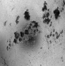


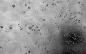

57 EV(primary MS) delta-n-p63(primary MS) EV(secondary MS) delta-n-p63(secondary MS) IMEC SUM102 MCF-7 EV (primary MS) delta-n-p63 (primary MS) MCF-10A The i nf l uences of ect opi c del t a- N- p63 on t he mammoshper e formation of breast cell lines IMEC(EV,P) IMEC(P63,P) IMEC(EV,S) IMEC(P63,S) SUM102(EV,P) SUM102(P63,P) SUM102(EV,S) SUM102(P63,S) MCF7(EV,P) MCF7(P63,P) MCF7(EV,S) MCF7(P63,S) MCF10A(EV,P) MCF10A(P63,P) 系列 1


58 EV (primary MS) delta-n-p63 knockdown(primary MS) MCF10A SUM102 EV mouse OCT4 mouse NANOG IMEC (primary) IMEC (secondary)
 Figure 14: over-expression of ectopic human delta-n-p63,")


59 EV mouse OCT4 mouse NANOG SUM102 (primary) SUM102 (secondary) MCF10A (primary) Figure 14: over-expression of ectopic human delta-n-p63, knocking down of endogenous delta-np63 and transfection of mouse oct3/4 and nanog, treatment of retinoic acid in breast cell lines had significant influences on formation of mammospheres. All breast cell lines were pre-treated with retinoic acid and DMSO as vehicle control, delta-n-p63 alpha and GFP empty vector adenoviruses as vehicle control, adenovirus expressing sirna against TP63 DNA binding domain and GFP empty vector adenovirus as vehicle control, mouse oct3/4 and nanog expressing plasmid and pcdna-3.1 as vehicle control. After treatment for 72 hours, all treated samples were harvested and plated on 24-well low binding plates at /well cellular density. Primary mammosphere were formed in 10 days to 2 weeks. The mammosphere structure were collected and digested with 0.25% Trypsin and EDTA for 10 min, and replated on 24-well low-binding plates for secondary mammosphere formation assay.
 P5(N,p63) P6(N,ev) P6(N,-p63) P5(P,ev) P5(P,p63) P6(P,ev) P6(P,-p63) Mouse")


 was treated with adenovirus expressing sirna")

60 Figure 15. P5(N,ev) P5(N,p63) P6(N,ev) P6(N,-p63) P5(P,ev) P5(P,p63) P6(P,ev) P6(P,-p63) Mouse oct3/4 Mouse nanog Mouse GAPDH Figure 15: The transcripts of oct3/4 and nanog could be detected in mouse stem cells and progenitor cells enriched subpopulations, and delta-n-p63 could regulate their mrna level, respectively. Mouse stem cells and progenitor cells enriched population were isolated from wide type and PATCH (-/+) B6/129 mice at age of 10 weeks. Stem cells enriched population (P6 fraction) was treated with adenovirus expressing sirna against TP63 DNA binding domain, progenitor cells enriched population (P5 fraction) was infected with delta-n-p63 alpha adenovirus, respectively. GFP empty vector was used as vehicle control. After infection for 72 hours, total RNA from all cell samples were harvested for semi-quantitative PCR analysis. Figure 16. P5 (EV) P5(P63) P6 (DMSO) P6 (RA)
 B6/129 mice at age of 10 weeks.")
 was infected with delta-n-p63 alpha adenovirus, and P6 fraction was treated with retinoic acid for 72 hours, respectively.")
61 P6 (EV) P6 (minus p63) Figure 16: Retinoic acid and delta-n-p63 have diverse effects on the mammosphere formation abilities of mouse mammary epithelial subpopulations. Mouse stem cells and progenitor cells enriched population were isolated from wide type and PATCH (-/+) B6/129 mice at age of 10 weeks. Stem cells enriched population (P6 fraction) was treated with adenovirus expressing sirna against TP63 DNA binding domain, progenitor cells enriched population (P5 fraction) was infected with delta-n-p63 alpha adenovirus, and P6 fraction was treated with retinoic acid for 72 hours, respectively. GFP empty vector and DMSO were used as vehicle control.
62 DOI: / CAN Nestin Is Expressed in the Basal/Myoepithelial Layer of the Mammary Gland and Is a Selective Marker of Basal Epithelial Breast Tumors Hua Li, Pratima Cherukuri, Na Li, et al. Cancer Res 2007;67: Published online January 18, Updated Version Access the most recent version of this article at: doi: / can Cited Articles Citing Articles This article cites 36 articles, 11 of which you can access for free at: This article has been cited by 7 HighWire-hosted articles. Access the articles at: alerts Sign up to receive free -alerts related to this article or journal. Reprints and Subscriptions Permissions To order reprints of this article or to subscribe to the journal, contact the AACR Publications Department at pubs@aacr.org. To request permission to re-use all or part of this article, contact the AACR Publications Department at permissions@aacr.org. Downloaded from cancerres.aacrjournals.org on February 8, 2013 Copyright 2007 American Association for Cancer Research
63 DOI: / CAN Research Article Nestin Is Expressed in the Basal/Myoepithelial Layer of the Mammary Gland and Is a Selective Marker of Basal Epithelial Breast Tumors Hua Li, 1 Pratima Cherukuri, 1 Na Li, 1 Victoria Cowling, 2 Michael Spinella, 1 Michael Cole, 2 Andrew K. Godwin, 4 Wendy Wells, 3 and James DiRenzo 1 1 Department of Pharmacology and Toxicology, Dartmouth Medical School, Hanover, New Hampshire; 2 Departments of Pharmacology and Toxicology and Genetics, Dartmouth Medical School, Norris Cotton Cancer Center; 3 Department of Pathology, Dartmouth Hitchcock Medical Center, Lebanon, New Hampshire; and 4 Medical Science Division, Fox Chase Cancer Center, Philadelphia, Pennsylvania Abstract Transcriptional profiling has identified five breast cancer subtypes, of which the basal epithelial is most aggressive and correlates with poor prognosis. These tumors display a high degree of cellular heterogeneity and lack established molecular targets, such as estrogen receptor-a, progesterone receptor, and Her2 overexpression, indicating a need for definitive diagnostic markers. We present evidence that nestin, a previously described marker of regenerative cells in diverse tissues, is expressed in the regenerative compartment of the normal human mammary gland. Colocalization studies indicate two distinct populations of mammary epithelia that express nestin: one expressing cytokeratin 14 (CK14) and #N-p63 and another expressing desmin. Immunohistochemical analysis indicates that #N-p63 and nestin are coordinately expressed during pregnancy in the murine mammary gland. In the embryonal carcinoma cell line NT2/ D1, ectopic #N-p63-A disrupts retinoic acid induced differentiation, thereby preserving expression of nestin; however, small interfering RNA mediated ablation of nestin is insufficient to promote differentiation, indicating that whereas nestin may identify cells within the regenerative compartment of the mammary gland, it is insufficient to block differentiation and preserve replicative capacity. Immunohistochemical analysis of basal epithelial breast tumors, including those shown to carry BRCA1 mutations, indicates robust expression of nestin and CK14, punctate expression of p63, and low to undetectable levels of desmin expression. Nestin was not detected in other breast cancer subtypes, indicating selectivity for basal epithelial breast tumors. These studies identify nestin as a selective marker of the basal breast cancer phenotype, which displays features of mammary progenitors. [Cancer Res 2007;67(2):501 10] Introduction Transcriptional profiling of large cohorts of human breast cancers has resulted in the identification of five distinct subtypes (1). These studies indicate that between 17% and 37% of human breast tumors display a basal epithelial phenotype (2 4). These tumors are highly aggressive, are poorly differentiated, and lack molecular targets Requests for reprints: James DiRenzo, Department of Pharmacology and Toxicology, Dartmouth Medical School, 7650 Remsen, Hanover, NH Phone: ; Fax: ; james.direnzo@dartmouth.edu. I2007 American Association for Cancer Research. doi: / can for endocrine or anti-her2therapy. The basal phenotype is associated with an early age of onset and short times to relapse and disease progression (5). A disproportionately high number of these tumors are detected during normal screening mammography intervals (6), reflecting the aggressive nature of these tumors. For these reasons, the basal epithelial subtype contributes disproportionately to breast cancer mortality (7). Improved understanding of the etiology of these tumors may help to identify selective markers and therapeutic targets that will improve detection, diagnosis, and treatment of the basal epithelial breast cancer subtype. Throughout reproductive life, the epithelial portion of the mammary gland undergoes multiple successive regenerative cycles characterized by cellular proliferation and terminal differentiation (8). During pregnancy, expansion of the epithelial compartment is thought to be initiated by the mitotic division of the mammary stem cells, resulting in production of a pool of mammary progenitors. The progenitor population enters a highly proliferative state that drives this expansion and is followed by cellular differentiation. The regenerative cycle ends with extensive apoptosis and tissue remodeling during post-lactation involution. In non-pregnant females, a less-pronounced cycle of proliferation and cell death occurs with each menstrual cycle (9). Continuous regenerative cycles within the epithelial portion of the mammary gland depend upon a population of mammary stem cells that retain their proliferative capacity and resist terminal differentiation. These features confer a prolonged replicative life span, suggesting that mammary stem cells may be capable of accumulating and harboring mutations and propagating these mutations into the progenitor pool. This further suggests that mammary stem cells may be the sites of breast cancer initiation, which is supported by the finding that the mammary stem cell fraction is amplified in the MMTV-Wnt1A murine breast cancer model (10). This observation is consistent with other studies that indicate breast cancer initiation is a condition of poorly regulated self-renewal (11) and has focused attention upon the oncogenic potential of genetic pathways that regulate self-renewal. To repopulate the mammary gland and to achieve cellular diversity during each regenerative cycle, while preserving selfrenewing capacity for subsequent regenerative cycles, the mitotic offspring of mammary stem cells must execute the decision to preserve or forfeit self-renewing capacity. There is abundant evidence that the p53 family member TP63 plays a critical role in this decision (12 15). The gene encoding TP63 uses proximal and distal promoters to produce trans-activating (TA-p63) and NH 2 - terminally deleted (DN-p63) isoforms (16). Other studies suggest that in the adult mammary gland, p63 is required for the preservation of self-renewal and cellular stasis (17). Mutations in Cancer Res 2007; 67: (2). January 15, 2007 Downloaded from cancerres.aacrjournals.org on February 8, 2013 Copyright 2007 American Association for Cancer Research
64 DOI: / CAN Cancer Research TP63 have been shown to underlie a broad spectrum of syndromes that have in common defects in cellular stasis of a variety of epithelial and apocrine structures (14). These defects are believed to result from a genetic program of non-regenerative differentiation that ultimately leads to the depletion of the regenerative compartment. Specifically relevant to the establishment and preservation of the mammary regenerative compartment are mutations within the a-specific COOH terminus of TP63 that underlie limb-mammary syndrome (OMIM# ; ref. 18). Patients bearing these mutations display severe to complete hypoplasia of the mammary epithelia. Consistent with these phenotypes, targeted ablation of TP63 in the mouse resulted in hypoplasia of both embryonic and adult epithelial and apocrine structures (13, 15). These studies indicate that TP63 is required for the establishment and preservation of the regenerative compartments of multiple epithelial structures. Additionally, studies using isoform-specific antibodies indicate that DN-p63 predominates in the basal epithelia of the mammary gland (19), which is a component of the mammary regenerative compartment. This indicates that DN-p63 may play an important role in the maintenance and preservation of cellular stasis during the mammary regenerative cycle. Nestin is an intermediate filament protein that is expressed in regenerative compartments of the central nervous system and other tissues (20). It has been shown to colocalize with p63, as detected by the pan-p63 monoclonal antibody 4A4, within a subset of the limbal epithelia of the cornea (21). This suggests that nestin may also be expressed in the regenerative compartment of epithelial structures and is consistent with multiple findings indicating that nestin is a marker of neural progenitors (22, 23). In this article, we report that nestin is expressed in two distinct cell types within the basal/myoepithelial layer of the mammary gland. Two-color immunofluorescence identifies a cell type in which nestin is coexpressed with cytokeratin 14 (CK14) and p63 and a second that is positive for desmin. Analysis of nestin and DN-p63 expression in the murine mammary gland during pregnancy indicates overlapping patterns of temporal and cell type specific regulation. We provide evidence that DN-p63-a is sufficient to block cellular differentiation and preserve nestin expression in the embryonal carcinoma cell line NT2/D1, and that small interfering RNA (sirna) mediated repression of nestin in NT2/D1 was insufficient to promote differentiation. Taken together, these observations suggest that nestin is expressed in the regenerative compartment in the mammary gland. Immunohistochemical analysis indicates that nestin is robustly expressed in basal epithelial breast tumors [defined here as triple negative for the estrogen receptor-a (ERa), the progesterone receptor (PR), and Her2] and undetectable in breast tumors representing other molecular classifications. We also present data indicating that nestin is strongly expressed in BRCA1-associated breast tumors, which is consistent with the finding that BRCA1-associated tumors possess a basal phenotype (3, 24). Further analysis of the triplenegative tumors indicates high levels of CK14, variable expression of p63, and undetectable levels of desmin, suggesting that these tumors arose from components of the basal epithelia that express nestin, CK14, and p63. This signature was absent in other breast cancer subtypes, indicating that nestin expression identifies the poor prognosis basal epithelial subtype. These studies also indicate that the highly aggressive and poorly differentiated basal breast cancer subtype displays biochemical features of the regenerative compartment of the mammary gland. Materials and Methods Human tissue samples. Normal human mammary gland samples derived from reduction mammoplasty were identified from archived samples within the Tissue and Tumor Bank at Dartmouth Hitchcock Medical Center. For breast tumors representing diverse subtypes, the files of the Department of Pathology at Dartmouth Hitchcock Medical Center were reviewed to identify formalin-fixed, paraffin-embedded (FFPE) samples representing tumors that were either ER /PR Her2, ER /PR /Her2 + [by fluorescence in situ hybridization (FISH)], or ER + /PR +. Identified tumors were evaluated to ensure that sufficient tissue existed within the paraffin blocks. Identification and collection of tissues and tumors were conducted in strict adherence with regulations related to the protection of patient identity. BRCA1-associated tumors were identified and selected from archived material obtained through the Family Risk Assessment Program at the Fox Chase Cancer Center (FCCC). These tumor tissue samples were derived from patients that had undergone genetic testing through the Clinical Molecular Genetics Laboratory at FCCC and were found to be carriers of a deleterious BRCA1 mutation. Murine tissue samples. C57/Black6 mice were mated at 8 weeks of age, and pregnancy was confirmed by the presence of the anogenital plug. Murine mammary glands were harvested by gross dissection at days 3, 7, 14, and 20 of pregnancy. Glands were fixed in Bouin s solution, dehydrated though a series of graded alcohols, and embedded in paraffin. Four- to 5-Am sections were prepared and fixed onto frosted glass microscope slides. Immunohistochemistry was done as described below. Immunohistochemistry. Tumors representing the basal breast cancer subtype were identified by negativity for ERa, PR, and Her2. Additionally, paraffin blocks representing tumors of the Her2subtype (ER /PR /Her2 + by FISH) and tumors with a luminal epithelial phenotype (ER + /PR + ) were identified for comparative analysis. Additionally, FFPE samples of normal human mammary gland derived from reduction mammoplasty were used for analysis of marker expression in the normal mammary gland. Briefly, 4-Am-thick series sections were cut and applied to charged glass slides (Superfrost Plus). Sections were deparaffinized in xylene, rehydrated through a series of graded alcohol, placed in 10 mmol/l citrate buffer (ph 6), and underwent antigen retrieval in a microwave oven for 15 min. Endogenous peroxidase was blocked with 0.3% hydrogen peroxide in distilled water for 10 min. Samples stained for nestin, desmin, and p63 were blocked in 5% donkey serum in 0.1% Triton X-100 in PBS. Samples stained with CK5 and CK14 were similarly blocked with 5% horse serum. All blocking was done for 30 min at 37jC. Immunohistochemistry was done using an avidin-biotin peroxidase system. The following primary antibodies were incubated for 45 min at 37jC: nestin (1:50, clone C-20; Santa Cruz Biotech, Santa Cruz, CA) or nestin (1:50, clone 10C2; Santa Cruz Biotech), desmin (1:50, clone Y-20; Santa Cruz, Santa Cruz, CA), pan-p63 (1:100, clone 4A4; BD Biosciences, San Diego, CA), CK14 (1:100, clone LL002; Neomarkers, Fremont, CA), and CK5 (1:25, clone XM26; Neomarkers). Detection of DNp63 isoforms was conducted using a rabbit polyclonal antibody raised during these studies and diluted 1:200. Following washes in PBST, a biotinylated secondary antibody (Vector Laboratories, Inc., Burlingame, CA) was applied for 30 min at 37jC. Detection of nestin and desmin staining was blocked in 5% donkey anti-goat IgG (1:200). Detection of pan-p63, CK5, and CK14 was blocked with horse anti-mouse IgG (1:400). Detection of the DN-p63 specific staining was achieved with a goat anti-rabbit IgG (1:400). Slides were incubated with streptavidin-linked peroxidase (1:400; Vector Laboratories) for 30 min at 37jC and developed with 3,3 -diaminobenzidine tetrahydrochloride staining kit (Vector Laboratories). Mayer s hematoxylin was applied as a counterstain. Two-color immunofluorescence. The pretreatment and preparation of slides, including deparaffinization, rehydration, and antigen retrieval, were identical to the protocols for immunohistochemistry. Blocking serum was applied for removing nonspecific binding accordingly. Nestin or desmin costained with pan-p63, CK5, and CK14 were blocked with 5% donkey serum. Then, the samples were incubated with two different primary antibodies together (nestin or desmin + pan-p63, CK5, CK14). Following a series of wash steps in PBST, the slides were incubated with two different Cancer Res 2007; 67: (2). January 15, Downloaded from cancerres.aacrjournals.org on February 8, 2013 Copyright 2007 American Association for Cancer Research
65 DOI: / CAN Nestin Identifies Basal Epithelial Breast Tumors Alexa Fluor conjugated secondary antibodies (1:200; Invitrogen, Carlsbad, CA) together. Nestin (C-20) or desmin costained with nestin (10C2), pan-p63 (4A4), CK5, and CK14 were blocked with donkey anti-goat-alexa Fluor 594 IgG and donkey anti-mouse-alexa Fluor 488. Slides were mounted with Vectorshield (Vector Laboratories) mounting solution with 4,6-diamidino- 2-phenylindole (DAPI) and imaged by fluorescence microscopy and confocal microscopy. Double staining of nestin plus desmin, CK14 + panp63 (4A4) is modified slightly due to identical host species of primary antibodies. The staining with primary antibodies was conducted sequentially. The first staining of the primary antibody was done as regular immunohistochemistry protocol. The only difference was to use Avidin- Alexa Fluor 488 (1:200; Invitrogen) to replace the streptavidin-peroxidase complex reagent followed by microwaving in 10 mmol/l citrate buffer (ph 6) for 15 min to remove nonspecific binding. Following treatment, slides were stained with the secondary primary antibody and detected with Alexa Fluor 594 conjugated IgG accordingly. Cell culture. The embryonal carcinoma cell line NT2/D1 was cultured as previously described, and differentiation studies were conducted by treating cells with either 0.01% DMSO (vehicle) or 1 Amol/L retinoic acid (RA). Cells were transfected using Fugene (Roche, Indianapolis, IN) according to the manufacturer s protocol. Ectopic expression of DN-p63-a was achieved by adenoviral infection. sirna-mediated repression of nestin was achieved by tranfection of a pool of four nestin-specific sirnas (ONTARGETplus SMARTpool, Dharmacon, Lafayette CO). A similar pool of four nonspecific sirnas was used as negative control (ONTARGETplus SMARTpool, Dharmacon). sirnas were transfected in amounts recommended by the manufacturer. Transfections were done using Oligofectamine (Invitrogen) according to manufacturer s protocol. Northern analysis. RNA was isolated using the RNeasy system (Qiagen, Valencia, CA). Seven to 10 Ag RNA was loaded per lane, and Northern blotting was conducted as previously described. Nestin mrna was detected using an f1-kb EcoRI fragment derived from an IMAGE clone # containing nestin cdna sequences. RA receptor h (RARh) mrna was detected using a 615-bp EcoRI fragment derived from the RARh cdna. Northern blotting procedures were as previously described (25). A2B5 immunodetection. Following treatment with DMSO or RA, NT2/ D1 cells were washed twice in cold PBS and incubated with an undiluted cell culture supernatant from a hybridoma that produces an anti-a2b5 immunoglobulin. Incubation was carried out for 30 min at 37jC, and samples were washed twice in PBS for 10 min each. Following washing, a 1:200 dilution of a goat anti-mouse-igg linked to Alexa Fluor 488 was incubated with the samples for 15 min at 37jC. Samples were washed, and nuclei were stained with DAPI before fluorescence imaging. Western blotting. Western analysis of TA-p63-g was done as previously described (26). DN-p63 specific Western blotting was conducted by blocking filters in TBST-5% milk for 1 h at room temperature. The rabbit anti-dn-p63 antibody was incubated with the filter for 1 h at room temperature in TBST- 1% milk. Following a series of washes in TBST, a horseradish peroxidase linked goat anti-rabbit secondary antibody was used to detect the rabbit IgG, and detection was carried by chemiluminescence. Results Nestin is expressed in the basal epithelial layer of the normal mammary gland. Several studies have indicated that DNp63 is highly expressed in the basal/myoepithelial layer of the mammary gland and other epithelial structures and is required for the preservation of self-renewal (17, 19, 27). Studies of the limbal epithelia of the cornea indicate that DN-p63-a is expressed in the self-renewing population, and this expression is repressed as cells forfeit their self-renewing capacity, enter a stage of transient amplification, and achieve terminal differentiation (28). Other studies show colocalization of nestin and p63 in a subset of cells within the limbal epithelia (21). This study suggested that nestin may be expressed in the regenerative compartment of the mammary gland. FFPE samples of normal human mammary gland tissue were sectioned and subjected to immunohistochemical analysis of the expression of nestin, the basal epithelial marker CK14, and p63. A goat polyclonal antibody directed against human nestin detected robust expression of nestin (Fig. 1A) in the subluminal compartment of the mammary gland. Similar studies using a mouse monoclonal antibody directed against a distinct epitope of nestin resulted in an identical pattern of expression (Fig. 1B). Immunohistochemical analysis of CK14 and p63 (Fig. 1C and D) confirms that each is present in the subluminal epithelia of the mammary duct. Interestingly, we noted that CK14 expression was restricted to the columnar basal epithelia of the ducts and was present at low to undetectable levels in the lobules (Fig. 1C, right). Two-color immunofluorescence using antibodies directed against two distinct epitopes of nestin resulted in a high degree of overlap, which increases confidence that the protein being detected is nestin (Fig. 1E). In mammary ducts and lobules, nestin was detected in two morphologically distinct cell types in the subluminal compartment (Fig. 1A, inset). The first is a subset of columnar basal epithelia (indicated by black arrowheads) in which cytoplasmic nestin staining surrounds the nucleus. A second filamentous cell type (indicated by red arrowheads) that is distributed along the periphery of the duct also stained positively for nestin. Importantly, regions within ducts and lobules are identifiable, in which the filamentous nestin-positive cell type is distinct and physically separate from the nestin-positive columnar epithelia. This observation indicates that nestin is expressed in two morphologically distinct subtypes in the subluminal compartment of the human mammary gland and raises the possibility that nestin may colocalize with CK14 and p63. Nestin independently colocalizes with basal/myoepithelial markers. To determine if nestin is expressed in the subluminal layer of the mammary gland, two-color immunofluorescence was used to measure the degree of colocalization between nestin and p63 or CK14. Results indicate colocalization of nestin with p63 (Fig. 2A, left), and confocal imaging indicates that the cytoplasmic nestin staining surrounds the nuclear p63 staining in multiple cells (Fig. 2A, right). To confirm these studies, we sought to show that nestin is coexpressed with the basal epithelial marker CK14. Consistent with previous studies, two-color immunofluorescence indicates that CK14 and p63 are coexpressed in cells within the columnar basal epithelia of the mammary gland (Fig. 2B, left). This result is also consistent with a recent study showing that CK14 is a direct transcriptional target of DN-p63 isoforms (29). Confocal imaging (Fig. 2B, right) clearly shows the cytoplasmic staining of CK14 surrounding the nuclear staining of p63. Two-color immunofluorescence also indicates that colocalization of nestin and CK14 are coexpressed in these cells (Fig. 2C). We also noted that no p63 or CK14 expression was detected in the nestin-positive filamentous cells that appear along the periphery of the ducts and lobules. The location and morphology of these cells suggests that they represent myoepithelia, a myoepithelial precursor, or myofibroblasts. Other studies have noted that nestin is coexpressed with the striated muscle neurofilament protein desmin in regenerating skeletal muscle (30). To determine if the filamentous nestinpositive cells were myoepithelial, two-color immunofluorescence was conducted to determine if nestin was coexpressed with desmin in these cells. Results (Fig. 2D) indicate that nestin and desmin are colocalized in the filamentous myoepithelial cells. Additionally, two-color immunofluorescence indicates no overlap between desmin and p63 (Fig. 3E) or desmin and CK14 (Fig. 3F). Taken together, these studies indicate that nestin is expressed in two Cancer Res 2007; 67: (2). January 15, 2007 Downloaded from cancerres.aacrjournals.org on February 8, 2013 Copyright 2007 American Association for Cancer Research

 and filamentous (red arrows). Sections were counterstained with hematoxylin.")


 and the mouse anti-nestin monoclonal antibody (middle) stain with identical patterns as displayed in the")

66 DOI: / CAN Cancer Research Figure 1. Nestin is expressed in two morphologically distinct subluminal mammary epithelial cell types. A, FFPE samples of normal human mammary gland, derived from reduction mammoplasty, were sectioned, applied to charged glass microscope slides, and subjected to immunohistochemical analysis for expression of nestin. Using a goat anti-nestin polyclonal antibody, staining of both ducts and lobules was observed in a layer of cells that is one cell removed from the luminal epithelia. Two specific cell morphologies were observed: columnar (black arrows) and filamentous (red arrows). Sections were counterstained with hematoxylin. B, to confirm the specificity of the goat anti-nestin polyclonal, similar analyses were conducted with a mouse anti-nestin monoclonal. Similar patterns were observed, confirming that the staining detected was due to the presence of nestin. Sections were counterstained with hematoxylin. C, staining with the basal epithelial marker CK14 was done using a mouse anti-human CK14 monoclonal antibody. Staining was detected in all ducts but only rarely in mammary lobules. Right, representative section of mammary lobule in which no CK14 staining is evident. Sections were counterstained with hematoxylin. D, detection of p63 by the pan-p63 monoclonal antibody 4A4 is restricted to the basal epithelia of mammary ducts and lobules. Sections were counterstained with hematoxylin. E, two-color immunofluorescence indicates that the goat anti-nestin polyclonal antibody (left) and the mouse anti-nestin monoclonal antibody (middle) stain with identical patterns as displayed in the merged image (right). morphologically and biochemically distinct cell types in the subluminal compartment of the mammary gland. The colocalization of nestin with p63 coupled to the abundant evidence that nestin and p63 are expressed in the regenerative compartments of diverse tissues may be consistent with a role for nestin in the regenerative compartment of the mammary gland. #N-p63 and nestin are coordinately regulated in the mouse mammary gland during pregnancy. Immunohistochemical analysis of p63 expression using the commercially available 4A4 monoclonal antibody indicates high levels of p63 expression in the subluminal compartment of the mammary gland. Because this antibody was raised against a region of p63 that is common to all p63 isoforms, it fails to distinguish between TA-p63 and DN-p63. To address this, we raised a rabbit polyclonal antibody against a 14- amino-acid peptide that is encoded by the DN-p63 specific exon 3. Affinity-purified fractions of this serum are capable of detecting DN-p63-a but not TA-p63-g when each was overexpressed in H1299 cells (Fig. 3A). Immunohistochemical analysis of normal human mammary gland using this antibody indicated that DN-p63 isoforms predominate in the subluminal epithelia of the mammary gland (Fig. 3B). This finding is consistent with previous studies (19) and is also consistent with the finding that DN-p63-a is the dominant isoform in both stem cells and transient amplifying cells of the limbal epithelia of the cornea (28). These studies coupled to the observation that p63 and nestin colocalize in the subluminal epithelia of the mammary gland suggest that nestin and DN-p63 may be coordinately regulated during the regenerative cycle of the mammary gland. To test this, mammary glands were harvested Cancer Res 2007; 67: (2). January 15, Downloaded from cancerres.aacrjournals.org on February 8, 2013 Copyright 2007 American Association for Cancer Research
 of this study indicated that the expression of nestin and DN-p63 was undetectable during early pregnancy (P3 and P7) but was detected in the columnar cells of the subluminal compartment at day 14")
.")


.")
 or 1 Amol/L RA indicated that RA-induced differentiation repressed nestin expression at 72and 96 h posttreatment and activated expression of RARb, a canonical RA target gene (Fig.")
 or DN-p63-a")
 indicate that GFP had no effect on repression of nestin or induction of RARh by RA. In contrast to GFP, ectopic DN-p63-a was sufficient to block RA-induced repression of nestin expression.")
 and nestin (Fig.")
67 DOI: / CAN Nestin Identifies Basal Epithelial Breast Tumors from mice at time points during pregnancy and analyzed for expression of DN-p63 and nestin. Results (Fig. 3C) of this study indicated that the expression of nestin and DN-p63 was undetectable during early pregnancy (P3 and P7) but was detected in the columnar cells of the subluminal compartment at day 14 of pregnancy (P14). Additionally, it was observed that by late pregnancy (P20), nestin expression was restricted to the myofibroblasts, and DN-p63 was undetectable. Importantly, staining for CK14 indicates that the basal epithelial cells are present throughout the time course of this pregnancy (Fig. 3C, right). These studies indicate that nestin and DN-p63 are coordinately regulated in the columnar basal epithelia during pregnancy in the mouse mammary gland. #N-p63-A blocks cellular differentiation and preserves nestin expression. Coordinated expression and colocalization of nestin and DN-p63 coupled to the established role for DN-p63 in the preservation of self-renewing capacity in the mammary gland and other epithelial structures indicate that nestin is expressed in cells with self-renewing capacity. This further suggests that expression of nestin may be differentially regulated during the preservation or forfeiture of self-renewal. To test this, we employed the NT2/D1 embryonal carcinoma cell culture model, in which robust levels of nestin are observed to decline in response to all-trans-ra induced cellular differentiation (31). Additionally, we noted that these cells did not express measurable levels of any DNp63 isoform (data not shown). Treatment of NT2/D1 cells with 0.01% DMSO (vehicle) or 1 Amol/L RA indicated that RA-induced differentiation repressed nestin expression at 72and 96 h posttreatment and activated expression of RARb, a canonical RA target gene (Fig. 4A). Consistent with these results, RA-induced differentiation leads to immunodetection of A2B5, a glycolipid that is a well-characterized marker of NT2/D1 differentiation (refs ; Fig. 4B). To determine if ectopic DN-p63-a is sufficient to block RA-induced repression of nestin expression, NT2/D1 cells were transfected with either green fluorescent protein (GFP; control) or DN-p63-a followed by treatment with either 0.01%DMSO (vehicle) or 1 Amol/L RA. RNA was collected at 0, 24, 48, 72, and 96 h posttreatment and subjected to Northern analysis. Results (Fig. 4C) indicate that GFP had no effect on repression of nestin or induction of RARh by RA. In contrast to GFP, ectopic DN-p63-a was sufficient to block RA-induced repression of nestin expression. Importantly, the induction of RARh by RA was still observed in the presence of DN-p63-a, but at somewhat lower levels, suggesting that DN-p63-a may elevate the threshold necessary for RA-induced differentiation. Immunodetection of A2B5 (Fig. 4D, top) and nestin (Fig. 4D, bottom) under identical conditions indicated that ectopic expression of DN-p63-a blocked differentiation of NT2/D1 cells by RA (Fig. 4D) and preserved expression of nestin. This result Figure 2. Nestin expression independently colocalizes with basal progenitor markers and with myoepithelial markers. A, two-color immunofluorescence of FFPE normal human mammary gland indicates that nestin and p63 are coexpressed in a subset of the basal epithelia of mammary ducts. Note (inset) that the red fluorescent signal indicating nestin surrounds the green nuclear signal that indicates p63. Right, representative confocal micrograph in which the cytoplasmic staining of nestin is observed to surround the nuclear staining of p63. B, two-color immunofluorescence indicates colocalization of CK14 and p63. Right, representative confocal micrograph showing the cytoplasmic staining of CK14 surrounding the nuclear staining of p63. C, two-color immunofluorescence indicates colocalization of nestin and CK14. Note the yellow staining that is the result of merged images of CK14 (red) and nestin (green). Right, representative confocal micrograph indicating substantial overlap of nestin and CK14. D, two-color immunofluorescence indicates colocalization of nestin and desmin in the filamentous cells arranged at the periphery of the ducts. E, two-color immunofluorescence indicates that desmin and p63 do not colocalize. Note the regions of desmin staining (red) that are distinct and physically separate from p63 staining (green). F, two-color immunofluorescence indicates that nestin and CK14 do not colocalize in the human mammary gland. Note the distinct red signal of desmin and the distinct green signal of CK14 along with the absence of a yellow signal that would indicate colocalization Cancer Res 2007; 67: (2). January 15, 2007 Downloaded from cancerres.aacrjournals.org on February 8, 2013 Copyright 2007 American Association for Cancer Research


68 DOI: / CAN Cancer Research Figure 3. Expression of DN-p63 and nestin are coordinately regulated in the murine mammary gland during pregnancy. A, Western analysis indicates that DN-p63-a, but not TA-p63-g, is selectively detected by an antisera raised against a DN-p63 specific epitope. H1299 cells were mock infected (control) or infected with adenoviruses programmed to express LacZ, DN-p63-a, and TA-p63-g. Cell lysates were subjected to Western analysis with an affinity-purified DN-p63 specific antisera or a a-specific polyclonal antibody (Cell Signaling, Danvers, MA). B, immunohistochemical analysis of normal human mammary tissue using an affinity-purified DN-p63 specific antisera indicates that DN-p63 isoforms are expressed in the basal epithelia of the mammary gland. C, immunohistochemical analysis of nestin and DN-p63 in the murine mammary gland during pregnancy indicates temporally coordinated expression of DN-p63 and nestin. Black arrows, immunodetection of DN-p63 and nestin in samples taken from pregnancy day 14. indicates that DN-p63-a opposes RA-mediated differentiation signals and is consistent with the role of DN-p63-a in the preservation of self-renewal. It also implies that nestin expression is reflective of the regenerative capacity of cells. The observation that DN-p63-a was able to block the repression of nestin during differentiation suggested that nestin expression may also oppose cellular differentiation. To test this, a pool of four nestin-directed sirnas or a pool or four negative control sirnas was transfected into NT2/D1 cells. Northern analysis indicates that nestin expression was substantially reduced by the nestin-specific pool at 24 h but began to recover by 48 h after transfection (Fig. 5A). Following transfection under these conditions, cells were treated with 0.01% DMSO or 1 Amol/L RA, and immunodetection of A2B5 was measured at 0, 24, 48, 72, 96, and 120 h posttreatment. Results (Fig. 5B) indicated that sirna-mediated repression of nestin over this time course was insufficient induce the A2B5 display or accelerate RA-induced A2B5 display. These results indicate that whereas repression of nestin occurs during differentiation induced by RA, direct ablation of nestin is insufficient to induce differentiation as measured by immunodetection of A2B5. Taken together, these studies indicate that DN-p63-a is able to oppose RA-induced cellular differentiation, thereby preserving expression of nestin and are consistent with previous work indicating that nestin expression is a feature of undifferentiated cells. Breast tumors with a basal epithelial phenotype express nestin, #N-p63, and CK14. The tumor stem cell theory of breast carcinogenesis implies that breast cancers may initiate within a population of cells capable of self-renewal (34). This may further suggest that highly aggressive and poorly differentiated tumors display features of stem cells. Based on this, we hypothesized that the poorly differentiated basal epithelial breast cancer subtype would express nestin and other markers of the regenerative compartment of the mammary gland. Tumors that were triple Cancer Res 2007; 67: (2). January 15, Downloaded from cancerres.aacrjournals.org on February 8, 2013 Copyright 2007 American Association for Cancer Research


 to uniform Figure 4. Ectopic DN-p63-a blocks differentiation and preserves expression of nestin.")

 or DN-p63-a, and cells were treated with DMSO or RA.")
69 DOI: / CAN Nestin Identifies Basal Epithelial Breast Tumors negative for ERa, PR, and Her2were selected for analysis of nestin, p63, and CK14 expression. For each marker, positivity was defined as detectable expression within the tumor and not merely at the periphery of the tumor. For example, a ductal carcinoma in situ (DCIS), in which the in tact basal/myoepithelial layer stains positive but the cells within the core of the DCIS do not, would be scored as negative. Results indicate that in 14 of 16 triple-negative tumors, nestin expression was readily detectable. Figure 6A shows a representative sample of nestin staining of a triple-negative tumor. Similarly, 16 of 16 basal breast tumors were positive for CK14 and indicated a range of signal intensities from intermediate to high. Figure 6B shows a representative sample of high-intensity CK14 staining of a basal epithelial tumor. Analysis of p63 expression in these tumors indicated that 8 of 16 tumors were positive and displayed a range of expression patterns from punctate (Fig. 6C, left) to intermediate (Fig. 6C, middle) to uniform Figure 4. Ectopic DN-p63-a blocks differentiation and preserves expression of nestin. A, Northern analysis of NT2/D1 cells treated with DMSO (vehicle) or 1 Amol/L RA shows that expression of nestin (top) declines at 72 and 96 h posttreatment. Middle, induction of RARh by RA, which serves as a control for the actions of RA. Bottom, distribution of the 28S and 18S ribosomal subunits, which serve as loading controls. B, treatment of NT2/D1 cells with RA results in immunodetection of the A2B5 glycolipid (green). Nuclei are stained with DAPI. C, ectopic expression of DN-p63-a preserves expression of nestin following RA treatment. NT2/D1 cells were transfected with EGFP (control) or DN-p63-a, and cells were treated with DMSO or RA. RNA was collected at 0, 24, 48, 72, and 96 h posttreatment. Northern analysis of nestin (top) indicates that DN-p63-a blocked RA-induced repression of nestin. Ectopic expression of DN-p63-a also blocked RA-induced expression of RARh. Bottom, distribution of the 28S and 18S ribosomal subunits, which serve as a loading control. D, ectopic DN-p63-a blocks RA-induced immunodetection of A2B5 and RA induced repression of nestin Cancer Res 2007; 67: (2). January 15, 2007 Downloaded from cancerres.aacrjournals.org on February 8, 2013 Copyright 2007 American Association for Cancer Research

 and 16 tumors with a")

, although limited by low sample numbers, indicate that nestin may be a selective marker of the basal breast cancer subtype.")
70 DOI: / CAN Cancer Research (Fig. 6C, right). Finally, analysis of these tumors indicated that none showed any evidence of desmin expression (data not shown). These studies indicate that basal epithelial breast tumors are positive for nestin, CK14, and p63 and negative for desmin and suggest that basal breast tumors have a phenotype that is similar to cells within the regenerative compartment of the mammary gland. To determine if nestin is a selective marker of the basal breast cancer subtype, we evaluated 16 tumors that were representative of the Her2subtype (ER /PR Her2 + by FISH) and 16 tumors with a luminal epithelial phenotype (ER + /PR + ). Under the same conditions in which nestin was detected in the basal tumors, we failed to detect nestin in these other subtypes. Consistent with our analysis of the basal breast tumors, a positive signal for nestin was defined as immunodetectable expression within the tumor. Nestin expression was detected at the periphery of ducts and in DCIS but not in the tumor itself, and these tumors were scored as negative. These studies (summarized in Table 1), although limited by low sample numbers, indicate that nestin may be a selective marker of the basal breast cancer subtype. Nestin expression is common in BRCA1-associated tumors. Global transcriptional profiling of human breast cancers has indicated that tumors that are associated with mutations in BRCA1 cluster within the basal breast cancer subtype (3). We therefore sought to determine if expression of nestin was detectable in BRCA1-associated tumors. Immunohistochemical analysis of these tumors with either the goat anti-nestin polyclonal antibody or the mouse anti-nestin monoclonal antibody indicated robust nestin (Fig. 6D) expression in six of eight individual cases. Similar to the basal breast tumors studied above, expression of CK14 and p63 was also detected in the BRCA1-associated tumors. These studies indicate that nestin expression is correlated with BRCA1 associated tumors and are consistent with the finding that BRCA1-associated tumors are classified as a basal breast cancer. This data coupled to our studies indicating that in normal mammary gland nestin is coexpressed with markers of the basal/myoepithelia may indicate that BRCA1-associated tumors display a progenitor-like phenotype. They further suggest that nestin may be a marker of BRCA1- associated tumors. Discussion We describe here the identification of nestin as a selective marker of the basal breast cancer subtype. We present evidence that nestin is expressed in two morphologically and biochemically distinct subtypes within the basal/myoepithelial layer of the normal human mammary gland. In one of these cell types, nestin is coexpressed with DN-p63, which coupled to the role of DN-p63 in preservation of self-renewal, suggesting that nestin may be Figure 5. sirna-mediated repression of nestin is insufficient to enhance differentiation of NT2/D1 cells. A, NT2/D1 cells were transfected with a pool of four nestin-specific sirnas or a pool of four nonspecific sirnas. Northern analysis indicates a significant reduction of nestin mrna at 24 h after transfection. B, A2B5 staining indicates that sirna-mediated repression of nestin was insufficient to stimulate A2B5 immunodetection or accelerate RA-mediated A2B5 immunodetection. Cells were transfected as above followed by treatment with DMSO or RA. At 120 h posttreatment, cells were stained for A2B5. Cancer Res 2007; 67: (2). January 15, Downloaded from cancerres.aacrjournals.org on February 8, 2013 Copyright 2007 American Association for Cancer Research


71 DOI: / CAN Nestin Identifies Basal Epithelial Breast Tumors Figure 6. Nestin, CK14, and p63 are selectively expressed in basal epithelial breast tumors and BRCA1-associated tumors. Tumors that were previously known to lack expression of ERa, PR, and Her2 were prescreened for expression of CK5/6 to confirm the basal epithelial phenotype. A, representative immunohistochemical staining of FFPE breast tumors with a basal phenotype indicates robust expression of nestin. Anti-nestin immunohistochemistry was done as described in Materials and Methods. B, representative immunohistochemical staining of FFPE breast tumors with a basal phenotype indicates robust expression of CK14. Anti-nestin immunohistochemistry was done as described in Materials and Methods. C, expression of p63 was detected in 8 of 16 breast tumors with a basal epithelial phenotype. Expression patterns ranged from punctate (left), to intermediate (middle), to uniform (right). BRCA1-associated breast tumors express robust levels of nestin, CK14, and p63. Breast tumors with confirmed mutations in BRCA1 were identified from the tissue and tumor bank at FCCC. D, immunohistochemical analyses reveal robust detection of nestin in BRCA1-associated tumors. E, immunohistochemical analyses reveal robust detection of CK14 in BRCA1- associated tumors. F, immunohistochemical analyses reveal intermediate detection of p63 in BRCA1-associated tumors. expressed in the regenerative compartment within the mammary gland. Analysis the mammary glands of pregnant mice indicated that the timing and cell type specificity of nestin and DN-p63 expression are coordinately regulated. Our data indicate that DNp63-a is sufficient to block RA-induced differentiation of NT2/D1 cells, and that this ability is linked to the preservation of nestin expression. Although the precise relevance of this observation to the relationship between DN-p63 isoforms and nestin in mammary progenitors is unclear, this finding does indicate that DN-p63-a is able to preserve a state of dedifferentiation in RA-treated NT2/D1 cells and in doing so preserves expression of nestin. Breast tumors representing the basal breast cancer subtype (ER /PR /Her2 ) express robust levels of nestin and CK14 and display a diverse pattern of DN-p63 expression, suggesting a basal/myoepithelial phenotype. This is likely to be consistent with the aggressive nature and poorly differentiated phenotype of the basal epithelial class of breast tumors. Our studies also identify nestin as a potential target for molecular detection and diagnosis of breast cancers with a basal phenotype, including those with known BRCA1 mutations. Analysis of p63 expression in basal epithelial breast tumors indicated that 8 of 16 samples were observed to express p63, whereas none of the Her2-associated or luminal tumor types showed any detectable expression of p63. The expression pattern of p63 varied from punctate to uniform, indicating that expression of p63 isoforms is unlikely to be a clinically useful marker. Although it is unclear if p63 expression identifies tumor stem cells, that p63 is required for self-renewal may indicate that these cells retain the capacity for self-renewal. Further analysis of mammary tumor stem cells will be required to determine if p63 expression underlies the self-renewing capacity of tumor stem cells in the basal epithelial subtype. Our study indicates that nestin is expressed in the basal/ myoepithelial layer of the mammary gland and is a selective marker of the basal epithelial breast cancer subtype. Although the precise function of nestin remains to be elucidated, several studies indicate that it may play a role in the regulation of mitosis (20, 35, 36). These findings coupled to the use of nestin as a marker of neural progenitors and the nestin promoter to selectively target neural progenitors suggest a role for nestin in the progenitor pools Table 1. Expression of nestin, CK14, and p63 in breast cancer subtypes Subtype Markers Nestin CK14 p63 Basal ERa /PR /Her2 14/16 (2.6e 9) 16/16 (3.77e 11) 8/16 (6.77e 5) Her2associated ERa /PR /Her2 + (FISH) 0/16 0/16 0/16 Luminal ERa + /PR + 0/16 0/16 0/ Cancer Res 2007; 67: (2). January 15, 2007 Downloaded from cancerres.aacrjournals.org on February 8, 2013 Copyright 2007 American Association for Cancer Research
Supplementary Table 3. 3 UTR primer sequences. Primer sequences used to amplify and clone the 3 UTR of each indicated gene are listed.
 Supplemental Figure 1. DLKI-DIO3 mirna/mrna complementarity. Complementarity between the indicated DLK1-DIO3 cluster mirnas and the UTR of SOX2, SOX9, HIF1A, ZEB1, ZEB2, STAT3 and CDH1with mirsvr and PhastCons
c Tuj1(-) apoptotic live 1 DIV 2 DIV 1 DIV 2 DIV Tuj1(+) Tuj1/GFP/DAPI Tuj1 DAPI GFP
 apoptotic live 1 DIV 2 DIV 1 DIV 2 DIV Tuj1(+) Tuj1/GFP/DAPI Tuj1 DAPI GFP") Supplementary Figure 1 Establishment of the gain- and loss-of-function experiments and cell survival assays. a Relative expression of mature mir-484 30 20 10 0 **** **** NCP mir- 484P NCP mir- 484P b Relative
Supplementary Figure 1 Establishment of the gain- and loss-of-function experiments and cell survival assays. a Relative expression of mature mir-484 30 20 10 0 **** **** NCP mir- 484P NCP mir- 484P b Relative
Abbreviations: P- paraffin-embedded section; C, cryosection; Bio-SA, biotin-streptavidin-conjugated fluorescein amplification.
 Supplementary Table 1. Sequence of primers for real time PCR. Gene Forward primer Reverse primer S25 5 -GTG GTC CAC ACT ACT CTC TGA GTT TC-3 5 - GAC TTT CCG GCA TCC TTC TTC-3 Mafa cds 5 -CTT CAG CAA GGA
Supplementary Figure 1 a
 Supplementary Figure a Normalized expression/tbp (A.U.).6... Trip-br transcripts Trans Trans Trans b..5. Trip-br Ctrl LPS Normalized expression/tbp (A.U.) c Trip-br transcripts. adipocytes.... Trans Trans
Nature Structural & Molecular Biology: doi: /nsmb Supplementary Figure 1
 Supplementary Figure 1 U1 inhibition causes a shift of RNA-seq reads from exons to introns. (a) Evidence for the high purity of 4-shU-labeled RNAs used for RNA-seq. HeLa cells transfected with control
Supplemental Data. Shin et al. Plant Cell. (2012) /tpc YFP N
 /tpc YFP N") MYC YFP N PIF5 YFP C N-TIC TIC Supplemental Data. Shin et al. Plant Cell. ()..5/tpc..95 Supplemental Figure. TIC interacts with MYC in the nucleus. Bimolecular fluorescence complementation assay using
MYC YFP N PIF5 YFP C N-TIC TIC Supplemental Data. Shin et al. Plant Cell. ()..5/tpc..95 Supplemental Figure. TIC interacts with MYC in the nucleus. Bimolecular fluorescence complementation assay using
a) Primary cultures derived from the pancreas of an 11-week-old Pdx1-Cre; K-MADM-p53
 Primary cultures derived from the pancreas of an 11-week-old Pdx1-Cre; K-MADM-p53") 1 2 3 4 5 6 7 8 9 10 Supplementary Figure 1. Induction of p53 LOH by MADM. a) Primary cultures derived from the pancreas of an 11-week-old Pdx1-Cre; K-MADM-p53 mouse revealed increased p53 KO/KO (green,
1 2 3 4 5 6 7 8 9 10 Supplementary Figure 1. Induction of p53 LOH by MADM. a) Primary cultures derived from the pancreas of an 11-week-old Pdx1-Cre; K-MADM-p53 mouse revealed increased p53 KO/KO (green,
Supplementary Materials
 Supplementary Materials 1 Supplementary Table 1. List of primers used for quantitative PCR analysis. Gene name Gene symbol Accession IDs Sequence range Product Primer sequences size (bp) β-actin Actb gi
Toluidin-Staining of mast cells Ear tissue was fixed with Carnoy (60% ethanol, 30% chloroform, 10% acetic acid) overnight at 4 C, afterwards
 overnight at 4 C, afterwards") Toluidin-Staining of mast cells Ear tissue was fixed with Carnoy (60% ethanol, 30% chloroform, 10% acetic acid) overnight at 4 C, afterwards incubated in 100 % ethanol overnight at 4 C and embedded in
Toluidin-Staining of mast cells Ear tissue was fixed with Carnoy (60% ethanol, 30% chloroform, 10% acetic acid) overnight at 4 C, afterwards incubated in 100 % ethanol overnight at 4 C and embedded in
Supplementary Appendix
 Supplementary Appendix This appendix has been provided by the authors to give readers additional information about their work. Supplement to: Sherman SI, Wirth LJ, Droz J-P, et al. Motesanib diphosphate
CD31 5'-AGA GAC GGT CTT GTC GCA GT-3' 5 ' -TAC TGG GCT TCG AGA GCA GT-3'
 Table S1. The primer sets used for real-time RT-PCR analysis. Gene Forward Reverse VEGF PDGFB TGF-β MCP-1 5'-GTT GCA GCA TGA ATC TGA GG-3' 5'-GGA GAC TCT TCG AGG AGC ACT T-3' 5'-GAA TCA GGC ATC GAG AGA
Figure S1. Analysis of genomic and cdna sequences of the targeted regions in WT-KI and
 Figure S1. Analysis of genomic and sequences of the targeted regions in and indicated mutant KI cells, with WT and corresponding mutant sequences underlined. (A) cells; (B) K21E-KI cells; (C) D33A-KI cells;
Supplementary Figure 1. ROS induces rapid Sod1 nuclear localization in a dosagedependent manner. WT yeast cells (SZy1051) were treated with 4NQO at
 were treated with 4NQO at") Supplementary Figure 1. ROS induces rapid Sod1 nuclear localization in a dosagedependent manner. WT yeast cells (SZy1051) were treated with 4NQO at different concentrations for 30 min and analyzed for
Supplementary Figure 1. ROS induces rapid Sod1 nuclear localization in a dosagedependent manner. WT yeast cells (SZy1051) were treated with 4NQO at different concentrations for 30 min and analyzed for
Supplementary Figures
 Supplementary Figures Supplementary Figure 1. H3F3B expression in lung cancer. a. Comparison of H3F3B expression in relapsed and non-relapsed lung cancer patients. b. Prognosis of two groups of lung cancer
Supplementary Document
 Supplementary Document 1. Supplementary Table legends 2. Supplementary Figure legends 3. Supplementary Tables 4. Supplementary Figures 5. Supplementary References 1. Supplementary Table legends Suppl.
Supplementary Figure 1 MicroRNA expression in human synovial fibroblasts from different locations. MicroRNA, which were identified by RNAseq as most
 Supplementary Figure 1 MicroRNA expression in human synovial fibroblasts from different locations. MicroRNA, which were identified by RNAseq as most differentially expressed between human synovial fibroblasts
TITLE: The Role of hcdc4 as a Tumor Suppressor Gene in Genomic Instability Underlying Prostate Cancer
 AD Award Number: TITLE: The Role of hcdc4 as a Tumor Suppressor Gene in Genomic Instability Underlying Prostate Cancer PRINCIPAL INVESTIGATOR: Audrey van Drogen, Ph.D. CONTRACTING ORGANIZATION: Sidney
Plasmids Western blot analysis and immunostaining Flow Cytometry Cell surface biotinylation RNA isolation and cdna synthesis
 Plasmids psuper-retro-s100a10 shrna1 was constructed by cloning the dsdna oligo 5 -GAT CCC CGT GGG CTT CCA GAG CTT CTT TCA AGA GAA GAA GCT CTG GAA GCC CAC TTT TTA-3 and 5 -AGC TTA AAA AGT GGG CTT CCA GAG
Supplementary Materials and Methods
 DD2 suppresses tumorigenicity of ovarian cancer cells by limiting cancer stem cell population Chunhua Han et al. Supplementary Materials and Methods Analysis of publicly available datasets: To analyze
TITLE: CYP1B1 Polymorphism as a Risk Factor for Race-Related Prostate Cancer
 AD Award Number: W81XWH-04-1-0579 TITLE: CYP1B1 Polymorphism as a Risk Factor for Race-Related Prostate Cancer PRINCIPAL INVESTIGATOR: Yuichiro Tanaka, Ph.D. CONTRACTING ORGANIZATION: Northern California
Table S1. Oligonucleotides used for the in-house RT-PCR assays targeting the M, H7 or N9. Assay (s) Target Name Sequence (5 3 ) Comments
 Target Name Sequence (5 3 ) Comments") SUPPLEMENTAL INFORMATION 2 3 Table S. Oligonucleotides used for the in-house RT-PCR assays targeting the M, H7 or N9 genes. Assay (s) Target Name Sequence (5 3 ) Comments CDC M InfA Forward (NS), CDC M
SUPPLEMENTAL INFORMATION 2 3 Table S. Oligonucleotides used for the in-house RT-PCR assays targeting the M, H7 or N9 genes. Assay (s) Target Name Sequence (5 3 ) Comments CDC M InfA Forward (NS), CDC M
Supplementary Table 2. Conserved regulatory elements in the promoters of CD36.
 Supplementary Table 1. RT-qPCR primers for CD3, PPARg and CEBP. Assay Forward Primer Reverse Primer 1A CAT TTG TGG CCT TGT GCT CTT TGA TGA GTC ACA GAA AGA ATC AAT TC 1B AGG AAA TGA ACT GAT GAG TCA CAG
BHP 2-7 and Nthy-ori 3-1 cells were grown in RPMI1640 medium (Hyclone) supplemented with 10% fetal bovine serum (Gibco), 2mM L-glutamine, and 100 U/mL
 supplemented with 10% fetal bovine serum (Gibco), 2mM L-glutamine, and 100 U/mL") 1 2 3 4 Materials and Methods Cell culture BHP 2-7 and Nthy-ori 3-1 cells were grown in RPMI1640 medium (Hyclone) 5 supplemented with 10% fetal bovine serum (Gibco), 2mM L-glutamine, and 100 U/mL 6 penicillin-streptomycin.
1 2 3 4 Materials and Methods Cell culture BHP 2-7 and Nthy-ori 3-1 cells were grown in RPMI1640 medium (Hyclone) 5 supplemented with 10% fetal bovine serum (Gibco), 2mM L-glutamine, and 100 U/mL 6 penicillin-streptomycin.
TITLE: A PSCA Promoter Based Avian Retroviral Transgene Model of Normal and Malignant Prostate
 AD Award Number: DAMD17-03-1-0163 TITLE: A PSCA Promoter Based Avian Retroviral Transgene Model of Normal and Malignant Prostate PRINCIPAL INVESTIGATOR: Robert Reiter, M.D. CONTRACTING ORGANIZATION: The
SUPPLEMENTARY INFORMATION
 doi: 10.1038/nature05883 SUPPLEMENTARY INFORMATION Supplemental Figure 1 Prostaglandin agonists and antagonists alter runx1/cmyb expression. a-e, Embryos were exposed to (b) PGE2 and (c) PGI2 (20μM) and
CONTRACTING ORGANIZATION: University of Mississippi Medical Center Jackson, MS 39216
 AD Award Number: W81XWH-12-1-0201 TITLE: Role of long non-coding RNAs in prostate cancer PRINCIPAL INVESTIGATOR: Yin-Yuan Mo CONTRACTING ORGANIZATION: University of Mississippi Medical Center Jackson,
Citation for published version (APA): Oosterveer, M. H. (2009). Control of metabolic flux by nutrient sensors Groningen: s.n.
: Oosterveer, M. H. (2009). Control of metabolic flux by nutrient sensors Groningen: s.n.") University of Groningen Control of metabolic flux by nutrient sensors Oosterveer, Maaike IMPORTANT NOTE: You are advised to consult the publisher's version (publisher's PDF) if you wish to cite from it.
University of Groningen Control of metabolic flux by nutrient sensors Oosterveer, Maaike IMPORTANT NOTE: You are advised to consult the publisher's version (publisher's PDF) if you wish to cite from it.
PREPARED FOR: U.S. Army Medical Research and Materiel Command Fort Detrick, Maryland
 AD Award Number: TITLE: PRINCIPAL INVESTIGATOR: Jeremy Chien, PhD CONTRACTING ORGANIZATION: Mayo Clinic, REPORT DATE: September 2012 TYPE OF REPORT: Annual PREPARED FOR: U.S. Army Medical Research and
TITLE: Neural Protein Synuclein (SNCG) in Breast Cancer Progression
 in Breast Cancer Progression") AD AWARD NUMBER: DAMD17-01-1-0352 TITLE: Neural Protein Synuclein (SNCG) in Breast Cancer Progression PRINCIPAL INVESTIGATOR: Yangfu Jiang, M.D., Ph.D. CONTRACTING ORGANIZATION: Long Island Jewish Medical
AD AWARD NUMBER: DAMD17-01-1-0352 TITLE: Neural Protein Synuclein (SNCG) in Breast Cancer Progression PRINCIPAL INVESTIGATOR: Yangfu Jiang, M.D., Ph.D. CONTRACTING ORGANIZATION: Long Island Jewish Medical
CONTRACTING ORGANIZATION: University of Southern California Los Angeles, CA 90033
 AD Award Number: W81XWH-07-01-0264 TITLE: Function of Klotho and is MicroRNA in Prostate Cancer and Aging PRINCIPAL INVESTIGATOR: Shao-Yao Ying, Ph.D. CONTRACTING ORGANIZATION: University of Southern California
BIOLOGY 621 Identification of the Snorks
 Name: Date: Block: BIOLOGY 621 Identification of the Snorks INTRODUCTION: In this simulation activity, you will examine the DNA sequence of a fictitious organism - the Snork. Snorks were discovered on
Award Number: W81XWH TITLE: Characterizing an EMT Signature in Breast Cancer. PRINCIPAL INVESTIGATOR: Melanie C.
 AD Award Number: W81XWH-08-1-0306 TITLE: Characterizing an EMT Signature in Breast Cancer PRINCIPAL INVESTIGATOR: Melanie C. Bocanegra CONTRACTING ORGANIZATION: Leland Stanford Junior University Stanford,
CONTRACTING ORGANIZATION: Mount Sinai School of Medicine New York, New York
 AD AWARD NUMBER: W81XWH-05-1-0475 TITLE: Restoration of Epithelial Polarity in Metastatic Tumors PRINCIPAL INVESTIGATOR: Sergei Sokol, Ph.D. CONTRACTING ORGANIZATION: Mount Sinai School of Medicine New
Supplementary Figure 1
 Supplementary Figure 1 Supplementary Figure 1. Lats1/2 deleted ihbs and ihps showed decreased transcripts of hepatocyte related genes (a and b) Western blots (a) and recombination PCR (b) of control and
TITLE: Crosstalk Between Cancer Cells and Bones Via the Hedgehog Pathway Determines Bone Metastasis of Breast Cancer
 AD Award Number: W81XWH-07-1-0400 TITLE: Crosstalk Between Cancer Cells and Bones Via the Hedgehog Pathway Determines Bone Metastasis of Breast Cancer PRINCIPAL INVESTIGATOR: Dr. Lalita Shevde-Samantrese
SUPPLEMENTARY DATA. Supplementary Table 1. Primer sequences for qrt-pcr
 Supplementary Table 1. Primer sequences for qrt-pcr Gene PRDM16 UCP1 PGC1α Dio2 Elovl3 Cidea Cox8b PPARγ AP2 mttfam CyCs Nampt NRF1 16s-rRNA Hexokinase 2, intron 9 β-actin Primer Sequences 5'-CCA CCA GCG
PREPARED FOR: U.S. Army Medical Research and Materiel Command Fort Detrick, MD
 AD Award Number: W81XWH-11-1-0126 TITLE: Chemical strategy to translate genetic/epigenetic mechanisms to breast cancer therapeutics PRINCIPAL INVESTIGATOR: Xiang-Dong Fu, PhD CONTRACTING ORGANIZATION:
Supplemental Information. Th17 Lymphocytes Induce Neuronal. Cell Death in a Human ipsc-based. Model of Parkinson's Disease
 Cell Stem Cell, Volume 23 Supplemental Information Th17 Lymphocytes Induce Neuronal Cell Death in a Human ipsc-based Model of Parkinson's Disease Annika Sommer, Franz Maxreiter, Florian Krach, Tanja Fadler,
CONTRACTING ORGANIZATION: University of California Lawrence Berkeley National Laboratory Berkeley, CA 94720
 AD Award Number: W81XWH-05-1-0526 TITLE: Effects of Extracellular Matix on DNA Repair in Vivo PRINCIPAL INVESTIGATOR: Aylin Rizki, Ph.D. CONTRACTING ORGANIZATION: University of California Lawrence Berkeley
Award Number: W81XWH TITLE: CYP1B1 Polymorphism as a Risk Factor for Race-Related Prostate Cancer
 AD Award Number: W81XWH-04-1-0579 TITLE: CYP1B1 Polymorphism as a Risk Factor for Race-Related Prostate Cancer PRINCIPAL INVESTIGATOR: Yuichiro Tanaka, Ph.D. Rajvir Dahiya, Ph.D. CONTRACTING ORGANIZATION:
Astaxanthin prevents and reverses diet-induced insulin resistance and. steatohepatitis in mice: A comparison with vitamin E
 Supplementary Information Astaxanthin prevents and reverses diet-induced insulin resistance and steatohepatitis in mice: A comparison with vitamin E Yinhua Ni, 1,2 Mayumi Nagashimada, 1 Fen Zhuge, 1 Lili
TITLE: Investigating the Role of TBX2 in the Inhibition of Senescence in Prostate Cancer
 AD Award Number: W81XWH-07-1-0155 TITLE: Investigating the Role of TBX2 in the Inhibition of Senescence in Prostate Cancer PRINCIPAL INVESTIGATOR: Srinivas Nandana CONTRACTING ORGANIZATION: Vanderbilt
PREPARED FOR: U.S. Army Medical Research and Materiel Command Fort Detrick, Maryland
 Page 1 09/10/2013 AD Award Number: W81XWH-12-1-0453 TITLE: The cytoplasm translocation of the androgen receptor cofactor p44 as a target for prostate cancer treatment PRINCIPAL INVESTIGATOR: Zhengxin Wang
PREPARED FOR: U.S. Army Medical Research and Materiel Command Fort Detrick, Maryland
 AD Award Number: W81XWH-12-1-0225 TITLE: Epstein Barr virus and blood brain barrier in Multiple Sclerosis PRINCIPAL INVESTIGATOR: Luwen Zhang CONTRACTING ORGANIZATION: University of Nebraska Lincoln, NE
Phylogenetic analysis of human and chicken importins. Only five of six importins were studied because
 Supplementary Figure S1 Phylogenetic analysis of human and chicken importins. Only five of six importins were studied because importin-α6 was shown to be testis-specific. Human and chicken importin protein
TITLE: Identification of New Serum Biomarkers for Early Breast Cancer Diagnosis and Prognosis Using Lipid Microarrays.
 AD Award Number: W81XWH-06-1-0690 TITLE: Identification of New Serum Biomarkers for Early Breast Cancer Diagnosis and Prognosis Using Lipid Microarrays. PRINCIPAL INVESTIGATOR: Guangwei Du, Ph.D. CONTRACTING
A smart acid nanosystem for ultrasensitive. live cell mrna imaging by the target-triggered intracellular self-assembly
 Electronic Supplementary Material (ESI) for Chemical Science. This journal is The Royal Society of Chemistry 2017 A smart ZnO@polydopamine-nucleic acid nanosystem for ultrasensitive live cell mrna imaging
Supplemental Information. Cancer-Associated Fibroblasts Neutralize. the Anti-tumor Effect of CSF1 Receptor Blockade
 Cancer Cell, Volume 32 Supplemental Information Cancer-Associated Fibroblasts Neutralize the Anti-tumor Effect of CSF1 Receptor Blockade by Inducing PMN-MDSC Infiltration of Tumors Vinit Kumar, Laxminarasimha
Nature Immunology: doi: /ni.3836
 Supplementary Figure 1 Recombinant LIGHT-VTP induces pericyte contractility and endothelial cell activation. (a) Western blot showing purification steps for full length murine LIGHT-VTP (CGKRK) protein:
Development of RT-qPCR-based molecular diagnostic assays for therapeutic target selection of breast cancer patients
 Development of RT-qPCR-based molecular diagnostic assays for therapeutic target selection of breast cancer patients Sangjung Park The Graduate School Yonsei University Department of Biomedical Laboratory
Culture Density (OD600) 0.1. Culture Density (OD600) Culture Density (OD600) Culture Density (OD600) Culture Density (OD600)
 0.1. Culture Density (OD600) Culture Density (OD600) Culture Density (OD600) Culture Density (OD600)") A. B. C. D. E. PA JSRI JSRI 2 PA DSAM DSAM 2 DSAM 3 PA LNAP LNAP 2 LNAP 3 PAO Fcor Fcor 2 Fcor 3 PAO Wtho Wtho 2 Wtho 3 Wtho 4 DTSB Low Iron 2 4 6 8 2 4 6 8 2 22 DTSB Low Iron 2 4 6 8 2 4 6 8 2 22 DTSB
A. B. C. D. E. PA JSRI JSRI 2 PA DSAM DSAM 2 DSAM 3 PA LNAP LNAP 2 LNAP 3 PAO Fcor Fcor 2 Fcor 3 PAO Wtho Wtho 2 Wtho 3 Wtho 4 DTSB Low Iron 2 4 6 8 2 4 6 8 2 22 DTSB Low Iron 2 4 6 8 2 4 6 8 2 22 DTSB
TITLE: Selective Oncolytic Therapy for Hypoxic Breast Cancer Cells. CONTRACTING ORGANIZATION: Ordway Research Institute Albany, New York 12208
 AD Award Number: W81XWH-06-1-0586 TITLE: Selective Oncolytic Therapy for Hypoxic Breast Cancer Cells PRINCIPAL INVESTIGATOR: Michael T. Fasullo, Ph.D. CONTRACTING ORGANIZATION: Ordway Research Institute
Supplementary Figure 1
 Supplementary Figure 1 3 3 3 1 1 Bregma -1.6mm 3 : Bregma Ref) Http://www.mbl.org/atlas165/atlas165_start.html Bregma -.18mm Supplementary Figure 1 Schematic representation of the utilized brain slice
TITLE: Properties of Leukemia Stem Cells in a Novel Model of CML Progression to Lymphoid Blast Crisis
 AD Award Number: W81XWH-05-1-0608 TITLE: Properties of Leukemia Stem Cells in a Novel Model of CML Progression to Lymphoid Blast Crisis PRINCIPAL INVESTIGATOR: Craig T. Jordan, Ph.D. CONTRACTING ORGANIZATION:
TITLE: Investigation of the Akt/PKB Kinase in the Development of Hormone-Independent Prostate Cancer
 AD Award Number: TITLE: Investigation of the Akt/PKB Kinase in the Development of Hormone-Independent Prostate Cancer PRINCIPAL INVESTIGATOR: Linda A. degraffenried, Ph.D. CONTRACTING ORGANIZATION: The
PREPARED FOR: U.S. Army Medical Research and Materiel Command Fort Detrick, Maryland
 AD Award Number: W81XWH-06-1-0093 TITLE: An Epigenetic Link to Prostate Cancer PRINCIPAL INVESTIGATOR: Raphael F Margueron, Ph.D. CONTRACTING ORGANIZATION: University of Medicine and Dentistry of New Jersey
TITLE: The Role Of Alternative Splicing In Breast Cancer Progression
 AD Award Number: W81XWH-06-1-0598 TITLE: The Role Of Alternative Splicing In Breast Cancer Progression PRINCIPAL INVESTIGATOR: Klemens J. Hertel, Ph.D. CONTRACTING ORGANIZATION: University of California,
TITLE: Evaluation of DNA Methylation as a Target for Intraductal Therapy for Ductal Carcinoma in Situ of the Breast
 AD AWARD NUMBER: DAMD17-02-1-0569 TITLE: Evaluation of DNA Methylation as a Target for Intraductal Therapy for Ductal Carcinoma in Situ of the Breast PRINCIPAL INVESTIGATOR: Kristin A. Skinner, M.D. CONTRACTING
AD (Leave blank) PREPARED FOR: U.S. Army Medical Research and Materiel Command Fort Detrick, Maryland
 PREPARED FOR: U.S. Army Medical Research and Materiel Command Fort Detrick, Maryland") AD (Leave blank) Award Number: W81XWH-06-2-0038 TITLE:T Cell Lipid rafts and complement Ligands for Diagnosis and Monitoring PRINCIPAL INVESTIGATOR: George C. Tsokos, MD CONTRACTING ORGANIZATION: Beth
AD (Leave blank) Award Number: W81XWH-06-2-0038 TITLE:T Cell Lipid rafts and complement Ligands for Diagnosis and Monitoring PRINCIPAL INVESTIGATOR: George C. Tsokos, MD CONTRACTING ORGANIZATION: Beth
Supplementary Figure 1
 Supplementary Figure 1 Supplementary Figure 1: Cryopreservation alters CD62L expression by CD4 T cells. Freshly isolated (left) or cryopreserved PBMCs (right) were stained with the mix of antibodies described
SUPPORTING INFORMATION
 SUPPORTING INFORMATION Biology is different in small volumes: endogenous signals shape phenotype of primary hepatocytes cultured in microfluidic channels Amranul Haque, Pantea Gheibi, Yandong Gao, Elena
Cross-talk between mineralocorticoid and angiotensin II signaling for cardiac
 ONLINE SUPPLEMENT TO Crosstalk between mineralocorticoid and angiotensin II signaling for cardiac remodeling An Di ZHANG,,3, Aurelie NGUYEN DINH CAT*,,3, Christelle SOUKASEUM *,,3, Brigitte ESCOUBET, 4,
Supplementary Figure 1a
 Supplementary Figure 1a Hours: E-cadherin TGF-β On TGF-β Off 0 12 24 36 48 24 48 72 Vimentin βactin Fig. S1a. Treatment of AML12 cells with TGF-β induces EMT. Treatment of AML12 cells with TGF-β results
TITLE: MiR-146-SIAH2-AR Signaling in Castration-Resistant Prostate Cancer
 AWARD NUMBER: W81XWH-14-1-0387 TITLE: MiR-146-SIAH2-AR Signaling in Castration-Resistant Prostate Cancer PRINCIPAL INVESTIGATOR: Dr. Goberdhan Dimri, PhD CONTRACTING ORGANIZATION: George Washington University,
TITLE: Inhibitors of Histone Deacetylases for Radiosensitization of Prostate Cancer
 AD Award Number: W81XWH-04-1-0170 TITLE: Inhibitors of Histone Deacetylases for Radiosensitization of Prostate Cancer PRINCIPAL INVESTIGATOR: Mira O. Jung, Ph.D. CONTRACTING ORGANIZATION: Georgetown University
PREPARED FOR: U.S. Army Medical Research and Materiel Command Fort Detrick, Maryland
 AD Award Number: DAMD17-03-1-0392 TITLE: The Role of Notch Signaling Pathway in Breast Cancer Pathogenesis PRINCIPAL INVESTIGATOR: Annapoorni Rangarajan, Ph.D. CONTRACTING ORGANIZATION: Indian Institute
TITLE: TREATMENT OF ENDOCRINE-RESISTANT BREAST CANCER WITH A SMALL MOLECULE C-MYC INHIBITOR
 AD Award Number: W81XWH-13-1-0159 TITLE: TREATMENT OF ENDOCRINE-RESISTANT BREAST CANCER WITH A SMALL MOLECULE C-MYC INHIBITOR PRINCIPAL INVESTIGATOR: Qin Feng CONTRACTING ORGANIZATION: Baylor College of
CONTRACTING ORGANIZATION: Johns Hopkins Bloomberg School of Public Health
 AD Award Number: W81XWH-12-1-0170 TITLE: Prospective Evaluation of Intraprostatic Inflammation and Focal Atrophy as a Predictor of Risk of High-Grade Prostate Cancer and Recurrence after Prostatectomy
TITLE: Induction of Ephs/Ephrins-Mediated Tumor Cells-Endothelial Cells Repulsion as an Anti-Cancer Therapeutic Approach
 AD Award Number: W81XWH-04-1-0661 TITLE: Induction of Ephs/Ephrins-Mediated Tumor Cells-Endothelial Cells Repulsion as an Anti-Cancer Therapeutic Approach PRINCIPAL INVESTIGATOR: Gerald Batist, M.D. CONTRACTING
Description of Supplementary Files. File Name: Supplementary Information Description: Supplementary Figures and Supplementary Tables
 Description of Supplementary Files File Name: Supplementary Information Description: Supplementary Figures and Supplementary Tables Supplementary Figure 1: (A), HCT116 IDH1-WT and IDH1-R132H cells were
Award Number: W81XWH TITLE: Dietary Fish Oil in Reducing Bone Metastasis of Breast Cancer
 AD Award Number: W81XWH-04-1-0693 TITLE: Dietary Fish Oil in Reducing Bone Metastasis of Breast Cancer PRINCIPAL INVESTIGATOR: Nandini Ghosh-Choudhury, Ph.D. CONTRACTING ORGANIZATION: University of Texas
TITLE: "Impact of Obesity on Tamoxifen Chemoprevention in a Model of Ductal Carcinoma in Situ"
 AD Award Number: W81XWH-09-1-0720 TITLE: "Impact of Obesity on Tamoxifen Chemoprevention in a Model of Ductal Carcinoma in Situ" PRINCIPAL INVESTIGATOR: Sarah Dunlap-Smith, Ph.D. CONTRACTING ORGANIZATION:
TITLE: 64Cu-DOTA-trastuzumab PET imaging in women with HER2 overexpressing breast cancer
 AD Award Number: W81XWH-10-1-0824 TITLE: 64Cu-DOTA-trastuzumab PET imaging in women with HER2 overexpressing breast cancer PRINCIPAL INVESTIGATOR: Joanne Mortimer, M.D. CONTRACTING ORGANIZATION: City of
Supplementary Information
 Supplementary Information Remodeling of heterochromatin structure slows neuropathological progression and prolongs survival in an animal model of Huntington s disease Junghee Lee, Yu Jin Hwang, Yunha Kim,
Roles of microrna-mediated Drug Resistance in Tumor Stem Cells of Small Cell Lung Carcinoma
 AD Award Number: W81XWH-12-1-0479 TITLE: Roles of microrna-mediated Drug Resistance in Tumor Stem Cells of Small Cell Lung Carcinoma PRINCIPAL INVESTIGATOR: Kounosuke Watabe, Ph.D. CONTRACTING ORGANIZATION:
PREPARED FOR: U.S. Army Medical Research and Materiel Command Fort Detrick, Maryland
 AD Award Number: W81XWH-12-1-0212 TITLE: Wnt/Beta-Catenin, Foxa2, and CXCR4 Axis Controls Prostate Cancer Progression PRINCIPAL INVESTIGATOR: Xiuping Yu CONTRACTING ORGANIZATION: Vanderbilt University
ice-cold 70% ethanol with gentle vortexing, incubated at -20 C for 4 hours, and washed with PBS.
 Cell cycle analysis For cell cycle analysis, single cell suspensions of E12.5 fetal liver cells were suspended in 4 ml ice-cold 7% ethanol with gentle vortexing, incubated at -2 C for 4 hours, and washed
CONTRACTING ORGANIZATION: Rush University Medical Center Chicago, IL 60612
 AD Award Number: W81XWH-11-1-0302 TITLE: Yin and Yang of Heparanase in breast Tumor Initiation PRINCIPAL INVESTIGATOR: Xiulong Xu, Ph.D. CONTRACTING ORGANIZATION: Rush University Medical Center Chicago,
TITLE: Effect of COX-2 (PGE2) and IL-6 on Prostate Cancer Bone Mets
 and IL-6 on Prostate Cancer Bone Mets") AD Award Number: W81XWH-05-1-0166 TITLE: Effect of COX-2 (PGE2) and IL-6 on Prostate Cancer Bone Mets PRINCIPAL INVESTIGATOR: Alice C. Levine, M.D. CONTRACTING ORGANIZATION: Mount Sinai School of Medicine
AD Award Number: W81XWH-05-1-0166 TITLE: Effect of COX-2 (PGE2) and IL-6 on Prostate Cancer Bone Mets PRINCIPAL INVESTIGATOR: Alice C. Levine, M.D. CONTRACTING ORGANIZATION: Mount Sinai School of Medicine
Formylpeptide receptor2 contributes to colon epithelial homeostasis, inflammation, and tumorigenesis
 Supplementary Data Formylpeptide receptor2 contributes to colon epithelial homeostasis, inflammation, and tumorigenesis Keqiang Chen, Mingyong Liu, Ying Liu, Teizo Yoshimura, Wei Shen, Yingying Le, Scott
TITLE: Dietary Genistein and Prostate Cancer Chemoprevention
 AD AWARD NUMBER: DAMD17-02-1-0662 TITLE: Dietary Genistein and Prostate Cancer Chemoprevention PRINCIPAL INVESTIGATOR: Coral A. Lamartiniere, Ph.D. CONTRACTING ORGANIZATION: The University of Alabama at
A basic helix loop helix transcription factor controls cell growth
 A basic helix loop helix transcription factor controls cell growth and size in root hairs Keke Yi 1,2, Benoît Menand 1,3, Elizabeth Bell 1, Liam Dolan 1,4 Supplementary note Low soil phosphate availability
TITLE: Genes Associated with Food Allergy and Eosinophilic Esophagitis
 AD Award Number: W81XWH-1-1-75 TITLE: Genes Associated with Food Allergy and Eosinophilic Esophagitis PRINCIPAL INVESTIGATOR: Dr. David Broide CONTRACTING ORGANIZATION: University of California, San Diego
TITLE: Adipose Estrogen and Increased Breast Cancer Risk in Obesity: Regulation by Leptin and Insulin
 AD Award Number: W81XWH-05-1-0497 TITLE: Adipose Estrogen and Increased Breast Cancer Risk in Obesity: Regulation by Leptin and Insulin PRINCIPAL INVESTIGATOR: Fahumiya Samad CONTRACTING ORGANIZATION:
Beta Thalassemia Case Study Introduction to Bioinformatics
 Beta Thalassemia Case Study Sami Khuri Department of Computer Science San José State University San José, California, USA sami.khuri@sjsu.edu www.cs.sjsu.edu/faculty/khuri Outline v Hemoglobin v Alpha
 www.lessonplansinc.com Topic: Protein Synthesis - Sentence Activity Summary: Students will simulate transcription and translation by building a sentence/polypeptide from words/amino acids. Goals & Objectives:
PREPARED FOR: U.S. Army Medical Research and Materiel Command Fort Detrick, Maryland
 AD Award Number: W81XWH-12-1-0212 TITLE: Wnt/beta-Catenin, Foxa2, and CXCR4 Axis Controls Prostate Cancer Progression PRINCIPAL INVESTIGATOR: Xiuping Yu CONTRACTING ORGANIZATION: Vanderbilt University
Journal of Cell Science Supplementary information. Arl8b +/- Arl8b -/- Inset B. electron density. genotype
 J. Cell Sci. : doi:.4/jcs.59: Supplementary information E9. A Arl8b /- Arl8b -/- Arl8b Arl8b non-specific band Gapdh Tbp E7.5 HE Inset B D Control al am hf C E Arl8b -/- al am hf E8.5 F low middle high
TITLE: The Role of HOX Proteins in Androgen-Independent Prostate Cancer
 AD Award Number: W81XWH-6-1-64 TITLE: The Role of HOX Proteins in Androgen-Independent Prostate Cancer PRINCIPAL INVESTIGATOR: Sunshine Daddario, B.A. CONTRACTING ORGANIZATION: University of Colorado Health
TITLE: Development of Antigen Presenting Cells for adoptive immunotherapy in prostate cancer
 AD Award Number: W8XWH-5-- TITLE: Development of Antigen Presenting Cells for adoptive immunotherapy in prostate cancer PRINCIPAL INVESTIGATOR: Mathias Oelke Ph.D. CONTRACTING ORGANIZATION: Johns Hopkins
Does EMT Contribute to Radiation Resistance in Human Breast Cancer?
 AD Award Number: W81XWH-10-1-0592 TITLE: Does EMT Contribute to Radiation Resistance in Human Breast Cancer? PRINCIPAL INVESTIGATOR: Anupama Munshi, Ph.D CONTRACTING ORGANIZATION: University of Oklahoma
PREPARED FOR: U.S. Army Medical Research and Materiel Command Fort Detrick, Maryland
 AD Award Number: W81XWH-12-1-0337 TITLE: Molecular Innovations Toward Theranostics of Aggressive Prostate Cancer PRINCIPAL INVESTIGATOR: Hsieh, Jer-Tsong CONTRACTING ORGANIZATION: University of Texas Southwestern
PREPARED FOR: U.S. Army Medical Research and Materiel Command Fort Detrick, Maryland
 AD Award Number: W81XWH-06-1-0524 TITLE: Elucidating and Modeling Irradiation Effects on Centrosomal and Chromosomal Stability within Breast Cancer PRINCIPAL INVESTIGATOR: Christopher A. Maxwell, Ph.D.
AD (Leave blank) TITLE: Genomic Characterization of Brain Metastasis in Non-Small Cell Lung Cancer Patients
 TITLE: Genomic Characterization of Brain Metastasis in Non-Small Cell Lung Cancer Patients") AD (Leave blank) Award Number: W81XWH-12-1-0444 TITLE: Genomic Characterization of Brain Metastasis in Non-Small Cell Lung Cancer Patients PRINCIPAL INVESTIGATOR: Mark A. Watson, MD PhD CONTRACTING ORGANIZATION:
AD (Leave blank) Award Number: W81XWH-12-1-0444 TITLE: Genomic Characterization of Brain Metastasis in Non-Small Cell Lung Cancer Patients PRINCIPAL INVESTIGATOR: Mark A. Watson, MD PhD CONTRACTING ORGANIZATION:
Award Number: W81XWH TITLE: Global Positioning Systems (GPS) Technology to Study Vector-Pathogen-Host Interactions
 Technology to Study Vector-Pathogen-Host Interactions") Award Number: W81XWH-11-2-0175 TITLE: Global Positioning Systems (GPS) Technology to Study Vector-Pathogen-Host Interactions PRINCIPAL INVESTIGATOR: Timothy P. Endy MD, MPH CONTRACTING ORGANIZATION: SUNY
Award Number: W81XWH-11-2-0175 TITLE: Global Positioning Systems (GPS) Technology to Study Vector-Pathogen-Host Interactions PRINCIPAL INVESTIGATOR: Timothy P. Endy MD, MPH CONTRACTING ORGANIZATION: SUNY
*To whom correspondence should be addressed. This PDF file includes:
 www.sciencemag.org/cgi/content/full/science.1212182/dc1 Supporting Online Material for Partial Retraction to Detection of an Infectious Retrovirus, XMRV, in Blood Cells of Patients with Chronic Fatigue
CONTRACTING ORGANIZATION: Vanderbilt University Medical Center Nashville, TN
 AD Award Number: W81XWH-04-1-0867 TITLE: A Myc-Driven in Vivo Model of Human Prostate Cancer PRINCIPAL INVESTIGATOR: Simon W. Hayward, Ph.D. CONTRACTING ORGANIZATION: Vanderbilt University Medical Center
The Chancellor, Masters and Scholars of the University of Cambridge, Clara East, The Old Schools, Cambridge CB2 1TN
 AWARD NUMBER: W81XWH-14-1-0110 TITLE: A Molecular Framework for Understanding DCIS PRINCIPAL INVESTIGATOR: Gregory Hannon CONTRACTING ORGANIZATION: The Chancellor, Masters and Scholars of the University
without LOI phenotype by breeding female wild-type C57BL/6J and male H19 +/.
 Sakatani et al. 1 Supporting Online Material Materials and methods Mice and genotyping: H19 mutant mice with C57BL/6J background carrying a deletion in the structural H19 gene (3 kb) and 10 kb of 5 flanking
Fort Detrick, Maryland
 Award Number: W81XWH-15-1-0636 TITLE: Effect of Diet on Gulf War Illness: A Pilot Study PRINCIPAL INVESTIGATOR: Ashok Tuteja, M.D. M.P.H CONTRACTING ORGANIZATION: Western Institute for Biomedical Research