Bioscience Research Print ISSN: Online ISSN:
|
|
|
- Colleen Kelley
- 5 years ago
- Views:
Transcription
:596-619.")

1 Available online freely at Bioscience Research Print ISSN: Online ISSN: Journal by Innovative Scientific Information & Services Network RESEARCH ARTICLE BIOSCIENCE RESEARCH, (1): OPEN ACCESS Toxicological and histopathological in-vivo studies for safe dose optimization of avastin and CCR2 antagonist nanoparticles Safaa H. Mohamed 1, Soheir E. Kotob 1, Hanaa H. Ahmed 1, Ahmed A. Abd-Rabou 1* and Mohamed S. Kishta 1 1 Hormones Department, Medical Research Division, National Research Centre, Dokki, Giza, Egypt, Affiliation ID: *Correspondence: ahmedchemia87@yahoo.com Accepted: 18 Aug.2018 Published online: 28 Feb We investigate hepatic and renal toxicity caused by different doses of bevacizumab (avastin; AV) and CCR2 inhibitor (CR) in their free and nano-counterparts in-vivo to optimize their doses for further therapeutic applications against cancer or age-related macular degeneration. AV and CR nanoparticles were synthesized, characterized, and used for treatment for 7 groups (6 male and 6 female rats in each one) of acute toxicity experiment and another 7 groups (3 subgroups in each one; 5 male and 5 female rats in each subgroup) of chronic toxicity experiment. Histopathological, biochemical, and blood analyses were performed. The liver and kidney function levels and hematology parameters showed no significant changes of groups treated with the low doses of the AV and CR therapeutic regimens compared with control group. The light microscope showed normal glomerular and liver morphology in the groups treated with the low doses, while there were some histological changes in the high doses of the used therapies associated with some significant increased levels of GPT, GOT, ALP, serum creatinine, and urea nitrogen. AV and CR increase the risk of injury in glomerular filtration barrier and hepatocytes in a dose-dependent manner. The injury may not only associate with the rising levels of kidney and liver function tests but also with structural and morphological changes. Keywords: Toxicity, Avastin nanoparticles, CCR2 antagonist nanoparticles, dose optimization. INTRODUCTION Bevacizumab, which available in the pharmaceutical market under the name of Avastin (AV), is used for treatment of many cancer types ( Folkman 1971)and eye aliments The American Society of Health-System Pharmacists. (2016). AV is a monoclonal antibody used as an angiogenesis inhibitor and it slows the growth of new blood vessels through its anti-vegf effect (Arao et al.,2011).by binding of the antibody to the VEGF, AV avoids the activation of VEGF receptor and then inhibits the angiogenesis, causing its anti-cancer effect [(Kim et al.,1993) However, angiogenesis is the common leading cause of cancer progression, targeting the VEGF is still tricky (Bottsford et al., 2012). In the past years, injection of an anticancer drug, bevacizumab (avastin; AV), is considered as a promising anti-vegf monoclonal antibody ( Ferrara et al.,2005) AV was listed on the World Health Organization's List of Essential Medicines (HOLEM) World Health Organization.( 2015).The wholesale cost in the developing countries is approximately $ per vial International Drug Price Indicator Guide. (2016) and in the United Kingdom costs
2 the NHS about British Medical Association. (2015).Although AV is listed in the HOLEM, it causes some side effects like much other chemotherapy. For colon, lung, glioblastoma, and renal cancer treatments, the administration route is intravenous (Machado et al.,2012, Mayer 2011 and Abd-Rabou 2017) For age-related macular degeneration, the route of administration is mainly by injection directly into the eye World Health Organization. (2013). In cancer, administration of AV is usually associated with headache, nose bleeding, and high blood pressure. Other plain side effects may be also included, such as bleeding, gastrointestinal perforation, and allergic reactions. When AV used for age-related macular degeneration, it may cause vision loss and retinal detachment The American Society of Health- System Pharmacists. (2016) Accordingly, dose optimization of the administrated dose of AV is very essential to minimize these burden side effects and to decrease liver and renal toxicity. One of the most commonly observed side effects caused by AV therapy is proteinuria (Izzedine et al.,2010).it elevated by a significant rate reached up to 21%- 63% (Zhu et al.,2007).which is the main reason of renal toxicity-related injury (Zhao et al.,2009). Additionally, there is an evidence indicated that VEGF inhibitors may involve in podocytes poisoning (Guan et al.,2006).it was recorded that inhibition of VEGF on the glomerular endothelium may alter the endothelial surface and persuade the development of thrombotic microangiopathy (Machado et al.,2012) It seems that, there is toxicity-related kidney dysfunction of the AV administration in high doses. However, the machineries related to the pathogenesis of kidney injury caused by AV are still obvious. Chemokines are a superfamily plays with their receptors in many pathological procedures like cancer (Conti and Rollins 2004 and Fang et al.,2012).to date, roughly fifty human chemokines have been identified. One of these chemokines is chemokine (C-C motif) ligand 2 (CCL2) which is also known as monocyte chemotactic protein-1 (MCP-1). In 1989, CCL2 was purified and cloned from human gliomas and myelomonocytic cells and was originating to participate in monocytes recruitment during inflammation and angiogenesis (Zachariae et al.,1990,tangirala et al.,1997 and Salcedo et al.,2000).ccl2 is produced by a variety of activating cells, such as lymphocytes and macrophages (Zachariae et al.,1990) Recent studies have reported that CCL2 is overexpressed in a majority of solid cancer types, including colon, liver, prostatic, and pancreatic cancers ( Wolf et al., 2012,Zhang et al.,2010 And Monti et al.,2003). Importantly, cancer microenvironment plays a critical role in cancer progression and chemo resistance (Liu and Vunjak 2016)The crosstalk between cancer cells and the tumor-infiltrating cells is also shown as an important mediator in cancer development and resistance. For example, CCL2, which secreted by many cancer cells, mediates immune inhibitory effects and facilitates cancer metastasis, thus blocking of CCL2-CCR2 signaling by specific inhibitors or antagonists augments CD8 + T-cellmediated responses and inhibits the metastatic process (Fridlender et al.,2010 and Qian et al., 2011).Pharmacokinetic analysis of CCR2 antagonist (CR) administration revealed a short half-life (1 h). After 9 h of CR injection, it was undetectable in blood circulation. Based on single dose analysis of CR, it was suggested that a single dose every 6 h would prevent complete clearance of the drug over a day. However, authors expect the possibility of CR accumulation in the blood that may cause toxicity ( Mitchell et al.,2013).lack of chemotherapy stability in addition to the presence of adverse effects are the main obstacles led scientists to develop novel tacking approaches against cancer using nanodrug delivery that selectively kill cancer cells.( Park et al., 2008).Many delivery strategies were developed by utilizing pegylated polymeric complexation (Chae et al.,2010 and Abd-Rabou et al.,2017).recently, we suggested that these methods increased the half-life and activity of anticancer agents, and reduced renal toxicity (Abd- Rabou AA et al.,2017) (Abd-Rabou et al.,2017) and (Abd-Rabou et al.,2016).therefore, an effective drug delivery technique needs to be industrialized to render AV and CR delivery less invasive and long-lasting. There are some nanosystems showed promise as drug delivery ones owing to their controlled and sustained release properties, subcellular size and biocompatibility with tissue and cells (Abd-Rabou et al.,2018 and Abd-Rabou et al.,2017).for AV, poly(dl-lactideco-glycolide) (PLGA) nanoparticles was used for its delivery with release of the encapsulated AV from the nanoparticles could last four weeks lifetime (Hao et al.,2009) Pan and his companions also created longlasting formulations (eight weeks) of AV through poly ethylene glycol (PEG)-coated PLGA Bioscience Research, 2019 volume 16(1):
3 nanoparticles (Pan et al.,2011).for CR, liposome targeted nano-delivery was approached to stimulate pulmonary endothelium and prevent metastasis (Marko et al.,2015) Liver function tests are essential biomarkers for hepatotoxicity caused by drugs. The glutamate pyruvate transaminase (GPT) or alanine aminotransferase (ALT) is a cellular enzyme mainly found in liver and kidney. High levels of GPT are observed in hepatic diseases and diseases of muscles and traumatisms. It is used in the diagnosis of the liver diseases. When GPT used in conjunction with aspartate aminotransferase (AST) or glutamate oxaloacetate (GOT) they aid in the diagnosis of infarcts in the myocardium, since the value of the ALT stays within the normal limits in the presence of elevated levels of AST. High concentration of GOT is found in heart muscle, liver, and skeletal muscle cells. Although an elevated level of GOT in the serum is not specific of the hepatic disease, it is used as a complementary diagnostic tool to verify the course of this disease with other enzymes like GPT and alkaline phosphatase (ALP) (Murray 1984). (Burtis et al.,1990)and(tietz et al., 1995) ALP is an enzyme present in almost all tissues of the organism, being particularly high in liver and kidney. Both increases and decreases of serum ALP are of importance clinically, ALP elevation is an indication of obstructive liver disease, hepatitis, hepatotoxicity caused by drugs and its decrease may be a cause of vitamin C deficiency (Burtis et al.,1990,tietz et al., 1995 and Wenger et al., 1984) Kidney function tests are indicators of kidney effectiveness and reflect renal toxicity caused by drugs. Urea is the final result of the metabolism of proteins. Urea elevation in blood (uremia) is an indicator of renal diseases or renal obstruction (Burtis et al.,1990,tietz et al., 1995 and, (Kaplan et al., 1984).Creatinine is the result of the degradation of the creatine, component of muscles, where it can be transformed into adenosine tri-phosphate (ATP), which is a source of high energy required for the cells bioenergetics. The creatinine production depends on the modification of the muscular mass, and it varies little and the levels usually are very stable. Creatinine is excreted by the kidneys. With progressive renal insufficiency, there is retention in blood urea and creatinine. Elevate creatinine level may be indicative of renal injury (Burtis et al.,1990 andtietz et al., 1995) Overall, we aimed, in this study, at exploring the acute and chronic toxicity of bevacizumab (avastin; AV) and CCR2 inhibitor (CR) in their free and nano-encapsulation counterparts on Wistar male and female rats. This was supplemented with biochemical and histopathological verifications, allowed us to optimize their doses for further applications used as medications against liver cancer model (Zoheir et al., 2015) and agerelated macular degeneration model World Health Organization. (2013). MATERIALS AND METHODS Materials Methoxy polyethylene glycol amine (mpeg- NH2, MW 5000 Da), 1-ethyl-3-(3- dimethylaminopropyl)-carbodiimide (EDC), N- hydroxysuccinimide (NHS), heparin, polyethylene glycol (MW 5000 Da), 2-(N morpholine) ethanesulfonic acid (MES), dimethyl sulfoxide (DMSO), Tween 80, and poly L-lysine (PLL), CCR2 antagonist RS : 6-Methyl-1 -[2-(5- methyl-2-phenyl-4-oxazolyl)ethyl]-spiro[4h-3,1- benzoxazine-4,4 -piperidin]-2(1h)-one(cr) were purchased from (Sigma-Aldrich, USA). Bevacizumab avastin (AV) was purchased from (Genentech Inc., USA). The GPT, GOT, ALP, urea, and creatinine diagnostic kits were purchased from (ChronoLab Systems, S.L., Barcelona, Spain). Ultrapure water (Millipore, Bedford, MA, USA) was used. Preparation of nano-void (NV), avastin nanoparticles (AV NPs), CR nanoparticles (CR NPs), and AV-CR nanoparticles (AV-CR NPs) According to the modified method that was recently established by the corresponding author [46], we have synthesized nano-void (NV), avastin nanoparticles (AV NPs), CR nanoparticles (CR NPs), and AV-CR nanoparticles (AV-CR NPs). To prepare PEG-exposed nanoparticles (nano-void; NV), we used a mixture of EDC and NHS. Briefly, heparin (HP: 0.1 mmol) was coupled with 0.1 mm of mpeg-nh2 (MW 5000 Da) using EDC (1.5 mmol) and NHS (1.6 mmol) in MES buffer (0.1 M, ph 5.5) at room temperature for 24 h stirring. Then, NV was prepared simply by mixing the polymer (PEG-heparin) with poly-l lysine buffer (PLL) overnight at 4 o C. The mix ratio (1: 6 v/v, PLL: PEG-heparin). To prepare the PEG-nanoparticles of avastin monoclonal antibody (AV NPs), firstly the PEGheparin polymer was synthesized as above and the core nanoparticles were prepared by mixing the polymer with PLL buffer overnight at 4 o C using this ratio (1:6 v/v, PLL: PEG-heparin). Then, AV Bioscience Research, 2019 volume 16(1):
4 NPs were synthesized by activating the amine groups in the formed nanoparticles mixture using EDC and NHS to bind with the carboxylic groups in 25 mg/ml monoclonal antibody (AV) by stirring overnight at 4 o C, after that amide bonds were formed and the AV NPs were functionalized. To prepare the PEG-nanoparticles of CCR2 antagonist RS (CR NPs), simply the PEGheparin polymer was synthesized as above and the CR nanoparticles were prepared by mixing the polymer with PLL buffer overnight at 4oC using this ratio (1: 6 v/v, PLL: PEG-heparin). Before adding PLL to form the CR NPs, 2 mg/ml CCR2 antagonist RS of CR was titrated to the PEG/heparin polymer. This mixture was vortexed for 5 min and subsequently sonicated for 5 min using a Sonics Vibra-cell sonifier VC750 equipped with a micro-tip (Newtown, CT) at amplitude = 35%, pulse-on = 5.0 s, and pulse-off = 3.0 s. The suspension was transferred to a round-bottom tube in a water bath with magnetic stirring overnight at room temperature. To prepare AV-CR NPs, firstly the CR NPs were synthesized as a core vehicle (as mentioned above) then the amine groups were activated using EDC and NHS to bind the carboxylic groups in 25 mg/ml monoclonal antibody (AV) by stirring overnight at 4 o C, after that amide bonds were formed and the AV-CR NPs were functionalized. NPs characterization Entrapment efficiency (EE) All NPs were dialyzed and the concentrations were measured of the nano-conjugated AV and CR after performing dialysis tubing technique for eliminating the impurities and the free drug using a membrane bag (Spectrum Laboratories, USA; molecular weight cut-off, MWCO: 25,000 Da). The free and conjugated forms of AV and CCR2 antagonist were detected with a variable wavelength detector using UV-based ELISA system. The calibration curves for quantification of these compounds were linear over the range of standard concentrations. Finally, entrapment efficiency (EE) was calculated. Transmission electron microscopic (TEM) Particle morphology of all nanoparticles was examined by transmission electron microscopy (TEM, Philips CM-10, FEI Inc., Hillsboro, OR, USA). 100 μg/ml of the nano-suspensions were dropped into Formvar-coated copper grids, and after complete drying, the samples were stained using 2% w/v uranyl acetate (Electron Microscopy Services, Ft. Washington, PA). Image capture and analysis was done using Digital Micrograph and Soft Imaging Viewer Software. Particle size distribution and zeta potential The particle size and zeta potential analyses of all nanoparticles were performed by photon correlation spectroscopy (PCS) and laser diffractometry (LD). For PCS measurements, 1mL of the nanoparticles solution was filled in the disposable transparent sizing clear cuvette and the size of sample was analyzed at 25 o C with a Malvern ZetaSizer (Malvern Instruments, Westborough, Massachusetts). In-vivo application Animals Male and female Albino rats of Wistar strain (body weight 120 g) were used in this study. Animals were obtained from the Animal House Colony at National Research Centre, Giza, Egypt. The animals were housed in a specific pathogen free barrier area in a room with controlled temperature (22±1 o C) and humidity (50±10%) and a 12 h light/dark cycle. The rats were allowed ad libitum access to water and standard laboratory diet consisting of casein 10%, salts mixture 4%, vitamins mixture 1%, corn oil 10% and cellulose 5% completed to 100 g with corn starch A.O.A.C.( 1995)The study protocol followed the guidelines approved by the Ethical Committee of the Medical Research of the National Research Centre (Approval No. 16/ 281). Acute toxicity study Forty six male and forty six female Wistar rats were enrolled in the current study and divided into 7 groups according to a single dose oral treatment(group 1: control; Group 2: AV; Group 3: AVNP; Group 4: CR; Group 5: CRNP; Group 6: AVCR; and Group 7: AVCR NP). All groups were treated once and scarified for blood and histopathological investigations after 14 days. The groups' information was tabulated in (Table 1). We illustrated the number of experiment trails, number of rats before and after drug injection, dietary intake, and dose optimization for chronic toxicity. Each group contains 6 male and 6 female Wistar rats. We used 2 trails for each group to reach the optimum safe dose, by which we did not observe any loss in the rats number and their dietary intake is normal. This section will be mentioned in details in the results. Bioscience Research, 2019 volume 16(1):
5 Chronic toxicity study The chronic toxicity study which lasted for 2 months assesses the effect of the tested candidates which administrated orally in a daily manner over this prolonged period of time. Ninety five male and ninety five female Wistar rats were enrolled in the current study and 6 different groups (AV, AVNP, CR, CRNP, AVCR, and AVCR NP) plus control. Each group had 15 males and 15 female rats. Each group was subdivided into 3 subgroups according to the administrated dose (High dose, H; moderate dose, M; and low dose, L). Each subgroup had 5 male and 5 female rats. According to the acute toxicity results, that will be discussed latter on, we used 5 mg/kg rat as the high dose of AV and 0.001mg/kg rat as the high dose of CR. Accordingly, the H, M, and L doses of AV were 5, 2.5, and 1.25 mg/kg rat and the H, M, and L doses of CR were 0.001, , mg/kg rat. We used these concentrations in both free- and nano-formulations of AV, CR, and AVCR. Blood Sampling and Biochemical Investigations At the end of the chronic toxicity experimental period, rats were fasted overnight and subjected to diethyl ether anesthesia. Blood samples were immediately collected from the retro-orbital venous plexus and divided into two portions, the first small portion was taken on EDTA tube (BD Company, Canada) for hematology profile analysis, while the second large portion was left to clot in clean dry test tubes, and then centrifuged at 1800 xg for ten minutes to obtain sera. The clear supernatant sera were then frozen at -20 ºC for the biochemical analyses. Hematology profiling. Medonic M32 analyzer included MPA micropipette adapter for sampling (Boule Diagnostics AB Company, Sweden) was used for hematology profiling of rat blood samples which collected using vacutainer tubes containing EDTA (BD Company, Canada). 20 μl of blood samples were collected from the vacutainer tubes and simply drawn into the micro-capillary tube connected with the MPA micro-pipette adapter for hematology profiling. Medonic M32 analyzer comes equipped with a high-precision shear-valve for getting accurate results of hematology analysis. Hematological analysis is concerned with the analysis of the cellular component of blood including tests that used for evaluation of red blood cell (RBC), hemoglobin (HGB), hematocrit (HCT), mean corpuscular volume (MCV), mean corpuscular hemoglobin (MCH), mean corpuscular hemoglobin concentration (MCHC), and red cell distribution width (RDW). Glutamate pyruvate transaminase (GPT). GPT catalyses the reversible transfer of an amino group from alanine to α-ketoglutarate forming glutamate and pyruvate. The pyruvate produced is reduced to lactate by lactate dehydrogenase (LDH) and NADH as shown in equations (1 and 2). The rate of decrease in concentration of NADH, measured photometrically, is proportional to the catalytic concentration of ALT present in the sample. The working reagent (WR) was prepared by mixing 4 volume R1 buffer (100 mmol/l TRIS ph 7.8, 1200 U/L lactate dehydrogenase (LDH), and 500 mmol/l L-Alanine) and 1 volume R2 Substrate (0.18 mmol/l NADH and 15 mmol/l α Ketoglutarate) (Murray,1984).The spectrophotometer was adjusted to zero with distilled water. 1mL of the WR plus 100 μl of the serum sample was pipetted into a cuvette. They were mixed and incubated for 1 minute. The initial absorbance (A) at 340 nm of the sample was read. Using the stopwatch the absorbances at 1 minute intervals thereafter for 3 minutes were taken. The difference between absorbances and the average absorbance differences per minute ( A/min) were calculated according the following equation (3). L-Alanine + α -Ketoglutarate ALT Glutamate + Pyruvate (1) Pyruvate + NADH + H + LDH Lactate + NAD (2) A/min * 1750 = U/L GPT (3) Glutamate oxaloacetate transferase(got). GOT catalyses the reversible transfer of an amino group from aspartate to α-ketoglutarate forming glutamate and oxalacetate. The oxalacetate produced is reduced to malate by malate dehydrogenase (MDH) and NADH as shown in equations (4 and 5). The rate of decrease in concentration of NADH, measured photometrically, is proportional to the catalytic concentration of GOT present in the sample [43]. The spectrophotometer was adjusted to zero with distilled water. To prepare the working reagent (WR), we have dissolved one tablet of R2 substrate (0.18 mmol/l NADH, 800 U/L Lactate dehydrogenase (LDH), 600 U/L Malate dehydrogenase (MDH), and12 mmol/l α Ketoglutarate) with one vial of R1 buffer (80 Bioscience Research, 2019 volume 16(1):
6 mmol/l TRIS ph 7.8 and 200 mmol/l L- Aspartate). A mixture of 1mL of the WR and 100 μl of serum sample was added to the cuvette and mixed for 1 minute. The initial absorbance (A) at 340 nm of the sample was read, and using the stopwatch the absorbances at 1 minute intervals thereafter for 3 minutes were taken. The difference between absorbances and the average absorbance differences per minute ( A/min) were calculated according the following equation (6). Aspartate + α-ketoglutarate AST Glutamate + Oxalacetate (4) Oxalacetate + NADH + H + MDH Malate + NAD (5) A/min * 1750 = U/L of GOT (6) Alkaline phosphatase (ALP). ALP catalyses the transfer of the phosphate group from p-nitrophenylphosphate to 2-amino-2- methyl-1-propanol (AMP), liberating p-nitrophenol according to the following equation (7). The rate of p-nitrophenol formation, measured photometrically, is proportional to the catalytic concentration of alkaline phosphatase present in the sample [46]. To prepare the working reagent (WR), we dissolved the contents of R2 Substrate (10 mmol/l p-nitrophenylphosphate (pnpp)) in the corresponding volume of R1 buffer (0.35 mol/l 2-Amino-2-methyl-1-propanol, 1 mmol/l Zinc sulfate, 2 mmol/l Magnesium acetate, and 2 mmol/l N-hydroxyethylethylenediaminetriacetic acid (EDTA)). The spectrophotometer was adjusted to zero with distilled water. A mixture of 1mL of the WR and 20 μl of serum sample was added to the cuvette and mixed for 1 minute. The initial absorbance (A) at 405 nm of the sample was read, and using the stopwatch the absorbances at 1 minute intervals thereafter for 3 minutes were taken. The difference between absorbances and the average absorbance differences per minute ( A/min) were calculated according the following equation (8). p-nitrophenylphosphate + AMP ALP p-nitrophenol + Phosphate (7) A/min * 2764 = U/L of ALP (8) Urea. Urea in the sample is hydrolized enzymatically into ammonia (NH3) and carbon dioxide (CO2). Ammonia ions formed react with α-ketoglutarate in a reaction catalysed by glutamate dehydrogenase (GLDH) with simultaneous oxidation of NADH to NAD + as shown in equations (9 and 10). The decrease in concentration of NADH is proportional to urea concentration in the sample1. To prepare the working reagent (WR), we dissolved the contents of R2 Enzymes (3750 U/L Urease, 6000 U/L Glutamate dehydrogenase (GLDH), and 0.32 mmol/l NADH) into the corresponding volume of R1 buffer (80 mmol/l TRIS ph 7.8 and 6 mmol/l α Ketoglutarate) and mixed gently to dissolve contents. The spectrophotometer was adjusted to zero with distilled water. A mixture of 1mL of the WR and 10 μl of serum sample or 10 μl of standard was added to the cuvette and mixed for 1 minute. The initial absorbance (A) at 340 nm of the sample was read after 30 s (A1) and the second one after 90 s (A2). The difference between absorbances were calculated according the following equations (11 and 12). Urea + H 2O + 2 H + Urease NH 3 + CO (9) 2 NH 3 + α - Ketoglutarate + NADH GLDH H 2O + NAD + + L-Glutamate (10) ΔA=A1 A (11) (ΔA Sample/ ΔA Standard) * 50 (Standard conc.) = mg/dl urea in the sample (12) Creatinine. The assay is based on the reaction of creatinine with sodium picrate. Creatinine reacts with alkaline picrate forming a red complex. The time interval chosen for measurements avoids interferences from other serum constituents. The intensity of the color formed is proportional to the creatinine concentration in the sample1. To prepare the working reagent (WR), we mixed equal volumes of R1 (17.5 mmol/l Picric acid) and R2 Alkaline reagent (0.29 mol/l Sodium hydroxide) and mixed gently to dissolve contents. The spectrophotometer was adjusted to zero with distilled water. A mixture of 1mL of the WR and 100 μl of serum sample or 100 μl of standard was added to the cuvette and mixed for 1 minute. The initial absorbance (A) at 492 nm of the sample was read after 30 s (A1) and the second one after 90 s (A2). The difference between absorbances was calculated according the following equations (13 and 14). ΔA= A1 A (13) (ΔA Sample- ΔA Blank/ ΔA Standard- ΔA Blank) * 2 (Standard conc.)=mg/dl creatininein the sample (14) Tissue sampling and Histopathological Investigation After blood collection, the rats were scarified and the whole liver and kidney of each rat was rapidly dissected, thoroughly washed with isotonic saline and dried on filter paper, stored in formalin saline (10%) for histopathological investigation. After fixation of liver and kidney samples of each Bioscience Research, 2019 volume 16(1):
7 rat in the different groups in 10% formalin saline for twenty four hours, washing was done in tap water. Then serial dilutions of alcohol (methyl, ethyl and absolute ethyl) were used for dehydration. Specimens were cleared in xylene and embedded in paraffin at 56ºC in hot air oven for twenty four hours. Paraffin bees wax tissue blocks were prepared for sectioning at 4 µm thickness by slidge microtome. The obtained tissue sections will be collected on glass slides, deparaffinized, and stained by hematoxylin and eosin stain. Eventually, examination was done through the light electric microscope.(banchroft et al.,1996) Statistical analysis Comparisons between treated groups versus control were made using a two-tailed Student s t test, and values of P < 0.05 were considered statistically significant. RESULTS Characterization of nanoparticles Figs. 1A-C showed the peak of the nano-void (NV) size distribution (Fig. 1A), TEM imaging (Fig. 1B), core size (109.9 nm) and shell size (161 nm) (Fig. 1C). These NV particles showed good stability with high negative charged zeta potential around -15 mv. Figs. 1D-F showed the peak of the AV NPs size distribution (Fig. 1D), TEM imaging (Fig. 1E), core size (52.3 nm) and shell size (72.02 nm) (Fig. 1F). These AV NPs showed high EE=86% and good stability with high negative charged zeta potential around -9 mv. Figs. 1G-I showed the peak of the CR NPs size distribution (Fig. 1G), TEM imaging (Fig. 1H), core size (57.7 nm) and shell size (73.3 nm) (Fig. 1I). These CR NPs showed high EE=74% and good stability with negative charged zeta potential around -3 mv. Figs. 1J-L showed the peak of the AVCR NPs size distribution (Fig. 1J), TEM imaging (Fig. 1K), core size (198.4 nm) and shell size (210 nm) (Fig. 1L). These AVCR NPs showed high EE=82% for AV and EE=75% for CR and good stability with positively charged zeta potential around 5 mv. The calibration curves of AV and CR after dialysis tubing were performed. The serial dilutions used for drawing the AV calibration curve were as follow: 0.39, 0.78, 1.56, and 3.12 mg/ml and for the CR calibration curve were as follow: , , , , and mg/ml. Dose optimization based on acute toxicity study All groups which enrolled in acute toxicity experiment were tabulated in (Table 1). We illustrated the number of experiment trails, number of rats before and after drug injection, dietary intake, and dose optimization for chronic toxicity. Briefly, 6 male and 6 female Wistar rats were used a control group (Group 1) and they were treated orally by a single dose of 0.5 ml distilled water (H2O). Group 1 ate 100% of the supplemented food and there was no loss in their number after 14 days. So, one trail for Group 1 is sufficient. The same number of rats (6/6) was enrolled in Group 2 which was treated orally with a single dose of 5 mg/ml AV dissolved in 0.5 ml H2O. Although there was no loss in their number after 14 days, their food intake was not completely efficient (80%). So, a second trail was mandatory to reach 100% dietary intake after oral injection of a single dose of 2.5 mg/ml AV dissolved in 0.5 ml H2O. The same number of rats (6/6) was enrolled in Group 3 which was treated orally with a single dose of 5 mg/ml AVNP dissolved in 0.5 ml H2O. Although there was no loss in their number after 14 days, their food intake was not completely efficient (70%). So, a second trail was mandatory to reach 100% dietary intake after oral injection of a single dose of 2.5 mg/ml AVNP dissolved in 0.5 ml H2O. The same number of rats (6/6) was enrolled in Group 4 which was treated orally with a single dose of 0.1 mg/ml CR dissolved in 0.5 ml H2O. Although there was no loss in their number after 14 days, their food intake was not completely efficient (40%). So, a second trail was mandatory to reach 100% dietary intake after oral injection of a single dose of mg/ml CR dissolved in 0.5 ml H2O. The same number of rats (6/6) was enrolled in Group 5 which was treated orally with a single dose of 0.1 mg/ml CRNP dissolved in 0.5 ml H2O. We observed a loss in their number after 14 days (one from males and one from females were dead) and their food intake was not good (10%). So, a second trail was mandatory to reach 100% dietary intake after oral injection of a single dose of mg/ml CRNP dissolved in 0.5 ml H2O with no death. The same number of rats (6/6) was enrolled in Group 6 which was treated orally with a single dose of 5 mg/ml AV and 0.1 mg/ml CR dissolved in 0.5 ml H2O. Bioscience Research, 2019 volume 16(1):
![Figure. (1): AV and CR NPs characterization. A-C] the peak of the nano-void (NV) size distribution (A), TEM imaging (B), core size and shell size (C).](/docs-images/96/127090169/images/8-0.jpg "D-F] the peak of the AV NPs size distribution (D), TEM imaging (E), core size and shell size (F). G-I] the peak of the CR NPs size distribution (G), TEM imaging (H), core size and shell size (I).")
![J-L] the peak of the AVCR NPs size distribution (J), TEM imaging (K), core size and shell size (L).](/docs-images/96/127090169/images/8-1.jpg "Table (1): Acute toxicity groups, number of experiment trails, number of rats before and after drug injection, dietary intake, and dose optimization for chronic toxicity Groups Trail Rat no.")
8 Figure. (1): AV and CR NPs characterization. A-C] the peak of the nano-void (NV) size distribution (A), TEM imaging (B), core size and shell size (C). D-F] the peak of the AV NPs size distribution (D), TEM imaging (E), core size and shell size (F). G-I] the peak of the CR NPs size distribution (G), TEM imaging (H), core size and shell size (I). J-L] the peak of the AVCR NPs size distribution (J), TEM imaging (K), core size and shell size (L). Table (1): Acute toxicity groups, number of experiment trails, number of rats before and after drug injection, dietary intake, and dose optimization for chronic toxicity Groups Trail Rat no. before injection (males/females) Drug Dose (mg/kg in 0.5mL H2O ) Dietary intake (%) Rat no. after injection (males/females) Group 1: 1 6/6 Control (H2O) H2O 100 6/6 Group 2: 1 6/6 AV /6 2 6/6 AV /6 Group 3: 1 6/6 AVNP /6 2 6/6 AVNP /6 Group 4: 1 6/6 CR /6 2 6/6 CR /6 Group 5: 1 6/6 CRNP /5* 2 6/6 CRNP /6 Group 6: 1 6/6 AVCR /6* 2 6/6 AVCR /6 Group 7: 1 6/6 AVCRNP /1* 2 6/6 AVCRNP /6 AV= avsatin, CR= CCR2 antagonist, NP= nanoparticles, %= percentage, H 2O= distilled water, * = death. We observed a loss in their number after 14 days (only one from males was dead) and their food intake was not good (30%). So, a second trail was mandatory to reach 100% dietary intake after oral injection of a single dose of 2.5 mg/ml AV and mg/ml CR dissolved in 0.5 ml H2O with no recorded death. The same number of rats (6/6) was enrolled in Group 7 which was treated orally with a single dose of AVCRNP (5 mg/ml AV and 0.1 mg/ml CR) dissolved in 0.5 ml H2O. We observed a dramatic loss in their number after 14 days (five from males and another five from females were dead) and their food intake was absolute zero (0%). So, a second trail was mandatory to reach 100% dietary intake after oral injection of a single dose of AVCRNP (2.5 mg/ml AV and mg/ml CR) dissolved in 0.5 ml H2O with no further recorded death. Bioscience Research, 2019 volume 16(1):
 circulating in rats' blood in the AV and AV NP treated groups and their subgroups depending on the drug dose (H: high, M: moderate, and L: low) versus")
 gradually in a dose-dependent pattern after AV treatment in male and female rats from high to low doses.")
9 Circulating glucose levels In Fig. 2A, we have tested the glucose levels (mg/dl) circulating in rats' blood in the AV and AV NP treated groups and their subgroups depending on the drug dose (H: high, M: moderate, and L: low) versus control group. We noted that there were slight alterations between groups but within the normal ranges of the circulating glucose levels. The control's glucose level was around 90 mg/ml and non-significantly decreased (P< 0.05) gradually in a dose-dependent pattern after AV treatment in male and female rats from high to low doses. Oppositely, there was a significant gradual increase (P> 0.05) in a dose-dependent manner after AVNP treatment specifically in male rats from high to low doses. In Fig. 2B, we have tested the glucose levels (mg/dl) circulating in rats' blood in the CR and CR NP treated groups and their subgroups depending on the drug dose (H: high, M: moderate, and L: low) versus control group. We observed that there were slight changes between groups but within the normal ranges of the circulating glucose levels. The control's glucose level was approximately 90 mg/ml and non-significantly decreased (P< 0.05) in a dose-dependent pattern after CR and its nano-formulation treatment specifically in male rats from high to low doses. On the other hand, there were non-significant changes (P< 0.05) of blood glucose levels in female rats compared to the control. In Fig. 2C, we have tested the glucose levels (mg/dl) circulating in rats' blood in the combinatorial regimens of AVCR and its nanoparticulate AVCR NP treated groups and their subgroups depending on the drug dose (H: high, M: moderate, and L: low) versus control group. We observed that there were slight changes between groups but within the normal ranges of the circulating glucose levels. The control's glucose level was approximately 90 mg/ml and non-significantly decreased (P< 0.05) in after AVCR treatment in male and female rats. Intriguingly, AVCR NP therapy dramatically decreased (P> 0.01) the blood glucose levels in male and female rats up to 50 mg/ml, with a noticed a significant dose-dependent decrease manner (P> 0.01) from high to low doses in the case of female rats treated with AVCR NPs. Hematology profile Using of Medonic M32 analyzer, rats' blood cell counted and hematological analysis was assessed, which is concerned with the analysis of the cellular component of blood including tests that used for evaluation of HGB (g/dl), MCH (pg), MCHC (g/dl), RBC (10 12 /L), MCV (fl), HCT (%), and RDW (%). Table 2 showed the hematology profile of male and female rats treated with 3 doses (high, moderate, and low) of AV (5, 2.5, and 1.25 mg/ml rat) and CR (0.001, , and mg/kg rat)-based therapeutic regimens. In the majority of the obtained results, there were non-significant changes (P< 0.05) observed in the treated groups and subgroups with free- and nano-av and/or CR therapeutic regimens compared to control. Figure. (2): Blood glucose levels in male and female rats treated with different doses (high: H, moderate: M, and low: L) of AV and CR-based therapeutic regimens. Bioscience Research, 2019 volume 16(1):
10 Table (2): Hematology profile of male and female rats treated with different doses of AV and CRbased therapeutic regimens. HGB, MCH, MCHC, RBC, MCV, HCT, RDW, Sex Dose Group g/dl pg g/dl /L fl % % Male CT CT SE Male High AV SE Moderate AV SE Low AV 9.40* * SE Female High AV SE Moderate AV SE Low AV SE Male High AVNP SE Moderate AVNP SE Low AVNP * SE Female High AVNP SE Moderate AVNP SE Low AVNP SE Male High CR SE Moderate CR SE Low CR SE Female High CR SE Moderate CR SE Low CR SE Male High CRNP SE Moderate CRNP SE Low CRNP Bioscience Research, 2019 volume 16(1):
11 SE Female High CRNP SE Moderate CRNP SE Low CRNP SE Male High AVCR SE Moderate AVCR SE Low AVCR SE Female High AVCR SE Moderate AVCR SE Low AVCR SE Male High AVCRNP SE Moderate AVCRNP SE Low AVCRNP SE Female High AVCRNP SE Moderate AVCRNP SE Low AVCRNP SE AV= avsatin, CR= CCR2 antagonist, NP= nanoparticles, HBG= hemoglobin, RBC = red blood cell, MCV = mean corpuscular volume, MCH = mean corpuscular hemoglobin, MCHC = mean corpuscular hemoglobin concentration, HCT = hematocrit, RDW = red cell distribution width. Strangely, we noticed a non-significant increase (P< 0.05) of the levels of HGB (g/dl), RBC (10 12 /L), and HCT (%) in the female rats treated with high and moderate doses of AVCR NP compared to control. We recorded significant decreases (P> 0.05) of HGB (g/dl) and RBC (10 12 /L) in male group treated with the low dose of AV compared with control. We observed that there was a significant decrease (P> 0.05) of MCHC (g/dl) in male group treated with the low dose of AVNP compared with control. In addition, there were non-significant increases (P< 0.05) of the levels of MCV (fl) in the female rats treated with the high dose of AV and CR, as well as in the male rats treated with the high dose of AV. Eventually, there were no significant changes in the levels of RDW (%) across the tested groups and control. Liver function profile We used the biochemical markers GOT, GPT, and ALP as indicators of liver status after male and female rat administration with AV, AVNP, CR, CRNP, AVCR, and AVCRNP (Table 3). High, moderate, and low doses were another factor for dose optimization procedure followed in the Bioscience Research, 2019 volume 16(1):
12 current study. Male and female rats treated with high, moderate, and low doses of AV showed no significant changes (P< 0.05) in GOT, GPT, and ALP compared to control, except those treated with high and moderate doses of AV show significant reduction in the levels of ALP (U/L) but within the normal ranges. Male and female rats treated with high doses of AVNP showed nonsignificant increases in ALP (U/L) compared to control. The elevation of ALP (U/L) in those female rats was associated with GOT (U/L) levels increment. Intriguingly, there were significant increases of ALP (U/L) and GOT (U/L) in male rats treated with the high dose of CR. There were non-significant increase in the levels of GOT, GPT, and ALP (U/L) in the male rats treated with the high dose of CRNP. There was general significant reduction (P> 0.05) in the majority of ALP (U/L) levels in the rats treated with CRNP, AVCR, and AVCR NP, but within the normal ranges. Male and female rats treated with the high dose of AVCR showed an increase in the levels of GOT (U/L) and GPT (U/L), respectively. Intriguingly, there were non-significant increases in the GOT (U/L) and GPT (U/L) levels when male and female rats treated with the high dose of AVCR NP. Kidney function profile Urea and creatinine were used as supportive biomarkers for renal toxicity (Table 4). Male rats treated with the high and moderate doses of AV showed non-significant increases in urea (U/L) compared to control. Male rats treated with the high dose of CR showed non-significant increases in urea (U/L) and creatinine (U/L) compared to control. Intriguingly, there was a significant increase in the levels of creatinine (U/L) when female rats treated with the high dose of CR. In addition, there were significant increases in the levels of creatinine (U/L) when male and female rats treated with the high dose of CRNP, while non-significant increase in urea (U/L) when male rats treated with CRNP. Intriguingly, there was a significant increase in the levels of creatinine (U/L) when male rats treated with the high dose of AVCR, while non-significant increase in urea (U/L) when male rats treated with AVCR. We also found that there was a significant increase in the levels of creatinine (U/L) when female rats treated with the high dose of AVCR NP, while non-significant increase in the levels of creatinine (U/L) when male rats treated with the same nano-platform. Table (3): Liver function profile of male and female rats treated with different doses of AV and CRbased therapeutic regimens. Sex Dose Group GOT, U/L GPT, U/L ALP, U/L Mean SE Mean SE Mean SE Male CT CT Male High AV * Moderate AV * Low AV Female High AV Moderate AV Low AV Male High AVNP Moderate AVNP Low AVNP Female High AVNP Moderate AVNP Low AVNP Male High CR 91.59* * Moderate CR Low CR Female High CR Moderate CR Bioscience Research, 2019 volume 16(1):
13 Low CR Male High CRNP Moderate CRNP Low CRNP * 7.05 Female High CRNP * 2.77 Moderate CRNP * Low CRNP * 7.53 Male High AVCR * 7.40 Moderate AVCR * 6.82 Low AVCR * Female High AVCR * Moderate AVCR Low AVCR 16.1* * 3.00 Male High AVCRNP Moderate AVCRNP Low AVCRNP 19.54* * 2.00 Female High AVCRNP * 8.34 Moderate AVCRNP * 8.67 Low AVCRNP Table (4): Kidney function profile of male and female rats treated with different doses of AV and CR-based therapeutic regimens. Sex Dose Group Urea, U/L Creatinine, U/L Mean SE Mean SE Male CT CT Male High AV Moderate AV Low AV Female High AV Moderate AV Low AV Male High AVNP Moderate AVNP Low AVNP Female High AVNP Moderate AVNP Low AVNP Male High CR Moderate CR Low CR Female High CR * 0.15 Moderate CR Low CR Male High CRNP * 0.23 Moderate CRNP Bioscience Research, 2019 volume 16(1):
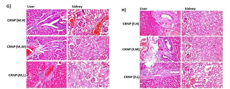
14 Low CRNP Female High CRNP * 0.10 Moderate CRNP Low CRNP Male High AVCR * 0.82 Moderate AVCR Low AVCR Female High AVCR Moderate AVCR Low AVCR Male High AVCRNP Moderate AVCRNP Low AVCRNP Female High AVCRNP * 0.08 Histopathological profile The obtained tissue sections taken from the rat liver were stained by hematoxylin and eosin stains for examination through the light electric microscope. Histopathological alterations were showed in (Fig. 3). Fig. 3A showed group of control versus male rats treated with AV. Control liver: There was no histopathological alteration and the normal histological structure of the central vein and surrounding hepatocytes in the parenchyma were recorded. Control kidney: There was no histopathological alteration and the normal histological structure of the glomeruli and tubules at the cortex were recorded. Male rats treated with AV: Liver with high dose of AV: Focal necrosis was detected in the parenchyma while the portal area showed inflammatory cells infiltration. Kidney with high dose of AV: Congestion was detected in the cortical blood vessels. Liver with moderate dose of AV: The portal area showed few inflammatory cells infiltration while the parenchyma had focal necrosis. Kidney with moderate dose of AV: There was no histopathological alteration. Liver with low dose of AV: The parenchyma showed focal necrosis with inflammatory cells infiltration associated with congestion in the central vein. Kidney with low dose of AV: Congestion was observed in the cortical blood vessels. Fig. 3B showed group of female rats treated with AV. Liver with high dose of AV: The portal area showed congestion in the portal vein Moderate AVCRNP Low AVCRNP with oedema and periductal fibrosis surrounding the bile ducts. Kidney with high dose of AV: There was no histopathological alteration. Liver with moderate dose of AV: The portal area showed inflammatory cells. Kidney with moderate dose of AV: There was no histopathological alteration. Liver with low dose of AV: Focal necrosis with inflammatory cells infiltration was detected in the parenchyma associated with oedema and inflammatory cells infiltration in the portal area. Kidney with low dose of AV: There was no histopathological alteration. Fig. 3C showed group of male rats treated with AVNP. Liver with high dose of AVNP: The parenchyma showed focal necrosis associated with inflammatory cells infiltration in the portal area. Kidney with high dose of AVNP: There was congestion in the cortical blood vessels. Liver with moderate dose of AVNP: The parenchyma showed focal inflammatory cells aggregation. Kidney with moderate dose of AVNP: There was no histopathological alteration. Liver with low dose of AVNP: Focal necroses with inflammatory cells infiltration were detected in the parenchyma. Kidney with low dose of AVNP: There was no histopathological alteration. Fig. 3D showed group of female rats treated with AVNP. Liver with high dose of AVNP: Focal inflammatory cells aggregation was detected in the parenchyma. Kidney with high dose of AVNP: There was no histopathological alteration. Liver with moderate dose of AVNP: The portal area showed inflammatory cells infiltration while the parenchyma had focal necrosis. Kidney with moderate dose of AVNP: There was no Bioscience Research, 2019 volume 16(1):
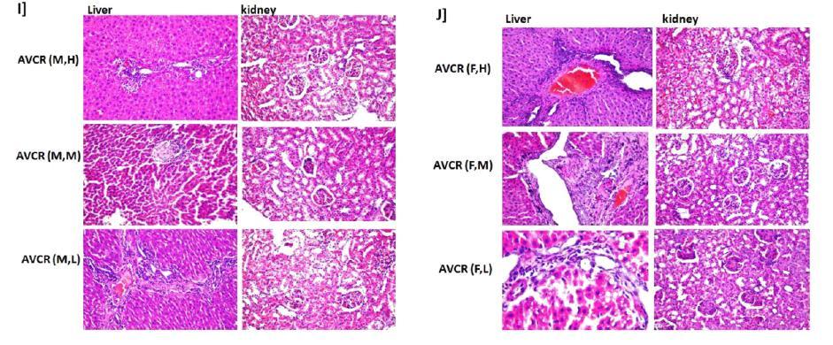
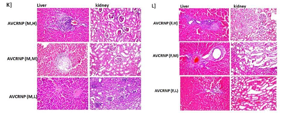
15 histopathological alteration. Liver with low dose of AVNP: There was no histopathological alteration. Kidney with low dose of AVNP: There was no histopathological alteration. Fig. 3E showed group of male rats treated with CR: Liver with high dose of CR: The portal area showed few inflammatory cells infiltration. Kidney with high dose of CR: There were focal haemorrhages in the corticomedullary portion. Liver with moderate dose of CR: There was congestion in the portal vein. Kidney with moderate dose of CR: There was no histopathological alteration. Liver with low dose of CR: Inflammatory cells infiltration was detected in the portal area. Kidney with low dose of CR: There was congestion in the cortical blood vessels. Fig. 3F showed group of female rats treated with CR: Liver with high dose of CR: The portal area showed massive inflammatory cells infiltration associated with focal necrosis in the parenchyma. Kidney with high dose of CR: The cortex showed congestion in the blood capillaries and glomeruli with degeneration in the tubular lining epithelium. Liver with moderate dose of CR: The portal area showed massive inflammatory cells infiltration as well as focal inflammatory cells aggregation in the parenchyma. Kidney with moderate dose of CR: There was perivascular inflammatory cells aggregation associated with tubular degeneration. Liver with low dose of CR: Inflammatory cells infiltration was detected in the portal area. Kidney with low dose of CR: There was congestion in the cortical blood vessels while the corticomedullary portion showed focal haemorrhages in between the degenerated tubules. Fig. 3G showed group of male rats treated with CRNP: Liver with high dose of CRNP: Massive inflammatory cells infiltration was detected in the portal area. Kidney with high dose of CRNP: Sever congestion was observed in the cortical blood vessels. Liver with moderate dose of CRNP: There were oedema and congestion in the portal vein at the portal area associated with periductal fibrosis surrounding the bile ducts. Kidney with moderate dose of CRNP: The glomeruli were congested while the tubules showed degeneration in the lining epithelium at the cortex. Liver with low dose of CRNP: The portal area showed focal inflammatory cells aggregation as well as diffuses inflammatory cells infiltration in the others with congestion in the portal vein. Kidney with low dose of CRNP: There was congestion in the glomeruli and degeneration in the lining epithelium of the cortical blood vessels. Fig. 3H showed group of female rats treated with CRNP: Liver with high dose of CRNP: Periductal fibrosis with inflammatory cells infiltration was detected surrounding the bile ducts at the portal area with congestion in the portal vein. Kidney with high dose of CRNP: The cortex showed degeneration in the lining epithelium of the tubules. Liver with moderate dose of CRNP: Oedema with few inflammatory cells infiltration was detected in the portal area associated with congestion in the portal vein as well as periductal fibrosis surrounding the bile ducts. Kidney with moderate dose of CRNP: There was degenerative change in the tubular lining epithelium of the cortex. Liver with low dose of CRNP: The portal area showed inflammatory cells infiltration. Kidney with low dose of CRNP: Congestion was detected in the cortical blood vessels. Fig. 3I showed group of male rats treated with AVCR: Liver with high dose of AVCR: The portal area showed inflammatory cells infiltration. Kidney with high dose of AVCR: There was no histopathological alteration. Liver with moderate dose of AVCR: There was focal necrosis in the hepatic parenchyma. Kidney with moderate dose of AVCR: There was no histopathological alteration. Liver with low dose of AVCR: The portal area showed inflammatory cells infiltration. Kidney with low dose of AVCR: There was degeneration in the tubular lining epithelium at the cortex. Fig. 3J showed group of female rats treated with AVCR: Liver with high dose of AVCR: The portal area showed inflammatory cells infiltration associated with congestion in the portal vein and hyperplasia in the bile ducts. Kidney with high dose of AVCR: The tubule at the cortex showed degenerative change in the lining epithelium. Liver with moderate dose of AVCR: The portal area showed fibrosis, inflammatory cells infiltration and hyperplasia in the bile ducts. Kidney with moderate dose of AVCR: There was no histopathological alteration. Liver with low dose of AVCR: Few inflammatory cells infiltration was detected in the portal area associated with fatty change in the adjacent surrounding hepatocytes. Kidney with low dose of AVCR: There was no histopathological alteration. Fig. 3K showed group of male rats treated with AVCRNP: Liver with high dose of AVCRNP: Massive inflammatory cells infiltration was detected in the portal area. Bioscience Research, 2019 volume 16(1):
: 596-619 611")
16 Bioscience Research, 2019 volume 16(1):
17 Bioscience Research, 2019 volume 16(1):
Protein & Enzyme Lab (BBT 314)
") Protein & Enzyme Lab (BBT 314) Experiment 3 A: Determination of the enzyme ALT or SGPT activity in serum by enzymatic method using Bioanalyzer Background: Alanine aminotransferase (glutamate pyruvate transaminase)
Protein & Enzyme Lab (BBT 314) Experiment 3 A: Determination of the enzyme ALT or SGPT activity in serum by enzymatic method using Bioanalyzer Background: Alanine aminotransferase (glutamate pyruvate transaminase)
Experiment 6. Determination of the enzyme ALT or SGPT activity in serum by enzymatic method using Biophotometer
 Experiment 6 Determination of the enzyme ALT or SGPT activity in serum by enzymatic method using Biophotometer Background: Alanine aminotransferase (glutamate pyruvate transaminase) belongs to the group
Biodegradable Zwitterionic Nanogels with Long. Circulation for Antitumor Drug Delivery
 Supporting Information Biodegradable Zwitterionic Nanogels with Long Circulation for Antitumor Drug Delivery Yongzhi Men, Shaojun Peng, Peng Yang, Qin Jiang, Yanhui Zhang, Bin Shen, Pin Dong, *, Zhiqing
Estimation of Serum Urea. BCH472 [Practical] 1
![Estimation of Serum Urea. BCH472 [Practical] 1](/thumbs/86/93847865.jpg "Estimation of Serum Urea. BCH472 [Practical] 1") Estimation of Serum Urea BCH472 [Practical] 1 -Urea: Urea is the highest non-protein nitrogen compound in the blood. Urea is the major excretory product of protein metabolism. It is formed by urea cycle
Estimation of Serum Urea BCH472 [Practical] 1 -Urea: Urea is the highest non-protein nitrogen compound in the blood. Urea is the major excretory product of protein metabolism. It is formed by urea cycle
BCH 447. Estimation of Serum Urea
 BCH 447 Estimation of Serum Urea 1 Objective: Estimation of Blood urea nitrogen (BUN) in serum sample. 2 -Urea: Urea is the highest non-protein nitrogen compound in the blood. Urea is the major excretory
Date... Name... Group... Urine sample (Tube No 2)
") Date... Name... Group... Instructions for the practical lesson on biochemistry Topic: Non-protein nitrogen compounds Task 1: Estimation of creatinine in serum and urine 1. Trichloroacetic acid 1.22 mol/l
Date... Name... Group... Instructions for the practical lesson on biochemistry Topic: Non-protein nitrogen compounds Task 1: Estimation of creatinine in serum and urine 1. Trichloroacetic acid 1.22 mol/l
Estimation of Serum Urea
 Estimation of Serum Urea 1 -Urea: Urea is the highest non-protein nitrogen compound in the blood. Urea is the major excretory product of protein metabolism. It is formed by urea cycle in the liver from
MATERIAL AND METHODS
 MATERIAL AND METHODS 3.1 SOURCE OF DATA The present data which is case controlled study was done during the period from 1 st Oct 2010 to 1 st Jan 2012. All the participants were recruited from outpatient
Methods of Enzyme Assay
 Methods of Enzyme Assay Introduction All enzyme assays measure either the consumption of substrate or production of product over time. Different enzymes require different estimation methods dependingon
BASIC ENZYMOLOGY 1.1
 BASIC ENZYMOLOGY 1.1 1.2 BASIC ENZYMOLOGY INTRODUCTION Enzymes are synthesized by all living organisms including man. These life essential substances accelerate the numerous metabolic reactions upon which
Estimation of Serum Urea and Urine Urea. Amal Alamri
 Estimation of Serum Urea and Urine Urea Amal Alamri Lecture Over view Urea Source and fate Blood Urea Urine Urea Urea Clearance BUN/Cr ratio Experiments Production 1-Estimation of Blood Urea Reabsorption
Methods of Enzyme Assay. By: Amal Alamri
 Methods of Enzyme Assay By: Amal Alamri Introduction: All enzyme assays measure either the consumption of substrate or production of product over time. Different enzymes require different estimation methods
Aspartate Transaminase (AST) Color Endpoint Assay Kit Manual Catalog #:
 Color Endpoint Assay Kit Manual Catalog #:") Aspartate Transaminase (AST) Color Endpoint Assay Kit Manual Catalog #: 5605-01 TABLE OF CONTENTS GENERAL INFORMATION... 2 Product Description... 2 Procedure Overview... 2 Kit Contents, Storage and Shelf
Aspartate Transaminase (AST) Color Endpoint Assay Kit Manual Catalog #: 5605-01 TABLE OF CONTENTS GENERAL INFORMATION... 2 Product Description... 2 Procedure Overview... 2 Kit Contents, Storage and Shelf
BIOO RESEARCH PRODUCTS. Aspartate Transaminase (AST) Color Endpoint Assay Kit Manual Catalog #:
 Color Endpoint Assay Kit Manual Catalog #:") BIOO RESEARCH PRODUCTS Aspartate Transaminase (AST) Color Endpoint Assay Kit Manual Catalog #: 5605-01 BIOO Scientific 2010 TABLE OF CONTENTS GENERAL INFORMATION... 1 Product Description... 1 Procedure
BIOO RESEARCH PRODUCTS Aspartate Transaminase (AST) Color Endpoint Assay Kit Manual Catalog #: 5605-01 BIOO Scientific 2010 TABLE OF CONTENTS GENERAL INFORMATION... 1 Product Description... 1 Procedure
Supplementary Table 1. Criteria for selection of normal control individuals among healthy volunteers
 Supplementary Table 1. Criteria for selection of normal control individuals among healthy volunteers Medical parameters Cut-off values BMI (kg/m 2 ) 25.0 Waist (cm) (Men and Women) (Men) 85, (Women) 90
Analytical test kits. Glutamine Lactic acids Malic acids Pyruvic acid Sucrose Sulfite Urea
 5 Analytical test kits Acetaldehyde Acetic acid Ammonia Arginine Ethanol Fructose Glucose Glutamine Lactic acids Malic acids Pyruvic acid Sucrose Sulfite Urea Principles & Features NZYTech test kits are
Urea Nitrogen (BUN) detection Kit
 detection Kit") K-ASSAY KAMIYA BIOMEDICAL COMPANY KAMIYA BIOMEDICAL COMPANY Urea Nitrogen (BUN) detection Kit For the quantitative determination of urea nitrogen in saliva and TCM Cat. No. KT-747 For Research Use Only.
K-ASSAY KAMIYA BIOMEDICAL COMPANY KAMIYA BIOMEDICAL COMPANY Urea Nitrogen (BUN) detection Kit For the quantitative determination of urea nitrogen in saliva and TCM Cat. No. KT-747 For Research Use Only.
Reduction of metastatic and angiogenic potency of malignant cancer by Eupatorium. fortunei via suppression of MMP-9 activity and VEGF production
 Supplementary Information Reduction of metastatic and angiogenic potency of malignant cancer by Eupatorium fortunei via suppression of MMP-9 activity and VEGF production Aeyung Kim, Minju Im, Nam-Hui Yim
Clinician Blood Panel Results
 Page 1 of 7 Blood Panel - Markers Out of Range and Patterns (Pattern: proprietary formula using one or more Blood Markers) Blood Panel: Check for Markers that are out of Lab Range ***NOTE*** Only one supplement
BIOO LIFE SCIENCE PRODUCTS
 BIOO LIFE SCIENCE PRODUCTS FOR REFERENCE PURPOSES This manual is for Reference Purposes Only. DO NOT use this protocol to run your assays. Periodically, optimizations and revisions are made to the kit
Non-Protein Nitrogenous Compounds. Non-Protein Nitrogenous Compounds. NPN s. Urea (BUN) Creatinine NH 3. University of Cincinnati MLS Program 1
 Creatinine NH 3. University of Cincinnati MLS Program 1") Non-Protein Nitrogenous Compounds NPN s Urea (BUN) Creatinine NH 3 Uric Acid Ammonia University of Cincinnati MLS Program 1 Urea Metabolic product derived from catabolism of proteins Proteolysis of proteins
Non-Protein Nitrogenous Compounds NPN s Urea (BUN) Creatinine NH 3 Uric Acid Ammonia University of Cincinnati MLS Program 1 Urea Metabolic product derived from catabolism of proteins Proteolysis of proteins
Liver Function Tests
 Liver Function Tests The liver is of vital importance in intermediary metabolism and in the detoxification and elimination of toxic substances. Damage to the organ may not obviously affects its activity
CoQ10(Coenzyme Q10) ELISA Kit
 ELISA Kit") CoQ10(Coenzyme Q10) ELISA Kit Catalogue No.: EU0196 Size: 48T/96T Reactivity: Universal Detection Range: 0.781-50ng/ml Sensitivity:
CoQ10(Coenzyme Q10) ELISA Kit Catalogue No.: EU0196 Size: 48T/96T Reactivity: Universal Detection Range: 0.781-50ng/ml Sensitivity:
Characterization and Modification of Low Molecular Water-Soluble Chitosan for Pharmaceutical Application
 Characterization and Modification of Low Molecular Water-Soluble Chitosan Bull. Korean Chem. Soc. 2003, Vol. 24, No. 9 1303 Characterization and Modification of Low Molecular Water-Soluble Chitosan for
TABLE OF CONTENTS GENERAL INFORMATION... 1
 BIOO RESEARCH PRODUCTS Glucose Assay Kit Manual Catalog #: 5611-01 BIOO Scientific Corp. 2011 TABLE OF CONTENTS GENERAL INFORMATION... 1 Product Description... 1 Procedure Overview... 1 Required Materials
Clinical Chemistry (CHE 221)
") Clinical Chemistry (CHE 221) Experiment # 14 Blood Alcohol Determination by Gas Chromatography and by Reaction with Alcohol Dehydrogenase Name Date Performed Date Submitted Partners Name(s) Partners Name(s)
Clinical Chemistry (CHE 221) Experiment # 14 Blood Alcohol Determination by Gas Chromatography and by Reaction with Alcohol Dehydrogenase Name Date Performed Date Submitted Partners Name(s) Partners Name(s)
Human Creatinine Urinary Detection Kit
 Human Creatinine Urinary CATALOG NO: IRAAKT2509 Detection Kit LOT NO: SAMPLE INTENDED USE The Urinary Creatinine kit is designed to quantitatively measure creatinine present in urine samples. BACKGROUND
Simultaneous Blood Brain Barrier Crossing and Protection for Stroke Treatment Based on Edaravone-Loaded Ceria Nanoparticles
 Supporting Information Simultaneous Blood Brain Barrier Crossing and Protection for Stroke Treatment Based on Edaravone-Loaded Ceria Nanoparticles Qunqun Bao, Ping Hu, * Yingying Xu, Tiansheng Cheng, Chenyang
Supplementary Information
 Electronic Supplementary Material (ESI) for Journal of Materials Chemistry B. This journal is The Royal Society of Chemistry 2017 Supplementary Information Geometrical Confinement Directed Albumin-Based
complemented with SipA ( SipA/pSipA) or SL1344 WT for 48 hours, after which the
 or SL1344 WT for 48 hours, after which the") P-gp expression (% of control) 12 1 8 6 4 2 * * Untreated SipA SipA/pSipA WT Supplementary Figure 1. SipA modulates the expression of P-gp in healthy murine intestinal epithelium in vivo. Salmonella Typhimurium
P-gp expression (% of control) 12 1 8 6 4 2 * * Untreated SipA SipA/pSipA WT Supplementary Figure 1. SipA modulates the expression of P-gp in healthy murine intestinal epithelium in vivo. Salmonella Typhimurium
Fluorescent Carbon Dots as Off-On Nanosensor for Ascorbic Acid
 Electronic Supplementary Material (ESI) for RSC Advances. This journal is The Royal Society of Chemistry 2014 Fluorescent Carbon Dots as Off-On Nanosensor for Ascorbic Acid Jun Gong, Xin Lu, Xueqin An*
Human Alpha 1 microglobulin ELISA Kit
 Human Alpha 1 microglobulin ELISA Kit Catalogue No.: EH4144 Size: 48T/96T Reactivity: Human Range:0.625-40ng/ml Sensitivity:
2. 2,4 Dinitro phenyl hydrazine (DNPH): I mm in 1N HCl. 5. Working standard: 1 in 20 dilution of the stock standard.
: I mm in 1N HCl. 5. Working standard: 1 in 20 dilution of the stock standard.") -1 Estimation of Alanine Transaminase (ALT) (Mohun and Cook, 1957) Reagents I. Buffered substrate: [100 mm phosphate buffer, 200mM DL-alanine; 2 mm 2-oxo glutarate.}- Dissolved 1.5 g di potassium hydrogen
-1 Estimation of Alanine Transaminase (ALT) (Mohun and Cook, 1957) Reagents I. Buffered substrate: [100 mm phosphate buffer, 200mM DL-alanine; 2 mm 2-oxo glutarate.}- Dissolved 1.5 g di potassium hydrogen
Glutathione Assay Kit
 Glutathione Assay Kit Catalog Number KA1649 250 assays Version: 02 Intended for research use only www.abnova.com Table of Contents Introduction... 3 Intended Use... 3 Background... 3 Principle of the Assay...
TECHNICAL BULLETIN. Sialic Acid Quantitation Kit. Catalog Number SIALICQ Storage Temperature 2 8 C
 Sialic Acid Quantitation Kit Catalog Number SIALICQ Storage Temperature 2 8 C TECHNICAL BULLETIN Product Description The Sialic Acid Quantitation Kit provides a rapid and accurate determination of total
ab Creatinine Assay Kit (Colorimetric)
") ab204537 Creatinine Assay Kit (Colorimetric) Instructions for Use For the quantitative determination of Creatinine in urine samples. This product is for research use only and is not intended for diagnostic
ab204537 Creatinine Assay Kit (Colorimetric) Instructions for Use For the quantitative determination of Creatinine in urine samples. This product is for research use only and is not intended for diagnostic
Clinician Blood Panel Results
 Page 1 of 8 Blood Panel - Markers Out of Range and Patterns (Pattern: proprietary formula using one or more Blood Markers) Blood Panel: Check for Markers that are out of Lab Range ***NOTE*** Only one supplement
For the rapid, sensitive and accurate measurement of Aspartate aminotransferase activity in various samples
 ab105135 Aspartate Aminotransferase Activity Assay Kit Instructions for Use For the rapid, sensitive and accurate measurement of Aspartate aminotransferase activity in various samples This product is for
HYPOGLYCAEMIC ACTION OF THE FLAVONOID FRACTION OF ARTOCARPUS HETEROPHYLLUS LEAF
 42 Research Paper ISSN 0189-6016 2006 Afr. J. Traditional, Complementary and Alternative Medicines www.africanethnomedicines.net HYPOGLYCAEMIC ACTION OF THE FLAVONOID FRACTION OF ARTOCARPUS HETEROPHYLLUS
Aac Reagent Set ELISA for the detection of Acidovorax avenae subsp. citrulli Catalog number: SRA 14800
 List of contents Lot number Aac Reagent Set Item 96 wells 500 wells 1000 wells 5000 wells Capture antibody 0.150 ml 0.275 ml 0.525 ml 2.525 ml Alkaline phosphatase enzyme conjugate 0.150 ml 0.275 ml 0.525
ratmdr1b PE ATPase Kit Assay Protocol jav CAT. NO. SBPE06
 ratmdr1b PE ATPase Kit Assay Protocol jav CAT. NO. SBPE06 Page 1 of 20 Determination of the interaction of drugs with the human ratmdr1b transporter using the PREDEASY TM ATPase Kit For the following membrane
Complete Medical History
 Lab Results for Ben Greenfield Last Test Date: Your medical history is not complete. Complete Medical History Complete Medical History What's Next Blood Draw Blood draw scheduled Complete your medical
Comparison of VACUETTE Heparin Gel Tubes for Common Chemistry Analytes
 Comparison of VACUETTE Heparin Gel Tubes for Common Chemistry Analytes Background: Greiner-Bio-One, Austria has been selling plastic evacuated tubes (VACUETTE ) for venous blood collection since 9. The
Clinician Blood Panel Results
 Page 1 of 8 Blood Panel - Markers Out of Range and Patterns (Pattern: proprietary formula using one or more Blood Markers) Blood Panel: Check for Markers that are out of Lab Range ***NOTE*** Only one supplement
Understanding Blood Tests
 PATIENT EDUCATION patienteducation.osumc.edu Your heart pumps the blood in your body through a system of blood vessels. Blood delivers oxygen and nutrients to all parts of the body. It also carries away
ARABINAN
 www.megazyme.com ARABINAN ASSAY PROCEDURE K-ARAB 08/18 (100 Assays per Kit) Megazyme 2018 INTRODUCTION: In the processing of apples and pears, the yield of juice can be dramatically improved by using enzymes
Blood Urea Nitrogen Enzymatic Kit Manual Catalog #:
 Blood Urea Nitrogen Enzymatic Kit Manual Catalog #: 5602-01 TABLE OF CONTENTS GENERAL INFORMATION... 2 Product Description... 2 Procedure Overview... 2 Kit Contents, Storage and Shelf Life... 3 Required
1. a)label the parts indicated above and give one function for structures Y and Z
label the parts indicated above and give one function for structures Y and Z") Excretory System 1 1. Excretory System a)label the parts indicated above and give one function for structures Y and Z W- renal cortex - X- renal medulla Y- renal pelvis collecting center of urine and then
Excretory System 1 1. Excretory System a)label the parts indicated above and give one function for structures Y and Z W- renal cortex - X- renal medulla Y- renal pelvis collecting center of urine and then
Creatinine (serum, plasma)
") Creatinine (serum, plasma) 1 Name and description of analyte 1.1 Name of analyte Creatinine 1.2 Alternative names None 1.3 Description of analyte Creatinine is a heterocyclic nitrogenous compound (IUPAC
Creatinine (serum, plasma) 1 Name and description of analyte 1.1 Name of analyte Creatinine 1.2 Alternative names None 1.3 Description of analyte Creatinine is a heterocyclic nitrogenous compound (IUPAC
Reagent Set DAS ELISA, Alkaline phosphatase label SRA 22001, SRA 23203, SRA 27703, SRA & SRA ToRSV, ArMV, GFLV, AnFBV and PDV
 List of contents Lot number Reagent Set Item 96 wells 500 wells 1000 wells 5000 wells Capture antibody 0.150 ml 0.275 ml 0.525 ml 2.525 ml Alkaline phosphatase enzyme conjugate 0.150 ml 0.275 ml 0.525
TRACP & ALP Assay Kit
 Cat. # MK301 For Research Use TRACP & ALP Assay Kit Product Manual Table of Contents I. Description...3 II. III. IV. Introduction...3 Components...4 Materials Required but not Provided...4 V. Storage...4
GLUCOSE OXIDASE
 www.megazyme.com GLUCOSE OXIDASE ASSAY PROCEDURE K-GLOX 11/16 (200 Assays per Kit) or (1960 Auto-Analyser Assays per Kit) or (2000 Microplate Assays per Kit) Megazyme 2016 INTRODUCTION: Glucose oxidase
4. Determination of fat content (AOAC, 2000) Reagents
 Reagents") 94 ANALYTICAL METHODS 1. Determination of moisture content (AOAC, 2000) 1. Dry the empty dish and lid in the oven at 105 C for 3 h and transfer to desiccator to cool. Weigh the empty dish and lid. 2. Weigh
94 ANALYTICAL METHODS 1. Determination of moisture content (AOAC, 2000) 1. Dry the empty dish and lid in the oven at 105 C for 3 h and transfer to desiccator to cool. Weigh the empty dish and lid. 2. Weigh
Nitrate/Nitrite Assay Kit Manual Catalog #
 BIOO RESEARCH PRODUCTS Nitrate/Nitrite Assay Kit Manual Catalog # 1305-01 This kit is manufactured to the international quality standard ISO 9001:2008. ISO CI#: SARA-2009-CA-0114-01-B BIOO Scientific Corp.2011
Organic Molecule Composition of Milk: Lab Investigation
 Name: Organic Molecule Composition of Milk: Lab Investigation Introduction & Background Milk & milk products have been a major food source from earliest recorded history. Milk is a natural, nutritionally
Canine Thyroid Stimulating Hormone, TSH ELISA Kit
 Canine Thyroid Stimulating Hormone, TSH ELISA Kit Catalog No: E0463c 96 Tests Operating instruction www.eiaab.com FOR RESEARCH USE ONLY; NOT FOR THERAPEUTIC OR DIAGNOSTIC APPLICATIONS! PLEASE READ THROUGH
Alanine Transaminase Assay Kit
 Alanine Transaminase Assay Kit Catalog Number KA1294 96 assays Version: 13 Intended for research use only www.abnova.com Table of Contents Introduction... 3 Background... 3 Principle of the Assay... 3
This revision also necessitates a change in the table numbering in the test for Organic Impurities.
 Methylphenidate Hydrochloride Extended-Release Tablets Type of Posting Notice of Intent to Revise Posting Date 27 Jul 2018 Targeted Official Date To Be Determined, Revision Bulletin Expert Committee Chemical
PVY Reagent Set Compound ELISA, Alkaline phosphatase label Potato virus Y Catalog number: SRA 20001
 List of contents Lot number PVY Reagent Set Item 96 wells 500 wells 1000 wells 5000 wells Capture antibody 0.150 ml 0.275 ml 0.525 ml 2.525 ml Detection antibody, bottle A 0.150 ml 0.275 ml 0.525 ml 2.525
RENAL FUNCTION ASSESSMENT ASSESSMENT OF GLOMERULAR FUNCTION ASSESSMENT OF TUBULAR FUNCTION
 Measured GFR (mgfr mgfr) and Estimated t GFR (egfr egfr) R. Mohammadi Biochemist (Ph.D.) Faculty member of Medical Faculty RENAL FUNCTION ASSESSMENT ASSESSMENT OF GLOMERULAR FUNCTION ASSESSMENT OF TUBULAR
Non-protein nitrogenous substances (NPN)
") Non-protein nitrogenous substances (NPN) A simple, inexpensive screening test a routine urinalysis is often the first test conducted if kidney problems are suspected. A small, randomly collected urine
Non-protein nitrogenous substances (NPN) A simple, inexpensive screening test a routine urinalysis is often the first test conducted if kidney problems are suspected. A small, randomly collected urine
TNF-alpha ELISA. For Research Use Only. Not For Use In Diagnostic Procedures.
 TNF-alpha ELISA For the quantitative determination of TNF-alpha in serum, plasma, buffered solution or cell culture medium. For Research Use Only. Not For Use In Diagnostic Procedures. Catalog Number:
PHYSICAL PROPERTIES AND DETECTION OF NORMAL CONSTITUENTS OF URINE
 PHYSICAL PROPERTIES AND DETECTION OF NORMAL CONSTITUENTS OF URINE - OBJECTIVES: 1- The simple examination of urine. 2- To detect some of the normal organic constituents of urine. 3- To detect some of the
Creatinine Test Code: CRE
 Creatinine Test Code: CRE Intended Use For in vitro diagnostic use only CARESIDE cartridges are used with the CARESIDE Analyzer to quantitatively measure creatinine concentration in anti-coagulated whole
Blueberry scorch virus Reagent Set DAS ELISA, Alkaline phosphatase label for BlScV Catalog number: SRA 19100
 List of contents Lot number Blueberry scorch virus Reagent Set Item 96 wells 500 wells 1000 wells 5000 wells Capture antibody 0.150 ml 0.275 ml 0.525 ml 2.525 ml Alkaline phosphatase enzyme conjugate 0.150
UREA CE Insert. 01 English - Ref.: 27. Ref.:27
 UREA CE Insert Ref.:27 Intended use. Colorimetric and enzymatic system for urea determination in samples of blood and urine, by end point reaction. Professional use. [Only for in vitro diagnostic use.]
Human Oxidized LDL ELISA Kit (MDA-LDL Quantitation), General
, General") Human Oxidized LDL ELISA Kit (MDA-LDL Quantitation), General For the detection and quantitation of human OxLDL in plasma, serum or other biological fluid samples Cat. No. KT-959 For Research Use Only.
Human Oxidized LDL ELISA Kit (MDA-LDL Quantitation), General For the detection and quantitation of human OxLDL in plasma, serum or other biological fluid samples Cat. No. KT-959 For Research Use Only.
Europium Labeling Kit
 Europium Labeling Kit Catalog Number KA2096 100ug *1 Version: 03 Intended for research use only www.abnova.com Table of Contents Introduction... 3 Intended Use... 3 Background... 3 Principle of the Assay...
Supplementary Files S1 Isolation of Monocytes S2 Haemolysis study Reagents Procedure S3 Cytotoxicity studies Trypan blue dye exclusion method
 Electronic Supplementary Material (ESI) for RSC Advances. This journal is The Royal Society of Chemistry 2014 Supplementary Files S1 Isolation of Monocytes A 3 ml volume of Histopaque 1083 solution was
EXCRETION QUESTIONS. Use the following information to answer the next two questions.
 EXCRETION QUESTIONS Use the following information to answer the next two questions. 1. Filtration occurs at the area labeled A. V B. X C. Y D. Z 2. The antidiuretic hormone (vasopressin) acts on the area
PAF Acetylhydrolase Assay Kit
 PAF Acetylhydrolase Assay Kit Catalog Number KA1354 96 assays Version: 04 Intended for research use only www.abnova.com Table of Contents Introduction... 3 Background... 3 Principle of the Assay... 3 General
Reagent Set DAS ELISA, Alkaline phosphatase label
 List of contents Lot number Item 96 wells 500 wells 1000 wells 5000 wells Capture antibody 0.150 ml 0.275 ml 0.525 ml 2.525 ml Alkaline phosphatase enzyme conjugate 0.150 ml 0.275 ml 0.525 ml 2.525 ml
ab Sphingomyelin Assay Kit
 ab133118 - Sphingomyelin Assay Kit Instructions for Use For the specific, sensitive and convenient method of quantifying Sphingomyelin in plasma or serum. This product is for research use only and is not
Potato leafroll virus (PLRV) Reagent Set DAS ELISA, Alkaline phosphatase label Catalog number: SRA List of contents
 Reagent Set DAS ELISA, Alkaline phosphatase label Catalog number: SRA List of contents") Potato leafroll virus (PLRV) Reagent Set List of contents Lot number Item 96 wells 500 wells 1000 wells 5000 wells Capture antibody 0.150 ml 0.275 ml 0.525 ml 2.525 ml Alkaline phosphatase enzyme conjugate
Potato leafroll virus (PLRV) Reagent Set List of contents Lot number Item 96 wells 500 wells 1000 wells 5000 wells Capture antibody 0.150 ml 0.275 ml 0.525 ml 2.525 ml Alkaline phosphatase enzyme conjugate
SIV p27 Antigen ELISA Catalog Number:
 INTENDED USE The RETRO-TEK SIV p27 Antigen ELISA is for research use only and is not intended for in vitro diagnostic use. The RETRO-TEK SIV p27 Antigen ELISA is an enzyme linked immunoassay used to detect
Supporting Information
 Supporting Information The Effects of Spacer Length and Composition on Aptamer-Mediated Cell-Specific Targeting with Nanoscale PEGylated Liposomal Doxorubicin Hang Xing +, [a] Ji Li +, [a] Weidong Xu,
Mouse C-Peptide ELISA Kit
 Mouse C-Peptide ELISA Kit Cat.No: DEIA4507 Lot. No. (See product label) Size 96T Intended Use The Mouse C-Peptide ELISA kit is for the quantitative determination of c-peptide in mouse serum, plasma, and
HBeAg and HBeAg Ab ELISA Kit
 HBeAg and HBeAg Ab ELISA Kit Catalog Number KA0290 96 assays Version: 17 Intended for research use only www.abnova.com Table of Contents Introduction... 3 Intended Use... 3 Principle of the Assay... 3
APPENDIX Heparin 2 mg heparin was dissolved in 0.9 % NaCl (10 ml). 200 µl of heparin was added to each 1 ml of blood to prevent coagulation.
. 200 µl of heparin was added to each 1 ml of blood to prevent coagulation.") APPENDIX 1 Preparation of reagents 1.1. Preparation of dosing solution Nonylphenol 15 mg of Nonylphenol was dissolved in olive oil (10 ml) and used as stock solution. The stock solution was serially diluted
APPENDIX 1 Preparation of reagents 1.1. Preparation of dosing solution Nonylphenol 15 mg of Nonylphenol was dissolved in olive oil (10 ml) and used as stock solution. The stock solution was serially diluted
Exendin-4 (Exenatide) ELISA Kit
 ELISA Kit") Exendin-4 (Exenatide) ELISA Kit Catalog: DEIABL227 For the quantitative determination of Exendin-4 in serum or plasma using competitive ELISA method For Research Use Only. Protocol Provided for Informational
Exendin-4 (Exenatide) ELISA Kit Catalog: DEIABL227 For the quantitative determination of Exendin-4 in serum or plasma using competitive ELISA method For Research Use Only. Protocol Provided for Informational
Hosta virus X ELISA KIT
 Hosta virus X ELISA KIT Cat. No.:DEIA9256 Pkg.Size:96T Intended use The Hosta virus X ELISA KIT is used to detect HVX in both leaves and roots of hosta plants. Reagents And Materials Provided 1. Capture
Influenza A IgG ELISA
 Influenza A IgG ELISA For the qualitative determination of IgG-class antibodies against Influenza A virus in human serum or plasma (citrate, heparin). For Research Use Only. Not For Use In Diagnostic Procedures.
NORMAL LABORATORY VALUES FOR CHILDREN
 Pediatric Drug Lookup Normal Laboratory Values for NORMAL LABORATORY VALUES FOR CHILDREN CHEMISTRY Normal Values Albumin 0-1 y 2.0-4.0 g/dl 1 y to adult 3.5-5.5 g/dl Ammonia Newborns 90-150 mcg/dl 40-120
BIOCHEMISTRY OF BLOOD
 BCH 471 BIOCHEMISTRY OF BLOOD Amal Alamri Experiment 1 Separation of Plasma and Serum from Whole Blood Whole Blood It is living tissue that circulates through the heart, arteries, veins, and capillaries
ENZYME MARKERS OF TOXICITY
 Why do we need enzyme markers? ENZYME MARKERS OF TOXICITY In vivo monitoring Serial sampling Early detection of metabolic changes Detection of organ-specific effects Establishment of NO EFFECT level Determination
02006B 1 vial 02006B 1 vial Store at -20 C. Lyophilized recombinant IL-2
 For detection and measurement of human interleukin 2 Catalog #02006 Catalog #02007 2 Plates 10 Plates Product Description The Human Interleukin 2 (IL-2) ELISA Kit is designed for the quantitative detection
1.2 Systematic Name: Orthophosphoric-monoester phosphohydrolase (alkaline optimum)
") Document Title Alkaline Phosphatase Page 1 of 6 Originating Department QA Approval Departments QA, QC Approval Date 5 th June 2017 Effective Date 8 th June 2017 1.0 PRODUCT DETAILS 1.1 Enzyme Name: Alkaline
Document Title Alkaline Phosphatase Page 1 of 6 Originating Department QA Approval Departments QA, QC Approval Date 5 th June 2017 Effective Date 8 th June 2017 1.0 PRODUCT DETAILS 1.1 Enzyme Name: Alkaline
Rat cholesterol ELISA Kit
 Rat cholesterol ELISA Kit Catalog No. CSB-E11706r (96T) This immunoassay kit allows for the in vitro quantitative determination of rat Cholesterol concentrations in serum, plasma and other biological fluids.
ACTG Laboratory Technologist Committee Revised Version 2.0 ACTG Lab Man Coulter HIV-1 p24 ELISA May 21, 2004
 Coulter HIV p24 1. PRINCIPLE The Human Immunodeficiency Virus Type 1 (HIV-1) is recognized as the etiologic agent of acquired immunodeficiency syndrome (AIDS). The virus is transmitted by sexual contact,
Clinical enzymology. University of Babylon College of pharmacy Second semester - biochemistry 3 rd class By Dr. Abdulhussien M. K.
 Clinical enzymology University of Babylon College of pharmacy Second semester - biochemistry 3 rd class 2014 2015 By Dr. Abdulhussien M. K. Aljebory Enzyme activity Enzyme assays usually depend on the
Human Carbamylated LDL ELISA Kit (CBL-LDL Quantitation)
") Product Manual Human Carbamylated LDL ELISA Kit (CBL-LDL Quantitation) Catalog Number MET-5032 96 assays FOR RESEARCH USE ONLY Not for use in diagnostic procedures Introduction Lipoproteins are submicroscopic
Product Manual Human Carbamylated LDL ELISA Kit (CBL-LDL Quantitation) Catalog Number MET-5032 96 assays FOR RESEARCH USE ONLY Not for use in diagnostic procedures Introduction Lipoproteins are submicroscopic
CONTEXT POINT 2: Plants and animals transport dissolved nutrients and gases in a fluid medium.
 CONTEXT POINT 2: Plants and animals transport dissolved nutrients and gases in a fluid medium. Identify the form(s) in which each of the following is carried in mammalian blood: Carbon dioxide 70% as hydrogen
Hepatitis A virus IgM ELISA Kit
 Hepatitis A virus IgM ELISA Kit Catalog Number KA0285 96 assays Version: 04 Intended for research use only www.abnova.com Table of Contents Introduction... 3 Intended Use... 3 Background... 3 Principle
enzymatic determinations of kecap Page 1 of 13
 enzymatic determinations of kecap Page 1 of 13 Contains enzymatic determinations of: - citrate - glycerol - glucose, fructose, starch - galactose - L-glutamic acid - formic acid - malate - ethanol - acetate
BCH 471. Experiment (5) Hemoglobin Estimation, Erythrocyte Sedimentation Rate (ESR) and Hematocrit (HCT)
 Hemoglobin Estimation, Erythrocyte Sedimentation Rate (ESR) and Hematocrit (HCT)") BCH 471 Experiment (5) Hemoglobin Estimation, Erythrocyte Sedimentation Rate (ESR) and Hematocrit (HCT) Objectives Quantitative determination of hemoglobin in a blood sample. Determination of erythrocyte
BCH 471 Experiment (5) Hemoglobin Estimation, Erythrocyte Sedimentation Rate (ESR) and Hematocrit (HCT) Objectives Quantitative determination of hemoglobin in a blood sample. Determination of erythrocyte
GB Translated English of Chinese Standard: GB NATIONAL STANDARD
 Translated English of Chinese Standard: GB5009.5-2016 www.chinesestandard.net Sales@ChineseStandard.net GB NATIONAL STANDARD OF THE PEOPLE S REPUBLIC OF CHINA GB 5009.5-2016 National food safety standard
STORE AT 4 o C Version 3
 Instruction Manual Advanced Protein Assay Reagent (Cat. # ADV01) ORDERING INFORMATION To order by phone: (303) - 322-2254 To order by Fax: (303) - 322-2257 To order by e-mail: cserve@cytoskeleton.com Technical
Tables of Normal Values (As of February 2005)
") Tables of Normal Values (As of February 2005) Note: Values and units of measurement listed in these Tables are derived from several resources. Substantial variation exists in the ranges quoted as normal
Tables of Normal Values (As of February 2005) Note: Values and units of measurement listed in these Tables are derived from several resources. Substantial variation exists in the ranges quoted as normal