Screening with New Modalities: Breast Ultrasound
|
|
|
- Magdalen Baker
- 6 years ago
- Views:
Transcription

1 Screening with New Modalities: Breast Ultrasound Wendie A. Berg, MD, PhD Professor of Radiology Magee-Womens Hospital of UPMC University of Pittsburgh School of Medicine
2 Disclosures No personal financial conflicts of interest Philips Healthcare loaning equipment for ultrasound clinical trial
3 Objectives Describe effect on cancer detection from adding screening US to mammography or tomosynthesis in women with dense breasts Discuss sources of false positives on screening US and ways to reduce them Compare outcomes from different methods of screening breast US
4 Evidence Supporting Screening Disease-specific mortality reduction Only studied for mammography Reduction in node-positive disease Increase in node-negative invasive cancers Reduction in interval cancers Fewer than 10% of all cancers diagnosed
5 Failure Analysis Webb ML et al Cancer 2013, epub 9/11/ invasive breast cancer dx f/u breast cancer deaths; median age 49 yr at dx 29% ca deaths were among women screened 19% screen detected 10% interval cancers 71% deaths among unscreened women
6 Interval Cancer Cancer dx by clinical symptoms in interval between recommended screenings Worse prognosis and worse outcome ~1/2 deaths in screened women diagnosed in their 40s are due to interval cancers
7 Mammography Failure Analysis #1 If not performed at all #2 High-risk women #3 Dense breasts
8 BI-RADS Density A. Almost entirely fatty B. Scattered fibroglandular density C. Heterogeneously dense which could obscure detection of small masses D. Extremely dense, which lowers the sensitivity of mammography
9


10 Breast Density as Function of Age Kerlikowske et al. JNCI 2007; 99: % of women of mammographic age have dense breasts
11 Masking of cancers with increasing breast density Increased risk of developing breast cancer
12 Interval Cancers and Breast Density Density Odds Ratio 95% CI < 10% % 2.1 (0.9, 5.2) 25-49% 3.6 (1.5, 8.7) 50-74% 5.6 (2.1, 15.3) 75% 17.8 (4.8, 65.9) p <.001 Boyd NF, et al. NEJM 2007;356:227-36
13 Referent Average Pt, Hazard Ratios A B C D Premeno Postmeno no HT Postmeno E+P Kerlikowske K et al J Clin Onc 2010;28:
14 Increased Deaths Chiu SY et al. Cancer Epidemiol Biomarkers Prev 2010;19: yr f/u Sweden 15,658 women % had dense breasts Increased breast cancer mortality with dense breasts RR 1.91 (95%CI ) Attributed to higher incidence Shorter sojourn time


15 24 States require some sort of density notification 3/13/16
16 Possible tests to add to mammography Modality vs. Mammography alone Absolute Cancer Detection per 1000 screens Clinical breast exam 0.3 Double Read or CAD 1 Tomosynthesis 1-2 Ultrasound 3-4 Molecular Breast Imaging, 7-8 CEDM MRI 10 Copyright Wendie Berg, MD, PhD
17 Unable to Tolerate MRI: ACRIN % (1 in 5.4) (95% CI 16.4 to 20.8%) women who had completed 3 years of screening with US and mammography were unable to undergo an MRI Berg WA et al. Radiology 2010;254:79-87
18 Ultrasound No radiation Not limited by dense tissue No injection of contrast or radioactive material Inexpensive
19 US to Replace Mammo? Berg WA et al JNCI 2016; 108, epub 12/18/ breast ca dx among 2809 women ACRIN US to detect one cancer, 127 for mammo Of 89 invasive cancers, 53 (60%) seen on US vs. 41 (46%) on mammography, p =.11 More likely node negative when found by US: 34/53 (64%) vs. 18/41 (44%), p =.003
20 US but not Mammo Inv Ca Detection Density, % US Mammo US, not Mammo 25 0/1 (0) 0/1 (0) 0/1 (0) /10 (60) 6/10 (60) 2/10 (20) /30 (53) 17/30 (57) 8/30 (27) /36 (61) 13/36 (36) 14/36 (39) >80 9/12 (75) 5/12 (42) 6/12 (50) P trend Berg WA et al JNCI 2016; 108, epub 12/18/15
21 False Positives Over 3 Years US Mammo P-value Recall Rate 515 (10.7%) 453 (9.4%).03 Biopsy Rate 266 (5.5%) 97 (2.0%) <.001 PPV Biopsies 31/266 (11.7%) 37/97 (38.1%) <.001 Berg WA et al JNCI 2016; 108, epub 12/18/15
22 US and Mammo Complementary Of 22 DCIS, 18 (82%) seen on mammography vs. 5 (23%) on US, p=.002 Sensitivity of mammography + US 0.76 ( ) vs ( ) mammo alone (p <.001) Berg WA et al JAMA 2012;307:


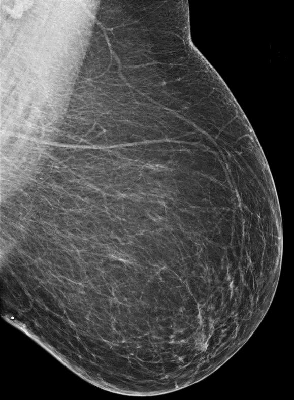
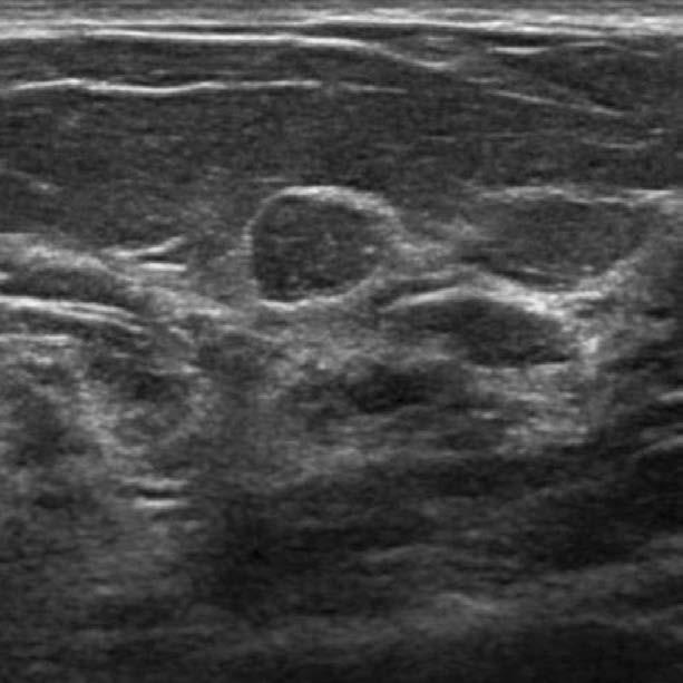
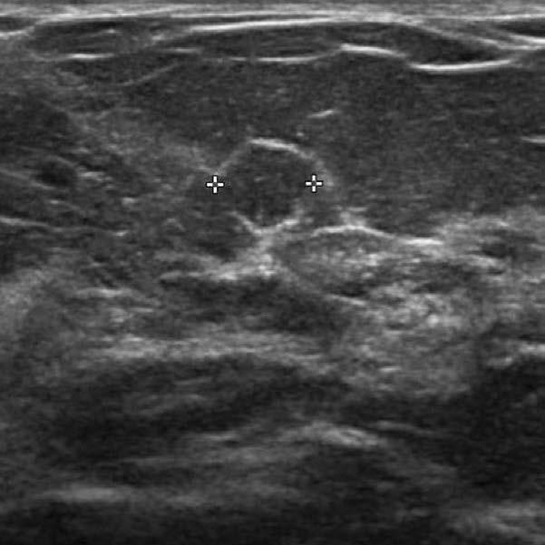
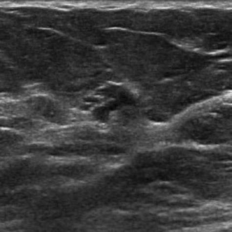
23 48F screening Courtesy Dr. Wei Yang, MD Anderson


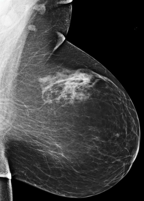
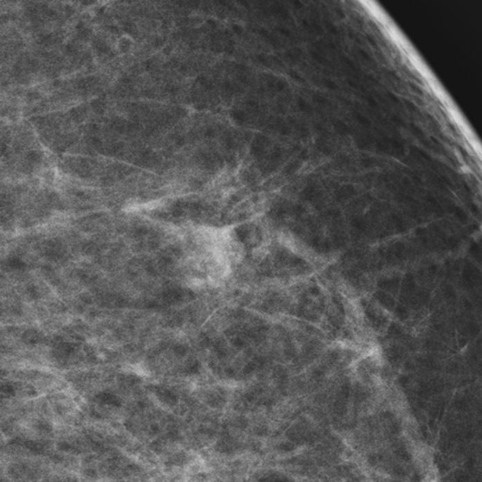
24 RT CC MAG RT ML MAG Courtesy Dr. Wei Yang, MD Anderson
25 Stereotactic biopsy: High nuclear grade DCIS solid type with comedo necrosis, with microinvasion, ER, PR-, HER2 + Skin-sparing mastectomy, 0/4 SLN
26 Supplemental US Physician Performed Technologist Performed Automated
27 Handheld US High-frequency transducer, MHz linear array Survey scanning transverse and sagittal Document 1 image per quadrant, 1 behind nipple for negative exam Lesions (all studies to date): Orthogonal views ± calipers; optional color or power Doppler image Positive test: BI-RADS 3 or higher assessment, or recommendation for further imaging (BI-RADS 0)
28 Physician Performed US: Multicenter Results Author N screens ICDR per 1000 Recall Rate (%) Bx Rate (% women) PPV3 Bx Performed Corsetti NS 449 (4.9) 50/623 (8.0) Berg yr (15.1) 207 (7.8) 14/264 (5.3) Berg yr (7.4) 242 (5.0) 21/276 (7.6) TOTAL 16, % 898 (5.4) 85/1163 (7.3) 4.9% of women had biopsies for benign findings
29 Tech-Performed US (USA): Prevalent Screens Author N ICDR per 1000 Recall Rate (%) Bx Rate (%) PPV3 Bx Performed Kaplan, , (9.5) 97 (5.2) 6/96 (6.3) Hooley, * (23.8) 46 (7.1) 3/58 (5.2) Weigert, , ,196 (13.8) 429 (5.0) 25/418 (6.7) Parris, , (12.3) 185 (3.3) 10/181 (5.5) Overall 16, ,206 (13.2) 757 (4.5) 47/753 (6.2) *analysis presented for women with negative screening mammograms Berg WA and Mendelson EB. Radiology 2014;272:12-27
30 Recalls: Tech-Performed HHUS 2,206/16,676 (13.2%) test positive on prevalence screen 1,399 (8.4%) all women BI-RADS (4.5%) all women BI-RADS 4 44/753 (5.8%) found to have cancer Only 43/16,676 (0.3%) recalled for additional evaluation (BI-RADS 0) prior to final assessment Berg WA and Mendelson EB. Radiology 2014;272:12-27
31 Disease Prevalence Affects Yield Moderate Risk* No Known Risks P-value Kolb /2914 (4.8 per 1000) 14/7901 (1.8 per 1000).011 Crystal /318 (12.5 per 1000) 3/1199 (2.5 per 1000) <.04 Overall 18/3232 (5.6 per 1000) 17/9100 (1.9 per 1000) *Personal hx of breast cancer or first-degree relative with breast cancer vs. no risks
32 Japan Tohno E et al Breast Cancer 2012;19: day educational program; results of training/testing for 415 technologists and 422 physicians Observers worse with experience < 100 cases Video sensitivity, still image sensitivity, and disease agreement for technologists > for MDs
33 Node-Negative Invasive Cancers Across 10 series, 475 cancers seen only on US, 415 (87.4%) invasive 273/303 (90.1%) with staging were node negative 22/91 (24%) ILC
34 By Participant, Yield/1000, ACRIN 6666 Year M+US M Supp. Yield, 95% CI P-value (2.1, 8.4) (0.9, 6.4) (0.9, 6.8).004 Supplemental yield of US is significant each year and similar for incidence and prevalence screens Berg WA et al JAMA 2012;307:
35 Weigert: Recalls Incidence Screens Year (12%) women screening US Recall rate 13.8% (n=1196): 767 (8.9%) BR 3; 429 (5.0%) BR 4,5; PPV3 5.6% 24 cancers, CDR 2.8 per 1000 Year 2 10,282 (17.9%) women Recall rate 12.7% (n=1310); CDR 2.3 per 1000 (24) Year (12.8%) women Recall rate 7.7% (n=316); CDR 2.7 per 1000 (11)


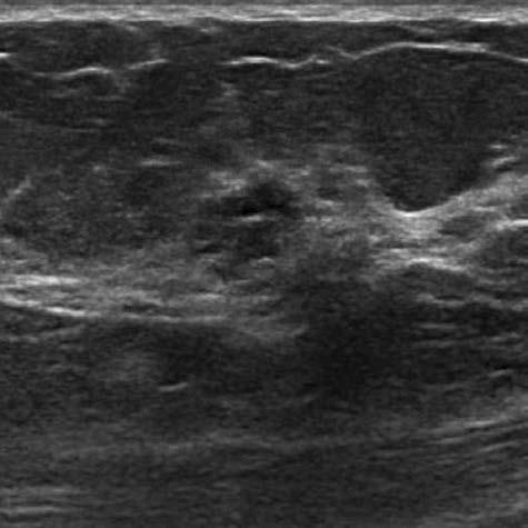
36 Radial Antiradial 60F, 5-yr risk 2.5%, 24-mo US: 12 mm grade 1 IDC-DCIS, N0 Courtesy WP Evans, III, MD


37 Radial Antiradial 75F personal hx Lt cancer 17 mm grade 3 IDC-DCIS, N0 Seen only on 24-month US Seen in retrospect on mammo Courtesy Gary Whitman, MD, MD Anderson


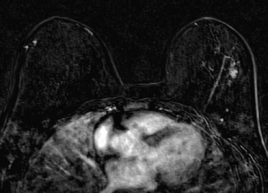
38 70F personal hx rt mastectomy, BRCA-1 mutation carrier 24 mo screen US+ 19 mm grade 3 IDC-DCIS, N0 Courtesy Dr. Mary Mahoney, U Cincinnati
39 ACRIN 6666: Breast Density Density n Yield per 1000 P-value 25% % % % >80% Berg WA, et al., RSNA 2009
40 Interval Cancer Rate: ACRIN 6666 Yr N Interval N Cancers (%) All Interval Ca Rate: 9/7473 screens = 1.2 per % of all cancers Berg WA et al JAMA 2012;307:
41 Interval Cancer Rate Italy Corsetti V et al Cancer 2011;47: Interval cancer rate in fatty breasts 1.0 per 1000 Interval cancer rate in dense breasts after adding screening US 1.1 per 1000
42 J-START Ohuchi N et al Lancet 2015, epub 11/4/2015 Asymptomatic women aged at 42 sites Randomized to M+US or M alone twice in 2 yrs 36,869 to intervention and 36,139 to control group
43 Results J-START first round Intervention Control P-value Sensitivity 91.1 ( ) 77.0 ( ).0004 Specificity 87.7 ( ) 91.4 ( ) <.0001 % Stage 0, I 144/184 (71.3) 79/117 (52.0).019 Interval Cancers 18 (0.05%) 35 (0.10%).034 Ohuchi N et al Lancet 2015, epub 11/4/2015
44 Time to Perform US: ACRIN 6666 Bilateral scan, not including time discussing results with patient nor creation of report Year Median (min) Mean SD
 Across all series, only 1 lesion had suspicious change yielding malignancy at 6-mo follow-up 12-month follow-up reasonable")
45 Reducing False Positives BI-RADS 3 lesions Prevalence of 15-20% of all patients having screening US in prior series (Barr et al; Hooley et al; Chae et al) Across all series, only 1 lesion had suspicious change yielding malignancy at 6-mo follow-up 12-month follow-up reasonable
46
47 Orthogonal Views Required for any mass for which future comparison is desirable Not necessary for simple cysts Incomplete characterization without this


48 RAD ARAD 53F Papillary DCIS with microinvasion Berg WA and Mendelson EB Radiology 2014;262: Courtesy Dr. Christophe Tourasse


49 50F invasive ductal carcinoma; echogenic rim in arad view only Berg WA and Mendelson EB Radiology ;262:
 post-menopausal participants, had cysts 73 using estrogen replacement 48 (66%) had cysts 1290 no HRT 489 (37.9%) had cysts (p<.0001, less common) 516/793 (65.")
50 Cysts ACRIN /2662 (47.1%) women over the three years 998 (37.5%) of 2659 year one 537/1363 (39.4%) post-menopausal participants, had cysts 73 using estrogen replacement 48 (66%) had cysts 1290 no HRT 489 (37.9%) had cysts (p<.0001, less common) 516/793 (65.1%) premenopausal women had cysts (p<.0001) Berg WA, et al Radiol Clin N Amer 2010;48:
51 Complicated Cysts ACRIN (14.1%) of 2662 participants 301 (80%) had at least one simple cyst 84 (22%) multiple, bilateral Overall 2/475 (0.42%) such lesions malignant Berg WA, et al Radiol Clin N Amer 2010;48:
52 Complicated Cysts N N Malignant (%) Kolb et al Venta et al Buchberger et al Berg et al Chang et al Daly et al ACRIN TOTAL (0.3) Berg WA et al Radiol Clin N Amer 2010,48:


53 53F incidental finding on US, aspirated, cytology: benign cyst with apocrine cells Cyst or Solid? Radial Antiradial


54 Radial Antiradial 51F strong FH, incidental finding on US Aspirated to resolution, thick cloudy yellow fluid, cyst


55 61F with new mass on mammography, prior ipsilateral cancer


56 Radial Antiradial 12 month follow-up US enlarged: 14-g US-guided bx papillary DCIS
57 BI-RADS 3 Chae EY et al AJR 2016;206: With mammographic abnormality, 4/184 (2.2%) malignant Without mammographic abnormality, 4/980 (0.4%) malignant (p=.025)
58 Clustered Microcysts 3.9 to 5.8% of US examinations 1/235 (0.4%) malignant across 5 series Mean age 48 years (32-71) Short-interval follow-up if uncertainty Caution if new mass on mammogram, post-menopausal woman not on HRT May merit biopsy Berg WA AJR 2005;185:952 Berg WA, et al Radiol Clin N Amer 2010;48:


59 48F new mass on screening mammogram
60 60F ipsilateral cancer elsewhere US-guided core biopsy DCIS, intermediate grade
61 Lesions Synchronous to New Cancer Kim SJ et al AJR 2008;191: /482 (11.4%) BI-RADS 3 lesions malignant 36/170 (21.2%) in same quadrant as 1º 12/122 (9.8%) in different quadrant 8/190 (4.2%) in contralateral breast
62 M-B Circumscribed Masses: US Berg WA et al Radiology 2013:268: women in ACRIN (6.2%) participants had 153 unique findings described as M-B masses on screening US over 3 annual screens 98 complicated cysts with debris 43 solid, circumscribed, oval masses 7 solid masses with 2-3 lobulations 5 clustered microcysts No malignancies (95%CI up to 2.4%)
63 Billing CPT codes 76641, unilateral complete right 76641, unilateral complete left Medicare reimbursement averages $165 Subject to deductible and copays
64 Billing ICD Inconclusive mammogram Applicable to dense breasts, NOS Inconclusive mammogram due to dense breasts

65 Automated Arm US A Tower B Y-axis Gantry & Transducer Carrier C X-axis Gantry D Ultrasound Machine Monitor E Touch Screen / Monitor F Transducer Holster G Patient Bed
66 Automated Arm Results Kelly KM et al Eur Radiol 2010; 20: women, 6425 exams, 8 facilities 40% women at intermediate risk 23 cancers mammography 46 cancers M+US Supplemental yield 3.6 per 1000 (95% CI 2.3 to 5.4) 10% recall rate 23/75 (31%) biopsies showed cancer

67 12 MHz Automated Breast US 15 cm footprint 3 acquisitions per breast in ~15 minutes 3D dataset Transverse Created coronal and sagittal displays
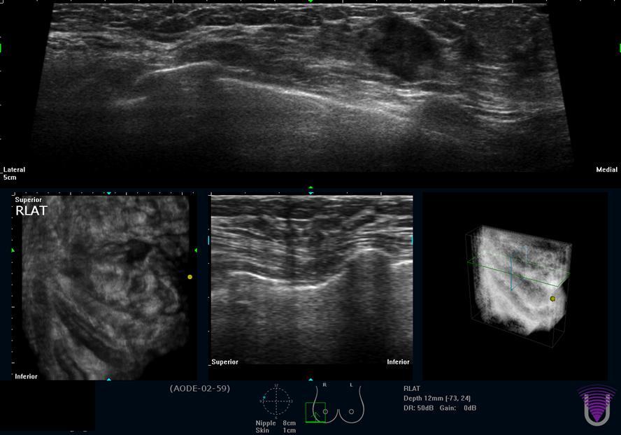
68
69 ABUS Results Brem RF et al Radiology 2015;273: ,318 women BI-RADS 1 or 2 mammo, dense breasts, automated whole breast US 30 (2/1000) cancers only by ABUS 25 detailed: 23 (92%) invasive, mean size 13 mm, 18 (78%) of those N0 20/23 (87%) ER+ 3/22 (14%) stage IIB or higher 13% absolute increase in recall rate immediate additional evaluation, not a final assessment
70 Time to acquire images HHUS vs. AUS HHUS 13 min (but range up to 90) Training to Yes, months, technologist AUS 15 min Minimal Sensitivity ~85% ~74% Number of images Time to interpret < 30 sec 5-10 min Recalls 13% Final assessment typically rendered 13% Incomplete, needs targeted US Interobserver Variability Κ = 0.53 (SE 0.02) Κ = 0.04 to 0.50
71 Is screening ultrasound still of benefit after tomosynthesis?
 US 23 cancers (ICDR 7.1/1000, 95%CI 4.2 to 10.0, p=0.006) False positive recall DBT 53 vs.")
72 ASTOUND trial Tagliafico AS et al JCO 2016;epub 3/9/ women with dense breasts, negative mammogram, 5 centers in Italy DBT 13 cancers (ICDR 4.0/ %CI 1.8 to 6.2) US 23 cancers (ICDR 7.1/1000, 95%CI 4.2 to 10.0, p=0.006) False positive recall DBT 53 vs. US 65 (p=0.26)
73 DBTUST study UPMC Pittsburgh UPMC Hamot, Erie Weinstein Imaging 6200 women DBT and technologist-performed screening US each year for three years NIH and PABCC funding
74 Three-Step Implementation 1) Does the woman have at least 10-yr life expectancy? No, then CBE only, with mammography only if warranted by symptoms

75 2) Is the patient at high risk for breast cancer and under age 70? Yes, then MRI annually beginning: When ascertained to be high risk Age 25 if BRCA1/2 or other pathogenic mutation 8yr after chest XRT if XRT before age 30 If unable to tolerate MRI, then US
76 3) Dense? Yes: Supplement annual mammography with US beginning at age No: Tomosynthesis beginning at age 40-45

77 Mam+US MRI Mammo
n Educational support from GE and Volpara n Reduce mortality n Healthy women will not be harmed
 Dense Breasts: What to Know and What to Do Wendie A. Berg, MD, PhD, FACR Professor of Radiology Magee-Womens Hospital of UPMC University of Pittsburgh School of Medicine wendieberg@gmail.com Disclosures
Current Status of Supplementary Screening With Breast Ultrasound
 Current Status of Supplementary Screening With Breast Ultrasound Stephen A. Feig, M.D., FACR Fong and Jean Tsai Professor of Women s Imaging Department of Radiologic Sciences University of California,
BREAST DENSITY WHAT IS IT? WHY IS IT IMPORTANT? & What IOWA SF250 Means to Patients and Providers
 BREAST DENSITY WHAT IS IT? WHY IS IT IMPORTANT? & What IOWA SF250 Means to Patients and Providers Arnold Honick, MD Radiology Consultants of Iowa, PLC ahonick@rciowa.com BREAST DENSITY LEGISLATION Nancy
Current Strategies in the Detection of Breast Cancer. Karla Kerlikowske, M.D. Professor of Medicine & Epidemiology and Biostatistics, UCSF
 Current Strategies in the Detection of Breast Cancer Karla Kerlikowske, M.D. Professor of Medicine & Epidemiology and Biostatistics, UCSF Outline ν Screening Film Mammography ν Film ν Digital ν Screening
Update in Breast Cancer Screening
 Disclosure information: Update in Breast Cancer Screening Karla Kerlikowske, MDDis Update in Breast Cancer Screening Grant/Research support from: National Cancer Institute and Grail - and - Karla Kerlikowske,
Dense Breasts. A Breast Cancer Risk Factor and Imaging Challenge
 Dense Breasts A Breast Cancer Risk Factor and Imaging Challenge Renee Pinsky, MD University of Michigan Department of Radiology Division of Breast Imaging No Disclosures QUIZ: ARE YOU DENSE? a. Breast
Breast density: imaging, risks and recommendations
 Breast density: imaging, risks and recommendations Maureen Baxter, MD Radiologist Director of Ruth J. Spear Breast Center Providence St. Vincent Medical Center Alison Conlin, MD/MPH Medical Oncologist
Supplemental Screening for Dense Breasts. Reagan Leverett, MD, MS
 Supplemental Screening for Dense Breasts Reagan Leverett, MD, MS Outline Anatomy and Density Risk of dense breasts Theory of Supplemental Screening Options for supplemental screening Tomosynthesis Ultrasound
Update in Breast Cancer Screening
 Disclosure information: Update in Breast Cancer Screening Karla Kerlikowske, MDDis Update in Breast Cancer Screening Grant/Research support from: National Cancer Institute - and - Karla Kerlikowske, MD
EARLY DETECTION: MAMMOGRAPHY AND SONOGRAPHY
 EARLY DETECTION: MAMMOGRAPHY AND SONOGRAPHY Elizabeth A. Rafferty, M.D. Avon Comprehensive Breast Center Massachusetts General Hospital Harvard Medical School Breast Cancer Screening Early detection of
Dense Breasts, Get Educated
 Dense Breasts, Get Educated What are Dense Breasts? The normal appearances to breasts, both visually and on mammography, varies greatly. On mammography, one of the important ways breasts differ is breast
5/24/16. Current Issues in Breast Cancer Screening. Breast cancer screening guidelines. Outline
 Disclosure information: An Evidence based Approach to Breast Cancer Karla Kerlikowske, MDDis Current Issues in Breast Cancer Screening Grant/Research support from: National Cancer Institute - and - Karla
EARLY DETECTION: MAMMOGRAPHY AND SONOGRAPHY
 EARLY DETECTION: MAMMOGRAPHY AND SONOGRAPHY Elizabeth A. Rafferty, M.D. Avon Comprehensive Breast Center Massachusetts General Hospital Harvard Medical School Breast Cancer Screening Early detection of
Pitfalls and Limitations of Breast MRI. Susan Orel Roth, MD Professor of Radiology University of Pennsylvania
 Pitfalls and Limitations of Breast MRI Susan Orel Roth, MD Professor of Radiology University of Pennsylvania Objectives Review the etiologies of false negative breast MRI examinations Discuss the limitations
What s New in Breast Imaging. Jennifer A. Harvey, M.D., FACR Professor of Radiology University of Virginia
 What s New in Breast Imaging Jennifer A. Harvey, M.D., FACR Professor of Radiology University of Virginia Disclosure Hologic, Inc. Shareholder and research agreement. Volpara Solutions, Ltd. Shareholder
Mammographic imaging of nonpalpable breast lesions. Malai Muttarak, MD Department of Radiology Chiang Mai University Chiang Mai, Thailand
 Mammographic imaging of nonpalpable breast lesions Malai Muttarak, MD Department of Radiology Chiang Mai University Chiang Mai, Thailand Introduction Contents Mammographic signs of nonpalpable breast cancer
Detection to Prediction: Imaging Markers of Breast Cancer Risk
 Detection to Prediction: Imaging Markers of Breast Cancer Risk Carrie B. Hruska, PhD, DABR Associate Professor of Medical Physics Mayo Clinic, Rochester, MN 2017 MFMER slide-1 Disclosure Per agreement
Challenges to Delivery of High Quality Mammography
 Challenges to Delivery of High Quality Mammography Overview of Current Challenges Barbara Monsees, Washington University Geographic Access, Equity and Impact on Quality Tracy Onega, Dartmouth Medical School
MANAGEMENT OF DENSE BREASTS. Nichole K Ingalls, MD, MPH NW Surgical Specialists September 25, 2015
 MANAGEMENT OF DENSE BREASTS Nichole K Ingalls, MD, MPH NW Surgical Specialists September 25, 2015 No financial disclosures National Cancer Institute National Cancer Institute Increased Cancer Risk... DENSITY
Why it matters and what to do
 Breast density: Why it matters and what to do Breast density is a frequent topic on social media, in the news and within medical literature - and your patients may be asking you about it. Dense breast
Emerging Techniques in Breast Imaging: Contrast-Enhanced Mammography and Fast MRI
 Emerging Techniques in Breast Imaging: Contrast-Enhanced Mammography and Fast MRI Lilian Wang, M.D. Breast Imaging Section Department of Radiology Northwestern Medicine Overview Rationale for new imaging
Real World Experience and Outcomes with Invenia ABUS (Automated Breast Ultrasound)
") Real World Experience and Outcomes with Invenia ABUS (Automated Breast Ultrasound) Background The gold standard for breast cancer screening and detection is mammography; it is also the only screening test
Real World Experience and Outcomes with Invenia ABUS (Automated Breast Ultrasound) Background The gold standard for breast cancer screening and detection is mammography; it is also the only screening test
Breast Cancer Screening and Diagnosis
 Breast Cancer Screening and Diagnosis Priya Thomas, MD Assistant Professor Clinical Cancer Prevention and Breast Medical Oncology University of Texas MD Anderson Cancer Center Disclosures Dr. Thomas has
RESEARCH ARTICLE. Woo Jung Choi, Joo Hee Cha*, Hak Hee Kim, Hee Jung Shin, Hyunji Kim, Eun Young Chae, Min Ji Hong. Abstract.
 DOI:http://dx.doi.org/10.7314/APJCP.2014.15.21.9101 RESEARCH ARTICLE Comparison of Automated Breast Volume Scanning and Hand- Held Ultrasound in the Detection of Breast Cancer: an Analysis of 5,566 Patient
Screening Options in Dense Breasts. Donna Plecha, M.D. Co-Director UHCMC Breast Centers Associate Professor of Radiology Director of Breast Imaging
 Screening Options in Dense Breasts Donna Plecha, M.D. Co-Director UHCMC Breast Centers Associate Professor of Radiology Director of Breast Imaging Dense Breasted Women Decreased sensitivity of mammography
Breast Ultrasound: Improving Your Skills & Patient Care
 Breast Ultrasound: Improving Your Skills & Patient Care Objectives Discuss US techniques available for image optimization. Review & compare the US appearances of benign & malignant masses. Cherie M. Kuzmiak,
Breast Density and Breast Tomosynthesis. How have they changed our lives?
 Breast Density and Breast Tomosynthesis How have they changed our lives? Renee W. Pinsky, MD Associate Professor of Radiology University of Michigan The only thing that is constant is change Heraclitus
SCREENING FOR BREAST CANCER BREAST IMAGING
 SCREENING FOR BREAST CANCER BREAST IMAGING Liane Philpotts, MD, FSBI, FACR Professor, Radiology and Biomedical Imaging Division Chief, Breast Imaging Dec. 5, 2017 Warner, E. NEJM 2011 Screening for
The latest developments - Automated Breast Volume Scanning. Dr. med. M. Golatta
 The latest developments - Automated Breast Volume Scanning Dr. med. M. Golatta Automated Breast Volume US: Why? o Mammography is limited in dense breasts: high false negative rate o Many of these tumors
Breast Imaging! Ravi Adhikary, MD!
 Breast Imaging! Ravi Adhikary, MD! ACS Estimated Cancers Statistics 2014! Breast! New Cases in Women! 232,670 (+67,570 in situ)! Deaths in Women! 40,000! Colon! 48,380! 24,040! Cervical! 12,360! 4,020!
BREAST MRI. Elizabeth A. Rafferty, M.D. Avon Comprehensive Breast Center Massachusetts General Hospital Harvard Medical School
 BREAST MRI Elizabeth A. Rafferty, M.D. Avon Comprehensive Breast Center Massachusetts General Hospital Harvard Medical School BREAST MRI Any assessment of the breast parenchyma requires the administration
Diagnosis and Treatment of Patients with Primary and Metastatic Breast Cancer
 Diagnosis and Treatment of Patients with Primary and Metastatic Breast Cancer Early Detection and Diagnosis Early Detection and Diagnosis Version 2005: Junkermann Version 2006 2009: Schreer / Albert Version
Supplemental Screening for Women with Dense Breast Tissue. Public Meeting December 13, 2013
 Supplemental Screening for Women with Dense Breast Tissue Public Meeting December 13, 2013 Agenda Meeting Convened 10am-10:15am Presentation of the Evidence and Voting Questions, Q&A 10:15am 11:15am Discussion
Financial Disclosures
 Financial Disclosures 3D Mammography: The Latest Developments in the Breast Imaging Arena I have no financial disclosures Dr. Katharine Lampen-Sachar Breast and Body Radiologist Radiology Associates of
Screening Mammography: The Controversy, Risk Assessment and Individualized Screening recommendations. Jonathan T. Sims MD, MBA
 Screening Mammography: The Controversy, Risk Assessment and Individualized Screening recommendations. Jonathan T. Sims MD, MBA I have no relevant Financial Disclosures Agenda Discuss the recent studies
Disclosures. Breast Cancer. Breast Imaging Modalities. Breast Cancer Screening. Breast Cancer 6/4/2014
 : Information for the Primary Care Physician Disclosures No financial relationships with commercial entities producing health care products/services. Roxsann Roberts, MD Section Chief, MRI Erlanger/EmCare
Update of Digital Breast Tomosynthesis. Susan Orel Roth, MD
 Update of Digital Breast Tomosynthesis Susan Orel Roth, MD NCI estimates that : Why DBT? Approximately 20% of breast cancers are missed at mammography screening Average recall rates approximately 10%
Bianca den Dekker, MD - PhD student. Prof dr R.M. Pijnappel Prof dr H.M. Verkooijen Dr M. Broeders
 Diagnostic value of Three-dimensional UltRasound in breast cancer screening participants referred with a BI-RADS 0 test result: a comparison of imaging strategies (TURBO) Bianca den Dekker, MD - PhD student
Ruud Pijnappel Professor of Radiology, UMC Utrecht. Chair Dutch Expert Centre for Screening Board EUSOBI
 Ruud Pijnappel Professor of Radiology, UMC Utrecht Best practice in Breast Imaging: what s new and what women need to know and Update on the Second Implementation Report of the 2003 Council Recommendation
Breast Density: Significance and Notification. Carol H. Lee Memorial Sloan-Kettering Cancer Center New York, NY
 Breast Density: Significance and Notification Carol H. Lee Memorial Sloan-Kettering Cancer Center New York, NY Significance of Breast Density Association with increased risk for breast cancer Decreased
Imaging Surveillance in Women with a History of Treated Breast Cancer. Wei Tse Yang, M.D.
 Imaging Surveillance in Women with a History of Treated Breast Cancer Wei Tse Yang, M.D. Breast Cancer 1. Extent 2. Response 3. Recurrence Surveillance Breast Cancer 1. Extent 2. Response Surveillance
Breast Cancer Screening and High Risk
 Breast Cancer Screening and High Risk Mary Freyvogel, DO Breast Surgeon Clinical Assistant Professor of Surgery University Hospitals Case Medical Center St. John Medical Center / Elyria Medical Center
Leonard M. Glassman MD
 BI-RADS The New BI-RADS Leonard M. Glassman MD FACR Former Chief of Breast Imaging American Institute for Radiologic Pathology Washington Radiology Associates, PC Breast Imaging Reporting and Data System
Breast Cancer. Most common cancer among women in the US. 2nd leading cause of death in women. Mortality rates though have declined
 Breast Cancer Most common cancer among women in the US 2nd leading cause of death in women Mortality rates though have declined 1 in 8 women will develop breast cancer Breast Cancer Breast cancer increases
Breast Cancer. Saima Saeed MD
 Breast Cancer Saima Saeed MD Breast Cancer Most common cancer among women in the US 2nd leading cause of death in women 1 in 8 women will develop breast cancer Incidence/mortality rates have declined Breast
Screening with Abbreviated Breast MRI (AB-MR)
") Screening with Abbreviated Breast MRI (AB-MR) Christopher Comstock, MD, FACR, FSBI Department of Radiology Memorial Sloan-Kettering Cancer Center New York, NY Outline History of our approach to screening
Screening with Abbreviated Breast MRI (AB-MR) Christopher Comstock, MD, FACR, FSBI Department of Radiology Memorial Sloan-Kettering Cancer Center New York, NY Outline History of our approach to screening
BREAST IMAGING and NEW IMAGING MODALITIES- A Surgeons view
 BREAST IMAGING and NEW IMAGING MODALITIES- A Surgeons view DR CHANTEL THORNTON SPECIALIST BREAST CANCER SURGEON BMSc (hons) MBBS (hons) FRACS Epworth Hospital, Richmond- Agora Centre for Women s Health
Prophylactic Mastectomy State of the Art
 Memorial Sloan-Kettering Cancer Center 1275 York Avenue, New York, NY 10065 6 th Brazilian Breast Cancer Conference Sao Paulo, Brazil 9 March 2012 Prophylactic Mastectomy State of the Art Monica Morrow
Does elastography change the indication to biopsy? IBDC
 Does elastography change the indication to biopsy? A LEXANDRA A THANASIOU, M D DEPARTMENT OF RADIOLOGY CURIE INSTITUTE PARIS, FRANCE IBDC Ultrasound Detected Cancers Physician-performed ultrasound increases
Outline. Digital Breast Tomosynthesis: Update and Pearls for Implementation. Tomosynthesis Dataset: 2D/3D (Hologic Combo Acquisition)
") Outline Digital Breast Tomosynthesis (DBT) the new standard of care Digital Breast Tomosynthesis: Update and Pearls for Implementation Emily F. Conant, M.D. Professor, Chief of Breast Imaging Department
Outline Digital Breast Tomosynthesis (DBT) the new standard of care Digital Breast Tomosynthesis: Update and Pearls for Implementation Emily F. Conant, M.D. Professor, Chief of Breast Imaging Department
Standard Breast Imaging Modalities. Lilian Wang, M.D. Breast Imaging Section Department of Radiology Northwestern Medicine
 Standard Breast Imaging Modalities Lilian Wang, M.D. Breast Imaging Section Department of Radiology Northwestern Medicine Overview Standard breast imaging modalities Mammography Ultrasound MRI Imaging
High Risk Screening: A Multimodality Approach
 High Risk Screening: A Multimodality Approach John Lewin, M.D., FACR, FSBI The Women s Imaging Center Denver, Colorado Disclosures Consultant to Hologic Previously received research funds from Hologic
BI-RADS Update. Martha B. Mainiero, MD, FACR, FSBI Brown University Rhode Island Hospital
 BI-RADS Update Martha B. Mainiero, MD, FACR, FSBI Brown University Rhode Island Hospital No Disclosures BI-RADS History 1980s Quality Issues ACR Accreditation BI-RADS 1994 2003 4 th Edition MRI, US January
BREAKING THE SOUND WAVES!
 BREAKING THE SOUND WAVES! SURVEY (HAND-HELD) VERSES AUTOMATIC BREAST ULTRASOUND WHAT S THE DIFFERENCE Patricia Gaspard (RT)(M)(R)(CRA)/Marie Barnett (RT)(M)(R) March 16, 2019 Hand Held vs ABUS (Automatic
Is Probably Benign Really Just Benign? Peter R Eby, MD, FSBI Virginia Mason Medical Center Seattle, WA
 Is Probably Benign Really Just Benign? Peter R Eby, MD, FSBI Virginia Mason Medical Center Seattle, WA Disclosures: CONSULTANT FOR DEVICOR MEDICAL ARS Question 1 Is probably benign really just benign?
Melissa Hartman, DO Women s Health Orlando VA Medical Center
 Melissa Hartman, DO Women s Health Orlando VA Medical Center Most common non-skin cancer and Second deadliest cancer in women Majority are diagnosed by abnormal screening study An approach to breast cancer
The Dilemma of Breast Density in Screening
 The Dilemma of Breast Density in Screening Priscilla J. Slanetz MD, MPH, FACR, FSBI Associate Professor of Radiology, Harvard Medical School Beth Israel Deaconess Medical Center, Boston, MA No financial
ACRIN 6666 IM Additional Evaluation: Additional Views/Targeted US
 Additional Evaluation: Additional Views/Targeted US For revised or corrected form check box and fax to 215-717-0936. Instructions: The form is completed based on recommendations (from ID form) for additional
Combined Screening With Ultrasound and Mammography vs Mammography Alone in Women at Elevated Risk of Breast Cancer JAMA. 2008;299(18):
:") ORIGINAL CONTRIBUTION Combined Screening With Ultrasound and vs Alone in Women at Elevated Risk of Breast Cancer Wendie A. Berg, MD, PhD Jeffrey D. Blume, PhD Jean B. Cormack, PhD Ellen B. Mendelson, MD
ORIGINAL CONTRIBUTION Combined Screening With Ultrasound and vs Alone in Women at Elevated Risk of Breast Cancer Wendie A. Berg, MD, PhD Jeffrey D. Blume, PhD Jean B. Cormack, PhD Ellen B. Mendelson, MD
Imaging in breast cancer. Mammography and Ultrasound Donya Farrokh.MD Radiologist Mashhad University of Medical Since
 Imaging in breast cancer Mammography and Ultrasound Donya Farrokh.MD Radiologist Mashhad University of Medical Since A mammogram report is a key component of the breast cancer diagnostic process. A mammogram
Outline. Abbreviated MRI and the Dense Breast. 2D Analog Screen Mammography. RCTs of Mammographic Screening
 Abbreviated MRI and the Dense Breast Christopher Comstock, MD FACR Breast Imaging Section Department of Radiology Memorial Sloan-Kettering Cancer Center New York, NY Outline History of mammographic screening
AB MR Interpretation Overview
 AB MR Interpretation Overview Goal of AB MR interpretation is to maintain high sensitivity and specificity In order to minimize false positives and short term follow ups, it is fundamental to focus only
Role of ultrasound in breast cancer screening. Daerim St. Mary Hospital Department of Surgery, Breast care center Dongwon-Kim, M.D.
 Role of ultrasound in breast cancer screening Daerim St. Mary Hospital Department of Surgery, Breast care center Dongwon-Kim, M.D. Backgrounds Ultrasound is essential tool in local clinic. US plays an
Breast Imaging: Multidisciplinary Approach. Madelene Lewis, MD Assistant Professor Associate Program Director Medical University of South Carolina
 Breast Imaging: Multidisciplinary Approach Madelene Lewis, MD Assistant Professor Associate Program Director Medical University of South Carolina No Disclosures Objectives Discuss a multidisciplinary breast
BREAST MRI. VASILIKI FILIPPI RADIOLOGIST CT MRI & PET/CT Departments Hygeia Hospital, Athens, Greece
 BREAST MRI VASILIKI FILIPPI RADIOLOGIST CT MRI & PET/CT Departments Hygeia Hospital, Athens, Greece Breast ΜR Imaging (MRM) Breast MR imaging is an extremely powerful diagnostic tool, that when used in
Breast cancer is the leading site of new cancer patients in Thai
 Original Article Can Automated Breast Volume Scanning be an Alternative Tool to Handheld Ultrasonography for Breast Cancer Screening? attawach Ariyaratrangsee, MD 1 ; Worachart Saksirinukul, MD 1 ; Thanyalak
GE Healthcare. Look differently. Invenia ABUS. Automated Breast Ultrasound
 GE Healthcare Look differently Invenia ABUS Automated Breast Ultrasound Invenia TM ABUS from GE Healthcare offers a view beyond mammography, with breast screening technology that looks differently. 40%
Aims and objectives. Page 2 of 10
 Diagnostic performance of automated breast volume scanner (ABVS) versus hand-held ultrasound (HHUS) as second look for breast lesions detected only on magnetic resonance imaging. Poster No.: C-1701 Congress:
Breast Imaging & You
 Breast Imaging & You What s Inside: Breast Imaging... 2 Digital Breast Tomosynthesis (DBT) mammograms... 4 Breast cancer screening... 6 Dense breast tissue... 8 Automated breast ultrasound (ABUS)... 9
FY16 BCCS Reimbursement Rates and Billing Guidelines Appendix B 2
 FY16 BCCS Reimbursement Rates and Billing Guidelines Appendix B 2 77053 Mammary ductogram or galactogram, single duct, Global Fee $59.05 May be billed with 77055, G0206, 77056, G0204, 76641, 76642 Billable
Breast Cancer: Selected Topics for the Primary Care Clinician
 Breast Cancer: Selected Topics for the Primary Care Clinician Leah Karliner, MD MAS October 2009 Primary Care Medicine: Principles and Practice OUTLINE Incidence and Mortality Risk Factors and Risk Reduction/Prevention
S. Murgo, MD. Chr St-Joseph, Mons Erasme Hospital, Brussels
 S. Murgo, MD Chr St-Joseph, Mons Erasme Hospital, Brussels? Introduction Mammography reports are sometimes ambiguous and indecisive. ACR has developped the BIRADS. BIRADS consists of a lexicon in order
Breast Imaging & You
 Breast Imaging & You What s Inside: Breast Imaging... 2 Digital Breast Tomosynthesis (DBT) mammograms... 4 Breast cancer screening... 6 Dense breast tissue... 8 Automated Breast Ultrasound (ABUS)... 9
When do you need PET/CT or MRI in early breast cancer?
 When do you need PET/CT or MRI in early breast cancer? Elizabeth A. Morris MD FACR Chief, Breast Imaging Service Memorial Sloan-Kettering Cancer Center NY, NY Objectives What is the role of MRI in initial
Angela Gilliam, MD University of Colorado Surgical Grand Rounds November 3, 2008
 Angela Gilliam, MD University of Colorado Surgical Grand Rounds November 3, 2008 Breast Cancer Most common cancer in American women 180,000 new cases per year Second most common cause of cancer death 44,000
AMSER Case of the Month: November 2018
 AMSER Case of the Month: November 2018 52 year old female with an abnormal screening mammogram Areeg Rehman, MS 4 Nova Southeastern University Rebecca T. Sivarajah, MD Penn State University College of
Frequently Asked Questions about Breast Density, Breast Cancer Risk, and the Breast Density Notification Law in California: A Consensus Document
 RSNA, 2013 Appendix E1 Frequently Asked Questions about Breast Density, Breast Cancer Risk, and the Breast Density Notification Law in California: A Consensus Document 1. I have been getting more questions
Since its introduction in 2000, digital mammography has become
 Review Article Smith A, PhD email : Andrew.smith@hologic.com Since its introduction in 2000, digital mammography has become an accepted standard of care in breast cancer screening and has paved the way
Epworth Healthcare Benign Breast Disease Symposium. Sat Nov 12 th 2016
 Epworth Healthcare Benign Breast Disease Symposium Breast cancer is common Sat Nov 12 th 2016 Benign breast disease is commoner, and anxiety about breast disease commoner still Breast Care Campaign UK
November 23, Dear Maryland Breast and Cervical Cancer Program Provider:
 STATE OF MARYLAND DHMH Maryland Department of Health and Mental Hygiene 201 W. Preston Street Baltimore, Maryland 21201 Martin O Malley, Governor Anthony G. Brown, Lt. Governor John M. Colmers, Secretary
Screening Breast Ultrasound: Past, Present, and Future
 Women s Imaging Review Brem et al. Breast Cancer Screening Ultrasound Women s Imaging Review FOCUS ON: Rachel F. Brem 1 Megan J. Lenihan Jennifer Lieberman Jessica Torrente Brem RF, Lenihan MJ, Lieberman
Armed Forces Institute of Pathology.
 Armed Forces Institute of Pathology www.radpath.com Armed Forces Institute of Pathology Breast Disease www.radpath.org Armed Forces Institute of Pathology Interpretation of Breast MRI Leonard M. Glassman
The Future of Breast MRI Improving Outcomes
 The Future of Breast MRI Improving Outcomes Connie Lehman MD PhD Professor of Radiology Harvard Medical School Director of Breast Imaging Massachusetts General Hospital Opportunities New technology provides
Screening Mammograms: Questions and Answers
 CANCER FACTS N a t i o n a l C a n c e r I n s t i t u t e N a t i o n a l I n s t i t u t e s o f H e a l t h D e p a r t m e n t o f H e a l t h a n d H u m a n S e r v i c e s Screening Mammograms:
New Imaging Modalities for better Screening and Diagnosis
 New Imaging Modalities for better Screening and Diagnosis Miri Sklair-Levy, MD Department of Diagnostic Imaging Sheba Medical Center, Sackler School of Medicine, Tel Aviv University Department of Diagnostic
Compressive Re-Sampling for Speckle Reduction in Medical Ultrasound
 Compressive Re-Sampling for Speckle Reduction in Medical Ultrasound Professor Richard Mammone Rutgers University Email Phone Number Christine Podilchuk, Lev Barinov, Ajit Jairaj and William Hulbert ClearView
Dr Robin Wilson, The Royal Marsden
 Screening: State of the Art High risk and dense breasts Robin Wilson Smart Breast Screening? 1 in 8 women in the will get breast cancer 8 in 9 will not 55% of breast cancers are not screen detected One
Breast Cancer Screening
 Breast Cancer Screening Eileen Rakovitch MD MSc FRCPC Sunnybrook Health Sciences Centre Medical Director, Louise Temerty Breast Cancer Centre LC Campbell Chair in Breast Cancer Research Associate Professor,
OPTO-ACOUSTIC BREAST IMAGING
 OPTO-ACOUSTIC BREAST IMAGING A Novel Fusion of Functional and Morphologic Imaging Reni S. Butler, MD A. Thomas Stavros, MD F. Lee Tucker, MD Michael J. Ulissey, MD PURPOSE 1. Explain opto-acoustic (OA)
Mammography and Other Screening Tests. for Breast Problems
 301.681.3400 OBGYNCWC.COM Mammography and Other Screening Tests What is a screening test? for Breast Problems A screening test is used to find diseases, such as cancer, in people who do not have signs
Breast Health and Imaging Glossary
 Contact: Lorna Vaughan HerSpace Breast Imaging & Biopsy Associates 300 State Route 35 South W. Long Branch, NJ 07764 732-571-9100, ext. 104 lorna@breast-imaging.com Breast Health and Imaging Glossary Women
ROLE OF MRI IN SCREENING, DIAGNOSIS AND MANAGEMENT OF BREAST CANCER. B.Zandi Professor of Radiology
 ROLE OF MRI IN SCREENING, DIAGNOSIS AND MANAGEMENT OF BREAST CANCER B.Zandi Professor of Radiology Introduction In the USA, Breast Cancer is : The Most Common Non-Skin Cancer The Second Leading cause of
Breast MRI: Friend or Foe?
 Breast MRI: Friend or Foe? UCSF Postgraduate Course May 18, 2013 Cheryl Ewing, MD Clinical Professor of Surgery UCSF Department of Surgery APPLEGATE HAS DOUBLE MASTECTOMY IN CANCER SCARE DIAGNOSED WITH
8/31/2016 HIDING IN PLAIN SITE, ARCHITECTURAL DISTORTIONS AND BREAST ASYMMETRIES ARCHITECTURAL DISTORTIONS ARCHITECTURAL DISTORTIONS
 HIDING IN PLAIN SITE, ARCHITECTURAL DISTORTIONS AND BREAST ASYMMETRIES DEBORAH THAMES R.T. (R)(M)(QM) ARCHITECTURAL DISTORTIONS Definition is disruption of the natural flow of breast pattern towards the
Triple Negative Breast Cancer: Clinical Presentation and Multimodality Imaging Characteristics
 Triple Negative Breast Cancer: Clinical Presentation and Multimodality Imaging Characteristics Poster No.: R-0141 Congress: RANZCR-AOCR 2012 Type: Scientific Exhibit Authors: O. H. Woo, S. Jang, K. R.
National Diagnostic Imaging Symposium 2013 SAM - Breast MRI 1
 National Diagnostic Imaging Symposium 2013 December 8-12, 2013 Disney s Yacht Club Resort Lake Buena Vista, Florida Self Assessment Module Questions, Answers and References Day SAM Title - Each SAM title
Here are examples of bilateral analog mammograms from the same patient including CC and MLO projections.
 Good afternoon. It s my pleasure to be discussing Diagnostic Breast Imaging over the next half hour. I m Wei Yang, Professor of Diagnostic Radiology and Chief, the Section of Breast Imaging as well as
LOBULAR CARCINOMA IN SITU: WHAT DOES IT MEAN? THE SURGEON'S PERSPECTIVE
 : WHAT DOES IT MEAN? THE SURGEON'S PERSPECTIVE Benjamin O. Anderson, M.D. Director, Breast Health Clinic Professor of Surgery and Global Health, University of Washington Joint Member, Fred Hutchinson Cancer
Case Scenario 1 History and Physical 3/15/13 Imaging Pathology
 Case Scenario 1 History and Physical 3/15/13 The patient is an 84 year old white female who presented with an abnormal mammogram. The patient has a five year history of refractory anemia with ringed sideroblasts