BI-RADS Update. Martha B. Mainiero, MD, FACR, FSBI Brown University Rhode Island Hospital
|
|
|
- Ophelia Floyd
- 6 years ago
- Views:
Transcription

1 BI-RADS Update Martha B. Mainiero, MD, FACR, FSBI Brown University Rhode Island Hospital


2 No Disclosures
3 BI-RADS History 1980s Quality Issues ACR Accreditation BI-RADS th Edition MRI, US January th Edition
4 In General More consistency of terms among 3 lexicons-mammo, US and MRI Elimination of descriptors that mean the same Can separate assessment from management
5 Lexicon MAMMOGRAPHY
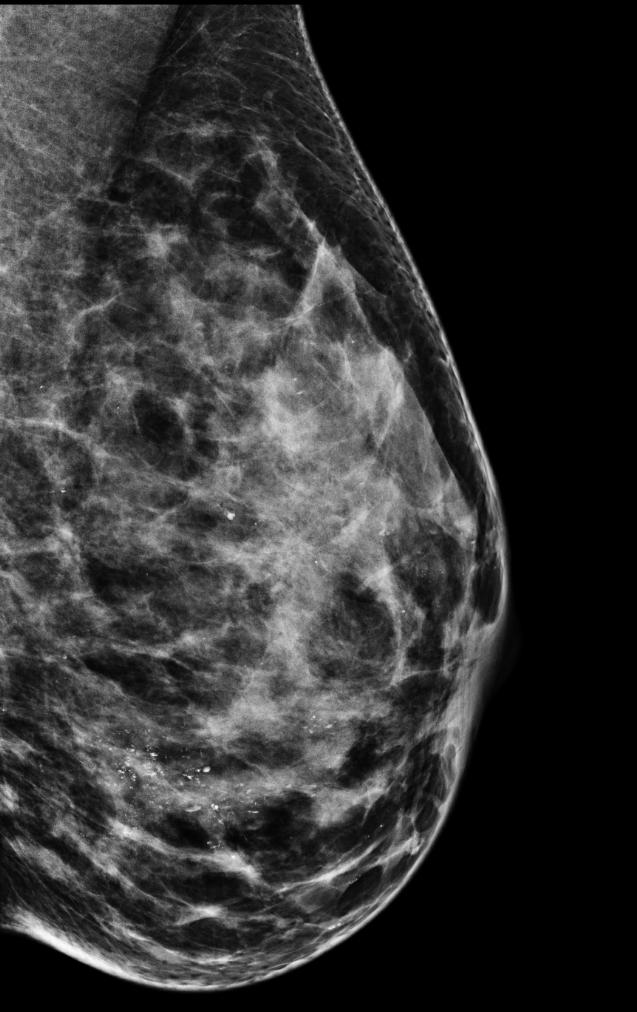
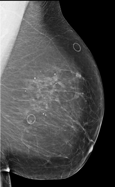
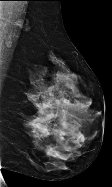
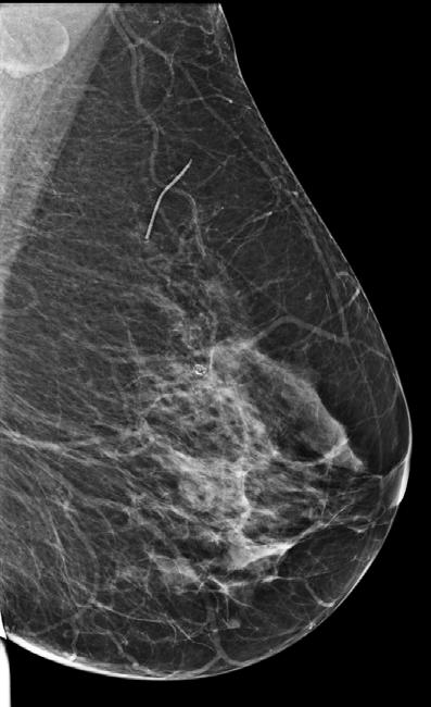
6 Breast Composition A. Fatty B. Scattered C. Heterogeneously Dense D. Extremely Dense 1, 2, 3, 4

7
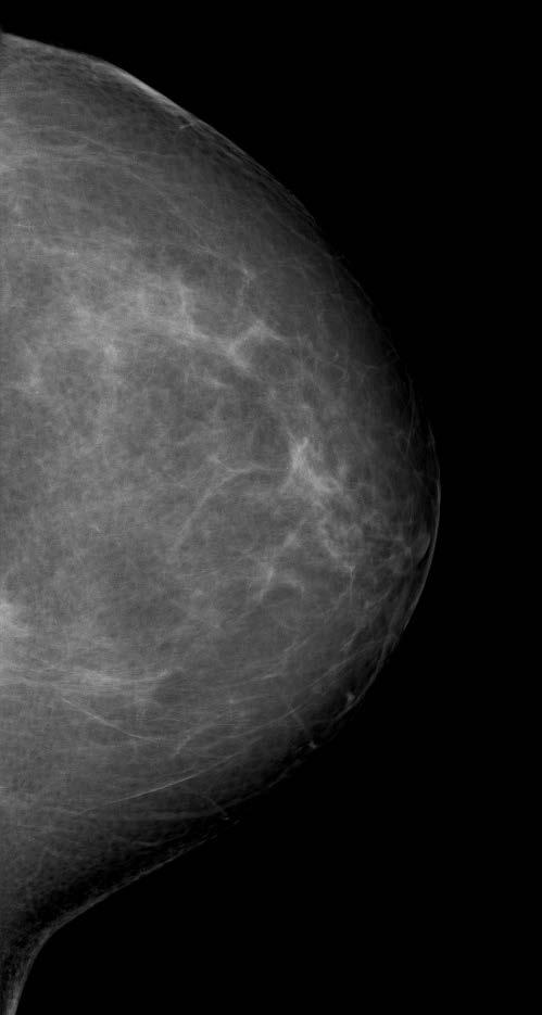
8 Breast Composition-A The breasts are almost entirely fatty The breast is almost entirely fat (<25% glandular)
9 Breast Composition-B There are scattered areas of fibroglandular density There are scattered fibroglandular densities (approximately 25-50% glandular)
10 Breast Composition-C The breasts are heterogeneously dense, which may obscure small masses The breast tissue is heterogeneously dense, which could obscure detection of small masses (approximately 51%-75% glandular)
11 Breast Composition-D The breasts are extremely dense, which lowers the sensitivity of mammography The breast tissue is extremely dense. This may lower the sensitivity of mammography (>75% glandular)

12 Mass Shape Margins

13 Masses-Shape Oval Round Irregular Lobulated

14 Masses-Margin (no change) Circumscribed Obscured Microlobulated Indistinct Spiculated

15 Masses-Density (no change) High Density Equal Density Low Density Fat Containing Use Asymmetry not Density when describing tissue that is asymmetric but not a mass
16 Calcifications Morphology Distribution
17 Morphology: Typically Benign Skin Vascular Coarse or Popcorn- Like Large Rod-Like Round <1mm Punctate <0.5mm Rim Dystrophic Milk of Calcium Suture

18 Rim Eggshell Lucent centered
19 Intermediate Concern (amorphous coarse heterogeneous)

20 Morphology: Suspicious Amorphous Coarse Heterogeneous Fine Pleomorphic Fine-linear or Finelinear branching

21 Suspicious Calcifications 4B Amorphous Coarse Heterogeneous Fine Pleomorphic 4C or 5 Fine-linear or Finelinear branching
22 Calcification Size Dystrophic Coarse Popcorn Coarse Heterogenous Fine Pleomorphic
23 Calcifications: Distribution Scattered Clustered Diffuse Regional >2cm Grouped 2cm Linear Segmental
24 Architectural Distortion
25 Asymmetries Asymmetry Area of fibroglandular tissue on 1 view Usually summation artifact Global Asymmetry At least one quadrant asymmetric fibroglandular tissue Usually normal variant
26
27 Asymmetries Focal Asymmetry 2 views, less than one quadrant 0.5-1% risk of malignancy if persists with no explanation after work up BI-RADS 3 on baseline study Developing asymmetry Focal asymmetry that is new or larger 15% risk of malignancy=bi-rads 4
28

29 Solitary Dilated Duct Rare Finding Consider additional imaging evaluation and tissue diagnosis unless benign etiology demonstrated
30 Lexicon ULTRASOUND

31 Masses-Shape Oval Round Irregular Lobulated

32 Masses-Margin Abrupt transition zone Circumscribed Angulated Microlobulated Spiculated Indistinct Echogenic transition zone

33 Masses-Orientation Parallel Not parallel Wider-than-tall Taller-than-wide

34 BIRADS 3 Solid, oval, circumscribed, parallel Complicated cyst Clustered microcysts
35 Tissue Composition-Screening Homogeneous background echotexture -Fat Homogeneous background echotexture -Fibroglandular tissue Heterogeneous background echotexture
36 Elasticity Soft Intermediate Hard

37 Lexicon MRI
38 Amount of Fibroglandular Tissue A. Almost entirely Fat B. Scattered areas of Fibroglandular Tissue C. Heterogeneous Fibroglandular Tissue D. Extreme Fibroglandular Tissue Density is a radiographic term (Mammography)

39 Background Parenchymal Enhancement Minimal Mild Moderate Marked
40 Finding-Focus, Mass, NME
41 Findings Current Lexicon Focus <5mm Mass any size Non-Mass Enhancement (NME) Retired Foci (as lesions ) Non-Mass-Like Enhancement
42 Mass (Nearly same as Mamm/US) Shape -round, oval, irregular Margins Circumscribed Non-Circumscribed Spiculated Irregular (no indistinct)


43 Focal area Linear Segmental Regional Diffuse NME Ductal

44 Internal Enhancement Patterns Homogeneous Heterogeneous Clumped (think pleomorphic) Clustered Ring Stippled, reticular/dendritic Central enahncement Enhancing septations
45 Kinetic Curve Assessment Fast >100% Medium % Slow <50% Persistent >10% Plateau Washout 10%
46 Category 0-Avoid! Need prior study Rare instance that mammo/us may confirm benign lymph node or fat necrosis
47 Category 3? New unique focus with benign morphology and kinetics Mass on initial exam with benign morphology and kinetics Not BPE, NME

48 Category 6 When known cancer actually seen on study Not for use when biopsy of additional area recommended
49 Reporting Guidance
50 Wording the Report Comparison to previous examination may be irrelevant Management recommendations have been separated from assessment categories to provide better flexibility

51 Location Clock Face, Quadrant Depth, Distance from the nipple
52 Assessment Categories 1. Negative 2. Benign 3. Probably Benign 4. Suspicious 5. Highly Suggestive of Malignancy
53 BI-RADS 3 Management 6 month follow-up until 1 year stability Then 12 months until 2-3 year stability Keep assessment BI-RADS 3 while in follow-up* * Unless resolves or becomes clearly benign
54 Concordant Management Recommendations 1,2 Age appropriate screening 3 Six or Twelve month Follow-up 4,5 Biopsy should be performed in the absence of clinical contraindication 6 Recommend surgical excision when clinically appropriate"
55 Discordant Imaging and Management Recommendations: Examples Suspicious nipple discharge, mammo and US negative. Known positive margins, MRI negative Post BCT imaging benign, on q 6 month mammography
56 Questions?

57 Thank You
Leonard M. Glassman MD
 BI-RADS The New BI-RADS Leonard M. Glassman MD FACR Former Chief of Breast Imaging American Institute for Radiologic Pathology Washington Radiology Associates, PC Breast Imaging Reporting and Data System
Imaging in breast cancer. Mammography and Ultrasound Donya Farrokh.MD Radiologist Mashhad University of Medical Since
 Imaging in breast cancer Mammography and Ultrasound Donya Farrokh.MD Radiologist Mashhad University of Medical Since A mammogram report is a key component of the breast cancer diagnostic process. A mammogram
S. Murgo, MD. Chr St-Joseph, Mons Erasme Hospital, Brussels
 S. Murgo, MD Chr St-Joseph, Mons Erasme Hospital, Brussels? Introduction Mammography reports are sometimes ambiguous and indecisive. ACR has developped the BIRADS. BIRADS consists of a lexicon in order
Amammography report is a key component of the breast
 Review Article Writing a Mammography Report Amammography report is a key component of the breast cancer diagnostic process. Although mammographic findings were not clearly differentiated between benign
ACRIN 6666 IM Additional Evaluation: Additional Views/Targeted US
 Additional Evaluation: Additional Views/Targeted US For revised or corrected form check box and fax to 215-717-0936. Instructions: The form is completed based on recommendations (from ID form) for additional
UW Radiology Review Course Breast Calcifications. BI-RADS 5 th Edition
 UW Radiology Review Course Breast Calcifications Grace Kalish, MD Vantage Radiology BI-RADS 5 th Edition Benign Skin Vascular Large rod like Coarse popcorn Suspicious Amorphous Coarse heterogenous Fine
Breast Imaging Lexicon
 9//201 200 BI RADS th Edition 201 BI RADS th Edition Breast Imaging Lexicon Mammographic Pathology and Assessment Categories Deborah Thames, R.T.(R)(M)(QM) The Advanced Health Education Center Nonmember:
Leonard M. Glassman MD Analysis of Breast Calcifications
 Importance of Calcification Leonard M. Glassman MD FACR American Institute for Radiologic Pathology Washington Radiology Associates, PC Washington DC 45% of all breast cancers present as calcification
MRI BI-RADS: How to make it out?
 MRI BI-RADS: How to make it out? Poster No.: C-1850 Congress: ECR 2016 Type: Educational Exhibit Authors: M. Ben Ammar, A. Ben Miled, O. Ghdes, S. Harguem, A. Gaja, N. Mnif; Tunis/TN Keywords: Breast,
ORIGINAL ARTICLE EVALUATION OF BREAST LESIONS USING X-RAY MAMMOGRAM WITH HISTOPATHOLOGICAL CORRELATION
 Available online at www.journalijmrr.com INTERNATIONAL JOURNAL OF MODERN RESEARCH AND REVIEWS IJMRR ISSN: 2347-8314 Int. J. Modn. Res. Revs. Volume 3, Issue 10, pp 807-814, October, 2015 ORIGINAL ARTICLE
8/31/2016 HIDING IN PLAIN SITE, ARCHITECTURAL DISTORTIONS AND BREAST ASYMMETRIES ARCHITECTURAL DISTORTIONS ARCHITECTURAL DISTORTIONS
 HIDING IN PLAIN SITE, ARCHITECTURAL DISTORTIONS AND BREAST ASYMMETRIES DEBORAH THAMES R.T. (R)(M)(QM) ARCHITECTURAL DISTORTIONS Definition is disruption of the natural flow of breast pattern towards the
Armed Forces Institute of Pathology.
 Armed Forces Institute of Pathology www.radpath.com Armed Forces Institute of Pathology Breast Disease www.radpath.org Armed Forces Institute of Pathology Evaluation of Breast Calcifications Leonard M.
AB MR Interpretation Overview
 AB MR Interpretation Overview Goal of AB MR interpretation is to maintain high sensitivity and specificity In order to minimize false positives and short term follow ups, it is fundamental to focus only
Armed Forces Institute of Pathology.
 Armed Forces Institute of Pathology www.radpath.com Armed Forces Institute of Pathology Breast Disease www.radpath.org Armed Forces Institute of Pathology Interpretation of Breast MRI Leonard M. Glassman
BI-RADS and Breast MRI. Kathy Borovicka, M.D. Thursday February 15, 2018
 BI-RADS and Breast MRI Kathy Borovicka, M.D. Thursday February 15, 2018 Learning Objectives Be familiar with the Breast Imaging Reporting and Data System (BI-RADS) Understand the components of a breast
Diagnostic Dilemmas of Breast Imaging
 Diagnostic Dilemmas of Breast Imaging Common Causes of Error in Breast Cancer Detection By: Jason Cord, M.D. Mammography: Initial Imaging The standard for detection of breast cancer Screening mammography
Lesion Imaging Characteristics Mass, Favoring Benign Circumscribed Margins Intramammary Lymph Node
 Lesion Imaging Characteristics Mass, Favoring Benign Circumscribed Margins Intramammary Lymph Node Oil Cyst Mass, Intermediate Concern Microlobulated Margins Obscured Margins Mass, Favoring Malignant Indistinct
Mammographic imaging of nonpalpable breast lesions. Malai Muttarak, MD Department of Radiology Chiang Mai University Chiang Mai, Thailand
 Mammographic imaging of nonpalpable breast lesions Malai Muttarak, MD Department of Radiology Chiang Mai University Chiang Mai, Thailand Introduction Contents Mammographic signs of nonpalpable breast cancer
Radiology Review Course Hotel del Coronado Coronado, California
 37 th Annual Radiology Review Course Hotel del Coronado Coronado, California Friday, April 21, 2017 - PM TABLE OF CONTENTS Friday, April 21, 2017 - PM SAM Session - Breast Imaging Update 12:45 PM 1:30
Ana Sofia Preto 19/06/2013
 Ana Sofia Preto 19/06/2013 Understanding the underlying pathophysiologic processes leading to the various types of calcifications Description and illustration of the several types of calcifications, according
PLACE LABEL HERE. ACRIN 6657 MRI Form: Pre-Treatment (MRI-1)
") M3 ACRIN 6657 MRI Form: Pre-Treatment (MRI-1) If this is a revised or corrected form,indicate by checking box. ACRIN Study 6657 Case # Instructions: In accordance with the protocol, four MRI exams are
M3 ACRIN 6657 MRI Form: Pre-Treatment (MRI-1) If this is a revised or corrected form,indicate by checking box. ACRIN Study 6657 Case # Instructions: In accordance with the protocol, four MRI exams are
Benign, Reactive and Inflammatory Lesions of the Breast
 Benign, Reactive and Inflammatory Lesions of the Breast Marilin Rosa, MD Associate Member Section Head of Breast Pathology Department of Anatomic Pathology Program Director, Breast Pathology Fellowship
Non-mass Enhancement on Breast MRI. Aditi A. Desai, MD Margaret Ann Mays, MD
 Non-mass Enhancement on Breast MRI Aditi A. Desai, MD Margaret Ann Mays, MD Breast MRI Important screening and diagnostic tool, given its high sensitivity for breast cancer detection Breast MRI - Indications
PLACE LABEL HERE BASELINE / PRE-TREATMENT. ACRIN 6657 Extension MRI Form: Baseline / Pre-Treatment MRI 1. o Unknown
 T1 ACRIN 6657 Extension MRI Form: Baseline / Pre-Treatment MRI 1 If this is a revised or corrected form, please box. ACRIN Study 6657 No. Instructions: In accordance with the protocol, four MRI exams are
OF HEMATOMA INTRACRANIAL HEMORRHAGE
 SCOPE TUTORIAL RADIOLOGY CT BRIAN BREASTS US MRCP Natrada Rawdhetubhai, M.D. Radiology Department, Lerdsin General Hospital CT OF HEMATOMA INTRACRANIAL HEMORRHAGE Intra-axial Extra-axial Intraventricular
Pitfalls and Limitations of Breast MRI. Susan Orel Roth, MD Professor of Radiology University of Pennsylvania
 Pitfalls and Limitations of Breast MRI Susan Orel Roth, MD Professor of Radiology University of Pennsylvania Objectives Review the etiologies of false negative breast MRI examinations Discuss the limitations
CDIS: what's beyond microcalcifications? - Pictorial essay
 CDIS: what's beyond microcalcifications? - Pictorial essay Poster No.: C-1096 Congress: ECR 2014 Type: Educational Exhibit Authors: R. N. Lucas, C. A. S. Ruano, I. Oliveira, J. M. G. Lourenco, Z. 1 1 1
Breast Cancer Screening with Mammography
 Progress in Public Health Breast Cancer Screening with Mammography JMAJ 44(7): 318 324, 2001 Tokiko ENDO Director, Department of Radiology, National Nagoya Hospital Abstract: Breast cancer has been increasing
MRI in breast cancer: diagnosis and intervention. Dr Sue Barter Addenbrookes Hospital, Cambridge UK
 MRI in breast cancer: diagnosis and intervention Dr Sue Barter Addenbrookes Hospital, Cambridge UK Intervention will be discussed in High Risk Screening! Indications UK and Europe: Breast MRI is well established
Hiding in Plain Sight / Site: Archictectural Distortions and Breast Asymmetries
 Hiding in Plain Sight / Site: Archictectural Distortions and Breast Asymmetries Dianne Georgian-Smith MD Associate Professor Harvard Med School Brigham and Women s Hospital Financial Disclosures Book Publication
Criteria of Malignancy. Evaluation Score
 30 5 Diagnostic Criteria Criteria of Malignancy Table 5.2 lists criteria in contrast-enhancing MR mammography that strongly indicate the presence of malignancy or are unspecific. Unifactorial evaluation
Ductal carcinoma in situ: ultrasound, mammography and MRI features with pathologic correlation
 Ductal carcinoma in situ: ultrasound, mammography and MRI features with pathologic correlation Poster No.: C-2252 Congress: ECR 2013 Type: Educational Exhibit Authors: L. Fernandes, H. A. M. R. Tinto,
Mammographically non-calcified ductal carcinoma in situ: sonographic features with pathological correlation in 35 patients
 Clinical Radiology (2009) 64, 628e636 ORIGINAL PAPER Mammographically non-calcified ductal carcinoma in situ: sonographic features with pathological correlation in 35 patients B. Mesurolle a, *, M. El-Khoury
Ultrasonography. Methods. Brief Description. Indications. Device-related Prerequisites. Technical Requirements. Evaluation Criteria
 1 Ultrasonography Brief Description Imaging modality using sound waves Tissue-specific wave reflection. Indications Evaluation of palpable breast nodules Evaluation of clinically occult mammographic findings
Breast calcification: Management and Pictorial Review
 Breast calcification: Management and Pictorial Review Poster No.: C-0692 Congress: ECR 2014 Type: Educational Exhibit Authors: V. de Lara Bendahan, M. F. Ramos Solis, A. Amador Gil, C. 1 2 3 2 4 4 Gómez
Pictorial Essay Singapore Med J 2009; 50(9) :
 :") 907 Pictorial Essay CME Article Breast calcifications: which are malignant? Muttarak M, Kongmebhol P, Sukhamwang N ABSTRACT Most calcifications depicted on mammograms are benign. However, calcifications
907 Pictorial Essay CME Article Breast calcifications: which are malignant? Muttarak M, Kongmebhol P, Sukhamwang N ABSTRACT Most calcifications depicted on mammograms are benign. However, calcifications
Pitfalls of Dynamic Contrast Enhanced MR Mammography (DCE-MRM) in Evaluation of Post-Biopsy Suspicious Breast Lesions
 in Evaluation of Post-Biopsy Suspicious Breast Lesions") Med. J. Cairo Univ., Vol. 86, No. 3, June: 1513-1522, 2018 www.medicaljournalofcairouniversity.net Pitfalls of Dynamic Contrast Enhanced MR Mammography (DCE-MRM) in Evaluation of Post-Biopsy Suspicious
Med. J. Cairo Univ., Vol. 86, No. 3, June: 1513-1522, 2018 www.medicaljournalofcairouniversity.net Pitfalls of Dynamic Contrast Enhanced MR Mammography (DCE-MRM) in Evaluation of Post-Biopsy Suspicious
The radiologic workup of a palpable breast mass
 Imaging in Practice CME CREDIT EDUCTIONL OJECTIVE: The reader will consider which breast masses require further workup and which imaging study is most appropriate Lauren Stein, MD Imaging Institute, Cleveland
Breast Ultrasound: Improving Your Skills & Patient Care
 Breast Ultrasound: Improving Your Skills & Patient Care Objectives Discuss US techniques available for image optimization. Review & compare the US appearances of benign & malignant masses. Cherie M. Kuzmiak,
Atypical Ductal Hyperplasia of the Breast:
 Atypical Ductal Hyperplasia of the Breast: Radiologic and Histopathologic Correlation 1 Ji Young Lee, M.D., Bo Kyoung Seo, M.D. 2, Jung Hyck Kim, M.D., Yu Whan Oh, M.D., Kyu Ran Cho, M.D., Eun Jeong Choi,
Practical and illustrated summary of updated BI-RADS for ultrasonography
 Practical and illustrated summary of updated I-RS for ultrasonography Jiyon Lee epartment of Radiology, NYU School of Medicine, NYU ancer Institute, reast Imaging enter, New York, NY, US The merican ollege
OPTO-ACOUSTIC BREAST IMAGING
 OPTO-ACOUSTIC BREAST IMAGING A Novel Fusion of Functional and Morphologic Imaging Reni S. Butler, MD A. Thomas Stavros, MD F. Lee Tucker, MD Michael J. Ulissey, MD PURPOSE 1. Explain opto-acoustic (OA)
Radiologic and pathologic correlation of non-mass like breast lesions on US and MRI: Benign, high risk, versus malignant
 Radiologic and pathologic correlation of non-mass like breast lesions on US and MRI: Benign, high risk, versus malignant Poster No.: C-1161 Congress: ECR 2013 Type: Educational Exhibit Authors: J. Kwak,
UNC Breast Imaging Division July 2018
 UNC Breast Imaging Division July 2018 This module is to educate residents on ACR BI-RADS Atlas 2 nd edition Ultrasound Sonomammographic anatomy Sonographic features most associated with malignancy Clinical
BI-RADS MRI: A Primer
 Erguvan- ogan et al. I- RS MRI Women s Imaging Pictorial Essay WOMEN S IMGING asak Erguvan-ogan 1 Gary J. Whitman 1 nne. Kushwaha 1,2 Michael J. Phelps 1,3 Peter J. empsey 1 Erguvan-ogan, Whitman GJ, Kushwaha,
Breast Imaging! Ravi Adhikary, MD!
 Breast Imaging! Ravi Adhikary, MD! ACS Estimated Cancers Statistics 2014! Breast! New Cases in Women! 232,670 (+67,570 in situ)! Deaths in Women! 40,000! Colon! 48,380! 24,040! Cervical! 12,360! 4,020!
BI-RADS 3 category, a pain in the neck for the radiologist which technique detects more cases?
 BI-RADS 3 category, a pain in the neck for the radiologist which technique detects more cases? Poster No.: B-0966 Congress: ECR 2013 Type: Scientific Paper Authors: J. Etxano Cantera, I. Simon-Yarza, G.
Alena Levit MD Avice O Connell MD University of Rochester, Rochester, NY
 Alena Levit MD Avice O Connell MD University of Rochester, Rochester, NY Purpose Review imaging spectrum of both common benign and malignant breast lesions Describe and demonstrate CT features with mammogram,
CURRENTLY IN THE United States, 431,284
 Increased Breast Calcifications in Women With ESRD on Dialysis: Implications for Breast Cancer Screening Mario Castellanos, MD, Seema Varma, MD, Kathleen Ahern, PhD, RN, Sue-Jane Grosso, MD, Shalom Buchbinder,
Imaging the Symptomatic Patient. Avice M.O Connell MD,FACR,FSBI Professor of Imaging Sciences Director, Women s Imaging University of Rochester
 Imaging the Symptomatic Patient Avice M.O Connell MD,FACR,FSBI Professor of Imaging Sciences Director, Women s Imaging University of Rochester The four most common symptoms Mass Pain Discharge Infection
MEDICAL IMAGING AND BREAST DISEASE HOW CAN WE HELP YOU?
 MEDICAL IMAGING AND BREAST DISEASE HOW CAN WE HELP YOU? Barbara M. Preston, M.D. SCREENING MAMMOGRAPHY AVERAGE RISK PATIENTS KAISER RECOMMENDATION: ALL WOMEN (INCLUDING TRANSGENDER FEMALES) Every 1-21
Index. C Calcifications fat necrosis 1, 61 fat necrosis 4, 69 nipple/peri-areolar involvement 1, 165
 A ADH. See Atypical ductal hyperplasia (ADH) American College of Radiology (ACR), BI-RADS background parenchymal enhancement, 8, 9, 81, 82 fibroglandular tissue guidelines, 6 American Joint Committee on
Intracystic papillary carcinoma of the breast
 Intracystic papillary carcinoma of the breast Poster No.: C-1932 Congress: ECR 2011 Type: Educational Exhibit Authors: V. Dimarelos, F. TZIKOS, N. Kotziamani, G. Rodokalakis, 1 2 3 1 1 1 2 T. MALKOTSI
Case Scenario 1 History and Physical 3/15/13 Imaging Pathology
 Case Scenario 1 History and Physical 3/15/13 The patient is an 84 year old white female who presented with an abnormal mammogram. The patient has a five year history of refractory anemia with ringed sideroblasts
AMSER Case of the Month: November 2018
 AMSER Case of the Month: November 2018 52 year old female with an abnormal screening mammogram Areeg Rehman, MS 4 Nova Southeastern University Rebecca T. Sivarajah, MD Penn State University College of
Triple-negative breast cancer: which typical features can we identify on conventional and MRI imaging?
 Triple-negative breast cancer: which typical features can we identify on conventional and MRI imaging? Poster No.: C-1862 Congress: ECR 2013 Type: Educational Exhibit Authors: V. Bertani 1, A. Gualano
Contrast-enhanced Breast MRI RSSA 2013
 Contrast-enhanced Breast MRI RSSA 2013 Prof. dr. Maurice van den Bosch University Medical Center Utrecht, the Netherlands Index 1) Breast cancer 2) Why MRI of the breast 3) Technique 4) Interpretation
Mimickers of breast malignancy
 Mimickers of breast malignancy Poster No.: C-0477 Congress: ECR 2010 Type: Educational Exhibit Topic: Breast Authors: S. H. Park, H.-Y. Choi; Incheon/KR Keywords: mimickers, breast malignancy, ultrasound
Breast MRI Update. Jeffrey C. Weinreb, MD, FACR Yale University School of Medicine
 Breast MRI Update Jeffrey C. Weinreb, MD, FACR jeffrey.weinreb@yale.edu Yale University School of Medicine I disclose the following financial relationships with relevant commercial interests: Bracco Bayer
AMSER Case of the Month: September 2018
 AMSER Case of the Month: September 2018 60-year-old woman with a left breast mass noted on screening mammography. Catherine McNulty, MS4 Tulane University School of Medicine Dr. Robin Sobolewski Breast
Pathologic outcomes of coarse heterogeneous calcifications detected on mammography
 Pathologic outcomes of coarse heterogeneous calcifications detected on mammography Poster No.: C-1957 Congress: ECR 2011 Type: Scientific Paper Authors: H. J. Lim, K. R. Cho, K. W. Hwang, B. K. Seo, O.
THYROID NODULES: THE ROLE OF ULTRASOUND
 THYROID NODULES: THE ROLE OF ULTRASOUND NOVEMBER 2017 DR. DEAN DURANT DEFINITION Thyroid nodule: Focal area within the thyroid gland with echogenicity different from surrounding parenchyma. THYROID NODULES
Case study 1. Rie Horii, M.D., Ph.D. Division of Pathology Cancer Institute Hospital, Japanese Foundation for Cancer Research
 NCCN/JCCNB Seminar in Japan April 15, 2012 Case study 1 Rie Horii, M.D., Ph.D. Division of Pathology Cancer Institute Hospital, Japanese Foundation for Cancer Research Present illness: A 50y.o.premenopausal
Invasive lobular carcinoma of the breast; spectrum of imaging findings.
 Invasive lobular carcinoma of the breast; spectrum of imaging findings. Poster No.: C-0847 Congress: ECR 2014 Type: Educational Exhibit Authors: D. Mandich, T. Diaz de Bustamante, L. Koren, M. Arroyo,
Sonographic Detection and Sonographically Guided Biopsy of Breast Microcalcifications
 Sonographic Detection and Sonographically Guided Biopsy of Breast Microcalcifications Mary Scott Soo 1 Jay A. Baker Eric L. Rosen OBJECTIVE. The purpose of this study was to evaluate the ability of sonography
ISSN X (Print) Research Article. *Corresponding author Dr. Amlendu Nagar
 Research Article. *Corresponding author Dr. Amlendu Nagar") Scholars Journal of Applied Medical Sciences (SJAMS) Sch. J. App. Med. Sci., 2015; 3(3A):1069-1073 Scholars Academic and Scientific Publisher (An International Publisher for Academic and Scientific Resources)
Scholars Journal of Applied Medical Sciences (SJAMS) Sch. J. App. Med. Sci., 2015; 3(3A):1069-1073 Scholars Academic and Scientific Publisher (An International Publisher for Academic and Scientific Resources)
BI-RADS Categorization As a Predictor of Malignancy 1
 Susan G. Orel, MD Nicole Kay, BA Carol Reynolds, MD Daniel C. Sullivan, MD BI-RADS Categorization As a Predictor of Malignancy 1 Index terms: Breast, biopsy, 00.1261 Breast neoplasms, localization, 00.125,
Evaluation of Breast Specimens Removed by Needle Localization Technique
 Evaluation of Breast Specimens Removed by Needle Localization Technique Specimen Handling: The breast specimen when received should be measured and grossly inspected for any orientation designated by the
Breast Imaging: Multidisciplinary Approach. Madelene Lewis, MD Assistant Professor Associate Program Director Medical University of South Carolina
 Breast Imaging: Multidisciplinary Approach Madelene Lewis, MD Assistant Professor Associate Program Director Medical University of South Carolina No Disclosures Objectives Discuss a multidisciplinary breast
Using T2-Weighted Sequences to More Accurately Characterize Breast Masses Seen on MRI
 Residents Section Pattern of the Month Westra et al. MRI of reast Masses Residents Section Pattern of the Month Downloaded from www.ajronline.org by 46.3.195.58 on 12/28/17 from IP address 46.3.195.58.
of Thyroid Lesions Comet Tail Crystals
 2 Ultrasound Features of Thyroid Lesions There are many different features indicating a certain benign or malignant tumor type, but many of these are overlapping signs. Combining several features is considered
Building Blocks for Effective Report Communication
 2016 SBI/ACR Breast Imaging Symposium Building Blocks for Effective Report Communication Richard L. Ellis, M.D. Breast Imaging Section Mayo Clinic Franciscan Health Care La Crosse, WI JW Marriott Austin
Radiologic Findings of Mucocele-like Tumors of the breast: Can we differentiate pure benign from associated with high risk lesions?
 Radiologic Findings of Mucocele-like Tumors of the breast: Can we differentiate pure benign from associated with high risk lesions? Poster No.: C-0332 Congress: ECR 2014 Type: Educational Exhibit Authors:
Fat Necrosis: A Grand Imposter
 Fat Necrosis: A Grand Imposter Poster No.: C-0751 Congress: ECR 2015 Type: Educational Exhibit Authors: L. C. Flores Salinas, Y. A. Ramirez Galvan, A. Garza Báez, C. M. Ferrara Chapa; Monterrey/MX Keywords:
Case-based discussion:
 Case-based discussion: Pailin Kongmebhol, M.D. Department of Radiology Faculty of Medicine Chiang Mai University There are many guidelines for managing thyroid nodules Two important guidelines: 2015 American
Detailed Program of the second BREAST IMAGING AND INTERVENTIONS PROGRAM am am : Clinician s requirements from breast imaging
 Detailed Program of the second BREAST IMAGING AND INTERVENTIONS PROGRAM 2012 Day one, 2 nd November BREAST IMAGING AND INTERVENTIONS PROGRAM 2012 9.00 AM 9.10 am Introduction 9.10 am - 9.30 am : Clinician
Intracystic Papillary Carcinoma of the Breast: Clinical and Radiological Findings with Histopathologic Correlation
 Intracystic Papillary Carcinoma of the Breast: Clinical and Radiological Findings with Histopathologic Correlation Poster No.: C-2078 Congress: ECR 2015 Type: Scientific Exhibit Authors: S. S. AlSharif
AACE/ACE Advanced Endocrine Neck Ultrasound Training Course 2016
 AACE/ACE Advanced Endocrine Neck Ultrasound Training Course 2016 This 9mm left inferior nodule should remind us all why we re here! There is no absolute number of images required for documentation
Cystic Masses of the Breast
 Residents Section Pattern of the Month Eisenberg ystic Masses of the reast Residents Section Pattern of the Month Residents inradiology Neely Hines 1 Priscilla J. Slanetz Ronald L. Eisenberg Hines N, Slanetz
Breast Health. Learning Objectives. Breast Anatomy. Poll Question. Breast Anatomy
 Learning Objectives Describe breast anatomy to a patient Breast Health Answer questions about causes of breast pain and masses Explain breast cancer screening/diagnostic modalities Appropriately triage
Segmental Breast Calcifications
 Residents Section Pattern of the Month Chen et al. Segmental reast Calcifications Residents Section Pattern of the Month Residents inradiology Po-Hao Chen 1 Erica T. Ghosh 1,2 Priscilla J. Slanetz 1,2
Standard Breast Imaging Modalities. Lilian Wang, M.D. Breast Imaging Section Department of Radiology Northwestern Medicine
 Standard Breast Imaging Modalities Lilian Wang, M.D. Breast Imaging Section Department of Radiology Northwestern Medicine Overview Standard breast imaging modalities Mammography Ultrasound MRI Imaging
Triple Receptor Negative Breast Cancer: Imaging and Clinical Characteristics
 Women s Imaging Original Research Krizmanich-Conniff et al. Triple Receptor Negative Breast Cancer Women s Imaging Original Research Kristin M. Krizmanich-Conniff 1 Chintana Paramagul 2 Stephanie K. Patterson
National Diagnostic Imaging Symposium 2013 SAM - Breast MRI 1
 National Diagnostic Imaging Symposium 2013 December 8-12, 2013 Disney s Yacht Club Resort Lake Buena Vista, Florida Self Assessment Module Questions, Answers and References Day SAM Title - Each SAM title
The role of MRI in assessment of asymmetrical breast densities
 The Egyptian Journal of Radiology and Nuclear Medicine (2010) 41, 501 508 Egyptian Society of Radiology and Nuclear Medicine The Egyptian Journal of Radiology and Nuclear Medicine www.elsevier.com/locate/ejrnm
Breast imaging of benign fat containing lesions
 Breast imaging of benign fat containing lesions Poster No.: C-1870 Congress: ECR 2017 Type: Educational Exhibit Authors: R. Aouini, I. Megdiche, D. Ben Hammadi, N. BEN MAMI, I. Attia, R. Neila, A. Zidi;
Role of Elastography in the Evaluation of Breast Lesions
 Original Article Radiology Section DOI: 10.7860/IJARS/2018/26510:2392 Role of Elastography in the Evaluation of Breast Lesions Annapurna Srirambhatla, Deepthi S, Balaji Vara Prasad, Prashanth Kumar KS
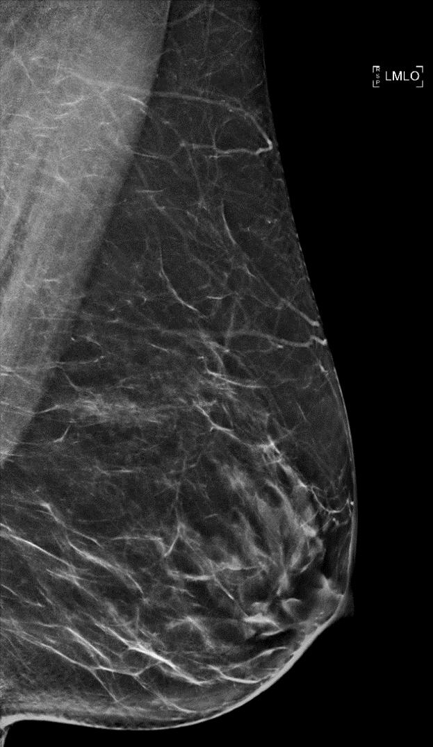
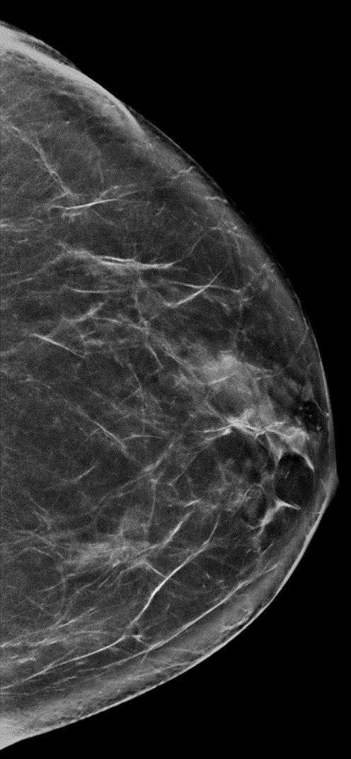
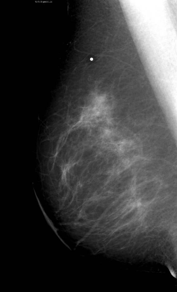
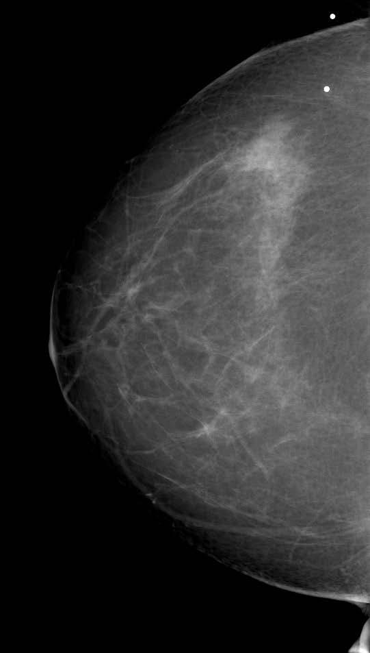
Here are examples of bilateral analog mammograms from the same patient including CC and MLO projections.
 Good afternoon. It s my pleasure to be discussing Diagnostic Breast Imaging over the next half hour. I m Wei Yang, Professor of Diagnostic Radiology and Chief, the Section of Breast Imaging as well as
Validation of the fifth edition BI-RADS ultrasound lexicon with comparison of fourth and fifth edition diagnostic performance using video clips
 Validation of the fifth edition BI-RADS ultrasound lexicon with comparison of fourth and fifth edition diagnostic performance using video clips Jung Hyun Yoon 1, Min Jung Kim 1, Hye Sun Lee 2, Sung Hun
AMSER Case of the Month: November 2018
 AMSER Case of the Month: November 2018 42 year old with right breast mass Rina Kiyota Petek Lake Erie College of Osteopathic Medicine, OMS-III Kossivi Dantey, MD Bibianna Klepchick, MD Matthew Hartman,
Positive Predictive Value of
 Note: This copy is for your personal non-commercial use only. To order presentation-ready copies for distribution to your colleagues or clients, contact us at www.rsna.org/rsnarights. Mary C. Mahoney,
Value of the BI-RADS classification in MR-Mammography for diagnosis of benign and malignant breast tumors
 Eur Radiol (2011) 21:2475 2483 DOI 10.1007/s00330-011-2210-7 BREAST Value of the BI-RADS classification in MR-Mammography for diagnosis of benign and malignant breast tumors Christian Sohns & Martin Scherrer
SIGNIFICANT OTHERS. Miscellaneous Benign Breast Conditions
 SIGNIFICANT OTHERS Miscellaneous Benign Breast Conditions Epworth HealthCare 1 FAT NECROSIS TRAUMATIC Cell rupture Seat-Belt injury Blunt trauma Iatrogenic injury Surgery, Flaps, Radiotherapy Pathology
Computer-Aided Detection and Diagnosis of Breast Abnormalities in Digital Mammography
 Computer-Aided Detection and Diagnosis of Breast Abnormalities in Digital Mammography Jelena Bozek, Kresimir Delac, Mislav Grgic University of Zagreb, Faculty of Electrical Engineering and Computing Department
Contents. Basic Ultrasound Principles and Terminology. Ultrasound Nodule Characteristics
 Contents Basic Ultrasound Principles and Terminology Basic Ultrasound Principles... 1 Ultrasound System... 2 Linear Transducer for Superficial Images and Ultrasound-Guided FNA... 3 Scanning Planes... 4
Policies, Standards, and Guidelines. Guidelines on Breast Ultrasound Examination and Reporting
 Policies, Standards, and Guidelines Guidelines on Breast Ultrasound Examination and Reporting Approved by Council June 2018 Approved: June 2018 Guidelines on Breast Ultrasound Examination and Reporting
CLINICAL SIGNIFICANCE OF BENIGN EPITHELIAL CHANGES
 Papillomas. Papillomas are composed of multiple branching fibrovascular cores, each having a connective tissue axis lined by luminal and myoepithelial cells ( Fig. 23-11 ). Growth occurs within a dilated
Evaluation of Abnormal Screening Mammograms
 342 Evaluation of Abnormal Screening Mammograms Ellen Shaw de Paredes, M.D. The purpose of routine screening mammography is to detect unsuspected cancer that has the potential to be cured. Abnormalities
Ultrasound of the Breast BASICS FOR THE ORDERING CLINICIAN
 Ultrasound of the Breast BASICS FOR THE ORDERING CLINICIAN Breast Ultrasound Anatomy Skin Breast Parenchyma Pectoralis Fascia Pectoralis Breast Ultrasound Anatomy Indications for Breast Ultrasound Palpable