Epworth Healthcare Benign Breast Disease Symposium. Sat Nov 12 th 2016
|
|
|
- Russell Neal
- 5 years ago
- Views:
Transcription

1 Epworth Healthcare Benign Breast Disease Symposium Breast cancer is common Sat Nov 12 th 2016 Benign breast disease is commoner, and anxiety about breast disease commoner still Breast Care Campaign UK commissioned a poll to enquire into women s knowledge and attitudes about breast health 47 % of the sample admitted to anxiety about their breasts 62 % were anxious about developing cancer, including 40 % of women in the age group, in whom cancer is rare
2 Over 90 % of patients who develop breast symptoms or signs have normal breasts or benign breast disease An understanding of the causes of benign breast conditions, their symptoms and their management will ensure that patients who have benign disease are treated correctly and are happy with their consultation Benign breast disease continues to cause considerable morbidity and anxiety, and with increasing patient awareness and expectation, the number of patients seeking referral for benign breast conditions is increasing Effective treatment includes making an accurate diagnosis followed by an adequate explanation of the condition and provision of relevant information related to both the diagnosis and how the condition is best managed Information given by word of mouth is easily forgotten, but can be backed up with a written leaflet

3 When a patient presents with a breast problem the basic question for the general practitioner is, Is there a chance that cancer is present, and, if not, can I manage these symptoms myself? Investigation of ANY breast symptom requires the diagnosis or exclusion of Breast Cancer 5 Almost every patient with a breast symptom fears that breast cancer is the cause 6 3

; and")
4 Triple Assessment of Breast Symptoms 12 practices identified as appropriate or inappropriate for the provision of breast cancer care in Australia. Not appropriate to confirm or exclude a diagnosis of breast cancer without undertaking the triple test, which involves: taking a patient history and clinical breast examination; imaging tests (mammogram and/or ultrasound); and biopsy to remove cells or tissue for examination 90% of breast symptoms are NOT due to CANCER 7 8 4

5 What do we know? >50% breast cancers found because of a breast change GP is the first port of call GP initiates the triple test by taking a history, performing a clinical examination and ordering appropriate imaging 10 5
 TRIPLE ASSESSMENT IS POSITIVE IF ANY COMPONENT IS INDETERMINATE, SUSPICIOUS OR")
6 Modern Diagnosis of Breast Disorders TRIPLE ASSESSMENT Clinical (History/Examination) Imaging (Mammography/Ultrasound +/- MRI) Biopsy (Fine Needle Aspiration/Core Biopsy) TRIPLE ASSESSMENT IS POSITIVE IF ANY COMPONENT IS INDETERMINATE, SUSPICIOUS OR MALIGNANT 6


7 Aims of Triple Assessment Maximise diagnostic accuracy Maximise preoperative diagnosis in breast cancer Minimise excisional biopsies for diagnosis Poor Incisions/ Poor Technique
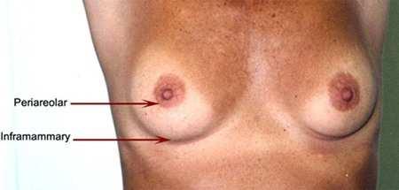
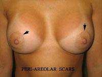
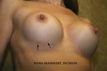
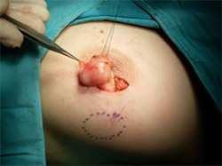
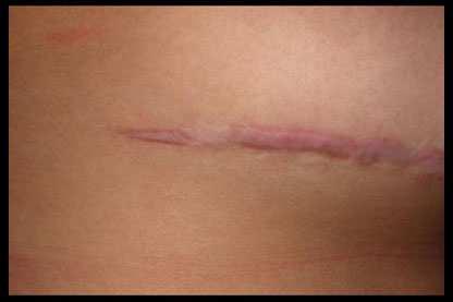
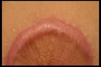
8 Recommended Incisions Poor Scarring
9 ALL palpable lumps require full triple assessment, irrespective of the perceived risk category of the patient. AIM Maximise diagnostic accuracy Maximise preoperative diagnosis in breast cancer Minimise excisional biopsies for diagnosis PROBABILITY OF DEVELOPING BREAST CANCER By age 25 By age30 By age 35 By age 40 By age 45 By age 50 By age 55 By age 60 By age 65 By age 70 By age 75 By age 80 By age 85 1 in 11,571 1 in 2,082 1 in in in in 54 1 in 32 1 in 22 1 in 16 1 in 13 1 in 11 1 in 9 1 in
10 If a woman is now AGE A woman has a 1 in 8 chance of being diagnosed with breast cancer by the age of 85 Her risk in the next 10 years is 1 in Age Number of new cases % New cases < % % % > % 75% breast cancers diagnosed in women over 50 10
11 Relative frequency of breast disorders Diffuse Nodularity Cyst Fibroadenoma For a triple test to be negative ALL three components: Clinical breast examination Medical Imaging Biopsy Must be benign or normal Cancer 11
12 The Triple Test Completion of the triple test is multidisciplinary: Clinician (GP/breast surgeon) Radiologist Pathologist The key is the review of all results and correlation with the presenting symptom One clinician must take responsibility The sensitivity or true positive rate of the triple test is 99.6%. That means that in women who have breast cancer, the triple test will detect that cancer in 99.6% of cases. The specificity of the triple test is 62%. That means that in women who have no cancer, you will get a normal result in 62% of cases. This gives a false positive rate of 38%. That means that in women who have no cancer, the triple test will be positive in 38% of cases. This is because we include those classified as indeterminate/equivocal in any one of the three components of the test as a positive outcome. For a test to be reliable in detecting an abnormality, you need a high sensitivity and thus the false positive rate will be high


13 HISTORY Sex Age Family History Personal Breast History Reproductive History Hormonal Treatment Previous Breast Imaging DOCUMENT ALL OF THE ABOVE
 Adopt a consistent")


14 CLINICAL BREAST EXAMINATION CLINICAL BREAST EXAMINATION (CBE) Adopt a consistent personal technique Always document findings Consider using standard 1-5 classification

15 Breast Imaging Women with breast symptoms should be referred for Diagnostic Imaging Assessment NOT to a Breast Screening Service Diagnostic vs Screening Mammography Screening mammography Patient should be asymptomatic Generally, the radiologist does not see films until the patient has left the radiology department Diagnostic mammography Patients with breast signs or symptoms (palpable lump, pain, nipple discharge) Patients with abnormality detected on screening mammogram Performed under the supervision of a radiologist Additional specialized mammographic views +/-U/S


16 Routine Mammography Consists of two images of each breast Craniocaudal (CC) Medial-lateral-oblique (MLO) 31 MLO 32 16


17 33 is ea se Sy CC m po si um /11/2016 Be ni gn Br ea st D Diagnostic Imaging Workup 34 17


18 Diagnostic Breast Imaging First imaging modality is ultrasound if: pt < age 35 years pregnancy and lactation (Add mammography if the lesion is suspicious on clinical or ultrasound examination) 35 >50 years: mammography and targeted ultrasound are complementary
19 Breast Density BIRADS Classification Breast Density Cannot be predicted based on physical exam Unrelated to breast size or consistency More common in younger women, during breast feeding, women using hormone replacement therapy 60% of women under 50 40% of women in their 50s 25% of women in their 60s have radiographically dense breasts 37 11/18/

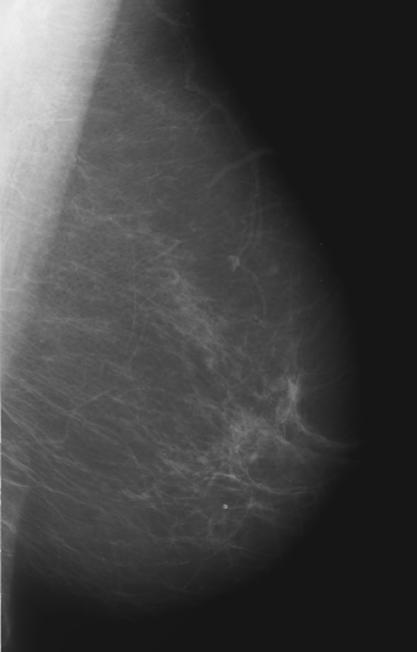
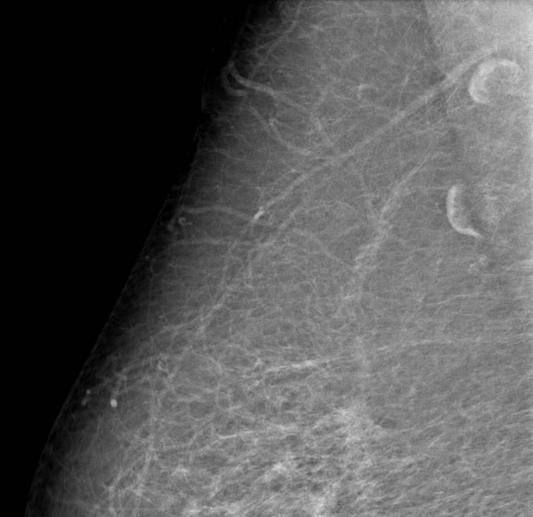
20 Mammographic Sensitivity 98% in women 50 with fatty breasts 30-69% sensitivity in women with dense breasts, particularly low if < 50 or at increased risk Kerlikowske et al JAMA 1996;276:33-38 Kolb et al Radiology 2002;225: Mandelson et al JNCI 2000;92:


21 Cancer in Fatty Breasts 11/18/2016 Cancer in Dense Breasts 41 11/18/


22 Digital Mammography Images captured electronically and stored digitally Digital images are viewed directly on a computer and radiologist can alter contrast / brightness / magnify without additional x-rays 44 22
23 Digital Mammography Digital mammography has been shown to be more accurate than film screen mammography in those: < 50 years of age Perimenopausal or premenopausal Dense breast tissue 45 Pisano E et al. N Engl J Med

24 Digital Breast Tomosynthesis A major factor contributing to the limited performance of mammography is the tissue superimposition that is created by the overlap of normal breast structures in a two-dimensional mammographic projection. These overlapping structures can obscure a lesion making it more difficult to perceive or rendering it completely mammographically occult. Tomosynthesis may improve cancer detection by mammography by enabling readers to detect lesions which are very difficult or impossible to visualize on conventional imaging due to overlying glandular tissue Tissue superimposition hides pathologies in 2D Tissue superimposition mimics pathologies in 2D 24


25 3D Principle of Operation X-ray tube moves in an arc across the breast A series of low dose images are acquired from different angles Total dose approximately the same as one 2D mammogram Projection images are reconstructed into 1 mm slices Reconstructed Slices { Arc of motion of x-ray tube, showing individual exposures BREAST ULTRASOUND Compression Paddle Compressed Breast Detector Housing 50 25
26 Breast Ultrasound Useful for the evaluation of: Palpable abnormality Mammographic abnormality Second look ultrasound very helpful for evaluating abnormal areas detected with MRI Surveillence following breast cancer surgery Ineffective in breast cancer screening (? even in those with dense breasts or high risk ) TAKE HOME MESSAGE ALL palpable lumps require full triple assessment, irrespective of the perceived risk category of the patient

27 Sensitivity (%) of Ultrasound and Mammography by age group in 975 cancers Age group Number of cases Ultrasound > Mammography The Breast, Surgeon Performed Ultrasound an Extension of the Clinical Examination sharpens your fingers -the stethoscope of the breast surgeon To confirm palpable lump corresponds to ultrasound abnormality (ie exclude dual pathology) Differentiate between solid and cystic lumps Management of breast abscesses Image assisted biopsies (FNA/core) **NOT a substitute for a formal diagnostic quality, radiologist reported ultrasound 54 27


28 Insist on Synoptic Imaging Reports Research indicates that synoptic reporting and more specifically the use of check lists is the most effective method of improving the content and completeness of reports. Classification 1. No significant abnormality 2. Benign findings 3. Indeterminate / equivocal findings 4. Suspicious findings of malignancy 5. Malignant findings When reporting the classification, both the number and the description of the classification should be stated
 MRI has a very")


29 Breast Magnetic Resonance Imaging (MRI ) MRI has a very high sensitivity and low specificity in screening. It picks up lots of abnormalities and many of them are not cancer False positive rate is high, leading to lots of unnecessary diagnostic tests

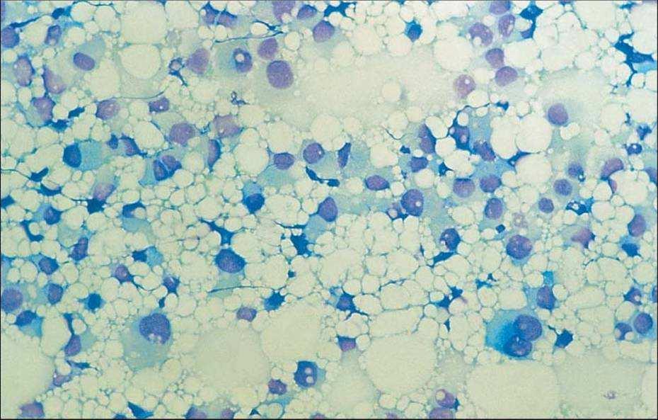
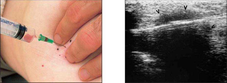
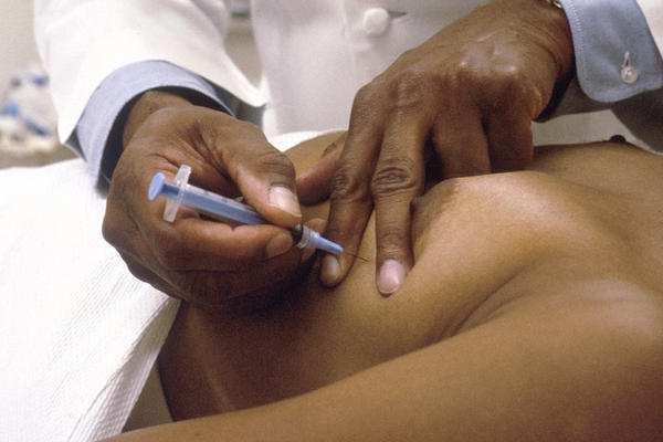
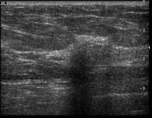
30 Needle Biopsy Fine Needle Aspiration ( FNA)

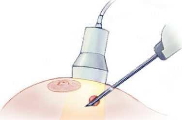
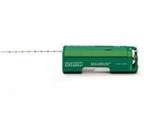
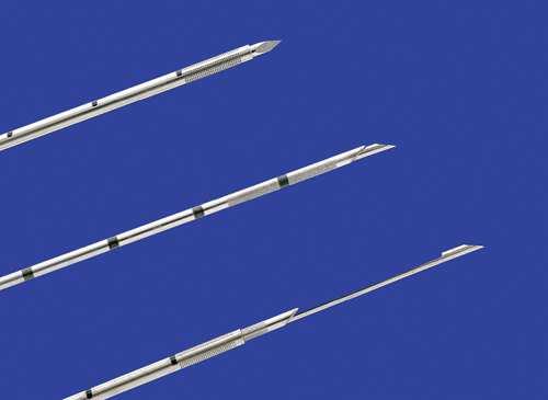
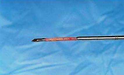
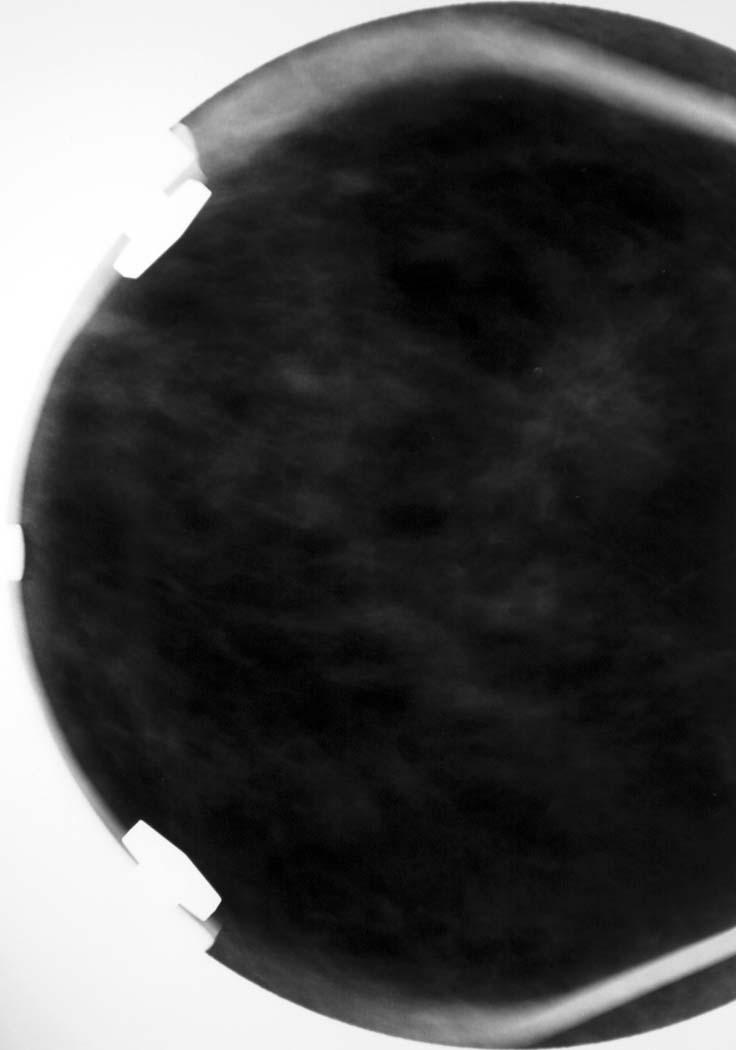
31 CORE BIOPSY


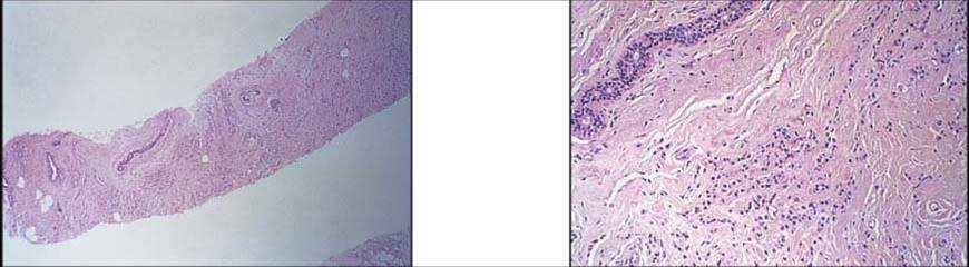
32 The sensitivity of needle biopsy is 90%, which may seem lower than expected This is largely due to sampling techniques ie the lesion is not accurately targeted/sampled on FNA or core biopsy It is vital that the results from biopsy correlate with both the clinical and imaging findings If this is done, the sensitivity increases to >99.6%

33 TAKE HOME MESSAGE ALL palpable lumps require full triple assessment, irrespective of the perceived risk category of the patient. MEDICOLEGAL CONSIDERATIONS The major source of litigation in Victoria 70% of all claims are breast related Breast cancer is the malignancy most often cited in delayed diagnosis claims against GPs 65 Single biggest risk for GPs 66 33

34 BREAST CANCER IS THE MALIGNANCY MOST OFTEN CITED IN DELAYED DIAGNOSIS CLAIMS AGAINST GPs Diagnostic delay is the most common cause of litigation for medico-legal negligence in the USA Reduced by strict adherence to protocol for triple assessment, failsafe mechanism for results and timescale for follow-up visits Nature of Claims False negative mammogram The patient complains of a lump but the clinician is not impressed by the findings (2nd most common scenario in medico-legal payouts) Poor cosmetic outcome following operation for benign breast disease False +ve cytology Failure to recommend adjuvant therapy
35 Nature of Claims 50% Failure to diagnose 26% Delay in diagnosis 9% 6% 3% 3% 3% Poor management Failure to warn Failure to manage Failure to advise result Failure to follow up 77% History of previous breast problems 41% Initial mammogram normal 41% Multiple GPs involved - holiday, locum, other GP in practice 27% Patient asked to return but did not 23% Breast feeding or pregnant 14% Patient returned but breast problem not followed-up 69 9% System to follow-up known malignancy failed 70 35
36 TAKE HOME MESSAGE ALL palpable lumps require full triple assessment, irrespective of the perceived risk category of the patient. For a triple test to be negative ALL three components: Clinical breast examination Medical Imaging Biopsy Must be benign or normal 71 36


37 RISK MANAGEMENT Ensure clear communication regarding risk Document ALL findings, results and discussions Adhere strictly to protocol for triple assessment Adopt a failsafe method for recall and follow up Thank you for your attention!
EARLY DETECTION: MAMMOGRAPHY AND SONOGRAPHY
 EARLY DETECTION: MAMMOGRAPHY AND SONOGRAPHY Elizabeth A. Rafferty, M.D. Avon Comprehensive Breast Center Massachusetts General Hospital Harvard Medical School Breast Cancer Screening Early detection of
EARLY DETECTION: MAMMOGRAPHY AND SONOGRAPHY
 EARLY DETECTION: MAMMOGRAPHY AND SONOGRAPHY Elizabeth A. Rafferty, M.D. Avon Comprehensive Breast Center Massachusetts General Hospital Harvard Medical School Breast Cancer Screening Early detection of
BREAST IMAGING and NEW IMAGING MODALITIES- A Surgeons view
 BREAST IMAGING and NEW IMAGING MODALITIES- A Surgeons view DR CHANTEL THORNTON SPECIALIST BREAST CANCER SURGEON BMSc (hons) MBBS (hons) FRACS Epworth Hospital, Richmond- Agora Centre for Women s Health
The Radiology Aspects
 REQUIREMENTS FOR INTERNATIONAL ACCREDITATION OF BREAST CENTERS/UNITS The Radiology Aspects Miri Sklair-Levy, Israel RADIOLOGY GUIDELINES FOR QUALITY ASSURANCE IN BREAST CANCER SCREENING AND DIAGNOSIS Radiologists
Breast Imaging & You
 Breast Imaging & You What s Inside: Breast Imaging... 2 Digital Breast Tomosynthesis (DBT) mammograms... 4 Breast cancer screening... 6 Dense breast tissue... 8 Automated Breast Ultrasound (ABUS)... 9
Breast Imaging & You
 Breast Imaging & You What s Inside: Breast Imaging... 2 Digital Breast Tomosynthesis (DBT) mammograms... 4 Breast cancer screening... 6 Dense breast tissue... 8 Automated breast ultrasound (ABUS)... 9
Melissa Hartman, DO Women s Health Orlando VA Medical Center
 Melissa Hartman, DO Women s Health Orlando VA Medical Center Most common non-skin cancer and Second deadliest cancer in women Majority are diagnosed by abnormal screening study An approach to breast cancer
Breast Cancer Screening and Diagnosis
 Breast Cancer Screening and Diagnosis Priya Thomas, MD Assistant Professor Clinical Cancer Prevention and Breast Medical Oncology University of Texas MD Anderson Cancer Center Disclosures Dr. Thomas has
1. Screening, Diagnosis and Surgical Management of Breast Cancer
 1. Screening, Diagnosis and Surgical Management of Breast Cancer Dr Melanie Walker, MBBS, FRACS (Breast Surgeon) Oncoplastic Breast Surgery Combination of optimal cancer surgery with plastic surgical techniques
Mammography. What is Mammography?
 Scan for mobile link. Mammography Mammography is a specific type of breast imaging that uses low-dose x-rays to detect cancer early before women experience symptoms when it is most treatable. Tell your
Breast Tomosynthesis. What is breast tomosynthesis?
 Scan for mobile link. Breast Tomosynthesis Breast tomosynthesis is an advanced form of mammography, a specific type of breast imaging that uses low-dose x-rays to detect cancer early when it is most treatable.
Breast Cancer Screening and High Risk
 Breast Cancer Screening and High Risk Mary Freyvogel, DO Breast Surgeon Clinical Assistant Professor of Surgery University Hospitals Case Medical Center St. John Medical Center / Elyria Medical Center
The radiologic workup of a palpable breast mass
 Imaging in Practice CME CREDIT EDUCTIONL OJECTIVE: The reader will consider which breast masses require further workup and which imaging study is most appropriate Lauren Stein, MD Imaging Institute, Cleveland
Standard Breast Imaging Modalities. Lilian Wang, M.D. Breast Imaging Section Department of Radiology Northwestern Medicine
 Standard Breast Imaging Modalities Lilian Wang, M.D. Breast Imaging Section Department of Radiology Northwestern Medicine Overview Standard breast imaging modalities Mammography Ultrasound MRI Imaging
Breast Imaging Update: Old Dog New Tricks
 Breast Imaging Update: Old Dog New Tricks Claire McKay, DO M&S Imaging Assoc. San Antonio, TX cmckayhart@juno.com Goals Describe modalities available, old and new Provide understanding of pros and cons
Financial Disclosures
 Financial Disclosures 3D Mammography: The Latest Developments in the Breast Imaging Arena I have no financial disclosures Dr. Katharine Lampen-Sachar Breast and Body Radiologist Radiology Associates of
Mammography. What is Mammography? What are some common uses of the procedure?
 Mammography What is Mammography? Mammography is a specific type of imaging that uses a low-dose x-ray system to examine breasts. A mammography exam, called a mammogram, is used to aid in the early detection
Breast imaging in general practice
 Breast series CLINICAL PRACTICE Breast imaging in general practice Nehmat Houssami, MBBS, FAFPHM, FASBP, PhD, is Associate Clinical Director, NSW Breast Cancer Institute, Westmead Hospital, Honorary Senior
Breast Cancer Imaging
 Breast Cancer Imaging I. Policy University Health Alliance (UHA) will cover breast imaging when such services meet the medical criteria guidelines (subject to limitations and exclusions) indicated below.
Policy Library Clinical Advantages of Digital Breast Tomosynthesis in Symptomatic Patients
 Policy Library Clinical Advantages of Digital Breast Tomosynthesis in Symptomatic Patients Version: 1 Approved by: Faculty of Clinical Radiology Council Date of approval: Click and type: day month and
Breast Health and Imaging Glossary
 Contact: Lorna Vaughan HerSpace Breast Imaging & Biopsy Associates 300 State Route 35 South W. Long Branch, NJ 07764 732-571-9100, ext. 104 lorna@breast-imaging.com Breast Health and Imaging Glossary Women
BreastScreen Victoria Annual Statistical Report
 BreastScreen Victoria Annual Statistical Report 005 Produced by: BreastScreen Victoria Coordination Unit Level, Pelham Street, Carlton South Victoria 05 PH 0 9660 6888 FX 0 966 88 EM info@breastscreen.org.au
Amammography report is a key component of the breast
 Review Article Writing a Mammography Report Amammography report is a key component of the breast cancer diagnostic process. Although mammographic findings were not clearly differentiated between benign
New Imaging Modalities for better Screening and Diagnosis
 New Imaging Modalities for better Screening and Diagnosis Miri Sklair-Levy, MD Department of Diagnostic Imaging Sheba Medical Center, Sackler School of Medicine, Tel Aviv University Department of Diagnostic
Current Strategies in the Detection of Breast Cancer. Karla Kerlikowske, M.D. Professor of Medicine & Epidemiology and Biostatistics, UCSF
 Current Strategies in the Detection of Breast Cancer Karla Kerlikowske, M.D. Professor of Medicine & Epidemiology and Biostatistics, UCSF Outline ν Screening Film Mammography ν Film ν Digital ν Screening
TOMOSYNTHESIS: WORTH ALL THE HYPE?
 X-Ray Associates of New Mexico, P.C. TOMOSYNTHESIS: WORTH ALL THE HYPE? MICHAEL N. LINVER, MD, FACR MAMMOGRAPHY: THE GOOD, THE PRETTY GOOD, & THE NOT SO GOOD MAMMOGRAPHY: THE GOOD, THE PRETTY GOOD, & THE
A GP S APPROACH TO BREAST LUMPS AND SYMPTOMS DR KK CHEUNG GPGC WORKSHOP
 A GP S APPROACH TO BREAST LUMPS AND SYMPTOMS DR KK CHEUNG GPGC WORKSHOP 18.08.18 HAVE A SYSTEM HISTORY EXAMINATION INVESTIGATION FOLLOW UP BREAST SYMPTOMS HISTORY DON T FORGET SKIN CHANGES AND NIPPLE CHANGES
Contrast-Enhanced Spectral Mammography
 Contrast-Enhanced Spectral Mammography Illuminating Breast Cancer Detection SenoBright HD TM gehealthcare.com/senobright Mammography is the most reliable imaging technique for breasts, but limitations
BR 1 Palpable breast lump
 BR 1 Palpable breast lump Palpable breast lump in patient 40 years of age or above MMG +/- spot compression or digital breast tomosynthesis over palpable findings Suspicious or malignant findings (BIRADS
Mammographic imaging of nonpalpable breast lesions. Malai Muttarak, MD Department of Radiology Chiang Mai University Chiang Mai, Thailand
 Mammographic imaging of nonpalpable breast lesions Malai Muttarak, MD Department of Radiology Chiang Mai University Chiang Mai, Thailand Introduction Contents Mammographic signs of nonpalpable breast cancer
Current issues and controversies in breast imaging. Kate Brown, South GP CME 2015
 Current issues and controversies in breast imaging Kate Brown, South GP CME 2015 JUDICIOUS USE OF RESOURCES IN REFERRALS FOR BREAST IMAGING THE DILEMMA How do target referrals for breast imaging? Want
Since its introduction in 2000, digital mammography has become
 Review Article Smith A, PhD email : Andrew.smith@hologic.com Since its introduction in 2000, digital mammography has become an accepted standard of care in breast cancer screening and has paved the way
Mammography and Other Screening Tests. for Breast Problems
 301.681.3400 OBGYNCWC.COM Mammography and Other Screening Tests What is a screening test? for Breast Problems A screening test is used to find diseases, such as cancer, in people who do not have signs
BreastScreen Queensland Gold Coast Service. General Practice Nurses Forum Thursday 4 May 2017
 BreastScreen Queensland Gold Coast Service General Practice Nurses Forum Thursday 4 May 2017 BreastScreen Queensland Gold Coast Service General Practice Nurses Forum Thursday 4 May 2017 BreastScreen in
WHAT TO EXPECT. Breast Tomosynthesis An additional screening tool in the fight against breast cancer HOLOGIC. The Women's Health Company
 WHAT TO EXPECT Breast Tomosynthesis An additional screening tool in the fight against breast cancer HOLOGIC The Women's Health Company ...,. Screening for breast cancer Doctors and scientists agree that
PAAF vs Core Biopsy en Lesiones Mamarias Case #1
 5/19/2014 PAAF vs Core Biopsy en Lesiones Mamarias Case #1 Fine Needle Aspiration Cytology of Breast: Correlation with Needle Core Biopsy 64-year-old woman Mass in breast Syed Hoda, MD CD31 Post-Radiation
BreastScreen Victoria Annual Statistical Report
 BreastScreen Victoria Annual Statistical Report 2010 BREASTSCREEN VICTORIA: ANNUAL STATISTICAL REPORT, 2010 Produced by: BreastScreen Victoria Coordination Unit Level 1, 31 Pelham Street, Carlton South
What s New in Breast Imaging. Jennifer A. Harvey, M.D., FACR Professor of Radiology University of Virginia
 What s New in Breast Imaging Jennifer A. Harvey, M.D., FACR Professor of Radiology University of Virginia Disclosure Hologic, Inc. Shareholder and research agreement. Volpara Solutions, Ltd. Shareholder
F r e q u e n t l y A s k e d Q u e s t i o n s. Mammograms
 Mammograms Q: What is a mammogram? A: A mammogram is a safe, low-dose x-ray exam of the breasts to look for changes that are not normal. The results are recorded on x-ray film or directly into a computer
Breast Cancer Screening
 Scan for mobile link. Breast Cancer Screening What is breast cancer screening? Screening examinations are tests performed to find disease before symptoms begin. The goal of screening is to detect disease
Dense Breasts, Get Educated
 Dense Breasts, Get Educated What are Dense Breasts? The normal appearances to breasts, both visually and on mammography, varies greatly. On mammography, one of the important ways breasts differ is breast
Benign phyllodes tumour
 Benign phyllodes tumour This leaflet tells you about benign phyllodes tumours. It explains what a benign phyllodes tumour is, and how it is diagnosed and treated. Benign breast conditions information provided
Updates in Mammography. Dr. Yang Faridah A. Aziz Department of Biomedical Imaging University Malaya Medical Centre
 Updates in Mammography Dr. Yang Faridah A. Aziz Department of Biomedical Imaging University Malaya Medical Centre Updates in Mammography Breast Imaging Dr. Yang Faridah A. Aziz Department of Biomedical
BIRADS 3 and 4 lesions viewed by ultrasound and not seen in digital mammograms and tomosynthesis.
 Original article Anales de Radiología México 2016 Jul;15(3):205-213. BIRADS 3 and 4 lesions viewed by ultrasound and not seen in digital mammograms and tomosynthesis. García-Quintanilla JF 1, González-Coronado
Corporate Medical Policy
 Corporate Medical Policy File Name: Origination: Last CAP Review: Next CAP Review: Last Review: digital_breast_tomosynthesis 3/2011 6/2016 6/2017 11/2016 Description of Procedure or Service Conventional
Dense Breasts. A Breast Cancer Risk Factor and Imaging Challenge
 Dense Breasts A Breast Cancer Risk Factor and Imaging Challenge Renee Pinsky, MD University of Michigan Department of Radiology Division of Breast Imaging No Disclosures QUIZ: ARE YOU DENSE? a. Breast
BREAST DENSITY WHAT IS IT? WHY IS IT IMPORTANT? & What IOWA SF250 Means to Patients and Providers
 BREAST DENSITY WHAT IS IT? WHY IS IT IMPORTANT? & What IOWA SF250 Means to Patients and Providers Arnold Honick, MD Radiology Consultants of Iowa, PLC ahonick@rciowa.com BREAST DENSITY LEGISLATION Nancy
Cairo/EG, Khartoum/SD, London/UK Biological effects, Diagnostic procedure, Ultrasound, Mammography, Breast /ecr2015/C-0107
 Role of sono-mammography in the evaluation of clinically palapble breast masses during pregnancy & lactation with differentaition between true patholgical & false physiological lobular hyperlpasia.sudanese
The London BreasT CenTre
 The London Breast Centre Contents Introduction 4 One-Stop Breast Care Clinic 5 Common breast problems 6 Hereditary breast cancer 8 Breast cancer and cancer screening 9 Breast diagnostic tests 10 Breast
Breast Tomosynthesis An additional screening tool in the fight against breast cancer
 What to Expect Breast Tomosynthesis An additional screening tool in the fight against breast cancer Every woman over 40 should be examined for breast cancer once a year. American Cancer Society What to
Breast Cancer: Selected Topics for the Primary Care Clinician
 Breast Cancer: Selected Topics for the Primary Care Clinician Leah Karliner, MD MAS October 2009 Primary Care Medicine: Principles and Practice OUTLINE Incidence and Mortality Risk Factors and Risk Reduction/Prevention
Jeddah Breast Cancer Pilot Screening Program, KSA
 Jeddah Breast Cancer Pilot Screening Program, KSA 7 th Global Summit on Cancer Therapy, Oct 5-7, 2015 Dubai, Crown Plaza Hotel Muna Baslaim, MD Consultant Surgeon Head of the Breast Unit, King Fahd General
ROLE OF MRI IN SCREENING, DIAGNOSIS AND MANAGEMENT OF BREAST CANCER. B.Zandi Professor of Radiology
 ROLE OF MRI IN SCREENING, DIAGNOSIS AND MANAGEMENT OF BREAST CANCER B.Zandi Professor of Radiology Introduction In the USA, Breast Cancer is : The Most Common Non-Skin Cancer The Second Leading cause of
Imaging in breast cancer. Mammography and Ultrasound Donya Farrokh.MD Radiologist Mashhad University of Medical Since
 Imaging in breast cancer Mammography and Ultrasound Donya Farrokh.MD Radiologist Mashhad University of Medical Since A mammogram report is a key component of the breast cancer diagnostic process. A mammogram
Breast Density. Information for Health Professionals
 Breast Density Information for Health Professionals BreastScreen NSW provides free screening mammography to asymptomatic women aged 50-74 every two years, with the aim of diagnosing breast cancer at an
BreastScreen Victoria Annual Statistical Report
 BreastScreen Victoria Annual Statistical Report CONTENTS FIGURES AND TABLES... Figures... Tables... 3 INTRODUCTION... 5 SECTION MAXIMISING PARTICIPATION... 6 Program acceptance... 6 Eligibility... 6 Inviting
FDA Executive Summary
 Meeting of the Radiological Devices Advisory Panel On October 24, 22, the panel will discuss, make recommendations, and vote on a premarket approval application supplement (P83/S) to expand the indications
CURRENT METHODS IN IMAGE GUIDED BREAST BIOPSY
 CURRENT METHODS IN IMAGE GUIDED BREAST BIOPSY Stuart Silver April 24, 2004 OBJECTIVES Review development of current techniques Discuss stereotactic breast biopsy Discuss US guided breast biopsy 1 OBJECTIVES
Breast Evaluation & Management Guidelines
 Breast Evaluation & Management Guidelines Pamela L. Kurtzhals, M.D. F.A.C.S. Head, Dept. of General Surgery Scripps Clinic, La Jolla Objective Review screening & diagnostic guidelines Focused patient complaints
Case 1. BREAST CANCER From Diagnosis to Treatment: The Role of Primary Care
 BREAST CANCER From Diagnosis to Treatment: The Role of Primary Care Leah Karliner, MD MAS University of California San Francisco Primary Care Medicine Update 2009 April 2009 Case 1 AR, a 60 year old African
CHAPTER 2 MAMMOGRAMS AND COMPUTER AIDED DETECTION
 9 CHAPTER 2 MAMMOGRAMS AND COMPUTER AIDED DETECTION 2.1 INTRODUCTION This chapter provides an introduction to mammogram and a description of the computer aided detection methods of mammography. This discussion
Screening Mammograms: Questions and Answers
 CANCER FACTS N a t i o n a l C a n c e r I n s t i t u t e N a t i o n a l I n s t i t u t e s o f H e a l t h D e p a r t m e n t o f H e a l t h a n d H u m a n S e r v i c e s Screening Mammograms:
Here are examples of bilateral analog mammograms from the same patient including CC and MLO projections.
 Good afternoon. It s my pleasure to be discussing Diagnostic Breast Imaging over the next half hour. I m Wei Yang, Professor of Diagnostic Radiology and Chief, the Section of Breast Imaging as well as
Breast Cancer Screening Clinical Practice Guideline. Kaiser Permanente National Breast Cancer Screening Guideline Development Team
 NATIONAL CLINICAL PRACTICE GUIDELINE Breast Cancer Screening Clinical Practice Guideline Kaiser Permanente National Breast Cancer Screening Guideline Development Team This guideline is informational only.
Q: Why is breast cancer a big deal?
 I hate breast cancer. As a radiologist who specializes in breast imaging, my career is devoted to the detection and diagnosis of breast cancer. I am passionate about women s health and my goal is to find
BreastScreen Victoria Annual Statistical Report
 BreastScreen Victoria Annual Statistical Report 29 BREASTSCREEN VICTORIA: ANNUAL STATISTICAL REPORT, 29 Produced by: BreastScreen Victoria Coordination Unit Level, 3 Pelham Street, Carlton South Victoria
Contrast-Enhanced Spectral Mammography helps to improve breast cancer diagnostics
 GE Healthcare Contrast-Enhanced Spectral Mammography helps to improve breast cancer diagnostics SenoBright TM mammography, Midwest U.S. hospital Simple test benefits clinicians and patients A hospital
Breast Cancer Screening and Treatment Mrs Belinda Scott Breast Surgeon Breast Associates Auckland
 Breast Cancer Screening and Treatment 2009 Mrs Belinda Scott Breast Surgeon Breast Associates Auckland BREAST CANCER THE PROBLEM 1.1 million women per year 410,000 deaths each year Increasing incidence
Tissue Breast Density
 Tissue Breast Density Reporting breast density within the letter to the patient is now mandated by VA law. Therefore, this website has been established by Peninsula Radiological Associates (PRA), the radiologists
International Day of Radiology 2016 Interview on Breast Imaging Australia / Dr. Michelle Reintals. Breast imaging in Australia
 International Day of Radiology 2016 Interview on Breast Imaging Australia / Dr. Michelle Reintals Breast imaging in Australia An interview with Dr. Michelle Reintals, Director of Breast at IMED Queensland
CURRENTLY FDA APPROVED ARE FULL FIELD DIGITAL MAMMOGRAPHY SYSTEMS AND FILM SCREEN STILL BEING USED AT SOME INSTITUTIONS
 ABBY DUROJAYE,M.D CURRENTLY FDA APPROVED ARE FULL FIELD DIGITAL MAMMOGRAPHY SYSTEMS AND FILM SCREEN STILL BEING USED AT SOME INSTITUTIONS BOTH HAVE BEEN SHOWN TO BE EFFECTIVE TOOLS EARLY DETECTION OF BREAST
Pitfalls and Limitations of Breast MRI. Susan Orel Roth, MD Professor of Radiology University of Pennsylvania
 Pitfalls and Limitations of Breast MRI Susan Orel Roth, MD Professor of Radiology University of Pennsylvania Objectives Review the etiologies of false negative breast MRI examinations Discuss the limitations
BARC/2013/E/019 BARC/2013/E/019. AUDIT OF MAMMOGRAPHY PERFORMED IN OUR HOSPITAL by Surita Kantharia Medical Division
 BARC/2013/E/019 BARC/2013/E/019 AUDIT OF MAMMOGRAPHY PERFORMED IN OUR HOSPITAL by Surita Kantharia Medical Division BARC/2013/E/019 GOVERNMENT OF INDIA ATOMIC ENERGY COMMISSION BARC/2013/E/019 AUDIT OF
Fundamentals of Breast Tomosynthesis
 Fundamentals of Breast Tomosynthesis Improving the Performance of Mammography Andrew Smith, Ph.D. This white paper is one in a series of research overviws on advanced technologies in women s healthcare.
Breast Cancer Diagnosis, Treatment and Follow-up
 Breast Cancer Diagnosis, Treatment and Follow-up What is breast cancer? Each of the body s organs, including the breast, is made up of many types of cells. Normally, healthy cells grow and divide to produce
Correlation between lesion type and the additional value of digital breast tomosynthesis
 Correlation between lesion type and the additional value of digital breast tomosynthesis Poster No.: C-1604 Congress: ECR 2011 Type: Scientific Exhibit Authors: C. Van Ongeval, L. Cockmartin, A. Van Steen,
Challenges to Delivery of High Quality Mammography
 Challenges to Delivery of High Quality Mammography Overview of Current Challenges Barbara Monsees, Washington University Geographic Access, Equity and Impact on Quality Tracy Onega, Dartmouth Medical School
Breast Imaging Donald L. Renfrew, MD
 This free educational material is provided by 333 N. Commercial Street, Suite 100, Neenah, WI 54956 Donald L. Renfrew, MD Breast cancer is the most frequent non-skin cancer diagnosis in women, with an
#46: DIGITAL TOMOSYNTHESIS: What is the Data Really Showing? TERMS (AKA) WHAT IS TOMOSYNTHESIS? 3/3/2014. Digital breast tomosynthesis =
 WHAT IS TOMOSYNTHESIS? 3/3/2014. Digital breast tomosynthesis =") #46: DIGITAL TOMOSYNTHESIS: What is the Data Really Showing? January K. Lopez, MD Hoag Breast Care Center Newport Beach, CA Disclosures: None TERMS (AKA) Digital breast tomosynthesis = DBT Tomo 3D Full
#46: DIGITAL TOMOSYNTHESIS: What is the Data Really Showing? January K. Lopez, MD Hoag Breast Care Center Newport Beach, CA Disclosures: None TERMS (AKA) Digital breast tomosynthesis = DBT Tomo 3D Full
DR AISHA A UMAR CHIEF CONSULTANT RADIOLOGIST NATIONAL HOSPITAL ABUJA.
 DR AISHA A UMAR CHIEF CONSULTANT RADIOLOGIST NATIONAL HOSPITAL ABUJA. OUTLINE WHY DO WE IMAGE WHOM TO IMAGE WHEN TO IMAGE HOW TO IMAGE WHAT TO IMAGE WITH PERSONAL EXPERIENCE CONCLUSION/RECOMMENDATIONS
Mammograms, Ultrasounds, MRI: Who gets what and why?
 Mammograms, Ultrasounds, MRI: Who gets what and why? Beast MRI Ultrasound Mammogram Kavita Dhamanaskar, MBBS, DNB, FRCP(C) Associate Professor @ McMaster University Radiologist @ Juravinski Hospital and
Plan. Lumps, Bumps, Leaking and Pain. Breast Cancer. Management of Breast Conditions. Palpable breast mass. Non-Palpable breast mass
 Lumps, Bumps, Leaking and Pain Management of Breast Conditions Rebecca A. Jackson, MD Obstetrics, Gynecology & Reproductive Sciences Epidemiology & Biostatistics University of California, San Francisco
Fat necrosis Benign breast conditions information
 Fat necrosis This leaflet tells you about fat necrosis. It explains what fat necrosis is, how it s diagnosed and what will happen if it needs to be followed up or treated. Benign breast conditions information
Benign Intraparenchymal Scarring in the DBT Era
 Benign Intraparenchymal Scarring in the DBT Era David Gruen, MD, MBA, FACR Director of Women s Imaging, Stamford Health, Stamford, CT With the advent of Digital Breast Tomosynthesis (DBT or 3D mammography),
Is Probably Benign Really Just Benign? Peter R Eby, MD, FSBI Virginia Mason Medical Center Seattle, WA
 Is Probably Benign Really Just Benign? Peter R Eby, MD, FSBI Virginia Mason Medical Center Seattle, WA Disclosures: CONSULTANT FOR DEVICOR MEDICAL ARS Question 1 Is probably benign really just benign?
A comparison of the accuracy of film-screen mammography, full-field digital mammography, and digital breast tomosynthesis
 Clinical Radiology xxx (2012) 1e6 Contents lists available at SciVerse ScienceDirect Clinical Radiology journal homepage: www.clinicalradiologyonline.net A comparison of the accuracy of film-screen mammography,
WHAT TO EXPECT. Genius 3D MAMMOGRAPHY Exam. The most exciting advancement in mammography in over 30 years
 WHAT TO EXPECT Genius 3D MAMMOGRAPHY Exam The most exciting advancement in mammography in over 30 years 91% of patients agree the quality of care provided by the facility was better with a Genius 3D MAMMOGRAPHY
Breast tomosynthesis reduces radiologist performance variability compared to digital mammography
 Breast tomosynthesis reduces radiologist performance variability compared to digital mammography Andrew Smith 1, Elizabeth Rafferty 2, Loren Niklason 1 1 Hologic, Inc., Bedford MA, USA 2 Massachusetts
THE BREAST CENTER AT MONTEFIORE NYACK HOSPITAL
 THE BREAST CENTER AT MONTEFIORE NYACK HOSPITAL COMPLETE BREAST CARE FROM THE TEAM THAT CARES I don t think I could get better care, more support, or encouragement at any of the bigger hospitals or cancer
Breast Magnetic Resonance Imaging (MRI) Westmead Breast Cancer Institute
 Westmead Breast Cancer Institute") Breast Magnetic Resonance Imaging (MRI) Westmead Breast Cancer Institute What is breast MRI? Breast MRI is a technique that uses a magnetic field to create an image of the breast tissue, using hundreds
Breast Magnetic Resonance Imaging (MRI) Westmead Breast Cancer Institute What is breast MRI? Breast MRI is a technique that uses a magnetic field to create an image of the breast tissue, using hundreds
Phyllodes tumours: borderline and malignant
 Phyllodes tumours: borderline and malignant This booklet is for people who would like more information about borderline or malignant phyllodes tumours. It describes what they are, the symptoms, how a diagnosis
A study on the usefulness of triple assessment in lumpy breasts in peri-menopausal women
 International Surgery Journal http://www.ijsurgery.com pissn 2349-3305 eissn 2349-2902 Research Article DOI: http://dx.doi.org/10.18203/2349-2902.isj20151249 A study on the usefulness of triple assessment
Digital Breast Tomosynthesis from a first idea to clinical routine
 International Master Programm Biomedical Engineering Digital Breast Tomosynthesis from a first idea to clinical routine Historical background 2D imaging of 3D objects has important limitations Jörg Barkhausen
Complete breast care from the team that cares. Breast Center
 Breast Center Complete breast care from the team that cares. Imaging Appointment: 845.348.8551 Surgical Consultation: 845.348.8507 nyackhospital.org/breastcenter 1 Complete breast care from the team that
Imaging Guidelines for Breast Cancer Screening
 Imaging Guidelines for Breast Cancer Screening Sarah Colwick, MD Dr. Sarah Colwick was born and raised in Sikeston, MO. She attended college and medical school at the University of Missouri-Kansas City
Intracystic papillary carcinoma of the breast
 Intracystic papillary carcinoma of the breast Poster No.: C-1932 Congress: ECR 2011 Type: Educational Exhibit Authors: V. Dimarelos, F. TZIKOS, N. Kotziamani, G. Rodokalakis, 1 2 3 1 1 1 2 T. MALKOTSI
ACCREDITATION DOCUMENT THE RADIOGRAPHER
 ACCREDITATION DOCUMENT THE RADIOGRAPHER Nijmegen, October 2012 1. Introduction An optimal quality of mammography is one of the fundamental requirements for successful breast cancer screening programmes.
Contrast-Enhanced Breast Tomosynthesis: Combining the Best of Both Worlds for Better Breast-Cancer Diagnosis
 Contrast-Enhanced Breast Tomosynthesis: Combining the Best of Both Worlds for Better Breast-Cancer Diagnosis T Wu (twu2@partners.org), E Rafferty, R Moore, D Kopans, Massachusetts General Hospital, Boston,
TMIST: Frequently Asked Questions
 TMIST: Frequently Asked Questions Key Topics for Site Investigators and Staff This document answers frequently asked questions about the Tomosynthesis Mammographic Imaging Screening Trial (TMIST/EA1151);
Emerging Techniques in Breast Imaging: Contrast-Enhanced Mammography and Fast MRI
 Emerging Techniques in Breast Imaging: Contrast-Enhanced Mammography and Fast MRI Lilian Wang, M.D. Breast Imaging Section Department of Radiology Northwestern Medicine Overview Rationale for new imaging