New Imaging Modalities for better Screening and Diagnosis
|
|
|
- Britton Nichols
- 6 years ago
- Views:
Transcription


1 New Imaging Modalities for better Screening and Diagnosis Miri Sklair-Levy, MD Department of Diagnostic Imaging Sheba Medical Center, Sackler School of Medicine, Tel Aviv University Department of Diagnostic Imaging Sheba Medical Center
2 Breast Cancer Statistics (Israel) Breast cancer is the most common female cancer About 4000 women are diagnosed with breast cancer each year and about 900 die of the disease Lifetime risk of developing breast cancer: 1 in 8 The earlier the disease is detected, the higher the chances of a cure - reaching nearly 90%. Breast cancer mortality rate is declining, probably due to improved screening and treatment
3 Breast Imaging Screening - asymptomatic women Diagnostic - work-up of a breast complaint or abnormal finding

4 Screening Mammography became clinically available in 1970 Mammography is the only screening test proven to reduce mortality FDA approved screening tool The only test proven to reduce mortality 15%-30%» Cancer. 2001;91(9):1724

5 MAMMOGRAPHY QUALITY CONTROL BI-RADS - The Breast Imaging Reporting and Data System - developed by the American College of Radiology To standardize the mammography report Findings, conclusions Mammography, extended for US, MRI The FDA mandates that all mammography reports should have the final BI-RADS assessment category
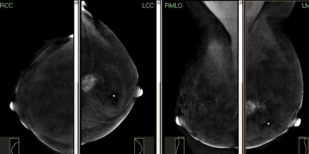
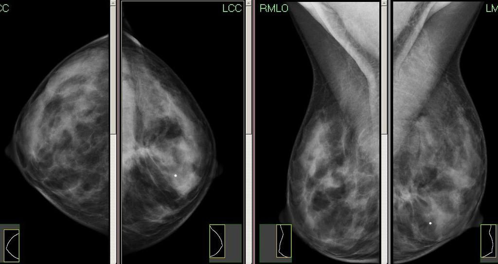
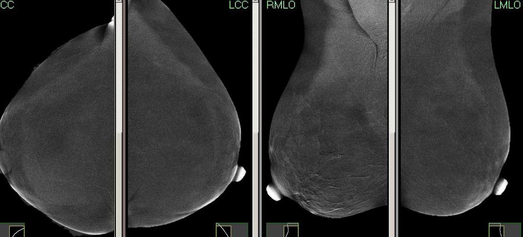
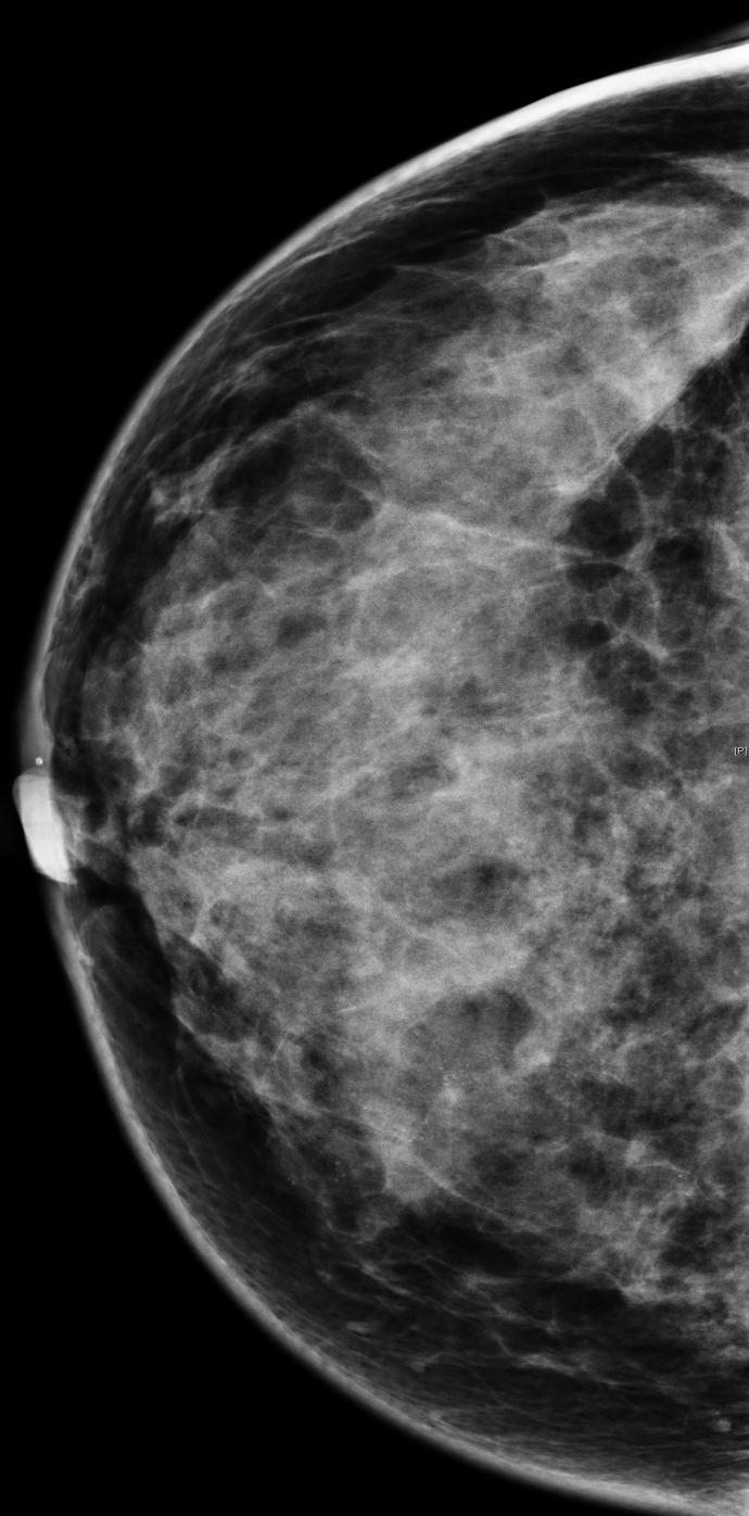
6 Mammography Limitations Dense breast Augmented breast Post op. breast
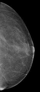
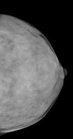
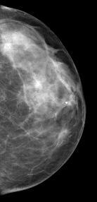
7 Mammography fatty dense
8 Mammography The prototype of the mammography unit was developed in Many technical advances have been made since then to improve Image quality To reduce the radiation dose Develop other breast imaging modalities To provide earlier diagnosis of breast disease More accurate assessment of disease extent and treatment response Improve the detection of recurrence
9 Diagnostic Performance of Digital versus Film Mammography for Breast-Cancer Screening Etta D. Pisano, M.D., Constantine Gatsonis, Ph.D., Edward Hendrick, Ph.D., Martin Yaffe, Ph.D., Janet K. Baum, M.D., Suddhasatta Acharyya, Ph.D., Emily F. Conant, M.D., Laurie L. Fajardo, M.D., Lawrence Bassett, M.D., Carl D'Orsi, M.D., Roberta Jong, M.D. and Murray Rebner, M.D. N Engl J Med Volume 353;17: October 27, 2005
10 Study Overview Study of 42,760 asymptomatic women The overall diagnostic accuracy of digital and film mammography as a means of screening for breast cancer was similar DIGITAL MAMMOGRAPHY was better Women under the age of 50 years Women with radiographically dense breasts Premenopausal or perimenopausal women
11 Full field digital mammography Advantages over film screen mammography Greater contrast resolution, especially in dense breasts. better visualization of skin and peripheral breast tissue The ability to post process the image by changing contrast and brightness, and by enlarging the image, increase the ability to detect subtle abnormalities. The ability to send images electronically (teleradiology). facilitating double reading The ability to store images in optical drives for future reference. Lower average radiation dose Eur J Radiol. 2007;64(3):419.
12 Computer-Aided Detection for Mammography - CAD
13 Computer-Aided Detection for Mammography Computer-aided detection (CAD) technology basically works like a second look. The computer marks abnormalities on the digitized films Detection, Diagnosis/classification The radiologist can decide whether the marked areas are suspicious and require further examination. The final interpretation is still made by the radiologist
14 Why do we need CAD Double reading could increase detection by - 15% Shortage of experienced radiologists The cost of true double reading
15 Computer-aided detection High Sensitivity - for the detection of cancers on screening mammograms. All cancers - 90% 86 88% for masses 98% for microcalcifications CAD has the potential to decrease the falsenegative rate from 31% to 19%
16 Computer-aided detection Specificity is a problem with CAD systems. CAD tends to mark a high number of "normal" areas as abnormalities High rate of false-positive The number will vary according to the level of sensitivity between 2 4 false prompts per study 1 - false prompt per image


17 69-year-old woman with invasive ductal carcinoma
18 CAD In Summary CAD may improve sensitivity of screening mammogram to a limited extent. Higher recall rate potential overdiagnosis. High costs associated with the equipment CAD has not been proven to improve mortality rates from breast cancer screening
19 Tomosynthesis
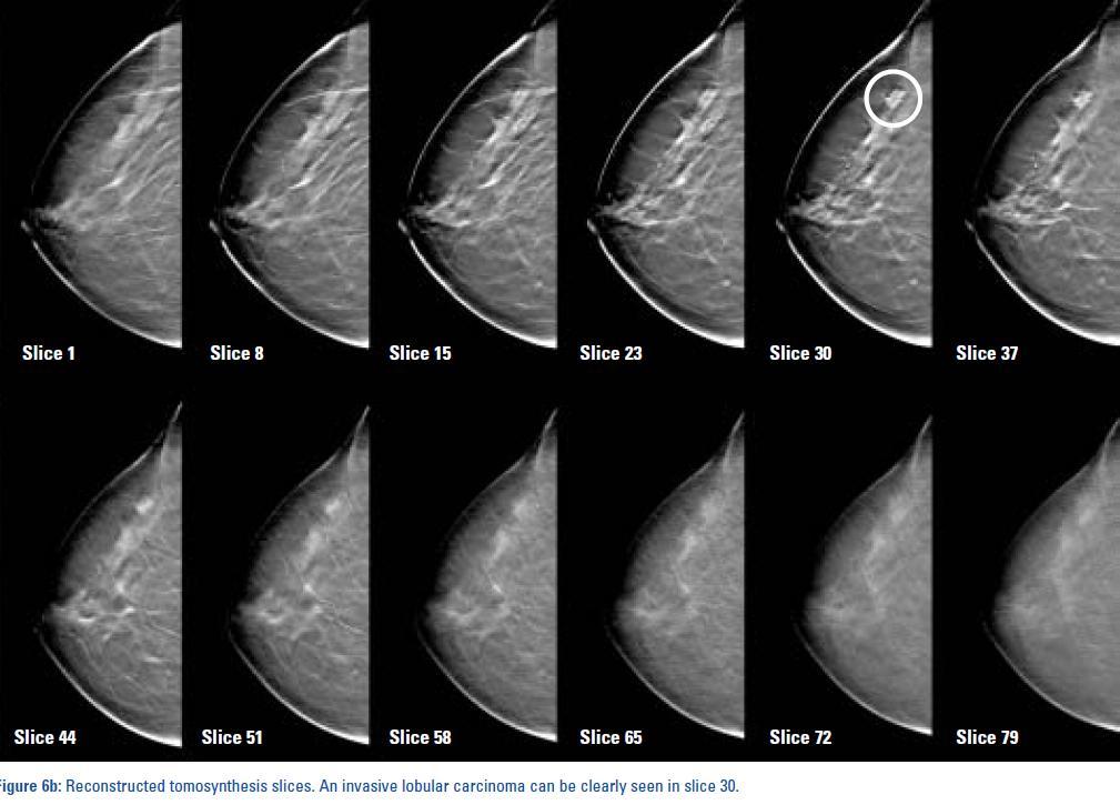
20 Tomosynthesis Tomosynthesis is a modification of digital mammography - moving x-ray source and digital detector A 3D volume of data acquired, reconstructed to thin sections Thin slice reconstruction improves the delineation of a lesion in the slice Reduce or eliminate the tissue overlap effect.
21
22 Screening setting Decrease recall rates Diagnostic setting Improves lesion characterization Tomosynthesis This technique shows promise in screening women with dense breast tissue and with high risk for breast cancer. Reading time twice for digital mammography. The examination longer exposure time of 10 seconds per acquisition compared to standard digital mammography, Increase the radiation dose Increase motion artifacts» AJR 2009;193(2):586» Eur Radiol. 2010;20(7):1545.» AJR. 2010;195(2):W172
23 Contrast-Enhanced Mammography
24 Dual Energy Contrast-Enhanced Mammography A pair of low- and high-energy images digital mammography system.( GE) Low energy exposures - conventional mammography kvp High energy - For iodine visualization kvp I.V ml/kg - iodine non-ionic contrast agent The total X-ray dose times the dose of a standard digital mammogram
25 Dual Energy Contrast-Enhanced Mammography 62 benign ; 80 malignant Sensitivity - MX+CEDM 93% ; MX - 78% No loss in specificity All 23 multifocal lesions were correctly detected by MX+CEDM vs. 16 and 15 lesions by MX and US respectively. Conclusion; Initial clinical results show that CEDM has better diagnostic accuracy than mammography alone and mammography+ultrasound Eur Radiol (2011) 21:


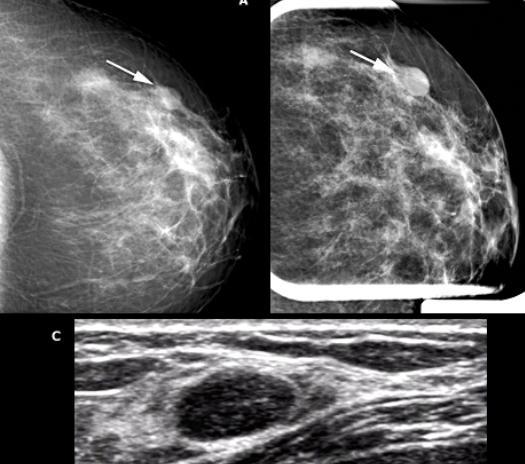
26 41y LT. breast palpable lump

27 43y LT. Axilla Lump
28 Dual Energy Contrast-Enhanced Mammography Diagnostic- identifying angiogenesis associated with a carcinoma in mammography. Problem solving in the case of equivocal mammography and ultrasound Advantage - fast imaging technique with immediate availability Analogous to contrast-enhanced MR imaging Screening - Potential to increase the cancer detection rate- dense breast
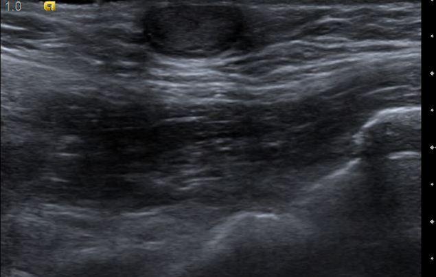
29 Breast Ultrasound


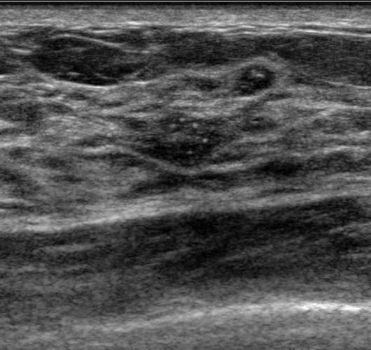
30 Breast US Indications The most useful adjunct to mammography Evaluation mammography findings cyst /solid Evaluation palpable lump (normal mammography) Breast examination in young women<30, pregnant,lactating Adjunct to mammography in dense breast US guided biopsy Not for screening?


31 Benign breast cysts Benign solid nodule


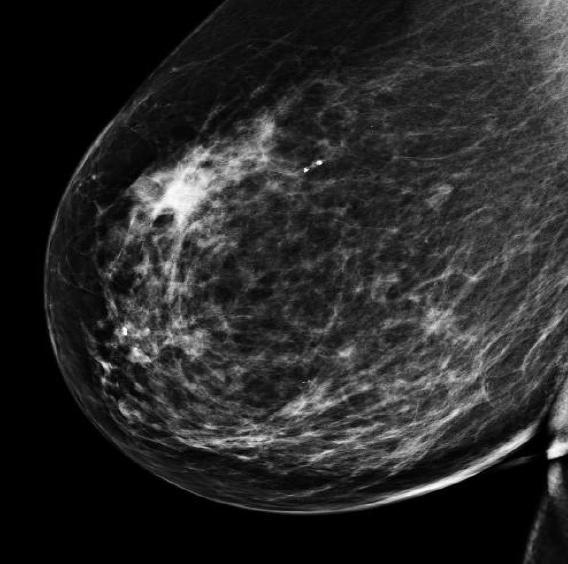
32 68 y, Lt. mastectomy, mammography reveals high breast density; carcinoma seen at ultrasound OCCULT CARCINOMA
33 Ultrasound And Screening Supplemental screening in women with dense breasts Limitation of mammography The recognition of the increased risk of breast cancer in women with dense breasts
34 US Screening? CHANGE 2007 US for screening breasts is an area of research 2011 Data support US screening plus mammography in women with dense breasts at high risk of breast cancer
35 Supplemental screening in women with dense breasts US screening of women with dense breasts Detects additional cancers per 1000 women screened False positives - biopsy positivity rate < 10% Radiology Improved breast cancer detection in asymptomatic women using 3D-automated breast ultrasound in mammographically dense breasts. Giuliano Clin Imaging. 2012
36 ELASTOGRAPHY ABVS Automated Breast Volume Scanner
37 US- elastography Palpation - assessment of shape and rigidity: US- elastography is a noninvasive imaging technique that can be used to depict relative tissue stiffness or displacement (strain) in response to a force Elastography = external compression (stress) deformation of the tissue (strain) The strain map of ultrasound elastography is superimposed on a conventional B-mode US


38 INVASIVE DUCT CARCINOMA 48 year palpable RT. Breast lump
39 Shear wave elasticity (SWE( Features measured with SW elastography; Quantitative elasticity kpa Reference values are not yet well established >50-70Kpa - malignant color scale linked to kpa Size ratios relative to B-mode imaging Shape at SW elastography Homogeneity of elasticity

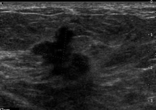
40 A 6-mm irregular hypoechoic mass in a 58-year-old Biopsy - infiltrating ductal carcinoma

41 An oval, circumscribed mass considered to be BI-RADS category 3 in a 67-women. Biopsy invasive ductal carcinoma.

42 Benign cyst - blue color : 0 kpa
43 In Summary Elastography Breast elastography is now an adjunct tool in breast ultrasonography. Easy to perform, short in a routine examination Applications Characterization of solid nodules BI-RADS 3 and BI-RADS 4a, in order to try to reduce unnecessary breast biopsies. Differentiation between solid and cystic lesions In the future - US elastography may be used to reduce biopsy rates for breast lesions


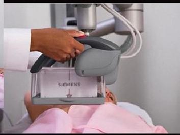
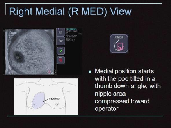
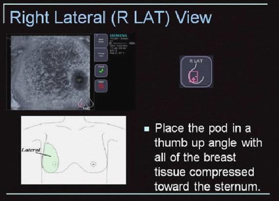
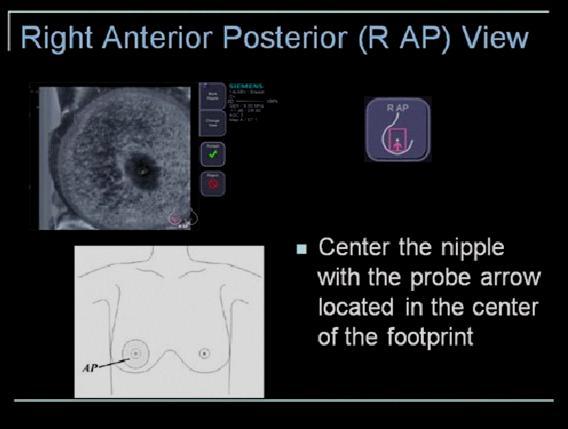
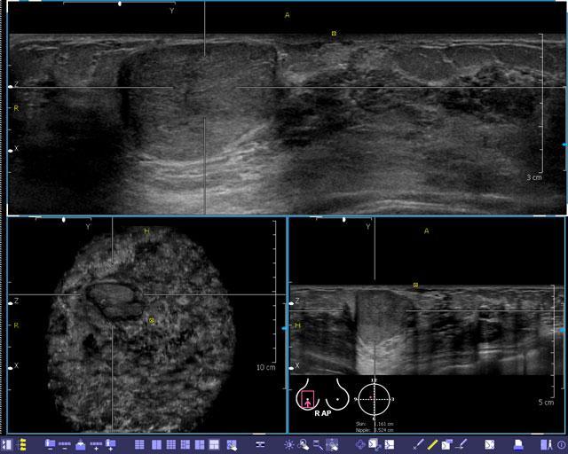
44 ABVS - Automated Breast Volume Scanner
45 US Ultrasound is a complementary technique to mammography Dense breasts and is part of the standard of care in diagnostic procedures. Manual US examinations time-consuming operator dependant Evaluation of already captured images.
46 ABVS Dedicated breast scanner - full-field volumes Nonphysician acquisition for 2 breasts in 15 min (60 seconds per view) Coronal, 3d full volume of the breast Conventional hand held US No operator dependent & variability - Scan consistent Improve clinical workflow Increased comfort for operator and patient
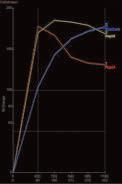
47 ABVS The FDA approved an automated ultrasound in Sep Used as an adjunct to mammography for asymptomatic women with dense breasts and a negative mammogram
48
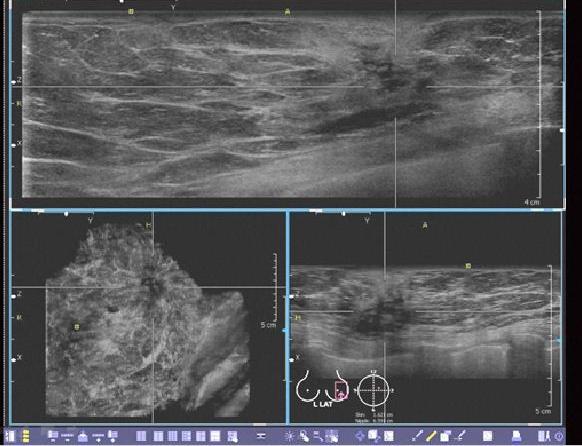
49 Fibroadenoma
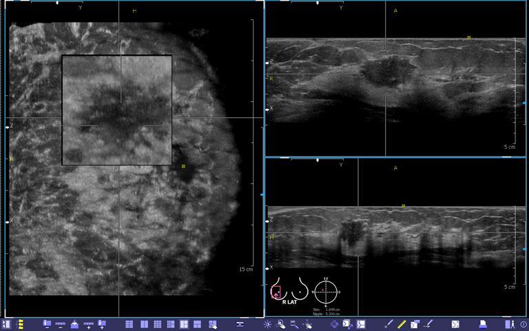
50 Carcinoma
51
52
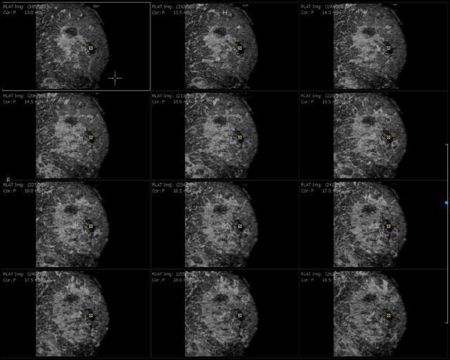
53 Breast MRI Mammography/US -are anatomic imaging The use of MRI for breast cancer detection is based on the concept of tumor angiogenesis or neovascularity Breast MRI relies on demonstrating the vascular characteristics of a lesion IV contrast. MRI evaluates morphology and enhancement patterns.
54 Vascular Characteristics Malignant lesions - angiogenesis, increased number of blood vessels Strong and fast enhancement Increased permeability of blood vessels and A-V shunts. Rapid washout of contrast
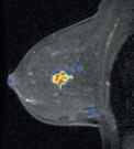
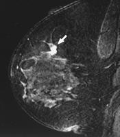
55 CADstream Angiogenesis Maps
56 Breast MRI Sensitivity % Specificity variable < 65% Overlap in the enhancement pattern of benign and malignant lesions» Acta Radiol. 2007;48(8):838» Top Magn Reson Imaging. 2008;19(3):143.
57 Indications for Breast MRI Diagnostic Extent of disease Pre-op Post lumpectomy r/o residual dis. - close or positive margins Response to neoadjuvant chemotherapy History of breast cancer r/o recurrence Search of occult primary with Ax LNs Silicone implant rupture Equivocal Exam Screening High risk/ Personal Hx of breast cancer

58 38 y- Family Hx. Mammography cluster microcalcificatoins

59 PREOP MRI Following MRI surgery changed from lumpectomy to mastectomy
60 Overview: Published Results Sensitivity MRI vs Mammography in Women with High Familial Risk for Breast Cancer AUTHOR # Pats # Ca Detected MRI Sensitivity-Specificity Mammography Sensitivity-Specificity Kuhl, % - 95% 32 % - 93% Tilanus-Lindthorst, % - 94% 0 % - NP Stoutjesdisjk, % - 93% 42 % - 96% Warner, % - 91% 28 % % Trecate, % - NP 0 % - NP Kriege, % - NP 40 % - NP Warner, % - 99% 36 % - 99% Leach, % - 81% 40 % - 93% Kuhl, % - NP 42 % - NP Kuhl, % - 97% 33 % - 97% Sardinelli, % - NP 59 % - NP Kuhl, Radiology 2007
61 Sensitivity in High-Risk Women Sensitivity of mammography: 28%-59% Sensitivity mammography + ultrasound + clinical breast examination: 49%-67% Sensitivity of MRI: 71%-100%


62 54 y, BRCA1, Rt mastectomy, screening MRI in left breast revealed 1 cm carcinoma not shown on mammography
63 Breast MRI Limitations Variable specificity - 30%-85% May lead to high false positive rate MRI-guided biopsy for lesions seen only on MRI

64 MRI-Guided Breast Biopsy Obtain a histologic diagnosis of lesions detected on MRI
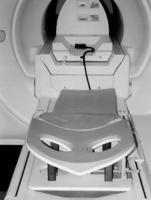
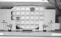
65 Nuclear Breast Imaging Functional breast imaging techniques Breast specific gamma imaging BSGI - Breast scintigraphy with 99mTc-SestaMIBI Positron emission mammography PEM - with 18F-2-deoxy-2-fluoro-D-glucose (FDG)


66 Breast Specific Gamma Imaging-BSGI Gamma camera in a mammographic configuration to provide high-resolution, functional images Image in positions comparable to mammography
67 Breast Specific Gamma Imaging-BSGI In several observational studies- compared to MRI Sensitivity - equal MRI -96% Sensitivity % for the detection of DCIS Specificity greater than MRI 59.5%» Radiology 2005;237(1):274» Radiology 2008;247(3):651» AJR 2009;192(2):379
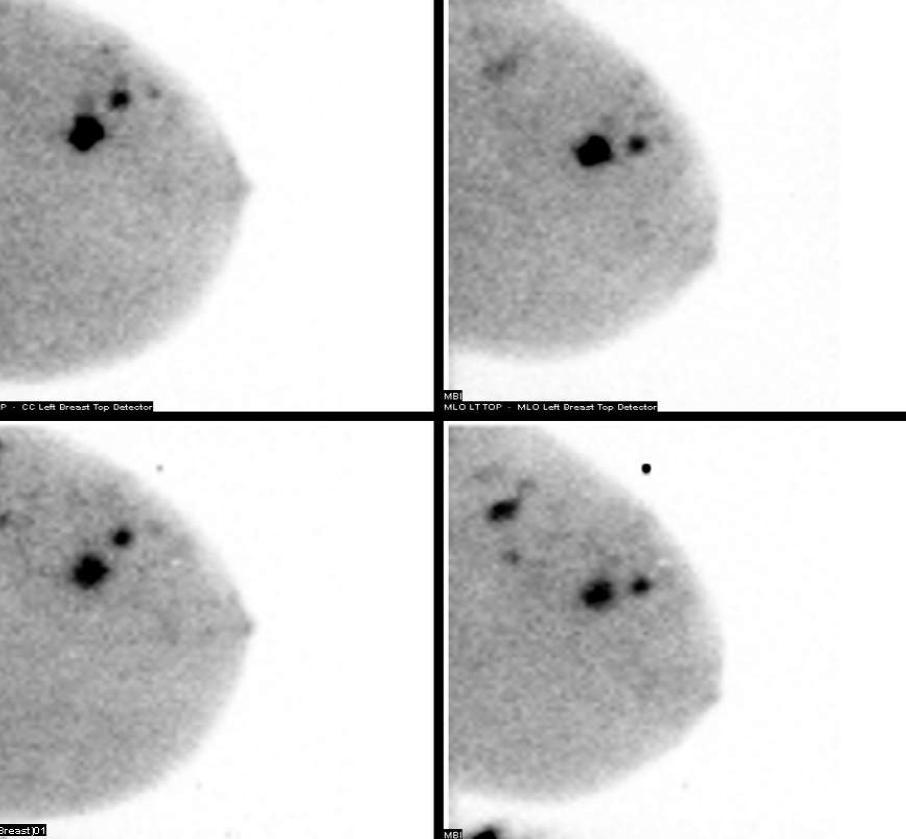
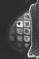
68 Mammography: Detected 2cm - MBI: Multiple foci of increased uptake
69 Nuclear Breast Imaging Diagnostic Indications similar to of breast MRI Extent of disease - preoperative assessment High risk screening Monitoring response to therapy About 15% of women for whom MRI is indicated, do not undergo the procedure for various reasons: Pacemaker Obesity can t lie down for the required period of time Claustrophobic renal problems. Screening-Not for screening - Radiation exposure and radiationrelated risks

70 PET -Positron emission tomography PET is primarily used as a modality to delineate the presence and/or extent of malignancy in patients known to have or suspected of having tumors
71 PET High sensitivity and specificity for tumors >8 mm in diameter clinically palpable and sometimes readily evident with conventional imaging. Limitations Accuracy is lower for non-palpable tumors or lesions smaller than 8 mm in size. FDG uptake can be poor in some well differentiated tumors and in lobular carcinomas

72 Dedicated PET Mammography The compression paddle from the mammography unit provides mild compression against the lower PET mammography detector. Less compression is used with the PET mammography unit than with conventional mammography
73 Positron emission mammography (PEM) High resolution- modification of PET 2 mm in-plane resolution Improve detection of small malignancies Sensitivity 86 91% Specificity 91-93%» Breast J. 2006;12(4):309 Indications Diagnostic-Preoperative assessment of disease extent NOT for screening PEM is still investigational.


74 41-year-old woman with a 1.8-cm mass in the left breast US - solid mass PET mammography image - single focus of increased FDG activity at the site of the mass. Pathology cm invasive ductal carcinoma
75 In summary Screening - Mammography Adjunct US (ABVS) Other modalities : NOT YET, Research Tomosynthesis, CEM, Population based screening anatomic / functional Diagnostic Not just mammography Multi-modality optimal breast cancer diagnosis Personalized medicine - Specific for women US, Automated 3D US, Contrast mammography, tomosynthesis, MRI, Molecular Breast Imaging
76 In summary Screening Mammography limitations Maybe reconsider as a single modality Women targeted approach Risk assessment Breast density Adjunct modalities US CEDM Tomosynthesis MRI Molecular breast imaging

77 Thank You
78 In summary Screening Diagnostic Mammography Ultrasound Mammography Contrast mx tomosynthesis US, ABVS,elastography ContrastMX Tomosynthesis MRI BRCA MRI Molecularbreast imaging(nuclearmedicine)
Breast Cancer Imaging
 Breast Cancer Imaging I. Policy University Health Alliance (UHA) will cover breast imaging when such services meet the medical criteria guidelines (subject to limitations and exclusions) indicated below.
Dr Robin Wilson, The Royal Marsden
 Screening: State of the Art High risk and dense breasts Robin Wilson Smart Breast Screening? 1 in 8 women in the will get breast cancer 8 in 9 will not 55% of breast cancers are not screen detected One
Here are examples of bilateral analog mammograms from the same patient including CC and MLO projections.
 Good afternoon. It s my pleasure to be discussing Diagnostic Breast Imaging over the next half hour. I m Wei Yang, Professor of Diagnostic Radiology and Chief, the Section of Breast Imaging as well as
Breast Imaging Update: Old Dog New Tricks
 Breast Imaging Update: Old Dog New Tricks Claire McKay, DO M&S Imaging Assoc. San Antonio, TX cmckayhart@juno.com Goals Describe modalities available, old and new Provide understanding of pros and cons
Emerging Techniques in Breast Imaging: Contrast-Enhanced Mammography and Fast MRI
 Emerging Techniques in Breast Imaging: Contrast-Enhanced Mammography and Fast MRI Lilian Wang, M.D. Breast Imaging Section Department of Radiology Northwestern Medicine Overview Rationale for new imaging
Updates in Mammography. Dr. Yang Faridah A. Aziz Department of Biomedical Imaging University Malaya Medical Centre
 Updates in Mammography Dr. Yang Faridah A. Aziz Department of Biomedical Imaging University Malaya Medical Centre Updates in Mammography Breast Imaging Dr. Yang Faridah A. Aziz Department of Biomedical
Standard Breast Imaging Modalities. Lilian Wang, M.D. Breast Imaging Section Department of Radiology Northwestern Medicine
 Standard Breast Imaging Modalities Lilian Wang, M.D. Breast Imaging Section Department of Radiology Northwestern Medicine Overview Standard breast imaging modalities Mammography Ultrasound MRI Imaging
The Radiology Aspects
 REQUIREMENTS FOR INTERNATIONAL ACCREDITATION OF BREAST CENTERS/UNITS The Radiology Aspects Miri Sklair-Levy, Israel RADIOLOGY GUIDELINES FOR QUALITY ASSURANCE IN BREAST CANCER SCREENING AND DIAGNOSIS Radiologists
Does elastography change the indication to biopsy? IBDC
 Does elastography change the indication to biopsy? A LEXANDRA A THANASIOU, M D DEPARTMENT OF RADIOLOGY CURIE INSTITUTE PARIS, FRANCE IBDC Ultrasound Detected Cancers Physician-performed ultrasound increases
Epworth Healthcare Benign Breast Disease Symposium. Sat Nov 12 th 2016
 Epworth Healthcare Benign Breast Disease Symposium Breast cancer is common Sat Nov 12 th 2016 Benign breast disease is commoner, and anxiety about breast disease commoner still Breast Care Campaign UK
BREAST MRI. VASILIKI FILIPPI RADIOLOGIST CT MRI & PET/CT Departments Hygeia Hospital, Athens, Greece
 BREAST MRI VASILIKI FILIPPI RADIOLOGIST CT MRI & PET/CT Departments Hygeia Hospital, Athens, Greece Breast ΜR Imaging (MRM) Breast MR imaging is an extremely powerful diagnostic tool, that when used in
Mammographic imaging of nonpalpable breast lesions. Malai Muttarak, MD Department of Radiology Chiang Mai University Chiang Mai, Thailand
 Mammographic imaging of nonpalpable breast lesions Malai Muttarak, MD Department of Radiology Chiang Mai University Chiang Mai, Thailand Introduction Contents Mammographic signs of nonpalpable breast cancer
Armed Forces Institute of Pathology.
 Armed Forces Institute of Pathology www.radpath.com Armed Forces Institute of Pathology Breast Disease www.radpath.org Armed Forces Institute of Pathology Interpretation of Breast MRI Leonard M. Glassman
EARLY DETECTION: MAMMOGRAPHY AND SONOGRAPHY
 EARLY DETECTION: MAMMOGRAPHY AND SONOGRAPHY Elizabeth A. Rafferty, M.D. Avon Comprehensive Breast Center Massachusetts General Hospital Harvard Medical School Breast Cancer Screening Early detection of
Current Status of Supplementary Screening With Breast Ultrasound
 Current Status of Supplementary Screening With Breast Ultrasound Stephen A. Feig, M.D., FACR Fong and Jean Tsai Professor of Women s Imaging Department of Radiologic Sciences University of California,
Breast Tomosynthesis. What is breast tomosynthesis?
 Scan for mobile link. Breast Tomosynthesis Breast tomosynthesis is an advanced form of mammography, a specific type of breast imaging that uses low-dose x-rays to detect cancer early when it is most treatable.
Contrast-Enhanced Spectral Mammography
 Contrast-Enhanced Spectral Mammography Illuminating Breast Cancer Detection SenoBright HD TM gehealthcare.com/senobright Mammography is the most reliable imaging technique for breasts, but limitations
Molecular Imaging and Breast Cancer
 Molecular Imaging and Breast Cancer Breast cancer forms in tissues of the breast usually in the ducts, tubes that carry milk to the nipple, and lobules, the glands that make milk. It occurs in both men
Mammography. What is Mammography?
 Scan for mobile link. Mammography Mammography is a specific type of breast imaging that uses low-dose x-rays to detect cancer early before women experience symptoms when it is most treatable. Tell your
BREAST MRI. Elizabeth A. Rafferty, M.D. Avon Comprehensive Breast Center Massachusetts General Hospital Harvard Medical School
 BREAST MRI Elizabeth A. Rafferty, M.D. Avon Comprehensive Breast Center Massachusetts General Hospital Harvard Medical School BREAST MRI Any assessment of the breast parenchyma requires the administration
Melissa Hartman, DO Women s Health Orlando VA Medical Center
 Melissa Hartman, DO Women s Health Orlando VA Medical Center Most common non-skin cancer and Second deadliest cancer in women Majority are diagnosed by abnormal screening study An approach to breast cancer
Imaging in breast cancer. Mammography and Ultrasound Donya Farrokh.MD Radiologist Mashhad University of Medical Since
 Imaging in breast cancer Mammography and Ultrasound Donya Farrokh.MD Radiologist Mashhad University of Medical Since A mammogram report is a key component of the breast cancer diagnostic process. A mammogram
Policy Library Clinical Advantages of Digital Breast Tomosynthesis in Symptomatic Patients
 Policy Library Clinical Advantages of Digital Breast Tomosynthesis in Symptomatic Patients Version: 1 Approved by: Faculty of Clinical Radiology Council Date of approval: Click and type: day month and
Breast Health and Imaging Glossary
 Contact: Lorna Vaughan HerSpace Breast Imaging & Biopsy Associates 300 State Route 35 South W. Long Branch, NJ 07764 732-571-9100, ext. 104 lorna@breast-imaging.com Breast Health and Imaging Glossary Women
Screening Mammograms: Questions and Answers
 CANCER FACTS N a t i o n a l C a n c e r I n s t i t u t e N a t i o n a l I n s t i t u t e s o f H e a l t h D e p a r t m e n t o f H e a l t h a n d H u m a n S e r v i c e s Screening Mammograms:
Pitfalls and Limitations of Breast MRI. Susan Orel Roth, MD Professor of Radiology University of Pennsylvania
 Pitfalls and Limitations of Breast MRI Susan Orel Roth, MD Professor of Radiology University of Pennsylvania Objectives Review the etiologies of false negative breast MRI examinations Discuss the limitations
Ge elastography cpt codes
 Ge elastography cpt codes Aetna considers digital mammography a medically necessary acceptable alternative to film mammography. Currently, there are no guideline recommendations from leading medical professional
Low Dose Molecular Breast Imaging
 Low Dose Molecular Breast Imaging Dr. M.K. O Connor Conflict of Interest Royalties - Gamma Medica Research funding GE Healthcare Research support MTTI Michael O Connor, Ph.D Dept. of Radiology Mayo Clinic
BREAST MRI. Elizabeth A. Rafferty, M.D. Avon Comprehensive Breast Center Massachusetts General Hospital Harvard Medical School
 BREAST MRI Elizabeth A. Rafferty, M.D. Avon Comprehensive Breast Center Massachusetts General Hospital Harvard Medical School BREAST MRI Any assessment of the breast parenchyma requires the administration
Breast Cancer Screening and Diagnosis
 Breast Cancer Screening and Diagnosis Priya Thomas, MD Assistant Professor Clinical Cancer Prevention and Breast Medical Oncology University of Texas MD Anderson Cancer Center Disclosures Dr. Thomas has
High Risk Screening: A Multimodality Approach
 High Risk Screening: A Multimodality Approach John Lewin, M.D., FACR, FSBI The Women s Imaging Center Denver, Colorado Disclosures Consultant to Hologic Previously received research funds from Hologic
Breast MRI Update. Jeffrey C. Weinreb, MD, FACR Yale University School of Medicine
 Breast MRI Update Jeffrey C. Weinreb, MD, FACR jeffrey.weinreb@yale.edu Yale University School of Medicine I disclose the following financial relationships with relevant commercial interests: Bracco Bayer
What s New in Breast Imaging. Jennifer A. Harvey, M.D., FACR Professor of Radiology University of Virginia
 What s New in Breast Imaging Jennifer A. Harvey, M.D., FACR Professor of Radiology University of Virginia Disclosure Hologic, Inc. Shareholder and research agreement. Volpara Solutions, Ltd. Shareholder
EARLY DETECTION: MAMMOGRAPHY AND SONOGRAPHY
 EARLY DETECTION: MAMMOGRAPHY AND SONOGRAPHY Elizabeth A. Rafferty, M.D. Avon Comprehensive Breast Center Massachusetts General Hospital Harvard Medical School Breast Cancer Screening Early detection of
ROLE OF MRI IN SCREENING, DIAGNOSIS AND MANAGEMENT OF BREAST CANCER. B.Zandi Professor of Radiology
 ROLE OF MRI IN SCREENING, DIAGNOSIS AND MANAGEMENT OF BREAST CANCER B.Zandi Professor of Radiology Introduction In the USA, Breast Cancer is : The Most Common Non-Skin Cancer The Second Leading cause of
Recent advances in breast imaging
 Recent advances in breast imaging Poster No.: C-1771 Congress: ECR 2013 Type: Educational Exhibit Authors: A. C. Pereira; PhD in Biomedicine, Faculty of Health Sciences, University of Beira Interior/PT
Recent advances in breast imaging
 Recent advances in breast imaging Poster No.: C-1771 Congress: ECR 2013 Type: Educational Exhibit Authors: A. C. Pereira; PhD in Biomedicine, Faculty of Health Sciences, University of Beira Interior/PT
11/1/2014. Radiologic incidentalomas Ordering pitfalls Newer technology and applications
 Bilal Tahir, MD Gitasree Borthakur, MD Indiana University School of Medicine Department of Radiology & Imaging Sciences October 31, 2014 ACP 2014 Dr. V. Aaron Nuclear (vaaron@iupui.edu) Dr. S. Westphal
Detection to Prediction: Imaging Markers of Breast Cancer Risk
 Detection to Prediction: Imaging Markers of Breast Cancer Risk Carrie B. Hruska, PhD, DABR Associate Professor of Medical Physics Mayo Clinic, Rochester, MN 2017 MFMER slide-1 Disclosure Per agreement
Financial Disclosures
 Financial Disclosures 3D Mammography: The Latest Developments in the Breast Imaging Arena I have no financial disclosures Dr. Katharine Lampen-Sachar Breast and Body Radiologist Radiology Associates of
BREAST IMAGING FOR SCREENING AND DIAGNOSING CANCER
 BREAST IMAGING FOR SCREENING AND DIAGNOSING CANCER UnitedHealthcare Commercial Medical Policy Policy Number: 2017T0375S Effective Date: June 1, 2017 Table of Contents Page INSTRUCTIONS FOR USE... 1 BENEFIT
Dense Breasts. A Breast Cancer Risk Factor and Imaging Challenge
 Dense Breasts A Breast Cancer Risk Factor and Imaging Challenge Renee Pinsky, MD University of Michigan Department of Radiology Division of Breast Imaging No Disclosures QUIZ: ARE YOU DENSE? a. Breast
Contrast-Enhanced Digital Mammography
 2015 ARRS Breast Symposium Contrast-Enhanced Digital Mammography John Lewin, M.D. Diversified Radiology of Colorado CEDM - Outline History Technique Literature Review / Cases Clinical Status Inexpensive,
Mammography. What is Mammography? What are some common uses of the procedure?
 Mammography What is Mammography? Mammography is a specific type of imaging that uses a low-dose x-ray system to examine breasts. A mammography exam, called a mammogram, is used to aid in the early detection
Breast Cancer Diagnosis, Treatment and Follow-up
 Breast Cancer Diagnosis, Treatment and Follow-up What is breast cancer? Each of the body s organs, including the breast, is made up of many types of cells. Normally, healthy cells grow and divide to produce
Medical Policy. MP Scintimammography and Gamma Imaging of the Breast and Axilla
 Medical Policy MP 6.01.18 BCBSA Ref. Policy: 6.01.18 Last Review: 09/19/2018 Effective Date: 09/19/2018 Section: Radiology Related Policies 9.01.502 Experimental / Investigational Services DISCLAIMER Our
Since its introduction in 2000, digital mammography has become
 Review Article Smith A, PhD email : Andrew.smith@hologic.com Since its introduction in 2000, digital mammography has become an accepted standard of care in breast cancer screening and has paved the way
Detailed Program of the second BREAST IMAGING AND INTERVENTIONS PROGRAM am am : Clinician s requirements from breast imaging
 Detailed Program of the second BREAST IMAGING AND INTERVENTIONS PROGRAM 2012 Day one, 2 nd November BREAST IMAGING AND INTERVENTIONS PROGRAM 2012 9.00 AM 9.10 am Introduction 9.10 am - 9.30 am : Clinician
Breast Imaging Donald L. Renfrew, MD
 This free educational material is provided by 333 N. Commercial Street, Suite 100, Neenah, WI 54956 Donald L. Renfrew, MD Breast cancer is the most frequent non-skin cancer diagnosis in women, with an
BI-RADS and Breast MRI. Kathy Borovicka, M.D. Thursday February 15, 2018
 BI-RADS and Breast MRI Kathy Borovicka, M.D. Thursday February 15, 2018 Learning Objectives Be familiar with the Breast Imaging Reporting and Data System (BI-RADS) Understand the components of a breast
Improving Methods for Breast Cancer Detection and Diagnosis. The National Cancer Institute (NCI) is funding numerous research projects to improve
 is funding numerous research projects to improve") CANCER FACTS N a t i o n a l C a n c e r I n s t i t u t e N a t i o n a l I n s t i t u t e s o f H e a l t h D e p a r t m e n t o f H e a l t h a n d H u m a n S e r v i c e s Improving Methods for
CANCER FACTS N a t i o n a l C a n c e r I n s t i t u t e N a t i o n a l I n s t i t u t e s o f H e a l t h D e p a r t m e n t o f H e a l t h a n d H u m a n S e r v i c e s Improving Methods for
Breast Imaging & You
 Breast Imaging & You What s Inside: Breast Imaging... 2 Digital Breast Tomosynthesis (DBT) mammograms... 4 Breast cancer screening... 6 Dense breast tissue... 8 Automated breast ultrasound (ABUS)... 9
Breast Imaging & You
 Breast Imaging & You What s Inside: Breast Imaging... 2 Digital Breast Tomosynthesis (DBT) mammograms... 4 Breast cancer screening... 6 Dense breast tissue... 8 Automated Breast Ultrasound (ABUS)... 9
Ultrasonography. Methods. Brief Description. Indications. Device-related Prerequisites. Technical Requirements. Evaluation Criteria
 1 Ultrasonography Brief Description Imaging modality using sound waves Tissue-specific wave reflection. Indications Evaluation of palpable breast nodules Evaluation of clinically occult mammographic findings
The latest developments - Automated Breast Volume Scanning. Dr. med. M. Golatta
 The latest developments - Automated Breast Volume Scanning Dr. med. M. Golatta Automated Breast Volume US: Why? o Mammography is limited in dense breasts: high false negative rate o Many of these tumors
Scintimammography and Gamma Imaging of the Breast and Axilla
 Scintimammography and Gamma Imaging of the Breast and Axilla Policy Number: 6.01.18 Last Review: 9/2017 Origination: 9/2006 Next Review: 9/2018 Policy Blue Cross and Blue Shield of Kansas City (Blue KC)
Contrast-Enhanced Spectral Mammography helps to improve breast cancer diagnostics
 GE Healthcare Contrast-Enhanced Spectral Mammography helps to improve breast cancer diagnostics SenoBright TM mammography, Midwest U.S. hospital Simple test benefits clinicians and patients A hospital
Medical Policy An independent licensee of the Blue Cross Blue Shield Association
 Scintimammography and Gamma Imaging of the Breast and Axilla Page 1 of 28 Medical Policy An independent licensee of the Blue Cross Blue Shield Association Title: Scintimammography and Gamma Imaging of
Diagnostic Dilemmas of Breast Imaging
 Diagnostic Dilemmas of Breast Imaging Common Causes of Error in Breast Cancer Detection By: Jason Cord, M.D. Mammography: Initial Imaging The standard for detection of breast cancer Screening mammography
Molecular Breast Imaging: History and Recent Developments
 Molecular Breast Imaging: History and Recent Developments Associate Professor, Department of Imaging Physics The University of Texas MD Anderson Cancer Center, Houston, Texas Educational Objectives 1.
Emerging Technologies in Breast Imaging
 June 30, 2016 Emerging Technologies in Breast Imaging Jay A. Baker, M.D. Division of Breast Imaging New Technologies in Breast Imaging Full Field Digital Mammography (FFDM) X-ray/Mammography Computer-Aided
Title: Opto-Acoustic Breast Imaging Imaging-Pathology Correlation of Opto-Acoustic
 Title: Opto-Acoustic Breast Imaging Imaging-Pathology Correlation of Opto-Acoustic Features Respecting Malignancy Authors: R S Butler, MD; A T Stavros, MD; P T Lavin, PhD; M J Ulissey, MD; F L Tucker,
BREAST IMAGING FOR SCREENING AND DIAGNOSING CANCER
 BREAST IMAGING FOR SCREENING AND DIAGNOSING CANCER UnitedHealthcare Oxford Clinical Policy Policy Number: DIAGNOSTIC 105.13 T2 Effective Date: December 1, 2017 Table of Contents Page INSTRUCTIONS FOR USE...
Breast Tomosynthesis An additional screening tool in the fight against breast cancer
 What to Expect Breast Tomosynthesis An additional screening tool in the fight against breast cancer Every woman over 40 should be examined for breast cancer once a year. American Cancer Society What to
WHAT TO EXPECT. Breast Tomosynthesis An additional screening tool in the fight against breast cancer HOLOGIC. The Women's Health Company
 WHAT TO EXPECT Breast Tomosynthesis An additional screening tool in the fight against breast cancer HOLOGIC The Women's Health Company ...,. Screening for breast cancer Doctors and scientists agree that
Mammograms, Ultrasounds, MRI: Who gets what and why?
 Mammograms, Ultrasounds, MRI: Who gets what and why? Beast MRI Ultrasound Mammogram Kavita Dhamanaskar, MBBS, DNB, FRCP(C) Associate Professor @ McMaster University Radiologist @ Juravinski Hospital and
Radiological assessment of neoadjuvent chemotherapy for breast cancer
 XV th Balkan Congress of Radiology Budapest, Hungary, October 12 15, 2017 Radiological assessment of neoadjuvent chemotherapy for breast cancer V. Bešlagić C l i n i c o f R a d i o l o g y, U n i v e
Breast Cancer. Most common cancer among women in the US. 2nd leading cause of death in women. Mortality rates though have declined
 Breast Cancer Most common cancer among women in the US 2nd leading cause of death in women Mortality rates though have declined 1 in 8 women will develop breast cancer Breast Cancer Breast cancer increases
MR sin plass i brystkreftdiagnostikk, dagens anbefalinger og fremtidsperspektiver
 MR sin plass i brystkreftdiagnostikk, dagens anbefalinger og fremtidsperspektiver Kathinka Kurz, MD, PhD, seksjonsoverlege SUS, kathinka.dehli.kurz@sus.no Technique - Subtraction Without contrast agent
Breast Cancer. Saima Saeed MD
 Breast Cancer Saima Saeed MD Breast Cancer Most common cancer among women in the US 2nd leading cause of death in women 1 in 8 women will develop breast cancer Incidence/mortality rates have declined Breast
PET/CT in Breast Cancer
 PET/CT in Breast Cancer Rodolfo Núñez Miller, M.D. Nuclear Medicine and Diagnostic Imaging Section Division of Human Health International Atomic Energy Agency Vienna, Austria Overview Introduction Locorregional
BREAST IMAGING FOR SCREENING AND DIAGNOSING CANCER
 Protocol: RAD029 Effective Date: March 1, 2018 BREAST IMAGING FOR SCREENING AND DIAGNOSING CANCER Table of Contents Page COMMERCIAL & MEDICAID COVERAGE RATIONALE... 1 MEDICARE COVERAGE RATIONALE... 5 DESCRIPTION
BREAST IMAGING and NEW IMAGING MODALITIES- A Surgeons view
 BREAST IMAGING and NEW IMAGING MODALITIES- A Surgeons view DR CHANTEL THORNTON SPECIALIST BREAST CANCER SURGEON BMSc (hons) MBBS (hons) FRACS Epworth Hospital, Richmond- Agora Centre for Women s Health
MRI in breast cancer: diagnosis and intervention. Dr Sue Barter Addenbrookes Hospital, Cambridge UK
 MRI in breast cancer: diagnosis and intervention Dr Sue Barter Addenbrookes Hospital, Cambridge UK Intervention will be discussed in High Risk Screening! Indications UK and Europe: Breast MRI is well established
Case Scenario 1 History and Physical 3/15/13 Imaging Pathology
 Case Scenario 1 History and Physical 3/15/13 The patient is an 84 year old white female who presented with an abnormal mammogram. The patient has a five year history of refractory anemia with ringed sideroblasts
Angela Gilliam, MD University of Colorado Surgical Grand Rounds November 3, 2008
 Angela Gilliam, MD University of Colorado Surgical Grand Rounds November 3, 2008 Breast Cancer Most common cancer in American women 180,000 new cases per year Second most common cause of cancer death 44,000
2/14/2019. Advances in Breast Imaging. Outline. Jiali Wang, Ph.D., DABR Medical Physicist. February 23, 2019
 Advances in Breast Imaging SCPMG Medical Imaging Technology & Informatics Jiali Wang, Ph.D., DABR Medical Physicist February 23, 2019 Outline History of breast imaging Advances in breast imaging a) Full-Field
Contrast-enhanced Breast MRI RSSA 2013
 Contrast-enhanced Breast MRI RSSA 2013 Prof. dr. Maurice van den Bosch University Medical Center Utrecht, the Netherlands Index 1) Breast cancer 2) Why MRI of the breast 3) Technique 4) Interpretation
Molecular Breast Imaging
 Molecular Breast Imaging Development of a Low-Dose Screening Test for Dense Breasts Conflict of Interest Royalties for technologies licensed to Gamma Medica Ideas Carrie B. Hruska, Ph.D. Department of
Throughout this policy, bracketed numbers link topics across multiple sections according to the indication numbers in the following list.
 Subject: Magnetic Resonance Imaging of the Breast Page: 1 of 33 Last Review Status/Date: September 2015 Magnetic Resonance Imaging of the Breast Description Magnetic resonance imaging (MRI) of the breast
WHICH INDICATION FOR BREAST MRI?
 WHICH INDICATION FOR BREAST MRI? Dr. P. De Visschere, Prof. Dr. G. Villeirs Genitourinary Radiology and Mammography University Hospital Gent Symposium Belgian Menopause Society 13/03/2010 Which Indication
Clinical Practice Guideline for the Indications for Use of Breast Magnetic Resonance Imaging (MRI)
") CIHRT Exhibit P-2595 Page 1 Question: Clinical Practice Guideline for the Indications for Use of Breast Magnetic Resonance Imaging (MRI) Eastern Health Breast Disease Site Group What are the current indications
CIHRT Exhibit P-2595 Page 1 Question: Clinical Practice Guideline for the Indications for Use of Breast Magnetic Resonance Imaging (MRI) Eastern Health Breast Disease Site Group What are the current indications
Evolution of diagnostic ultrasound systems Current achievements in breast ultrasound
 Evolution of diagnostic ultrasound systems Current achievements in breast ultrasound Dr. Ayumi Izumori, M. D. Department of Breast Surgery, Takamatsu Heiwa Hospital Tokushima Breast Care Clinic, Japan
MP Magnetic Resonance Imaging for Detection and Diagnosis of Breast Cancer
 Medical Policy MP 6.01.29 BCBSA Ref. Policy: 6.01.29 Last Review: 09/19/2018 Effective Date: 09/19/2018 Section: Radiology Related Policies 6.01.45 Computer-Aided Evaluation of Malignancy With Magnetic
Positron Emission Mammography. Description
 Subject: Positron Emission Mammography Page: 1 of 12 Last Review Status/Date: December 2016 Positron Emission Mammography Description Positron emission mammography (PEM) is a form of positron emission
Breast Cancer Screening
 Scan for mobile link. Breast Cancer Screening What is breast cancer screening? Screening examinations are tests performed to find disease before symptoms begin. The goal of screening is to detect disease
CURRENTLY FDA APPROVED ARE FULL FIELD DIGITAL MAMMOGRAPHY SYSTEMS AND FILM SCREEN STILL BEING USED AT SOME INSTITUTIONS
 ABBY DUROJAYE,M.D CURRENTLY FDA APPROVED ARE FULL FIELD DIGITAL MAMMOGRAPHY SYSTEMS AND FILM SCREEN STILL BEING USED AT SOME INSTITUTIONS BOTH HAVE BEEN SHOWN TO BE EFFECTIVE TOOLS EARLY DETECTION OF BREAST
Malignant transformation of fibroadenomas
 Malignant transformation of fibroadenomas Poster No.: C-2503 Congress: ECR 2013 Type: Educational Exhibit Authors: L. N. Elias, M. A. Rudner, L. M. Yano, P. C. Moraes, Y. 1 1 1 1 1 1 2 1 2 Chang, M. B.
CT Laser Mammography CTLM
 CT Laser Mammography CTLM Scanning for life IMAGINE...A breast imaging method that is painless, non-invasive, does not touch the breast, uses no harmful ionizing radiation, can be repeated as often as
DR AISHA A UMAR CHIEF CONSULTANT RADIOLOGIST NATIONAL HOSPITAL ABUJA.
 DR AISHA A UMAR CHIEF CONSULTANT RADIOLOGIST NATIONAL HOSPITAL ABUJA. OUTLINE WHY DO WE IMAGE WHOM TO IMAGE WHEN TO IMAGE HOW TO IMAGE WHAT TO IMAGE WITH PERSONAL EXPERIENCE CONCLUSION/RECOMMENDATIONS
Amammography report is a key component of the breast
 Review Article Writing a Mammography Report Amammography report is a key component of the breast cancer diagnostic process. Although mammographic findings were not clearly differentiated between benign
Large health system benefits from multiple scanners for breast MRI
 Publication for the Philips MRI Community Issue 42 DECEMBER 2010 Large health system benefits from multiple scanners for breast MRI Breast MR and MR-guided biopsy are daily practice at WellStar Health
The Cure Starts Here 2/9/2015. You can get up and use the restroom anytime, but be discreet please. Objectives
 Objectives The Cure Starts Here Deborah Thames R.T. (R)(M)(QM) Staging a Patient through Imaging Modalities Knowing the Choices with BI-RADs and the Lexicon Treatment Options and Managing Patient Care
Disclosures. Breast Cancer. Breast Imaging Modalities. Breast Cancer Screening. Breast Cancer 6/4/2014
 : Information for the Primary Care Physician Disclosures No financial relationships with commercial entities producing health care products/services. Roxsann Roberts, MD Section Chief, MRI Erlanger/EmCare
Contrast enhanced spectral mammography: A literature review
 Contrast enhanced spectral mammography: A literature review Poster No.: R-0172 Congress: RANZCR-AOCR 2012 Type: Authors: Keywords: DOI: Educational Exhibit S. Buzynski, D. Taylor; Perth/AU Breast, Digital
BR 1 Palpable breast lump
 BR 1 Palpable breast lump Palpable breast lump in patient 40 years of age or above MMG +/- spot compression or digital breast tomosynthesis over palpable findings Suspicious or malignant findings (BIRADS
BIRADS 3 and 4 lesions viewed by ultrasound and not seen in digital mammograms and tomosynthesis.
 Original article Anales de Radiología México 2016 Jul;15(3):205-213. BIRADS 3 and 4 lesions viewed by ultrasound and not seen in digital mammograms and tomosynthesis. García-Quintanilla JF 1, González-Coronado
Screening with Abbreviated Breast MRI (AB-MR)
") Screening with Abbreviated Breast MRI (AB-MR) Christopher Comstock, MD, FACR, FSBI Department of Radiology Memorial Sloan-Kettering Cancer Center New York, NY Outline History of our approach to screening
Screening with Abbreviated Breast MRI (AB-MR) Christopher Comstock, MD, FACR, FSBI Department of Radiology Memorial Sloan-Kettering Cancer Center New York, NY Outline History of our approach to screening
Medical Policy An independent licensee of the Blue Cross Blue Shield Association
 Magnetic Resonance Imaging (MRI) of the Breast Page 1 of 51 Medical Policy An independent licensee of the Blue Cross Blue Shield Association Title: Magnetic Resonance Imaging (MRI) of the Breast Professional
Breast Cancer Update 2018 The Latest in Diagnosis and Treatment SARATH K, PALAKODETI, DO, FAACS GENERAL, BREAST, AND COSMETIC SURGEON TOLEDO CLINIC
 Breast Cancer Update 2018 The Latest in Diagnosis and Treatment SARATH K, PALAKODETI, DO, FAACS GENERAL, BREAST, AND COSMETIC SURGEON TOLEDO CLINIC Objectives Identify breast lesions and masses, and know