BREAKING THE SOUND WAVES!
|
|
|
- Audra Arabella Carpenter
- 5 years ago
- Views:
Transcription
1 BREAKING THE SOUND WAVES! SURVEY (HAND-HELD) VERSES AUTOMATIC BREAST ULTRASOUND WHAT S THE DIFFERENCE Patricia Gaspard (RT)(M)(R)(CRA)/Marie Barnett (RT)(M)(R) March 16, 2019
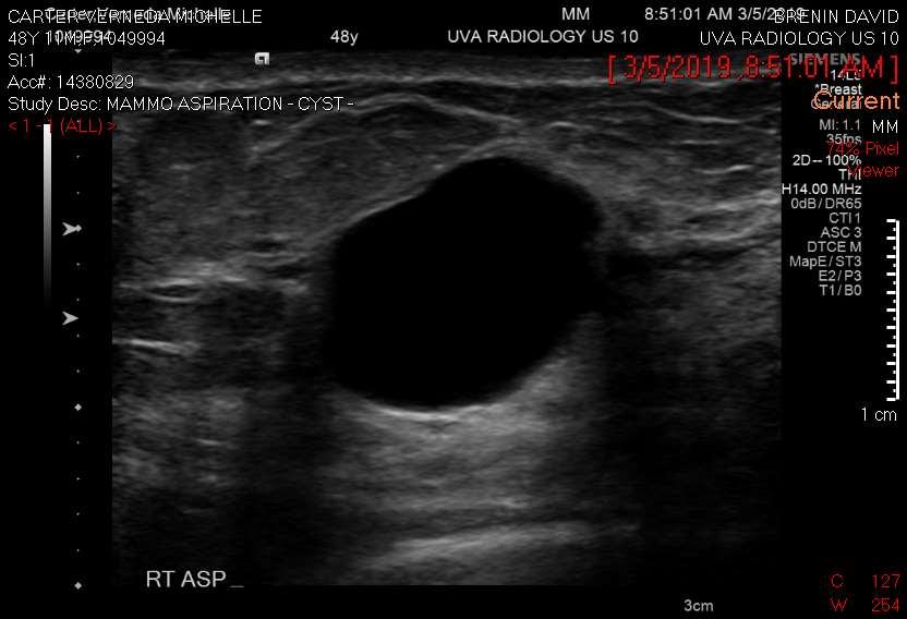
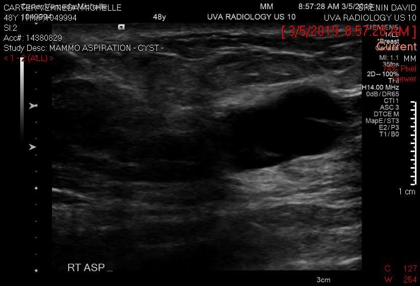
2 Hand Held vs ABUS (Automatic Breast Ultrasound ) Objectives After completing this course, the participant should be able: to describe when and why screening breast ultrasound should be used to describe the scanning technique of screening breast ultrasound to describe the differences between Automated Breast ultrasound and Hand held ultrasound to discuss the ultrasound findings of cystic and solid breast masses technologist role in assisting with ultrasound
3 Breast Ultrasound Who, What, When, Where and Why Another imaging tool to define breast masses (solid versus fluid filled) A tool specific for patients with dense breast/additional diagnostic tool No radiation associated/uses sound waves Fast imaging/fast processing Has a Recording option

4 Ultrasound Waves in Bellagio Water Show Utube video
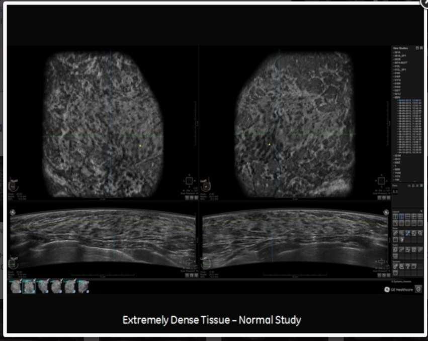
5 Breast Density Many states have laws that require additional imaging for patients with dense breast Mammography is very sensitive to breast tissue that is mostly fatty replaced which is why most screening starts at age 40 Dense breast can hide masses 4 Categories of Breast Density Age is a factor


6 Breast Density Categories Many systems use A, B, C, D to describe the density ratings, with A being fatty and D being extremely dense A B Fatty scattered areas of dense glandular heterogenously dense Dense
7 Types of Breast Ultrasounds Screening Diagnostic Survey Hand held Survey Automated Unilateral/Axilla Mass Specific Research Focused Ultrasound
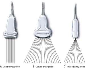
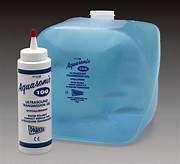
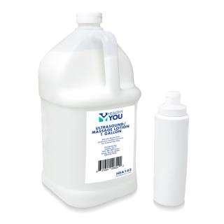
8 Key Points on Ultrasound Point No Radiation Requires Transducer Contact Solution Produces shades of grey Performer Note Uses Sound Waves Wand instrument that includes crystals in tip for breast scanning Gel for Hand Held/Lotion for Automated Masses are normally dark Tech or Rad for Hand Held Mammo tech/sonographer Automated
9
10
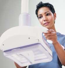
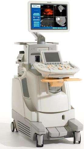
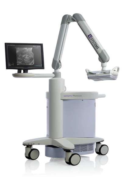
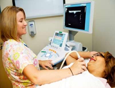
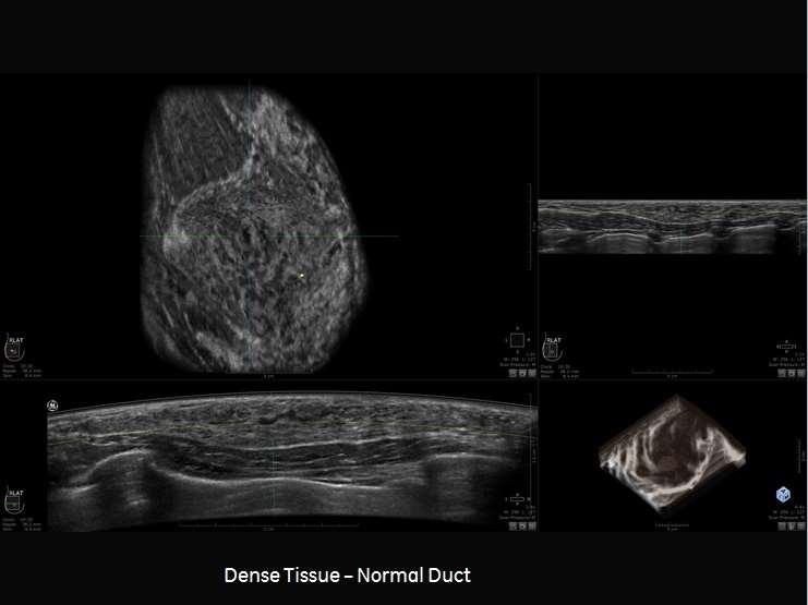
11 HOW IS ULTRASOUND PEFORMED Breast ultrasound uses sound waves to make a computer picture of the inside of the breast. A gel is put on the skin of the breast, and a mechanical wand called a transducer is moved over the skin. The transducer sends out sound waves and picks up the echoes as they bounce off body tissues. The echoes are made into a picture on a computer screen. Automated ultrasound is an option that uses a much larger transducer to take hundreds of images that cover nearly the entire breast.
12 Hand Held vs Automated Ultrasound Hand held Advantages Real time Images Seems more personalized Immediate answers Known method Has video option Disadvantages Takes time Not great for Screening Requires highly trained operator Low reproductivity Automated Advantages Covers large area transducer 3D/Projections/Volumes Less False Negatives Perfect for Screening Images can be easily manipulated by radiologist post survey Fast Not operator dependent Disadvantages Can be painful Requires formalized training for radiologist Physicians don t trust it yet


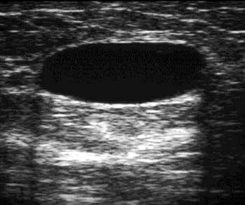
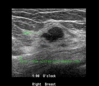
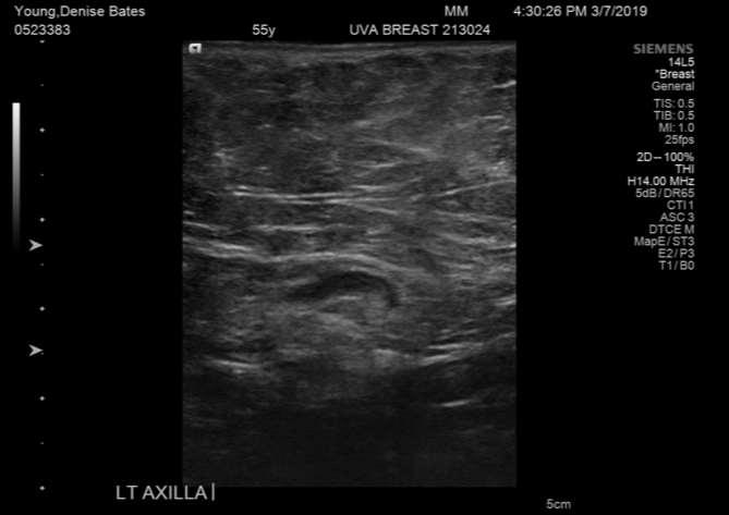
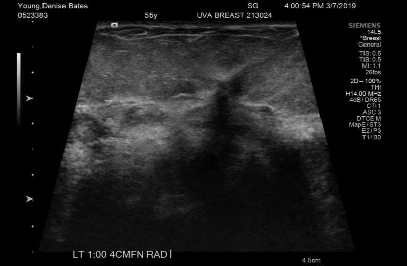
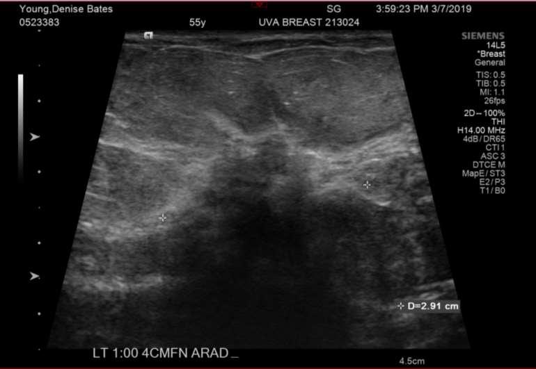
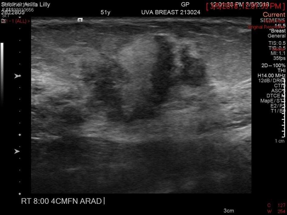
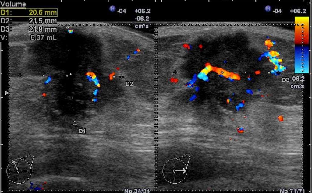
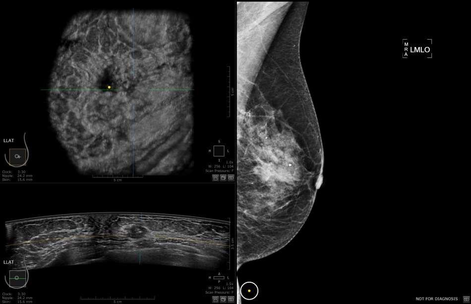
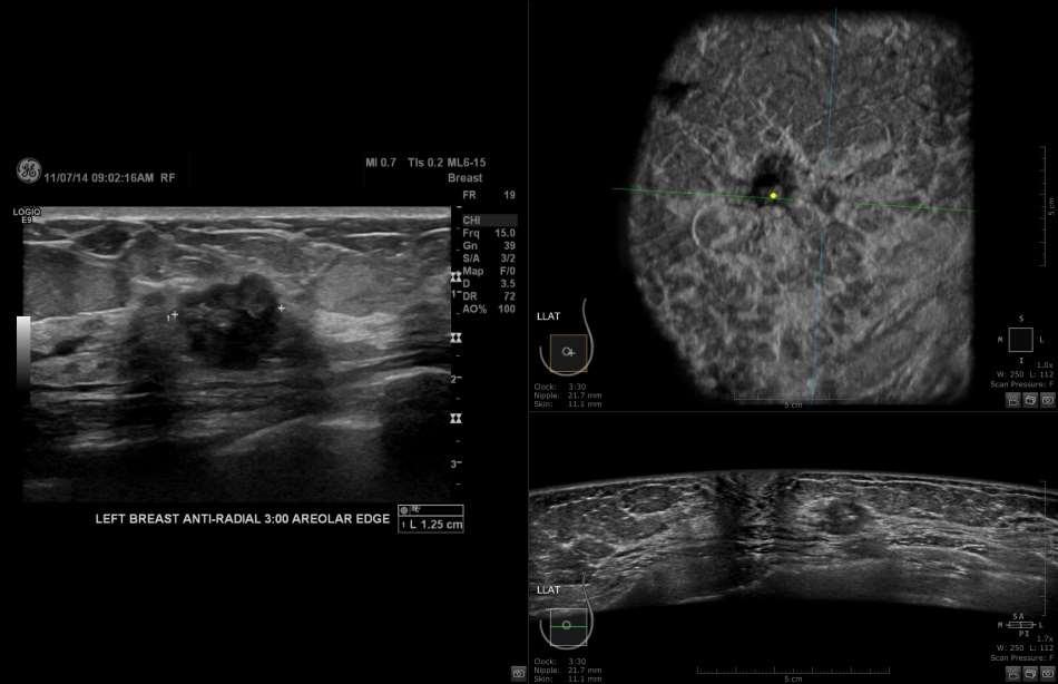
13 Ultrasound Masses Benign Masses Malignant Lymph node Define borders Wide Irregular shape Tall Increase color White border Bulls eye sign
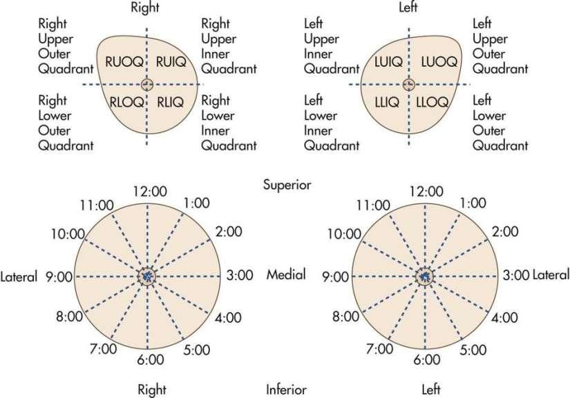
14 Anti radial / coronal Radial /sagittal Transverse Longitudinal
15
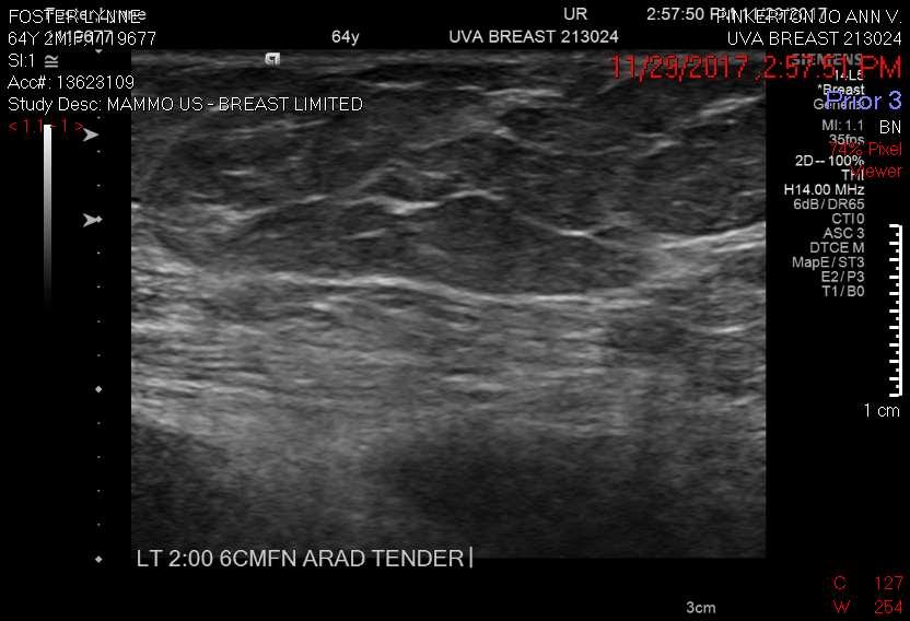
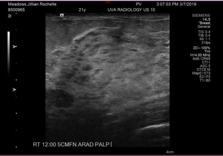
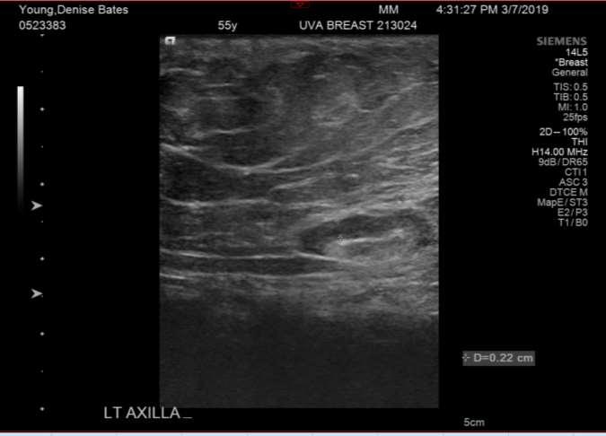
16 HAND HELD SONOGRAPHY
17
18
19
20
21
22
23
24
25
26
27

28 AUTOMATED IMAGES
29
30
31
32
33
34 Technologist Role Prep the Room Prep the Patient Chaperone w/male scanners Explain to the patient about ultrasound Perform the exam using appropriate markings, measurements and power(color) Discharge patient Clean Room LET S TRY IT
35 to describe when and why screening breast ultrasound should be used to describe the scanning technique of screening breast ultrasound to describe the differences between Automated Breast ultrasound and Hand held ultrasound to discuss the ultrasound findings of cystic and solid breast masses technologist role in assisting in ultrasound
Breast Imaging & You
 Breast Imaging & You What s Inside: Breast Imaging... 2 Digital Breast Tomosynthesis (DBT) mammograms... 4 Breast cancer screening... 6 Dense breast tissue... 8 Automated breast ultrasound (ABUS)... 9
Breast Imaging & You
 Breast Imaging & You What s Inside: Breast Imaging... 2 Digital Breast Tomosynthesis (DBT) mammograms... 4 Breast cancer screening... 6 Dense breast tissue... 8 Automated Breast Ultrasound (ABUS)... 9
Ultrasound imaging is a noninvasive medical test that helps physicians diagnose and treat medical conditions.
 CAROTID ULTRASOUND What is Carotid Ultrasound Imaging? Ultrasound imaging, also called ultrasound scanning or sonography, involves exposing part of the body to highfrequency sound waves to produce pictures
Breast density. Understand what it is and why it matters
 Breast density Understand what it is and why it matters What is breast density? Breasts are composed of fibrous, glandular and fatty tissues. Breast density is a term that refers to the proportion of these
Abdominal Ultrasound
 Abdominal Ultrasound What is Ultrasound Imaging of the Abdomen? What are some common uses of the procedure? How should I prepare? What does the equipment look like? How does the procedure work? How is
The latest developments - Automated Breast Volume Scanning. Dr. med. M. Golatta
 The latest developments - Automated Breast Volume Scanning Dr. med. M. Golatta Automated Breast Volume US: Why? o Mammography is limited in dense breasts: high false negative rate o Many of these tumors
Improving Methods for Breast Cancer Detection and Diagnosis. The National Cancer Institute (NCI) is funding numerous research projects to improve
 is funding numerous research projects to improve") CANCER FACTS N a t i o n a l C a n c e r I n s t i t u t e N a t i o n a l I n s t i t u t e s o f H e a l t h D e p a r t m e n t o f H e a l t h a n d H u m a n S e r v i c e s Improving Methods for
CANCER FACTS N a t i o n a l C a n c e r I n s t i t u t e N a t i o n a l I n s t i t u t e s o f H e a l t h D e p a r t m e n t o f H e a l t h a n d H u m a n S e r v i c e s Improving Methods for
Current Status of Supplementary Screening With Breast Ultrasound
 Current Status of Supplementary Screening With Breast Ultrasound Stephen A. Feig, M.D., FACR Fong and Jean Tsai Professor of Women s Imaging Department of Radiologic Sciences University of California,
Ultrasound - Musculoskeletal
 Ultrasound - Musculoskeletal What is Ultrasound Imaging of the Musculoskeletal System? Ultrasound imaging, also called ultrasound scanning or sonography, involves exposing part of the body to high-frequency
Breast Ultrasound: Improving Your Skills & Patient Care
 Breast Ultrasound: Improving Your Skills & Patient Care Objectives Discuss US techniques available for image optimization. Review & compare the US appearances of benign & malignant masses. Cherie M. Kuzmiak,
Supplemental Screening for Dense Breasts. Reagan Leverett, MD, MS
 Supplemental Screening for Dense Breasts Reagan Leverett, MD, MS Outline Anatomy and Density Risk of dense breasts Theory of Supplemental Screening Options for supplemental screening Tomosynthesis Ultrasound
BI-RADS Update. Martha B. Mainiero, MD, FACR, FSBI Brown University Rhode Island Hospital
 BI-RADS Update Martha B. Mainiero, MD, FACR, FSBI Brown University Rhode Island Hospital No Disclosures BI-RADS History 1980s Quality Issues ACR Accreditation BI-RADS 1994 2003 4 th Edition MRI, US January
Ultrasound - Prostate
 Scan for mobile link. Ultrasound - Prostate Ultrasound of the prostate uses sound waves to produce pictures of a man s prostate gland and to help diagnose symptoms such as difficulty urinating or an elevated
Breast and Ovarian Cancer
 Patient Education Breast and Ovarian Cancer Screening and detection The goal of screening for cancer is to find it as early as possible, when it is easiest to cure. This handout describes the symptoms
Cancer , The Patient Education Institute, Inc. ocf80101 Last reviewed: 06/08/2016 1
 Cancer Introduction Cancer begins in your cells, which are the building blocks of your body. Extra cells can form a mass called a tumor. Some tumors aren t cancerous, while other ones are. Cells from cancerous
Human Systems. Technology - Ultrasounds
 Human Systems Technology - Ultrasounds What is General Ultrasound Imaging? Ultrasound imaging, also called ultrasound scanning or sonography, involves exposing part of the body to high-frequency sound
Leonard M. Glassman MD
 BI-RADS The New BI-RADS Leonard M. Glassman MD FACR Former Chief of Breast Imaging American Institute for Radiologic Pathology Washington Radiology Associates, PC Breast Imaging Reporting and Data System
What Does Mammography Follow Up Involve?
 IOWA RADIOLOGY 1 What Does Mammography Follow Up Involve? 515-226-9810 Ankeny Clive Downtown Des Moines Lakeview IOWA RADIOLOGY 2 Table of Contents Introduction... 1 Imaging... 2 Mammography and Ultrasound...
BREAST DENSITY WHAT IS IT? WHY IS IT IMPORTANT? & What IOWA SF250 Means to Patients and Providers
 BREAST DENSITY WHAT IS IT? WHY IS IT IMPORTANT? & What IOWA SF250 Means to Patients and Providers Arnold Honick, MD Radiology Consultants of Iowa, PLC ahonick@rciowa.com BREAST DENSITY LEGISLATION Nancy
1 Fundamentals. Basic Definitions and Physics Principles. Fundamentals
 1 To become versed in the language of ultrasonography, it is necessary to review some of the basic principles of physics. The wave physics principles of ordinary (i.e., audible) sound apply to ultrasound
Imaging Guidelines for Breast Cancer Screening
 Imaging Guidelines for Breast Cancer Screening Sarah Colwick, MD Dr. Sarah Colwick was born and raised in Sikeston, MO. She attended college and medical school at the University of Missouri-Kansas City
COMENIUS-Project: SM&CLIL Radiation & Medicine
 Medical imaging refers to the techniques and processes used to create images of the human body (or parts thereof) for clinical purposes. Thanks to modern mathematics and computer technology, medical imaging
GE Healthcare. Look differently. Invenia ABUS. Automated Breast Ultrasound
 GE Healthcare Look differently Invenia ABUS Automated Breast Ultrasound Invenia TM ABUS from GE Healthcare offers a view beyond mammography, with breast screening technology that looks differently. 40%
Having an Ultrasound Scan
 Having an Ultrasound Scan Information for Patients In this leaflet: Introduction 2 What is an Ultrasound scan?....2 How does it work?... 2 Are there any risks?.2 What do I need to do before my scan?.....3
8/31/2016 HIDING IN PLAIN SITE, ARCHITECTURAL DISTORTIONS AND BREAST ASYMMETRIES ARCHITECTURAL DISTORTIONS ARCHITECTURAL DISTORTIONS
 HIDING IN PLAIN SITE, ARCHITECTURAL DISTORTIONS AND BREAST ASYMMETRIES DEBORAH THAMES R.T. (R)(M)(QM) ARCHITECTURAL DISTORTIONS Definition is disruption of the natural flow of breast pattern towards the
An abdominal ultrasound produces a picture of the organs and other structures in the upper abdomen.
 Scan for mobile link. Ultrasound - Abdomen Ultrasound imaging of the abdomen uses sound waves to produce pictures of the structures within the upper abdomen. It is used to help diagnose pain or distention
Breast Cancer. What is breast cancer?
 Scan for mobile link. Breast Cancer Breast cancer is a malignant tumor in or around breast tissue. It usually begins as a lump or calcium deposit that develops from abnormal cell growth. Most breast lumps
F r e q u e n t l y A s k e d Q u e s t i o n s. Mammograms
 Mammograms Q: What is a mammogram? A: A mammogram is a safe, low-dose x-ray exam of the breasts to look for changes that are not normal. The results are recorded on x-ray film or directly into a computer
Imaging in breast cancer. Mammography and Ultrasound Donya Farrokh.MD Radiologist Mashhad University of Medical Since
 Imaging in breast cancer Mammography and Ultrasound Donya Farrokh.MD Radiologist Mashhad University of Medical Since A mammogram report is a key component of the breast cancer diagnostic process. A mammogram
Breast Cancer Early Detection and Diagnosis
 Breast Cancer Early Detection and Diagnosis Can Breast Cancer Be Found Early? Breast cancer is sometimes found after symptoms appear, but many women with breast cancer have no symptoms. This is why regular
What is Digital Thermography?
 Hello to everyone, Just to inform you that I am booking appointments for thermography screening. It will be held Saturday June 16th at my clinic in Caledon. www.remediesforhealth.ca for directions for
Breast Imaging! Ravi Adhikary, MD!
 Breast Imaging! Ravi Adhikary, MD! ACS Estimated Cancers Statistics 2014! Breast! New Cases in Women! 232,670 (+67,570 in situ)! Deaths in Women! 40,000! Colon! 48,380! 24,040! Cervical! 12,360! 4,020!
Henda s Law. Supplemental screening for women with dense breast tissue and increased risk
 . Henda s Law Supplemental screening for women with dense breast tissue and increased risk The 2011 Texas Legislature passed House Bill 2102 which is effective 1st September 2011. The law is informally
Breast Cancer. What is breast cancer?
 Scan for mobile link. Breast Cancer Breast cancer is a malignant tumor in or around breast tissue. It usually begins as a lump or calcium deposit that develops from abnormal cell growth. Most breast lumps
Children's (Pediatric) Ultrasound - Abdomen
 Ultrasound - Abdomen") Scan for mobile link. Children's (Pediatric) Ultrasound - Abdomen Children s (pediatric) ultrasound imaging of the abdomen is a safe, noninvasive test that uses sound waves to produce a clear picture of
Scan for mobile link. Children's (Pediatric) Ultrasound - Abdomen Children s (pediatric) ultrasound imaging of the abdomen is a safe, noninvasive test that uses sound waves to produce a clear picture of
Pelvic Ultrasound.
 Pelvic Ultrasound Before Your Exam: Drink 32 oz. of water one hour before your examination time. Try to drink all the liquid within 30 minutes. Do not urinate before the exam. Arrive for your exam with
Tissue Breast Density
 Tissue Breast Density Reporting breast density within the letter to the patient is now mandated by VA law. Therefore, this website has been established by Peninsula Radiological Associates (PRA), the radiologists
Children's (Pediatric) Contrast-enhanced Voiding Urosonography
 Contrast-enhanced Voiding Urosonography") Scan for mobile link. Children's (Pediatric) Contrast-enhanced Voiding Urosonography Pediatric contrast-enhanced voiding urosonography uses ultrasound to examine a child's bladder and urinary tract. It
Scan for mobile link. Children's (Pediatric) Contrast-enhanced Voiding Urosonography Pediatric contrast-enhanced voiding urosonography uses ultrasound to examine a child's bladder and urinary tract. It
Armed Forces Institute of Pathology.
 Armed Forces Institute of Pathology www.radpath.com Armed Forces Institute of Pathology Breast Disease www.radpath.org Armed Forces Institute of Pathology Interpretation of Breast MRI Leonard M. Glassman
Screening with New Modalities: Breast Ultrasound
 Screening with New Modalities: Breast Ultrasound Wendie A. Berg, MD, PhD Professor of Radiology Magee-Womens Hospital of UPMC University of Pittsburgh School of Medicine Disclosures No personal financial
Galactography (Ductography)
") Scan for mobile link. Galactography (Ductography) Galactography uses mammography and an injection of contrast material to create pictures of the inside of the breast s milk ducts. It is most commonly used
Scan for mobile link. Galactography (Ductography) Galactography uses mammography and an injection of contrast material to create pictures of the inside of the breast s milk ducts. It is most commonly used
Pitfalls and Limitations of Breast MRI. Susan Orel Roth, MD Professor of Radiology University of Pennsylvania
 Pitfalls and Limitations of Breast MRI Susan Orel Roth, MD Professor of Radiology University of Pennsylvania Objectives Review the etiologies of false negative breast MRI examinations Discuss the limitations
Breast Density It's the Law
 Last year Iowa became the 30th state in the last 12 years to require that density information be added to the written mammogram report to the patient. This report is sent directly from the interpreting
Screening Mammograms: Questions and Answers
 CANCER FACTS N a t i o n a l C a n c e r I n s t i t u t e N a t i o n a l I n s t i t u t e s o f H e a l t h D e p a r t m e n t o f H e a l t h a n d H u m a n S e r v i c e s Screening Mammograms:
Standard Breast Imaging Modalities. Lilian Wang, M.D. Breast Imaging Section Department of Radiology Northwestern Medicine
 Standard Breast Imaging Modalities Lilian Wang, M.D. Breast Imaging Section Department of Radiology Northwestern Medicine Overview Standard breast imaging modalities Mammography Ultrasound MRI Imaging
Annual Screening Mammogram and its Relation to Breast Density
 International Journal of Medical Research & Health Sciences Available online at www.ijmrhs.com ISSN No: 2319-5886 International Journal of Medical Research & Health Sciences, 2017, 6(11): 83-90 I J M R
Q: Why is breast cancer a big deal?
 I hate breast cancer. As a radiologist who specializes in breast imaging, my career is devoted to the detection and diagnosis of breast cancer. I am passionate about women s health and my goal is to find
Ultrasonography of the Neck as an Adjunct to FNA. Nicole Massoll M.D.
 Ultrasonography of the Neck as an Adjunct to FNA Nicole Massoll M.D. Basic Features of Head and Neck Ultrasound and Anatomy Nicole Massoll M.D. University of Arkansas for Medical Sciences, Little Rock
Ultrasound Physics & Doppler
 Ultrasound Physics & Doppler Endocrine University 2018 Mark Lupo, MD, FACE, ECNU Objectives Review the essential components of ultrasound physics in neck sonography Demonstrate the importance of ultrasound
WOMENCARE A Healthy Woman is a Powerful Woman (407) Mammography
 Mammography") Mammography WOMENCARE A Healthy Woman is a Powerful Woman (407) 898-1500 Mammography is an X-ray technique used to study the breasts. It can help doctors find breast cancer at an early stage (when treatment
Mammography WOMENCARE A Healthy Woman is a Powerful Woman (407) 898-1500 Mammography is an X-ray technique used to study the breasts. It can help doctors find breast cancer at an early stage (when treatment
Ultrasound Knobology
 Ultrasound Knobology Raj Dasgupta MD, FACP, FCCP, FASSM Assistant Professor of Clinical Medicine Pulmonary / Critical Care / Sleep Medicine University of Southern California (USC) Objectives Physics of
Ultrasound. Information for patients and families
 Ultrasound Information for patients and families Read this booklet to learn: what an ultrasound is the different types and how to prepare what to bring to your ultrasound appointment what to expect where
S. Murgo, MD. Chr St-Joseph, Mons Erasme Hospital, Brussels
 S. Murgo, MD Chr St-Joseph, Mons Erasme Hospital, Brussels? Introduction Mammography reports are sometimes ambiguous and indecisive. ACR has developped the BIRADS. BIRADS consists of a lexicon in order
Dense Breasts, Get Educated
 Dense Breasts, Get Educated What are Dense Breasts? The normal appearances to breasts, both visually and on mammography, varies greatly. On mammography, one of the important ways breasts differ is breast
INFORMATION for PATIENTS
 INFORMATION for PATIENTS What is MRI? Magnetic Resonance Imaging uses a computer, magnetic fields and radio waves to generate images of the body. It can be used for virtually all parts of the body, generating
When You Need To Know More.
 www.siemens.com/ultrasound When You Need To Know More. ACUSON S2000 Ultrasound System Table of Contents Powerful Imaging 01 Penetrating Insight 02 03 Revealing Perspectives 04 05 Smart Workflow 06 Ergonomics
Full ultrasound breast volumes. Faster scans. Streamlined workflow. ACUSON S2000 Automated Breast Volume Scanner. Answers for life.
 Full ultrasound breast volumes. Faster scans. Streamlined workflow. ACUSON S2000 Automated Breast Volume Scanner Answers for life. 1 ACQUIRE An automated whole breast solution. Reduced acquisition time.
ACRIN 6666 IM Additional Evaluation: Additional Views/Targeted US
 Additional Evaluation: Additional Views/Targeted US For revised or corrected form check box and fax to 215-717-0936. Instructions: The form is completed based on recommendations (from ID form) for additional
Mammography. What is Mammography? What are some common uses of the procedure?
 Mammography What is Mammography? Mammography is a specific type of imaging that uses a low-dose x-ray system to examine breasts. A mammography exam, called a mammogram, is used to aid in the early detection
CONTENTS. Test Number cpd Tanya Reynolds (Nat. Dip. Diag. Rad., B. Tech. Diag. Rad., B. Tech. Ultrasound)
") CONTENTS page 1-15 page 16 BASIC 2-DIMENSIONAL ULTRASOUND PRINCIPLES Multiple Choice Test Test Number cpd 41640 Tanya Reynolds (Nat. Dip. Diag. Rad., B. Tech. Diag. Rad., B. Tech. Ultrasound) Tanya is
CONTENTS page 1-15 page 16 BASIC 2-DIMENSIONAL ULTRASOUND PRINCIPLES Multiple Choice Test Test Number cpd 41640 Tanya Reynolds (Nat. Dip. Diag. Rad., B. Tech. Diag. Rad., B. Tech. Ultrasound) Tanya is
The Radiology Aspects
 REQUIREMENTS FOR INTERNATIONAL ACCREDITATION OF BREAST CENTERS/UNITS The Radiology Aspects Miri Sklair-Levy, Israel RADIOLOGY GUIDELINES FOR QUALITY ASSURANCE IN BREAST CANCER SCREENING AND DIAGNOSIS Radiologists
Ruud Pijnappel Professor of Radiology, UMC Utrecht. Chair Dutch Expert Centre for Screening Board EUSOBI
 Ruud Pijnappel Professor of Radiology, UMC Utrecht Best practice in Breast Imaging: what s new and what women need to know and Update on the Second Implementation Report of the 2003 Council Recommendation
What Is Nuclear Medicine?
 What Is Nuclear Medicine? An Introductory Guide For Patients And Their Families What is nuclear medicine? Nuclear medicine is a type of medical imaging that uses small amounts of radioactive material (called
Look differently. Invenia ABUS. Automated Breast Ultrasound
 Look differently. Invenia ABUS Automated Breast Ultrasound InveniaTM ABUS from GE Healthcare offers a view beyond mammography, with breast screening technology that looks differently. 40 % The unseen risk.
Patient Education. Women s Imaging
 Patient Education Women s Imaging What you should know about your Gynecologic (GYN) Ultrasound A gynecologic ultrasound is performed to examine your pelvic area to evaluate for some of the following symptoms:
ISSN X (Print) Research Article. *Corresponding author Dr. Amlendu Nagar
 Research Article. *Corresponding author Dr. Amlendu Nagar") Scholars Journal of Applied Medical Sciences (SJAMS) Sch. J. App. Med. Sci., 2015; 3(3A):1069-1073 Scholars Academic and Scientific Publisher (An International Publisher for Academic and Scientific Resources)
Scholars Journal of Applied Medical Sciences (SJAMS) Sch. J. App. Med. Sci., 2015; 3(3A):1069-1073 Scholars Academic and Scientific Publisher (An International Publisher for Academic and Scientific Resources)
Patient Education. Ultrasound
 Patient Education Ultrasound What you should know about your Abdominal Ultrasound. An abdominal ultrasound is performed to examine your abdominal organs such as liver, spleen, gall bladder or pancreas.
Basic Training Programme. 16 Februrary 2018, ROTTERDAM. Pre and Post-Course Test Answers
 Basic Training Programme 16 Februrary 2018, ROTTERDAM Pre and Post-Course Test Answers Your details: Name: Conference registration number/ BT delegate number: Email address: Are you already performing
General Ultrasound. What is General Ultrasound Imaging?
 Scan for mobile link. General Ultrasound What is General Ultrasound Imaging? Ultrasound is safe and painless, and produces pictures of the inside of the body using sound waves. Ultrasound imaging, also
General Ultrasound. What is General Ultrasound Imaging?
 Scan for mobile link. General Ultrasound Ultrasound imaging uses sound waves to produce pictures of the inside of the body. It is used to help diagnose the causes of pain, swelling and infection in the
A GP S APPROACH TO BREAST LUMPS AND SYMPTOMS DR KK CHEUNG GPGC WORKSHOP
 A GP S APPROACH TO BREAST LUMPS AND SYMPTOMS DR KK CHEUNG GPGC WORKSHOP 18.08.18 HAVE A SYSTEM HISTORY EXAMINATION INVESTIGATION FOLLOW UP BREAST SYMPTOMS HISTORY DON T FORGET SKIN CHANGES AND NIPPLE CHANGES
Supplemental Screening for Women with Dense Breast Tissue. Public Meeting December 13, 2013
 Supplemental Screening for Women with Dense Breast Tissue Public Meeting December 13, 2013 Agenda Meeting Convened 10am-10:15am Presentation of the Evidence and Voting Questions, Q&A 10:15am 11:15am Discussion
Crisis or Opportunity?
 Crisis or Opportunity? Breast Density Notification, Workflow and the Implementation of Ultrasound Gerald R. Kolb 1 Breast density notification is becoming a reality in many states across the US by virtue
The early detection programme for breast cancer
 Making a wellinformed decision The early detection programme for breast cancer Why are women offered a mammogram through the quality controlled screening programme? Women between 50 and 69 years of age
Breast Tomosynthesis. What is breast tomosynthesis?
 Scan for mobile link. Breast Tomosynthesis Breast tomosynthesis is an advanced form of mammography, a specific type of breast imaging that uses low-dose x-rays to detect cancer early when it is most treatable.
Mammography. What is Mammography?
 Scan for mobile link. Mammography Mammography is a specific type of breast imaging that uses low-dose x-rays to detect cancer early before women experience symptoms when it is most treatable. Tell your
Breast Magnetic Resonance Imaging (MRI) Westmead Breast Cancer Institute
 Westmead Breast Cancer Institute") Breast Magnetic Resonance Imaging (MRI) Westmead Breast Cancer Institute What is breast MRI? Breast MRI is a technique that uses a magnetic field to create an image of the breast tissue, using hundreds
Breast Magnetic Resonance Imaging (MRI) Westmead Breast Cancer Institute What is breast MRI? Breast MRI is a technique that uses a magnetic field to create an image of the breast tissue, using hundreds
Amammography report is a key component of the breast
 Review Article Writing a Mammography Report Amammography report is a key component of the breast cancer diagnostic process. Although mammographic findings were not clearly differentiated between benign
Breast Density and Screening
 Breast Density and Screening Alberta Breast Cancer Screening Program Version: Jan 2019 What is this Booklet About? This is a guide to help you understand breast density and how it may affect you. Having
Breast Cancer Screening
 Scan for mobile link. Breast Cancer Screening What is breast cancer screening? Screening examinations are tests performed to find disease before symptoms begin. The goal of screening is to detect disease
3D Automated breast ultrasound (ABUS): pictorial review of applications and clinical utility.
: pictorial review of applications and clinical utility.") 3D Automated breast ultrasound (ABUS): pictorial review of applications and clinical utility. Poster No.: C-1182 Congress: ECR 2013 Type: Educational Exhibit Authors: A. Domingo, C. Cusido, F. V. Gras,
3D Automated breast ultrasound (ABUS): pictorial review of applications and clinical utility. Poster No.: C-1182 Congress: ECR 2013 Type: Educational Exhibit Authors: A. Domingo, C. Cusido, F. V. Gras,
Screening for Breast Cancer
 Understanding Task Force Recommendations Screening for Breast Cancer U.S. Preventive Services Task Force (Task Force) has issued a final recommendation statement on Screening for Breast Cancer. se final
Mammography. The Lebanese Society of Obstetrics and Gynecology. Women s health promotion series
 The Lebanese Society of Obstetrics and Gynecology Women s health promotion series Mammography When breast cancer is diagnosed at an early stage it could be treated and the patient would have a high chance
RADIOLOGIC AND IMAGING SCIENCE (RIS)
") Kent State University Catalog 2017-2018 1 RADIOLOGIC AND IMAGING SCIENCE (RIS) RIS 34001 INTRODUCTION TO DIAGNOSTIC MEDICAL SONOGRAPHY 1 Credit Provides an introduction to diagnostic medical sonography.
Kent State University Catalog 2017-2018 1 RADIOLOGIC AND IMAGING SCIENCE (RIS) RIS 34001 INTRODUCTION TO DIAGNOSTIC MEDICAL SONOGRAPHY 1 Credit Provides an introduction to diagnostic medical sonography.
IN THE COUNCIL OF THE DISTRICT OF COLUMBIA
 1 2 3 4 5 6 7 8 9 10 11 12 13 14 15 16 17 18 19 20 21 22 23 24 25 26 27 28 A BILL 22-960 IN THE COUNCIL OF THE DISTRICT OF COLUMBIA To require health care facilities to notify patients of mammogram results
Jaundice , The Patient Education Institute, Inc. syf80102 Last reviewed: 05/05/2017 1
 Jaundice Introduction Jaundice causes your skin and the whites of your eyes to turn yellow. Too much bilirubin causes jaundice. Bilirubin is a yellow chemical in hemoglobin, the substance that carries
Hepatobiliary Ultrasound Rimon Bengiamin, MD, RDMS Assistant Clinical Professor Director of Emergency Ultrasound UCSF Fresno. Objectives. Why?
 Hepatobiliary Ultrasound Rimon Bengiamin, MD, RDMS Assistant Clinical Professor Director of Emergency Ultrasound UCSF Fresno Objectives Discuss the goals of point-of-care biliary ultrasound Review the
Challenges to Delivery of High Quality Mammography
 Challenges to Delivery of High Quality Mammography Overview of Current Challenges Barbara Monsees, Washington University Geographic Access, Equity and Impact on Quality Tracy Onega, Dartmouth Medical School
Intravascular Ultrasound
 Scan for mobile link. Intravascular Ultrasound Intravascular ultrasound (IVUS) uses a transducer or probe to generate sound waves and produce pictures of the coronary arteries. IVUS can show the entire
The radiologic workup of a palpable breast mass
 Imaging in Practice CME CREDIT EDUCTIONL OJECTIVE: The reader will consider which breast masses require further workup and which imaging study is most appropriate Lauren Stein, MD Imaging Institute, Cleveland
Thyroid Nodules: What to do next?
 Thyroid Nodules: What to do next? Ally P. H. Prebtani Professor of Medicine Internal Medicine, Endocrinology & Metabolism McMaster University Canada Copyright 2017 by Sea Courses Inc. All rights reserved.
Alice Fung, MD Oregon Health and Science University
 Alice Fung, MD Oregon Health and Science University Disclosure Comments The speaker Alice Fung, MD Has relevant financial relationships to disclose. Received honorarium from (Guerbet). This individual
for Pacific women Early detection is your best protection
 for Pacific women Early detection is your best protection BreastScreen Aotearoa (BSA) aims to reduce the number of women who die from breast cancer offers free mammograms (breast x-rays) every two years
Ultrasound Tomography (UST) a form of ABUS
 a form of ABUS") Ultrasound Tomography (UST) a form of ABUS Ultrasound Tomography: A Breast Imaging Modality Whose Time Has Come Neb Duric 1,2,, Peter Littrup 2,3, Olivier Roy 2, Cuiping Li 2, Steve Schmidt 2, Heather
Ultrasound Tomography (UST) a form of ABUS Ultrasound Tomography: A Breast Imaging Modality Whose Time Has Come Neb Duric 1,2,, Peter Littrup 2,3, Olivier Roy 2, Cuiping Li 2, Steve Schmidt 2, Heather
WELCOME! Introduction to Bedside Ultrasound
 WELCOME! Introduction to Bedside Ultrasound TEACHERS University of California-Irvine School of Medicine Nathan Molina nathan.d.molina@gmail.com Trevor Plescia taplescia90@gmail.com Jack Silva jpsilva42@gmail.com
InformatIon. for PatIents
 InformatIon for PatIents 1165 Union Street NE #100 Salem, OR 97301 P: (503) 588-2674 / (888) 999-7030 F: (503) 391-1200 www.diagnosticimagingofsalem.com What is MRI? Magnetic Resonance Imaging uses a computer,
Ultrasound Principles cycle Frequency Wavelength Period Velocity
 ! Teresa S. Wu, MD, FACEP Director, EM Ultrasound Program & Fellowship Co-Director, Simulation Based Training Program & Fellowship Associate Program Director, EM Residency Program Maricopa Medical Center
ULTRASOUND SCAN. Patient Information Leaflet
 Patient Information Leaflet ULTRASOUND SCAN Introduction We have received a request from your referring doctor for you to have an Ultrasound scan. This leaflet is designed to give you some information
Terminology Tissue Appearance
 By Marc Nielsen, MD Advantages/Disadvantages Generation of Image Ultrasound Machine/Transducer selection Modes of Ultrasound Terminology Tissue Appearance Scanning Technique Real-time Portable No ionizing
Emerging Techniques in Breast Imaging: Contrast-Enhanced Mammography and Fast MRI
 Emerging Techniques in Breast Imaging: Contrast-Enhanced Mammography and Fast MRI Lilian Wang, M.D. Breast Imaging Section Department of Radiology Northwestern Medicine Overview Rationale for new imaging