Maintaining a quality mammography facility: Overview of ACR and MQSA Requirements and Update on BI-RADS 5 TH Edition
|
|
|
- Hilary Hall
- 6 years ago
- Views:
Transcription
1 Maintaining a quality mammography facility: Overview of ACR and MQSA Requirements and Update on BI-RADS 5 TH Edition Sandra S. Rao, M.D. Northwestern Medicine I have nothing to disclose.
2 Objectives: Review accreditation requirements Understand how to successfully obtain and renew FDA certification Describe changes in the 5 th Edition BI- RADS for Assessment Categories and recommendations Discuss the update in the BI-RADS classification of breast densitiy

3 MQSA FDA Certification and ACR Accreditation With a special thank you for providing information to: Judith A. Wolfman, M.D., F.A.C.R.
4 Breast Imaging: Accreditation & Certification Mammography Film-Screen Digital Ultrasound Stereotactic Unit Must be Accredited by ACR and Certified by FDA Voluntary ACR Accreditation MRI Voluntary ACR Accreditation, May, 2010; Required for CMS reimbursement Jan, 2012
5 Mammography Accreditation & Certification How did we get here? Professional Concerns about quality of mammography Voluntary Accreditation Programs Illinois Radiological Society, 1986 ACR Accreditation Program, 1987

6 and Public Concern & Advocacy
7 History of Mammography Accreditation & Certification 1992 President Bush signed the Mammography Quality Standards Act 1994 FDA s Interim Rules required all mammography facilities in the US to be accredited, certified and inspected QC regs mirrored the ACR requirements 1999 FDA s Final Rules went into effect with new detailed requirements, including direct patient notification
8 ?? MQSA, FDA, ACR??? MQSA = Mammography Quality Standards Act - Legislation passed by US Congress on 10/23/92 - Stipulated that as of October 1, 1994, all mammography facilities were to be accredited by an approved accreditation body & certified by the FDA FDA = Food & Drug Administration Approves the Accreditation Body Certifies the Mammography facility Issues an MQSA Certificate when the accreditation body notifies the FDA that the facility has been accredited. Certification valid for 3 years Performs annual Inspections Penalty for performing mammography without certification are severe (up to $10K per exam performed) ACR = American College of Radiology Mammography Accreditation Program Certified by FDA as accreditation body Must meet MQSA requirements as published by FDA
9 MQSA Final Regulations Proposed Regulations Published 4/3/96 Final Regulations Published 10/28/97 Implementation 4/28/99 Reauthorization signed into law 10/9/98 Requires lay summary of results to all patients Release of original mammograms temporary and permanent
10 Facility Requirements Under MQSA DEPARTMENT OF HEALTH AND HUMAN SERVICES Food and Drug Administration 21 CFR Part 900 Quality Mammography Standards AGENCY: Food and Drug Administration, HHS. ACTION: Final rule As Amended by Federal Register Notice 10/22/98, 4/15/99, 6/17/99 DATES: This regulation is effective April 28, 1999; except Sec (b)(8)(i), (e)(4)(iii)(b),(e)(5)(i)(b) which become effective October 28, A certificate is required before operation - 6-month provisional if just starting out; - 3-year full certificate after accreditation Mammo facilities must be: Accredited, every 3 yrs Certified, every 3 yrs Inspected, every year Complementary, not duplicative
11 ACR Accreditation is required by the FDA for which of the following Breast Imaging modalities???: 1. Mammography 2. Breast Ultrasound 3. Breast MRI 4. Stereotactic Breast Biopsy 5. All of the above
12 ACR Accreditation is required by the FDA for which of the following Breast Imaging modalities: 1. Mammography 2. Breast Ultrasound 3. Breast MRI 4. Stereotactic Breast Biopsy 5. All of the above
13 FDA Certification Requirements The Mammography Facility must meet the following Quality Standards as described in Section of the final as published in the Federal Register: (a) - Personnel (b) - Mammography equipment (c) - Medical records & reports (d) - Quality Assurance general (e) - Quality Assurance equipment (f) - Quality Assurance Medical outcomes audit (g) - Mammographic Procedure for examinees with implants (h) - Consumer complaint mechanism (f) Additional Clinical Image Review and Notification (f) Additional mammography review and patient notification
14 ACR Mammography Accreditation for FDA Certification Application requires documentation of: -Personnel Qualifications -Equipment and documentation of QC program -Physicist inspection report Phantom and Clinical Image Submission: -ACR Phantom obtained with TLD for documentation of image quality and dose - Clinical Images: Fatty and Dense => 2-view mammogram of each type
15 FDA Certification Requirements The Mammography Facility must meet the following Quality Standards as described in Section of the final as published in the Federal Register: (a) - Personnel (b) - Mammography equipment (c) - Medical records & reports (d) - Quality Assurance general (e) - Quality Assurance equipment (f) - Quality Assurance Medical outcomes audit (g) - Mammographic Procedure for examinees with implants (h) - Consumer complaint mechanism (f) Additional Clinical Image Review and Notification (f) Additional mammography review and patient notification
16 Personnel: State license Interpreting Physicians Initial Qualifications Diagnostic radiology certification OR 3 months of formal training 60 hours of category I CME in mammography 240+ mammograms interpreted under the direct supervision In the 6 months immediately prior to independent interpretation OR If board certified at first allowable time, within the last 2 years of residency
17 Interpreting Physicians Continuing Requirements Continuing experience Interpret at least 960 examinations/ 24 months Continuing education At least 15 category I CME credits/ 36 months At least 6 of the 15 CME units must be in each mammographic modality used 8 hours of training in each mammographic modality before independent use, i.e. film vs. digital vs. tomosynthesis
18 Interpreting Physicians Reestablishing Qualifications Continuing experience Interpret or multi-read under direct supervision EITHER 240 examinations OR enough examinations to bring total to 960/24 months Interpretations performed in immediate 6 months prior to resuming independent reading Continuing education Bring total CME credits up to 15 unit/36 months
19 Radiologic Technologists State license OR Initial Qualifications Certified by FDA-approved body 40 hours of documented mammography training Performing at least 25 examinations under direct supervision 8 hours of training in each mammographic modality to be used
20 Radiologic Technologists Continuing Requirements Continuing experience Perform at least 200 examinations/ 24 months Continuing education At least 15 CEUs/ 36 months At least 6 of the 15 CEUs must be in each mammographic modality used 8 hours of training in each mammographic modality before independent use, i.e. film vs. digital
21 Radiologic Technologists Reestablishing Qualifications Continuing experience Complete 25 examinations under direct supervision Continuing education Bring total CEUs up to 15 credits/36 months
22 Medical Physicists Initial Qualifications Licensed or approved by a State OR Certified by FDA-approved body Master s or higher degree in a physical science 20 semester hours of physics 20 hours of documented mammography survey training One facility and 10 units surveyed 8 hours of training in each modality surveyed before surveying independently
23 Medical Physicists Continuing Requirements Continuing experience Survey at least 2 facilities & 6 units/ 24 months Continuing education At least 15 CEUs/ 36 months At least 6 of the 15 CEUs must be in each mammographic modality used 8 hours of training in each mammographic modality before independent use
24 Medical Physicists Reestablishing Qualifications Direct Supervision Continuing experience Bring total number of supervised surveys up to 2 facilities and 6 units / 24 months Continuing education Bring total CEUs up to 15 credits/36 months
25 To meet the initial qualifications for the interpretation of mammography, the physician must interpret how many mammography exams under the supervision of a qualified radiologist?
26 To meet the initial qualifications for the interpretation of mammography, the physician must interpret how many mammography exams under the supervision of a qualified radiologist?
27 FDA Certification Requirements The Mammography Facility must meet the following Quality Standards as described in Section of the final as published in the Federal Register: (a) - Personnel (b) - Mammography equipment (c) - Medical records & reports (d) - Quality Assurance general (e) - Quality Assurance equipment (f) - Quality Assurance Medical outcomes audit (g) - Mammographic Procedure for examinees with implants (h) - Consumer complaint mechanism (f) Additional Clinical Image Review and Notification (f) Additional mammography review and patient notification
28 Equipment Standards Specific Requirements Equipment must be designed for mammography Motion of tube-image receptor assembly Capable of being fixed in any position No unintended motion during power interruption Image receptor sizes for each screen-film unit 18 x 24 cm and 24 x 30 cm cassettes 18 x 24 cm and 24 x 30 cm moving grid
29 Equipment Standards Specific Requirements (cont.) Light fields Provide average illumination of 160 lux at 100 cm Magnification capacity Systems designed for magnification provide at least one magnification in the range of 1.4 to 2.0
30 Equipment Standards Specific Requirements (cont.) Breast Compression Devices Compression paddles For each image receptor size Flat and parallel to the breast support table, OR If paddle not designed to be flat and parallel, meet manufacturer s design and maintenance requirements Hands-free initial power driven compression Operable from both sides of patient Fine adjustment controls
31 Equipment Standards Specific Requirements (cont.) Technique factor selection and display Manual selection of mas available Technique factors indicated before exposure Automatic exposure control AEC operable in all clinically used modes Flexible positioning of detector Selected optical density may be varied from normal (zero) setting
32 Equipment Standards Specific Requirements (cont.) Display of selected focal spot and/or target material X-ray film designed for mammography Intensifying screens matched to film Film processing solutions meet film requirements High-intensity lighting (hot-lights) available Film masking devices available
33 FDA Certification Requirements The Mammography Facility must meet the following Quality Standards as described in Section of the final as published in the Federal Register: (a) - Personnel (b) - Mammography equipment (c) - Medical records & reports (d) - Quality Assurance general (e) - Quality Assurance equipment (f) - Quality Assurance Medical outcomes audit (g) - Mammographic Procedure for examinees with implants (h) - Consumer complaint mechanism (f) Additional Clinical Image Review and Notification (f) Additional mammography review and patient notification
34 Reporting and Recordkeeping Standards Overall Assessment of Findings Report identifies interpreting physician Assessment Categories: Negative Benign Probably Benign Suspicious Highly Suggestive of Malignancy Incomplete: Need additional imaging evaluation
35 Reporting and Recordkeeping Standards Communication of Results Report sent to referring health care provider (or patient with no health care provider) within 30 days Suspicious or Highly suggestive of malignancy results communicated to health care provider and patient as soon as possible (ideally, within 3-5 days) Summary report written in lay language sent to all patients within 30 days Self-referred patients receive both the lay language letter and the mammography report

36 Reporting and Recordkeeping Standards: Mammographic Image information Patient name, Patient identifier Facility name & location Date of Exam Technologist ID Mammo Unit ID View & Laterality Technical Factors
37 Medical Records and Reports Medical reports and films kept by facilities for: 5 years, OR 10 years if no additional mammography performed on patient at that facility, OR Longer if required by state or local law Original films transferred ( temporary or permanent) upon request of patient or patient s representative Fee charged may not exceed documented cost incurred by the facility
38 Medical Records and Reports (cont.) Inspectors will ask to see: Randomly selected records Sample reports sent to referring health care provider Template of lay summary of mammography report sent to patient System for communicating findings Randomly selected reports Interpreting physicians identified Assessment Categories
39 FDA Certification Requirements The Mammography Facility must meet the following Quality Standards as described in Section of the final as published in the Federal Register: (a) - Personnel (b) - Mammography equipment (c) - Medical records & reports (d) - Quality Assurance general (e) - Quality Assurance equipment (f) - Quality Assurance Medical outcomes audit (g) - Mammographic Procedure for examinees with implants (h) - Consumer complaint mechanism (f) Additional Clinical Image Review and Notification (f) Additional mammography review and patient notification
40 General Administrative Requirements Responsible staff: Lead interpreting physician Interpreting physicians Medical physicist Quality control technologist Responsible for meeting quality assurance recordkeeping requirements
41 FDA Certification Requirements The Mammography Facility must meet the following Quality Standards as described in Section of the final as published in the Federal Register: (a) - Personnel (b) - Mammography equipment (c) - Medical records & reports (d) - Quality Assurance general (e) - Quality Assurance equipment (f) - Quality Assurance Medical outcomes audit (g) - Mammographic Procedure for examinees with implants (h) - Consumer complaint mechanism (f) Additional Clinical Image Review and Notification (f) Additional mammography review and patient notification
42 Equipment Quality Assurance Specifications for: Tests to be performed Frequency Action limits Facility generally free to choose test procedures that meet its needs For non-screen-film equipment, follow manufacturer s quality assurance instructions
43 Equipment Quality Assurance Tests Other Than Annual (SFM) Daily tests: Film processor Weekly tests: Phantom image Quarterly tests: Fixer retention Repeat analysis Semiannual tests: Darkroom fog Screen-film contact Compression device performance


44 ACR Phantom on Screen-Film Same Target-Filter and kvp: Mo-Mo 26 kvp Underexposed Proper Exposure Overexposed 56 mas Lower Contrast 104 mas 200 mas Lower Contrast
45 In order to pass must see 4 largest fibers 3 largest speck groups 3 largest masses Subtract artifacts if they appear Fiber-like Speck-like Mass-like Phantom Image Quality Evaluation
46 Equipment Quality Assurance Annual Tests (SFM) Automatic exposure control Kilovoltage peak accuracy Focal spot condition Beam quality and half-value layer Breast entrance air kerma and AEC reproducibility Dosimetry X-ray field/ light field/ image receptor/ compression paddle alignment Uniformity of screen speed System artifacts Radiation output Decompression
47 Equipment Quality Assurance Mobile Units (SFM) Performance testing of mobile units for image quality accuracy At each location and before imaging patients Examples of acceptable tests Phantom testing Acceptable limits for variation in mas Other tests
48 Equipment Quality Assurance Test Results (SFM) Use of test results Compare results to action limits For results out of limits Identify source of problem Take corrective action and document
49 Equipment Quality Assurance Corrective Action Time Frames Before use of failed component for: Processor Phantom Fog Screen-film contact Compression Dose Other modality receptors Mobile unit Within 30 days for all other tests
50 Quality Assurance: Digital Mammography Medical Physicist & Tech QC Must use manufacturer s QC procedures Most failures result in stopping clinical imaging until failure can be corrected Know when to use raw vs. processed
51 Screen-Film vs. Digital Mammography Image Receptor Resolution or pixelation Contrast differences between SFM and FFDM SNR replaces Optical Density as metric for judging image quality Image postprocessing

52 Image Acquisition: Film-Screen Imaging Chain Film Film Film Film Processor Alternator File Room Film Acquisition Processing Display Storage *E.Berns, modified

53 Digital Image Formation X-ray Tube Computer Digital Detector Signal Readout A/D X-ray Unit Softcopy Display E. Berns

54 Digital Imaging Chain Signal readout For- Processing image RAW PACS archive Digital detector For- Presentation Image Processed RWS FFDM computer => Dissociated RWS: -Adjust contrast / brightness - Zoom or electronic magnification -additional processing *E.Berns, modified
55 Basic Digital Mammography Image Receptor Paddle Breast Grid Scintillator converts x-rays to visible light Electronic Detector Counts Photons Photon count per pixel = signal value on monitor Pixel Width Electric current Computer (E. Berns)
 Computed Radiography (Fuji) Tiled CCD Arrays (LORAD) - discontinued E.")
56 Approaches to Digital Mammography Silicon Diode Array (GE) Slot Scanning Phosphor-CCD (Fischer) Selenium with Silicon Diode Array (Lorad, Siemens, Others) Computed Radiography (Fuji) Tiled CCD Arrays (LORAD) - discontinued E. Berns
57 Processing Steps for Digital Images True Raw Image Raw Image (Bad Pixel and Gain- Corrected) Processed Image (Thicknessequalized)

58 Digital Image Formation Goal: Algorithm: Thickness Compensation To visualize the entire breast with a single window setting. 1. Add a nearly-equivalent thickness [of water] to raise gray levels. * Note: This is only applied near the skin-line Raw TC-Processed Line-Profile through Breast Compensated Thickness { Breast Thickness {

59 Thickness Equalization Algorithm Skin Line Uniformly Thick Part of Breast Modified Portion of Image Processed Raw Digital Image
60 Approaches to Digital Image Display Detector Size (cm) vs. Image size (pixels) 5370 Fischer 29 Lorad & Siemens Fischer Lorad & Siemens Monitor 23 GE 2304 GE

 70 mm (7.")
61 Resolution SFM FFDM Effectively lp/mm in contact mode GE Fischer & Siemens Lorad Fuji (CR) 100 mm (5 lp/mm) 54 mm (9.3 lp/mm) 70 mm (7.1 lp/mm) 62 mm (8.1 lp/mm)
62 Quality Assurance: Digital Technologist Tests FFDM Only Monitor Cleaning Laser printer sensitometry Contrast-to-Noise Ratio (CNR) System Resolution & Scan Speed Flat Field and/or Detector Calibrations Uniformity SNR Checks Clean CR Reader and Work Area Viewing Conditions & Monitor QC MTF Check AOP or AEC Check Phantom Daily erasure of plates Inspection and cleaning of plates Cleaning air vents


63 ACR Phantom on Digital Technique factors matched to FSM: Mo-Mo 26 kvp Underexposed Proper Exposure Overexposed 50 mas Lower SNR 100 mas 200 mas Higher SNR
64 Image Quality in Screen-film and Digital Mammography In screen-film mammography, image quality is determined by film optical density (OD) In digital mammography, image quality is determined by signal-to-noise ratios
65 Quality Control: Digital Medical Physicist Tests FFDM (specified by Manufacturer) Viewing conditions check and setting Monitor calibration - Artifact evaluation Image quality SMPTE pattern AOP Mode and SNR Check Analysis of RWS screen uniformity ACR Phantom & Contrast-to-Noise Ratio (CNR) System resolution/scan speed uniformity Check Flat field test MTF measurement Geometric distortion and resolution uniformity Collimation Assessment System artifacts X-ray field size and Chest wall missed tissue Image display monitor(s) check Tech Review Compression paddle alignment Ghost Image Linearity, reproducibility, and accuracy SNR and CNR Softcopy Workstation QC AEC Thickness Tracking CR reader sensitivity ( S number) Pixel Correction CR reader shading correction Detector Calibration Imaging plate fogging test Verification of AEC with CR cassettes on each unit
66 RWS Monitor Quality Control SMPTE Pattern Luminance Levels RWS Screen Uniformity Clinical Image Check Viewing Conditions Check
67 RWS SMPTE Patterns: 5% & 95% contrast boxes 10% contrast boxes High contrast line-pair resolution

68 High Contrast Line Pair Patterns 1 pixel/line Vert Horiz 2 pixel/line Vert Horiz 4 pixels/line Vert Horiz

69 Calibrate monitors each day Record min and max luminance Luminance Levels Measure luminance at: Compare to baseline must be within action limits Or

70 RWS Clinical Image Check Does the background match? Is the background dark enough? Does the dense tissue area match? Is the dense tissue light enough? Is the contrast adequate?
71 RWS Clinical Image Check
72 RWS Clinical Image Check

73 RWS Clinical Image Check
74 Equipment Quality Assurance Medical Physicist Survey All annual QC tests for all mammography modalities Review of facility s equipment test records and corrective actions Survey report Includes summary and recommendations for corrective action Dated and signed by qualified medical physicist who performed or supervised the survey Sent to facility within 30 days (earlier for major problems)
75 Equipment Quality Assurance Mammography Equipment Evaluations Conducted or supervised by medical physicist On newly installed equipment prior to use on patients After disassembly and reassembly of equipment After major component change or repair Problems detected must be corrected before equipment is put into service Facility must maintain documentation for system
76 Infection Control Facility must establish procedures for cleaning and disinfecting mammography equipment Procedures must comply with existing federal, state, and local laws Procedures must comply with manufacturer s recommendaiton for cleaning and disinfection
77 Inspection of QA and QC Programs Inspector will confirm that facility has: Established a QA program Designated qualified personnel The inspector will check records for: Daily processor quality control tests Weekly phantom image tests Quarterly repeat analysis test Semiannual darkroom fog tests Digital QC performed as per manufacturer
78 Inspection of QA and QC Programs (cont.) The inspector will check records for (cont): Semiannual screen-film contact tests Semiannual compression tests Personnel responsibilities and procedures for QA/QC testing Other QA-related written policies, procedures and records Mammography technique charts
79 Inspection of QA and QC Programs (cont.) The QC records should show that tests were: Conducted at regulated frequencies Conducted according to the appropriate manual or manuals Followed up by any corrective actions shown to be necessary Corrective actions must be documented
80 FDA Certification Requirements The Mammography Facility must meet the following Quality Standards as described in Section of the final as published in the Federal Register: (a) - Personnel (b) - Mammography equipment (c) - Medical records & reports (d) - Quality Assurance general (e) - Quality Assurance equipment (f) - Quality Assurance Medical outcomes audit (g) - Mammographic Procedure for examinees with implants (h) - Consumer complaint mechanism (f) Additional Clinical Image Review and Notification (f) Additional mammography review and patient notification
81 Mammography Medical Outcomes Audit System required to track all positive mammograms Positive = Final Assessment of Suspicious or Highly Suggestive of Malignancy Obtain pathology results for all biopsies performed Facility assigns at least one physician to ensure that: Data is collected for the facility and individual interpreting physician analyzed once every 12 months Follow-up actions are documented, if any are taken Effective tool for maintaining or improving the quality of interpretations
82 Medical Audit and Outcome Analysis Records Track all positive mammograms (separation of screening vs diagnostic not required) Correlate findings with pathology results for all biopsies performed Include cancers of which the facility becomes aware and correlate with the imaging Inspector will: Examine tracking system Ask how biopsy results obtained Ask to see examples of reports obtained by the facility Confirm that analysis is performed annually
83 FDA Certification Requirements The Mammography Facility must meet the following Quality Standards as described in Section of the final as published in the Federal Register: (a) - Personnel (b) - Mammography equipment (c) - Medical records & reports (d) - Quality Assurance general (e) - Quality Assurance equipment (f) - Quality Assurance Medical outcomes audit (g)-Mammographic Procedure for examinees with implants (h) - Consumer complaint mechanism (f) Additional Clinical Image Review and Notification (f) Additional mammography review and patient notification
84 Breast Implant Imaging To ensure adequate examinations of the estimated 2 million women with breast implants Primary requirements Inquire about implants before the exam is performed Maximize visualization of breast tissue Radiologic technologists qualifying after April 28, 1999, must have breast imaging training
85 FDA Certification Requirements The Mammography Facility must meet the following Quality Standards as described in Section of the final as published in the Federal Register: (a) - Personnel (b) - Mammography equipment (c) - Medical records & reports (d) - Quality Assurance general (e) - Quality Assurance equipment (f) - Quality Assurance Medical outcomes audit (g) - Mammographic Procedure for examinees with implants (h) - Consumer complaint mechanism (f) Additional Clinical Image Review and Notification (f) Additional mammography review and patient notification
86 Consumer Complaints Facility must develop written system for collecting and resolving complaints Complaints that cannot be resolved at facility are referred to the accreditation body Complaints not resolved by accreditation body are referred to the FDA Serious complaints are reports of events that could adversely affect clinical outcomes Facility must maintain records of serious complaints for at least 3 years
87 MQSA requires that the Mammography Medical Outcome audit include tracking of the following Assessment Categories: 1. BI-RADS 0 2. BI-RADS 3 3. BI-RADS 4 4. BI-RADS 5
88 MQSA requires that the Mammography Medical Outcome audit include tracking of the following Assessment Categories: 1. BI-RADS 0 2. BI-RADS 3 3. BI-RADS 4 4. BI-RADS 5
89 Additional Mammography Review and Patient Notification If FDA believes mammography quality compromised Facility provides clinical images for additional review to accreditation body or an entity designated by the FDA Based on review, FDA determines whether to notify affected patients and referring health care providers.

90 For Complete MQSA-FDA Information:
91 Breast Imaging Center of Excellence ACR recognition of breast imaging centers that achieve excellence by accreditation in all of ACR s voluntary breast imaging accreditation programs and modules, including: Mammography (ACR or an FDA-approved state accrediting body) Stereotactic Breast Biopsy by ACR Breast Ultrasound by ACR including biopsy module (MRI currently not included)
92 ACR Breast Ultrasound Accreditation Breast Ultrasound Ultrasound-Guided Breast Biopsy Module
93 ACR Breast Ultrasound Accreditation: Physician Qualifications All physicians who supervise, perform &/or interpret breast ultrasound exams must: Be licensed medical practioners Have a thorough understanding of indications for breast US exams Be familiar with the basic physical principles & limitations of US technology Be familiar with alternative & complimentary imaging & diagnostic procedures Be capable of correlating with mammography and other procedures
94 ACR Breast Ultrasound Accreditation: Physician Qualifications All physicians who supervise, perform &/or interpret breast ultrasound exams must (cont): Have thorough understanding of US technology, power output, equipment calibration and safety Be able to demonstrate familiarity with breast anatomy, physiology and pathology Be familiar with interpretation & documentation in accordance with the ACR Practice Guideline for Communication of Diagnostic Imaging Findings

95 ACR Breast Ultrasound Accreditation: Additional Physician Qualifications* *
96 ACR Ultrasound-Guided Breast Biopsy Accreditation: Physician Qualifications* *
97 ACR Breast Ultrasound Accreditation: Technologist Qualifications* *
98 ACR Breast Ultrasound Accreditation: Technologist Qualifications* Physician not required to be present during breast US performed by ARDMS or ARRT techs with certification in breast US Physician must be in dept. during breast US exams performed by ARRT techs without advance registry in breast US Ultimately, physician responsible for appropriate images and interpretation *
99 ACR Breast Ultrasound Accreditation: Equipment* High resolution, real time, linear arrays Operating at a center frequency of at least 10 MHz and preferably higher (highest frequency capable of adequate penetration to the depth of interest should be used) Capable of electronic focal zone(s) adjustment *

100 ACR Breast Ultrasound Accreditation: Quality Control* The following QC should be performed as recommended in the ACR Technical Standard for Diagnostic Medical Physics Performance Monitoring of Real Time US Equipment: *
101 ACR Ultrasound-Guided Breast Biopsy Accreditation Module: Outcome Data* Total # procedures Total # CA s found Total # benign lesions Total # repeat bx s, reason & type of bx Insufficient sample Discordance Celllular atypia, radial scar other Complications by type of bx (ie CNB, FNAC): Hematoma, infection, pneumothorax *
102 ACR Ultrasound-Guided Breast Biopsy Accreditation Module: Biopsy Report* ACR strongly recommends the biopsy report include: Pathology results Imaging/pathologic concordance Follow-up recommendations based on pathology results *
103 ACR Breast Ultrasound Accreditation: Exam Identification and Labeling* *
104 ACR Breast Ultrasound Accreditation: Accreditation Testing* Facilities should submit best work All images should be submitted on film or high-quality photographic paper FNAC only for cytology of a solid mass, not cyst aspiration *

105 ACR Breast Ultrasound Accreditation: Clinical Images* If cyst or mass not marked = failure Marking more than one will also result in failure Evaluation of the quality of mammogram is not part of the assessment *
106 ACR Ultrasound-Guided Breast Biopsy Accreditation Module: Clinical Images* Films submitted should demonstrate that physicians performing the procedure possess the skill necessary for appropriate needle positioning. Position of the needle relative to the solid mass must be easily identified on the prebiopsy and biopsy images. If mass is not marked or more than one mass marked will result in failure *

107 ACR Ultrasound-Guided Breast Biopsy Accreditation Module: Clinical Images* *
108 ACR Breast Ultrasound Accreditation: Fees* *
109 ACR Stereotactic Breast Biopsy Accreditation: Overview* Evaluate & feedback of facility s: Staff qualifications Equipment QC & QA Accuracy of needle placement Image quality & dose Clinical & phantom images Currently exempt from MQSA regulations, but standards are consistent for possible future regulatory inclusion *
110 ACR Stereotactic Breast Biopsy Accreditation: Physician Qualifications* Interpreting Physician Collaborative Setting Setting in which both radiologists and surgeons conduct stereotactic breast biopsy procedure. Both physicians have joint responsibility for: Patient selection Quality assurance including medical audit Radiologist responsible for: Mammographic interpretation Oversight of all QC & QA Supervision of the radiologic technologist & physicist Radiologist must be qualified under MQSA *

111 ACR Stereotactic Breast Biopsy Accreditation: Physician Qualifications* Interpreting Physician Collaborative Setting *
112 ACR Stereotactic Breast Biopsy Accreditation: Physician Qualifications* Interpreting Physician Independent Setting Setting in which either radiologist or other physician conduct stereotactic breast biopsy procedure. The physician s responsibilities include: Patient selection (including documentation of correlative clinical breast exam) Quality assurance including medical audit Oversight of all QC Supervision of the radiologic technologist & physicist Post-biopsy management of the patient Radiologist also responsible for: Mammographic interpretation Documentation of correlative breast exam Referring patient to surgeon for follow-up of indicated lesions Radiologist must be qualified under MQSA
113 ACR Stereotactic Breast Biopsy Accreditation: Physician Qualifications* *
114 ACR Stereotactic Breast Biopsy Accreditation: Technologist Qualifications* *
115 ACR Stereotactic Breast Biopsy Accreditation: Medical Physicist* *
116 ACR Stereotactic Breast Biopsy Accreditation: Equipment* -Specifically designed dedicated stereotactic breast biopsy units - Mammographic units using a specially designed add-on device for breast biopsy - Mammographic units exclusively using lateral arm devices, but only if the needle can be seen in relation to the lesion in 2 views. *
117 ACR Stereotactic Breast Biopsy Accreditation: Quality Control* ACR Stereotactic Breast Biopsy Quality Control Manual *

118 ACR Stereotactic Breast Biopsy Accreditation: Quality Control* *


119 ACR Stereotactic Breast Biopsy Accreditation: Quality Control* *

120 ACR Stereotactic Breast Biopsy Accreditation: Quality Control* *
121 ACR Stereotactic Breast Biopsy Accreditation: Outcome Data* Total # procedures Total # CA s found Total # benign lesions Total # repeat bx s, reason & type of bx Insufficient sample Discordance Celllular atypia, radial scar other Complications by type of bx (ie CNB, FNAC): Hematoma, infection, other *
122 ACR Stereotactic Breast Biopsy Accreditation: Clinical Images* Facility must submit: One calcification case that demonstrates accurate needle placement The case s corresponding mammograms (high quality copies are acceptable) Calcifications must be identified on both the mammograms and all biopsy images Submitted images should demonstrate that physician possesses the skills necessary for appropriate needle biopsy Lead interpreting physician must review and approve clinical images *

123 ACR Stereotactic Breast Biopsy Accreditation: Image Identification* *

124 ACR Stereotactic Breast Biopsy Accreditation: Phantom Image & Dose* *
125 ACR Stereotactic Breast Biopsy Accreditation: Fees* *
126 ACR Breast MRI Accreditation: Overview* Evaluate & feedback of facility s: Staff qualifications Equipment QC & QA MR safety policies Image quality Clinical & phantom images *
127 ACR Breast MRI Accreditation: Overview* Facilities performing breast MRI must have the capacity to perform mammographic correlation, directed breast US, and MRIguided intervention OR create a referral arrangement with a cooperating facility that could provide these services. Recommend that the cooperating facility be accredited by the ACR in breast MRI. *
128 Medicare Improvement for Patients and Providers Act (MIPPA) 2008 All facilities that bill for advanced diagnostic imaging services under technical component of part B Medicare must be accredited by January 1, 2012 to qualify for Medicare reimbursement Affects providers of MRI, CT, PET and nuclear medicine for outpatient Medicare beneficiaries
129 Medicare Improvement for Patients and Providers Act (MIPPA) 2008 Each facility must: have a process for all patients to obtain copies of their records and images that is HIPAA compliant. Have a procedure for documenting the qualifications of facility s personnel from the primary source when appropriate for licenses & certification. Make publically available a notification for patients, family members or consumers that they may file a written complaint with the ACR
130 ACR Breast MRI Accreditation: Physician Qualifications* Interpreting Physician: Must have knowledge & expertise in breast disease and breast imaging diagnosis. The physician s responsibilities include: Reviewing all indications for the examination Specifying the pulse sequences to be performed Specifying the use and dosage of contrast agents Ensuring that a physician is present and immediately available when contrast is administered to patients Generating official interpretations (final reports) Assurring the quality of both the images & interpretations All physicians supervising and/or interpreting breast MRI exams must meet minimum criteria
131 ACR Breast MRI Accreditation: Physician Qualifications* Interpreting Physician: Initial Qualifications:» Board Certified Certification in Radiology or Diagnostic Radiology by the ABR, AOBR or Royal College of Physicians & Surgeons of Canada» AND If board certified before 2008, must also meet 1 of the following: Oversight, interpretation and reporting of 150 breast MRI exams in past 36 mos or Interpretation and reporting of 100 breast MRI exams in a supervised situation» AND 15 hours of Cat. 1 CME in Breast MRI» OR Not Board Certified Completion of an ACGME or AOA approved diagnostic radiology residency Interpretation and reporting of 100 breast MRI exams in the last 36 months in a supervised setting, and 15 hours of Category 1 CME in MRI (including clinical applications of MRI in breast imaging, MRI artifacts, safety, and instrumentation
132 ACR Breast MRI Accreditation: Physician Qualifications* Interpreting Physician: Continuing Qualifications: Upon renewal, 75 breast MRI exams in the prior 36 months* Must meet one of the following: 1. MOC requirements for the ABR 2. Completes 150 hours (including 75 hours Category 1 CME) in the prior 36 months 3. Completes 15 hours CME (half of which is Cat 1) in the prior 36 months specific to the imaging modality or organ system.
133 ACR Breast MRI Accreditation : Technologist Qualifications* Initial Qualifications: -Registered in MRI by: -American Registry of Radiologic Technologists (ARRT), or -American Registry of MRI Technologists (ARMRIT), or -Canadian Association of Medical Radiation Technologists (CAMRT) OR -Registered in radiography by ARRT and/or unlimited state license, and -6 months supervised clinical MRI scanning experience OR -Associate s or bachelor s degree in an allied health field, and -Certification in another Clinical imaging field (eg. ARDMS or NMTCB), and -6 months supervised clinical MRI scanning experience AND Licensure in the state in which he/she practices AND Supervised experience in breast MRI, and Supervised experience in the intravenous administration of MR contrast (if administered by tech) *
134 ACR Breast MRI Accreditation: Technologist Qualifications* Continuing Qualifications: - Upon renewal, 50 breast MRI exams in the prior 24 months* - Continuing education: 1. Registered technologists - In compliance with CE requirements of their certifying organization for the imaging modality in which they perform services - CE includes credits pertinent to the technologist s ACR accredited clinical practice 2. State Licensed technologists: - 24 hours of CE every 2 years - CE is relevant to imaging and the radiologic sciences, patient care - CE includes credits pertinent to the technoligst s ACR accredited clinical practice 3. All others: - 24 hours of CE every 2 years - CE is relevant to imaging and the radiologic sciences, patient care - CE includes credits pertinent to the technoligst s ACR accredited clinical practice

135 ACR Breast MRI Accreditation: Medical Physicist* *
136 ACR Breast MRI Accreditation: Medical Physicist or MR Scientist* In addition, must: - Be familiar with MRI safety for patients, personnel, and the public; the FDA s guidance for MR diagnostic devices; and other regulations pertaining to the performance of the equipment - Be knowledgeable in the field of MR physics and familiar with MRI technology, including function, clinical uses, and performance specifications, as well as calibration processes and limitations of the performance testing hardware, procedures, and algorithms. - Have a working understanding of clinical imaging protocols and methods of their optimization. This proficiency should be maintained by participation in CE programs of sufficient frequency to ensure familiarity with current concepts, equipment and procedures. Is responsible for all surveys of the breast MRI equipment. *
137 ACR Breast MRI Accreditation: Equipment* No minimum requirement for field strength. Must have: - dedicate, bilateral breast coil - capable of simultaneous, bilateral imaging - meet all state and federal performance requirements, including those for: - maximum static magnetic field strength - maximum rate of change of magnetic field strength (db/dt) - maximum radiofrequency power deposition (specific absorption rate) - maximum auditory noise levels **Must have equipment to perform mammographic correlation, directed breast ultrasound, and MRI-guided intervention OR, referral arrangement with and ACR accredited breast imaging facility *

138 ACR Breast MRI Accreditation: Quality Control* 2004 ACR Magnetic Resonance Imaging Quality Control Manual *

139 Breast MRI Technologist s QC
140 ACR Breast MRI Accreditation Quality Assurance Accuracy of interpretation Complications and adverse events Medical outcomes audit program Reporting MRI BI-RADS Accreditation Testing Clinical Images *

141 ACR Breast MRI Accreditation Clinical Images : -submit 2 bilateral breast MRI cases from different patients: 1. Known, enhancing, biopsy-proven carcinoma 2. BI-RADS category 1 or 2 -All images of a case from same patient to include localizer or scout and the following 4 sequences:

142 ACR Practice Guidelines and Technical Standards For additional information & references:
143 BI-RADS 5 TH EDITION Assessment categories and recommendations Most notably, the management recommendations have been removed /disassociated from the BI-RADS assessment category
144 Assessment categories: 1 and 2 1= Negative 2= Benign finding Both have same management recommendation: routine mammography screening
145 Use category 1 when no specific findings are described in the report Use category 2 when at least one benign finding is described in the report May have benign findings and use category 1 if findings are not described in the report
146 Category 3 = probably benign Not used for indeterminate category Use for specific imaging findings known to have greater than essentially 0% but less than 2% likelihood of representing malignancy Only use this category after completion of a full diagnostic breast imaging examination, not on a screening mammogram or screening ultrasound
147 BI-RADS category 3: Probably benign Initial short interval follow up in 6 months Then short interval follow up in 6 months at time of annual exam Then follow up in 12 months at time of annual BUT still give the report an assessment of category 3 if mammographic or sonographic surveillance is ongoing
148 Biopsy of a probably benign lesion If there is patient or referring clinician concern about a lesion that is considered probably benign, still make the category BIRADS 3: probably benign but add a line that tissue diagnosis will be performed due to patient or referring clinician concern
149 BI-RADS category 4 Findings prompting breast interventional procedure, from aspiration to biopsy Optional to divide category into three subdivisions: 1. 4A: low suspicion for malignancy > 2% to </= 10% likelihood of malignancy 2. 4B: moderate suspicion for malignancy >10% to </= 50% likelihood of malignancy 3. 4C: high suspicion for malignancy > 50% to < 95% more malignant than benign Edition 4 stated consider biopsy and the 5 th Edition states in management biopsy should be performed in the absence of clinical contraindication
150 BI-RADS category 5 Highly suggestive of malignancy In past category was used for non palpable breast lesions that went straight to surgical excision. Today most lesions undergo percutaneous biopsy Current rational to use category 5: to identify lesions for which any nonmalignant percutaneous tissue diagnosis is automatically considered discordant
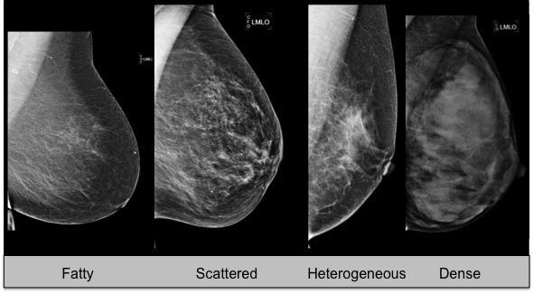
151 >/= 95% likelihood of malignancy Management: 4 th edition stated appropriate action should be take and 5 th edition states biopsy should be performed in the absence of clinical contraindication
152 BI-RADS category 6 Special circumstance when breast imaging is performed after a tissue diagnosis of malignancy but prior to complete surgical excision Examples: second opinions target lesion already biopsied or monitoring response to neo-adjuvant chemotherapy
153 Special cases regarding category 6: Post lumpectomy with positive resection margins and finding on imaging *** if positive margins and benign findings on mammography, it is BI-RADS 2 with additional sentence pathology report suggests possibility of residual tumor despite absence of mammographic correlate
154 Edition 4 stated Appropriate action should be taken and Edition 5 states surgical excision when clinically appropriate If there is a suspicious finding on the imaging study in addition to the known malignancy, then use category 4 or 5 Second opinions that require additional imaging work up should be category 0 Do not include category 6 in audit
155 BI-RADS category 0 Usually used in screening mammography or sonography 1. Additional imaging evaluation needed 2. Need previous exams for comparison if needed to make a final assessment 3. Technical repeat examination
156 When using category 0 to indicate need for prior exams, the facility needs to have in place tracking procedure to ensure that the patient will be recalled for imaging if films are not available in 30 days
157 Assessment and management for breast MRI Category 1 = negative Category 2 = benign; findings are described in the report Management: annual screening breast MRI and mammography in line with established guidelines for high risk screening
158 Category 3 = probably benign Evolving category Follow up of foci: if not considered BPE Follow up of masses: evaluate on morphology and kinetics and if considered benign may follow versus biopsy Follow up of NME: no, needs biopsy Interval follow up at 6, 12, 24 months
159 Category 4: suspicious and recommend tissue sampling - Not subdivided into 4A, 4B, 4C Category 5: highly suggestive of malignancy - Tissue sampling is recommended Category 6: Known biopsy proven malignancy
160 BREAST DENSITY (composition) breast composition measurement is about the volume of attenuating tissue in the breast 1. Removed the old area-based percentage thresholds 2. Switched from using numbers for the categories to letters
161 a. The breasts are almost entirely fatty b. There are scattered areas of fibroglandular density c. The breasts are heterogeneously dense, which may obscure small masses d. The breasts are extremely dense, which lowers the sensitivity of mammography
162
Update on MQSA and Mammography Accreditation
 MQSA - Who s Who Update on MQSA and Mammography Accreditation The Law: Mammography Quality Standards Act (MQSA) The Regulator: US Food and Drug Administration (FDA) Priscilla F. Butler, M.S. Senior Director,
HISTORY OF MQSA AND ACR
 HISTORY OF MQSA AND ACR DEBORAH THAMES R.T. (R)(M)(QM) WHY MQSA? In the United States, there was a lack of standards in mammography imaging. Reporting Imaging Type of imaging screening/diagnostic Equipment
Overview. ACR Update on FFDM Accreditation. New ACR Activities. QC Today. QC Tomorrow. ACR Breast Imaging Centers of Excellence BICOE
 ACR Update on FFDM Accreditation Overview New ACR Activities Eric Berns, PhD University of Colorado Hospital Denver Health Medical Center Denver, CO QC Today QC Tomorrow *No financial disclosures to report
BICOE Breast Imaging Center of Excellence. What is it? - Requirements. National Mammography Database. What do you get? ACR Accreditation in:
 BICOE Breast Imaging Center of Excellence What is it? - Requirements William Geiser, MS DABR Senior Medical Physicist MD Anderson Cancer Center Houston, Texas wgeiser@mdanderson.org ACR Accreditation in:
BICOE Stereotactic Breast Biopsy and Breast Ultrasound Accreditation. Introduction. Educational Objectives
 BICOE Stereotactic Breast Biopsy and Breast Ultrasound Accreditation William Geiser, MS DABR Senior Medical Physicist MD Anderson Cancer Center Houston, Texas wgeiser@mdanderson.org 1 Introduction Objectives
Overview. ACR Accreditation Update in Mammography. New ACR Activities. Requirements Today. What s New For Tomorrow. ACR: Recognized by FDA and CMS
 ACR Accreditation Update in Mammography Eric Berns, PhD University of Colorado Hospital Denver Health Medical Center Denver, CO *No financial disclosures to report Overview New ACR Activities Requirements
Mammography Quality Control: A Refresher
 Mammography Quality Control: A Refresher MATT WAIT, MS, DABR Objectives Attendees will re-familiarize themselves with the purpose of quality assurance and quality control Attendees will re-familiarize
BICOE Stereotactic Breast Biopsy and Breast Ultrasound Accreditation. Introduction. Educational Objectives
 BICOE Stereotactic Breast Biopsy and Breast Ultrasound Accreditation William Geiser, MS DABR Senior Medical Physicist MD Anderson Cancer Center Houston, Texas wgeiser@mdanderson.org 1 Introduction Objectives
Mammography. Background and Perspective. Mammography Evolution. Background and Perspective. T.R. Nelson, Ph.D. x41433
 - 2015 Background and Perspective 2005 (in US) Women Men Mammography Invasive Breast Cancer Diagnosed 211,240 1,690 Noninvasive Breast Cancer Diagnosed 58,940 Deaths from Breast Cancer 40,410 460 T.R.
The American College of Radiology Breast Ultrasound Accreditation Program: Frequently Asked Questions (Revised: July 31, 2017)
") The American College of Radiology Breast Ultrasound Accreditation Program: Frequently Asked Questions (Revised: July 31, 2017) Table of Contents APPLICATION - GENERAL... 1 MOVED FACILITIES AND UNITS...
The American College of Radiology Breast Ultrasound Accreditation Program: Frequently Asked Questions (Revised: July 31, 2017) Table of Contents APPLICATION - GENERAL... 1 MOVED FACILITIES AND UNITS...
ACR MRI Accreditation Program. ACR MRI Accreditation Program Update. Educational Objectives. ACR accreditation. History. New Modular Program
 ACR MRI Accreditation Program Update Donna M. Reeve, MS, DABR, DABMP Department of Imaging Physics University of Texas M.D. Anderson Cancer Center Educational Objectives Present requirements of the new
Q. Where can I find out if my state currently requires stereotactic breast biopsy accreditation?
 The American College of Radiology Stereotactic Breast Biopsy Accreditation Program: Frequently Asked Questions (Revised: September 7, 2017; updated questions in red) Table of Contents Application - General...
ACCREDITATION CASE REVIEW 2: US AND US-GUIDED BIOPSY CRITERIA
 ACCREDITATION CASE REVIEW 2: US AND US-GUIDED BIOPSY CRITERIA Stamatia Destounis, MD, FSBI, FACR Elizabeth Wende Breast Care, LLC Clinical Professor University of Rochester School of Medicine and Dentistry
Implementation of the 2012 ACR CT QC Manual in a Community Hospital Setting BRUCE E. HASSELQUIST, PH.D., DABR, DABSNM ASPIRUS WAUSAU HOSPITAL
 Implementation of the 2012 ACR CT QC Manual in a Community Hospital Setting BRUCE E. HASSELQUIST, PH.D., DABR, DABSNM ASPIRUS WAUSAU HOSPITAL Conflict of Interest Disclaimer Employee of Aspirus Wausau
REGULATION: QUALITY ASSURANCE PROGRAMS FOR MEDICAL DIAGNOSTIC X-RAY INSTALLATIONS N.J.A.C. 7:28-22
 REGULATION: QUALITY ASSURANCE PROGRAMS FOR MEDICAL DIAGNOSTIC X-RAY INSTALLATIONS N.J.A.C. 7:28-22 New Jersey Department of Environmental Protection Bureau of X-ray Compliance PO Box 420, Mail Code 25-01
Hong Kong College of Radiologists Mammography Statement
 Hong Kong College of Radiologists Mammography Statement The Hong Kong College of Radiologists would like to give the following comments concerning mammography. Mammography screening: Breast cancer is the
Accreditation Case Review: Mammography and Stereotactic Biopsy
 Accreditation Case Review: Mammography and Stereotactic Biopsy Brett T. Parkinson MD Breast Imaging Director Breast Care Services Intermountain Healthcare Chair, ACR Committee on Mammography Accreditation
Disclosures. Outline. Learning Objectives. Introduction. Introduction. Stereotactic Breast Biopsy vs Mammography: Image Quality and Dose.
 Disclosures Stereotactic Biopsy vs Mammography: and Dose None Vikas Patel, PhD, DABR Upstate Medical Physics 2014 Annual Meeting The American Association of Physicists in Medicine Austin, TX Learning Objectives
NONE. Disclosures. Accreditation Update
 ACR Ultrasound Accreditation: Requirements and Pitfalls Presented to: American Association of Physicists in Medicine Presented by: Jennifer Walter RDMS,RVT, RT(R) ACR Quality & Safety August 03, 2016 Disclosures
RADIATION PROTECTION IN DIAGNOSTIC AND INTERVENTIONAL RADIOLOGY. L19: Optimization of Protection in Mammography
 IAEA Training Material on Radiation Protection in Diagnostic and Interventional Radiology RADIATION PROTECTION IN DIAGNOSTIC AND INTERVENTIONAL RADIOLOGY L19: Optimization of Protection in Mammography
Kish chakrabarti, Ph.D. Senior Physicist CDRH/FDA
 Facility Certification Extension Requirements, Quality Assurance and Medical Physicists role for Hologic Selenia Dimensions Digital Breast Tomosynthesis (DBT) System Kish chakrabarti, Ph.D. Senior Physicist
The Radiology Aspects
 REQUIREMENTS FOR INTERNATIONAL ACCREDITATION OF BREAST CENTERS/UNITS The Radiology Aspects Miri Sklair-Levy, Israel RADIOLOGY GUIDELINES FOR QUALITY ASSURANCE IN BREAST CANCER SCREENING AND DIAGNOSIS Radiologists
MRI FAQs. After reading these documents and checking your protocols, you can apply online here: https://acredit.acr.org.
 MRI FAQs Application - General Q. My facility plans to apply for ACR MRI Accreditation, where do I start? A. Start by reading the following documents, available on the ACR website: The Diagnostic Modality
Introduction 1. Executive Summary 5
 Roman_pages 20-09-2005 21:01 Pagina IX Table of contents Introduction 1 Executive Summary 5 1. Epidemiological guidelines for quality assurance in breast cancer screening 15 1.10 Introduction 17 1.20 Local
ACR MRI Accreditation: Medical Physicist Role in the Application Process
 ACR MRI Accreditation: Medical Physicist Role in the Application Process Donna M. Reeve, MS, DABR, DABMP Department of Imaging Physics University of Texas M.D. Anderson Cancer Center Educational Objectives
The American College of Radiology Digital Mammography QC Manual: Frequently Asked Questions (Revised 12/12/2018; new and updated items in red)
") The American College of Radiology Digital Mammography QC Manual: Frequently Asked Questions (Revised 12/12/2018; new and updated items in red) Table of Contents General... 1 Applicability... 3 Transitioning
The American College of Radiology Digital Mammography QC Manual: Frequently Asked Questions (Revised 12/12/2018; new and updated items in red) Table of Contents General... 1 Applicability... 3 Transitioning
AAPM Annual Meeting. ACR Accreditation Update in CT
 AAPM Annual Meeting ACR Accreditation Updates in CT, Ultrasound, Mammography and MRI: ACR Accreditation Update in CT Michael McNitt-Gray, PhD, DABR, FAAPM Professor, Department of Radiological Sciences
Update on the ACR FFDM QC Manual
 Update on the ACR FFDM QC Manual Eric Berns, PhD University of Colorado Hospital Denver Health Medical Center Denver, CO AAPM 2011 Vancouver, Canada 9000 8500 8000 7500 7000 6500 6000 5500 5000 4500 4000
ACR Breast MRI Accreditation Program - DRAFT
 ACR Breast MRI Accreditation Program - DRAFT Donna M. Reeve, MS, DABR, DABMP Department of Imaging Physics Educational Objectives Provide an overview of the ACR Breast MRI Accreditation Program (BMRAP)
UPMC 1 Delineation of Privileges Request Criteria Summary Sheet
 UPMC 1 Facility: Specialty: UPMC Altoona Radiology KNOWLEDGE TRAINING Successful Completion of an ACGME/AOA, accredited program. The successful completion of an approved (ACGME/AOA) post graduate residency
CLINICAL PRIVILEGE WHITE PAPER
 Vascular Procedure ultrasound 57 Procedure 57 CLINICAL PRIVILEGE WHITE PAPER Vascular ultrasound Background Vascular ultrasound is a noninvasive procedure used to diagnose and locate disorders of the vascular
Session 2: The Role of Specialist Radiology Technologists
 Session 2: The Role of Specialist Radiology Technologists Louise M. Henderson, MSPH PhD Assistant Professor, Department of Radiology The University of North Carolina, Chapel Hill Overview Role of the technologist
Updates in Mammography. Dr. Yang Faridah A. Aziz Department of Biomedical Imaging University Malaya Medical Centre
 Updates in Mammography Dr. Yang Faridah A. Aziz Department of Biomedical Imaging University Malaya Medical Centre Updates in Mammography Breast Imaging Dr. Yang Faridah A. Aziz Department of Biomedical
ACR Accreditation Toolkit for Validation Site Surveys
 ACR Accreditation Toolkit for Validation Site Surveys The ACR will be performing unannounced validation site surveys as part of the accreditation process. This checklist is designed to assist you in gathering
Challenges to Delivery of High Quality Mammography
 Challenges to Delivery of High Quality Mammography Overview of Current Challenges Barbara Monsees, Washington University Geographic Access, Equity and Impact on Quality Tracy Onega, Dartmouth Medical School
Mammography. What is Mammography? What are some common uses of the procedure?
 Mammography What is Mammography? Mammography is a specific type of imaging that uses a low-dose x-ray system to examine breasts. A mammography exam, called a mammogram, is used to aid in the early detection
Implementation of a New Tomosynthesis Program: A Physicists Perspective
 Implementation of a New Tomosynthesis Program: A Physicists Perspective Bill Geiser, MS DABR Senior Medical Physicist wgeiser@mdanderson.org 1 Conflict of Interest None of the authors nor their immediate
9/3/2015. New Mammographic Modality Training. Mammographer. Qualified Radiologic Technologist. Tomosynthesis Training
 FDA Required Breast Tomosynthesis Training New Mammographic Modality Training On any new mammography technology, such as breast tomosynthesis, the Mammography Quality Standards Act (MQSA) (http:fda.gov/radiation-emitting
CT Quality Control Manual FAQs
 CT Quality Control Manual FAQs General Question: How often will the QC Manual be updated and how will those updates be communicated? Answer: The ACR CT Physics Subcommittee will review any comments, issues
Opportunities and Innovations in Digital Mammography John M. Sandrik, Ph.D. GE Healthcare Milwaukee, WI
 Opportunities and Innovations in Digital Mammography John M. Sandrik, Ph.D. GE Healthcare Milwaukee, WI john.sandrik@med.ge.com with many thanks to Vince Polkus, Advanced Applications Product Mgr. 1 Content
25 TEXAS ADMINSTRATIVE CODE (TAC) (x) Certification of Mammography Systems and Mammography Machines Used for Interventional Breast Radiography
 (x) Certification of Mammography Systems and Mammography Machines Used for Interventional Breast Radiography") 25 TEXAS ADMINSTRATIVE CODE (TAC) 289.230 Certification of Mammography Systems and Mammography Machines Used for Interventional Breast Radiography Texas Regulations for Control of Radiation (effective
25 TEXAS ADMINSTRATIVE CODE (TAC) 289.230 Certification of Mammography Systems and Mammography Machines Used for Interventional Breast Radiography Texas Regulations for Control of Radiation (effective
Breast Imaging Essentials
 Breast Imaging Essentials Module 1 Transcript 2016 ASRT. All rights reserved. Breast Imaging Essentials Module 1 - Fundamentals of Breast Imaging 1. ASRT Animation 2. Welcome Welcome to Module 1 of Breast
MQSA Physicist Qualifications. Jon J. Erickson, Ph.D., DABR
 2027 North 36 th Street Saint Joseph, Mo 64506 (86) 390-90 (800) 306-4477 MQSA Physicist Qualifications Jon J. Erickson, Ph.D., DABR Contents Missouri Mammography Physicist Initial Qualifications Certificate
EARLY DETECTION: MAMMOGRAPHY AND SONOGRAPHY
 EARLY DETECTION: MAMMOGRAPHY AND SONOGRAPHY Elizabeth A. Rafferty, M.D. Avon Comprehensive Breast Center Massachusetts General Hospital Harvard Medical School Breast Cancer Screening Early detection of
Medical Diagnostic Imaging
 Medical Diagnostic Imaging Laboratories Medical Diagnostic Imaging Lab Name Location Person in Charge Programs Served Courses Served Patient Care and Management (2) Introduction to MDI Radiographic Technique
Digital mammography imaging from Carestream Health solutions for great workflow, productivity, and patient care.
 Digital Mammography Imaging on KODAK CR Systems Digital mammography imaging from Carestream Health solutions for great workflow, productivity, and patient care. Commercial distribution of the CR Mammography
The American College of Radiology Breast MRI Accreditation Program: Frequently Asked Questions (Updated: October 4, 2017)
") The American College of Radiology Breast MRI Accreditation Program: Frequently Asked Questions (Updated: October 4, 2017) To navigate in this document, either click a section in the Table of Contents or
The American College of Radiology Breast MRI Accreditation Program: Frequently Asked Questions (Updated: October 4, 2017) To navigate in this document, either click a section in the Table of Contents or
Mammography. What is Mammography?
 Scan for mobile link. Mammography Mammography is a specific type of breast imaging that uses low-dose x-rays to detect cancer early before women experience symptoms when it is most treatable. Tell your
EARLY DETECTION: MAMMOGRAPHY AND SONOGRAPHY
 EARLY DETECTION: MAMMOGRAPHY AND SONOGRAPHY Elizabeth A. Rafferty, M.D. Avon Comprehensive Breast Center Massachusetts General Hospital Harvard Medical School Breast Cancer Screening Early detection of
Ultrasound Accreditation Program Requirements
 Ultrasound Accreditation Program Requirements OVERVIEW... 2 MANDATORY ACCREDITATION TIME REQUIREMENTS... 2 PERSONNEL QUALIFICATIONS... 2 PHYSICIAN QUALIFICATIONS... 3 TECHNOLOGIST QUALIFICATIONS... 4 QUALITY
Routine Quality Assurance Cookbook
 This Cookbook is a companion guide to the AIUM Routine Quality Assurance (QA) for Diagnostic Ultrasound Equipment document, which outlines the basic QA requirements for AIUM-accredited practices. The Guide
Dental Extraoral X-ray Systems
 Dental Extraoral X-ray Systems PROPOSED REVISIONS TO 4732.XXXX, 1.0 4732.#### DENTAL EXTRAORAL X-RAY SYSTEMS; STATIONARY AND MOBILE. Subpart 1. Applicability. A registrant s x-ray system used for dental
Debbie Childs RDMS, RVT Sonographer Murphy Medical Center Murphy, NC
 Debbie Childs RDMS, RVT Sonographer Murphy Medical Center Murphy, NC Worked at Murphy Medical Center as a sonographer for 18 years Registered in Abdomen, OB/GYN, Breast, & Vascular Ultrasound ACR Accredited
Contrast-Enhanced Spectral Mammography
 Contrast-Enhanced Spectral Mammography Illuminating Breast Cancer Detection SenoBright HD TM gehealthcare.com/senobright Mammography is the most reliable imaging technique for breasts, but limitations
Breast Tomosynthesis. What is breast tomosynthesis?
 Scan for mobile link. Breast Tomosynthesis Breast tomosynthesis is an advanced form of mammography, a specific type of breast imaging that uses low-dose x-rays to detect cancer early when it is most treatable.
Radiation Physics Consultants, Inc. Medical Physicist. Mammography Qualifications, Continuing Experience and Continuing Education
 Radiation Physics Consultants, Inc. Mammography Qualifications, Continuing Experience and Continuing Education For: Steven T. Nicholas, M.S. DABMP MAP ID s. Each medical physicist who provides medical
Multicenter Trial, Phase I Assessment of 2-D FFDM Versus Combo of 2-D and 3-D Tomosynthesis in Breast Cancer Screening
 ACRIN PA 4006: Comparison of Full Field Digital Mammography with Digital Breast Tomosynthesis Image Acquisition in Relation to Screening Call-Back Rate Imaging Manual Multicenter Trial, Phase I Assessment
The Practice Standards for Medical Imaging and Radiation Therapy. Sonography Practice Standards
 The Practice Standards for Medical Imaging and Radiation Therapy Sonography Practice Standards 2017 American Society of Radiologic Technologists. All rights reserved. Reprinting all or part of this document
MAMMOGRAPHY QC: MORE THAN PHANTOM IMAGES. Stephanie Schofield RTR QC Technologist Nova Scotia Health Authority
 1 MAMMOGRAPHY QC: MORE THAN PHANTOM IMAGES Stephanie Schofield RTR QC Technologist Nova Scotia Health Authority Disclaimer 2 I have nothing to disclose. Outline 3 QC tests for the mammography technologist
Standards for Radiation Oncology
 Standards for Radiation Oncology Radiation Oncology is the independent field of medicine which deals with the therapeutic applications of radiant energy and its modifiers as well as the study and management
Agenda: Dental Cone Beam Imaging
 Cone Beam Imaging Agenda: Dental Cone Beam Imaging *Definition and Functionality *Usage and diagnostics benefits *Comparative radiation information *Federal regulatory responsibilities: manufacturing *State
Mammography: Analysis and Advanced Techniques
 Mammography: Analysis and Advanced Techniques March 25- Anaheim, CA & Simulcast June 3 - Indianapolis, IN & Simulcast Oct 7 - Montreal, QC Canada & Simulcast Dec 9 - Webinar only a division of Herzing
POSITIONING ACR REQUIREMENTS IMAGE REVIEW CATEGORIES DEFICIENCIES IN POSITIONING ACR REQUIREMENTS 3/28/2016 NUMBER 1 REASON FOR ACR FAILURE
 CERTIFYING AGENCIES MAMMOGRAPHY CLINICAL IMAGE EVALUATION Pam Fulmer, BA RT (R)(M)(QM) FDA approved certifying states States can only certify facilities within their state borders Illinois Iowa South Carolina
Dental Intraoral X-ray Systems
 Dental Intraoral X-ray Systems PROPOSED REVISIONS TO 4732.XXXX, 2.0 4732.#### DENTAL INTRAORAL X-RAY SYSTEMS; STATIONARY AND MOBILE. Commented [JC(1]: Based on part 4732.0880. Subpart 1. Applicability.
Mammography: Analysis and Advanced Techniques $50. Special Price. 8 credit seminar October 28, 2017 Knoxville, TN. a division of Herzing University
 Mammography: Analysis and Advanced Techniques $50 Special Price 8 credit seminar October 28, 2017 Knoxville, TN a division of Herzing University Seminar Schedule 8:00 am Registration and Coffee 8:30 am
Contrast-Enhanced Digital Mammography
 2015 ARRS Breast Symposium Contrast-Enhanced Digital Mammography John Lewin, M.D. Diversified Radiology of Colorado CEDM - Outline History Technique Literature Review / Cases Clinical Status Inexpensive,
created by high-voltage devices Examples include medical and dental x-rays, light, microwaves and nuclear energy
 What is radiation? Radiation is energy emitted from a source, that travels through space and can penetrate matter. Listed below are two types that we are exposed to and contribute to our overall radiation
Overview of the 2005 IOM Report Improving Breast Imaging Quality Standards
 Overview of the 2005 IOM Report Improving Breast Imaging Quality Standards Goals of 2005 Report Study commissioned by US Congress in preparation for reauthorization of MQSA in 2007. to determine whether
Scope of Practice for the Diagnostic Ultrasound Professional
 Scope of Practice for the Diagnostic Ultrasound Professional Copyright 1993-2000 Society of Diagnostic Medical Sonographers, Dallas, Texas USA: All Rights Reserved Worldwide. Organizations which endorse
SenoBright Contrast Enhanced Spectral Mammography Technology. Ann-Katherine Carton Sylvie Saab-Puong Matt Suminski
 SenoBright Contrast Enhanced Spectral Mammography Technology Ann-Katherine Carton Sylvie Saab-Puong Matt Suminski White Paper October 2012 SenoBright Contrast Enhanced Spectral Mammography Technology Ann-Katherine
Education: Bachelor s degree from an approved program recognized by the Joint Review Committee on Education in Radiologic Technology (JRCERT).
.") PART II HAWAII HEALTH SYSTEMS CORPORATION 5.800 STATE OF HAWAII 5.801 5.802 Minimum Qualification Specifications 5.803 for the Classes: 5.804 5.805 IMAGING SPECIALIST I, II, III, IV, V & VI Education:
PART II HAWAII HEALTH SYSTEMS CORPORATION 5.800 STATE OF HAWAII 5.801 5.802 Minimum Qualification Specifications 5.803 for the Classes: 5.804 5.805 IMAGING SPECIALIST I, II, III, IV, V & VI Education:
Preparing for Medical Physics Components of the ABR Core Examination
 Preparing for Medical Physics Components of the ABR Core Examination The ABR core examination for radiologists contains material on medical physics. This content is based on the medical physics that is
Diagnostic Imaging Providers Privileging Guidelines
 Diagnostic Imaging Providers Privileging Guidelines The following guidelines are intended to promote reasonable and consistent quality and safety standards for the provision of imaging services. These
Breast Cancer Imaging
 Breast Cancer Imaging I. Policy University Health Alliance (UHA) will cover breast imaging when such services meet the medical criteria guidelines (subject to limitations and exclusions) indicated below.
ASRT Position Statements
 ASRT Position Statements Effective June 25, 2017 Contents Collective Bargaining Units... 1 Conjoint Evaluation of Educational Programs... 1 Degree Requirements for Medical Imaging and Radiation Therapy
The IAC Standards and Guidelines for Dental CT Accreditation
 The IAC Standards and Guidelines for Dental CT Accreditation Table of Contents All entries in Table of Contents are linked to the corresponding sections. Introduction... 3 Part A: Organization... 4 Section
Prepublication Requirements
 Issued Prepublication Requirements Standards Revisions for Organizations Providing Fluoroscopy Services The Joint Commission has approved the following revisions for prepublication. While revised requirements
The Mammography Examination
 JANUARY 2015 5 The Mammography Examination The purpose of The American Registry of Radiologic Technologists (ARRT ) Mammography Examination is to assess the knowledge and cognitive skills underlying the
FDA Executive Summary
 Meeting of the Radiological Devices Advisory Panel On October 24, 22, the panel will discuss, make recommendations, and vote on a premarket approval application supplement (P83/S) to expand the indications
WHAT TO EXPECT. Breast Tomosynthesis An additional screening tool in the fight against breast cancer HOLOGIC. The Women's Health Company
 WHAT TO EXPECT Breast Tomosynthesis An additional screening tool in the fight against breast cancer HOLOGIC The Women's Health Company ...,. Screening for breast cancer Doctors and scientists agree that
Detailed Program of the second BREAST IMAGING AND INTERVENTIONS PROGRAM am am : Clinician s requirements from breast imaging
 Detailed Program of the second BREAST IMAGING AND INTERVENTIONS PROGRAM 2012 Day one, 2 nd November BREAST IMAGING AND INTERVENTIONS PROGRAM 2012 9.00 AM 9.10 am Introduction 9.10 am - 9.30 am : Clinician
Improving Methods for Breast Cancer Detection and Diagnosis. The National Cancer Institute (NCI) is funding numerous research projects to improve
 is funding numerous research projects to improve") CANCER FACTS N a t i o n a l C a n c e r I n s t i t u t e N a t i o n a l I n s t i t u t e s o f H e a l t h D e p a r t m e n t o f H e a l t h a n d H u m a n S e r v i c e s Improving Methods for
CANCER FACTS N a t i o n a l C a n c e r I n s t i t u t e N a t i o n a l I n s t i t u t e s o f H e a l t h D e p a r t m e n t o f H e a l t h a n d H u m a n S e r v i c e s Improving Methods for
Highmark Privileging Application. 1. Complete this application if you provide any diagnostic
 Highmark Privileging Application INSTRUCTIONS 1. Complete this application if you provide any diagnostic imaging services. 2. Please complete a separate application for each physical location where imaging
History of MQSA ACR creates testing procedures Oct Mammography Quality Standards Act Dec 1993 Interim Regulations Oct All mammog
 15 Years of MQSA, What works, what doesn t Jerry Soen, MS DABR History of MQSA 1990-9191 ACR creates testing procedures Oct. 1992 Mammography Quality Standards Act Dec 1993 Interim Regulations Oct. 1994
S. Murgo, MD. Chr St-Joseph, Mons Erasme Hospital, Brussels
 S. Murgo, MD Chr St-Joseph, Mons Erasme Hospital, Brussels? Introduction Mammography reports are sometimes ambiguous and indecisive. ACR has developped the BIRADS. BIRADS consists of a lexicon in order
Screening Mammograms: Questions and Answers
 CANCER FACTS N a t i o n a l C a n c e r I n s t i t u t e N a t i o n a l I n s t i t u t e s o f H e a l t h D e p a r t m e n t o f H e a l t h a n d H u m a n S e r v i c e s Screening Mammograms:
Since its introduction in 2000, digital mammography has become
 Review Article Smith A, PhD email : Andrew.smith@hologic.com Since its introduction in 2000, digital mammography has become an accepted standard of care in breast cancer screening and has paved the way
STRUCTURED EDUCATION REQUIREMENTS IMPLEMENTATION DATE: JULY 1, 2017
 STRUCTURED EDUCATION REQUIREMENTS Bone Densitometry The purpose of structured education is to provide the opportunity for individuals to develop mastery of discipline-specific knowledge that, when coupled
BI-RADS Update. Martha B. Mainiero, MD, FACR, FSBI Brown University Rhode Island Hospital
 BI-RADS Update Martha B. Mainiero, MD, FACR, FSBI Brown University Rhode Island Hospital No Disclosures BI-RADS History 1980s Quality Issues ACR Accreditation BI-RADS 1994 2003 4 th Edition MRI, US January
ACRIN 6666 IM Additional Evaluation: Additional Views/Targeted US
 Additional Evaluation: Additional Views/Targeted US For revised or corrected form check box and fax to 215-717-0936. Instructions: The form is completed based on recommendations (from ID form) for additional
Breast Imaging & You
 Breast Imaging & You What s Inside: Breast Imaging... 2 Digital Breast Tomosynthesis (DBT) mammograms... 4 Breast cancer screening... 6 Dense breast tissue... 8 Automated Breast Ultrasound (ABUS)... 9
Hong Kong Association of Medical Physics. Guide to Candidates
 Hong Kong Association of Medical Physics Certification of Medical Physicists Professional Assessment Guide to Candidates PA App Form (r2017.1) Contents Chapter 1 p. 3 Professional Assessment System and
The New Jersey Radiographic Quality Assurance Program at 5 Years
 The New Jersey Radiographic Quality Assurance Program at 5 Years Julie Timins, MD a, Paul Orlando, BS b, Jill Lipoti, PhD b Purpose: Five years ago, the New Jersey Bureau of Radiological Health decided
DESCRIPTION: Percentage of final reports for screening mammograms that are classified as probably benign
 Quality ID #146 (NQF 0508): Radiology: Inappropriate Use of Probably Benign Assessment Category in Screening Mammograms National Quality Strategy Domain: Efficiency and Cost Reduction 2018 OPTIONS F INDIVIDUAL
Steven Tate Nicholas
 Hereby awards this Certificate of Completion to Steven Tate Nicholas 3 hours of HOLOGIC's Online Tomosynthesis Training For Medical Physicists in accordance with MQSA guidance for Digital 3-D Mammography
MR QA/QC for MRgRT. Rick Layman, PhD, DABR Department of Radiology July 13, 2015
 MR QA/QC for MRgRT Rick Layman, PhD, DABR Department of Radiology July 13, 2015 The Ohio State University Comprehensive Cancer Center Arthur G. James Cancer Hospital and Richard J. Solove Research Institute
Melissa Hartman, DO Women s Health Orlando VA Medical Center
 Melissa Hartman, DO Women s Health Orlando VA Medical Center Most common non-skin cancer and Second deadliest cancer in women Majority are diagnosed by abnormal screening study An approach to breast cancer
Breast Tomosynthesis An additional screening tool in the fight against breast cancer
 What to Expect Breast Tomosynthesis An additional screening tool in the fight against breast cancer Every woman over 40 should be examined for breast cancer once a year. American Cancer Society What to
CURRENTLY FDA APPROVED ARE FULL FIELD DIGITAL MAMMOGRAPHY SYSTEMS AND FILM SCREEN STILL BEING USED AT SOME INSTITUTIONS
 ABBY DUROJAYE,M.D CURRENTLY FDA APPROVED ARE FULL FIELD DIGITAL MAMMOGRAPHY SYSTEMS AND FILM SCREEN STILL BEING USED AT SOME INSTITUTIONS BOTH HAVE BEEN SHOWN TO BE EFFECTIVE TOOLS EARLY DETECTION OF BREAST
ACR Accreditation Update in MRI
 ACR Accreditation Update in MRI Whole Body Systems Extremity (MSK) Ron Price Vanderbilt University Medical Center Nashville, TN Dedicated Breast MRI Accreditation Update 1. ACR MRI Accreditation: Overview,