Accreditation Case Review: Mammography and Stereotactic Biopsy
|
|
|
- Blaze Mark Hubbard
- 6 years ago
- Views:
Transcription

1
2 Accreditation Case Review: Mammography and Stereotactic Biopsy Brett T. Parkinson MD Breast Imaging Director Breast Care Services Intermountain Healthcare Chair, ACR Committee on Mammography Accreditation April, 2016

3 The Janice Beesley Hartvigsen Breast Care Center at Intermountain Medical Center
4 Clinical Image Evaluation Scoring Procedures for Screen Film and Full Field Digital Mammography Mammography Accreditation Program
5 Accreditation Objectives Improve quality of mammography Reason for clinical image review -Quality of patient imaging is key -Provide feedback for improvement Uniform assessment of performance

6 Course Objectives Reasons for failed clinical images - Categories - Deficiencies - Problem solving and correcting deficiencies - Improving overall quality of images FS and FFDM criteria are generally same

7


8

9
10
11 Imaging Category Failure Rate (%) Positioning 1250 (20) Exposure 944 (15) Compression 887 (14) Sharpness 806 (13) Contrast 785 (13) Artifacts 703 (11) Labeling 465 (8) Noise 288 (5) Total 6128 (100)
12 Breast Density Past: The Old Descriptors
")


13 Type 1 Almost entirely fat (<10% fibroglandular) Fatty Type 2 Scattered fibroglandular densities (10%-50%) Type 3 Heterogene-ously dense (51%-75% fibroglandular) Dense Type 4 Extremely dense (>75% fibroglandular)
14 Breast Density Present: BI-RADS 2013
15 Breast Composition Categories The breasts are almost entirely fatty There are scattered areas of fibroglandular density The breast are heterogeneously dense, which may obscure small masses The breasts are extremely dense, which lowers the sensitivity of mammography (ACR BI-RADS ATLAS, 2013)
16 Fatty

17
18 Scattered

19
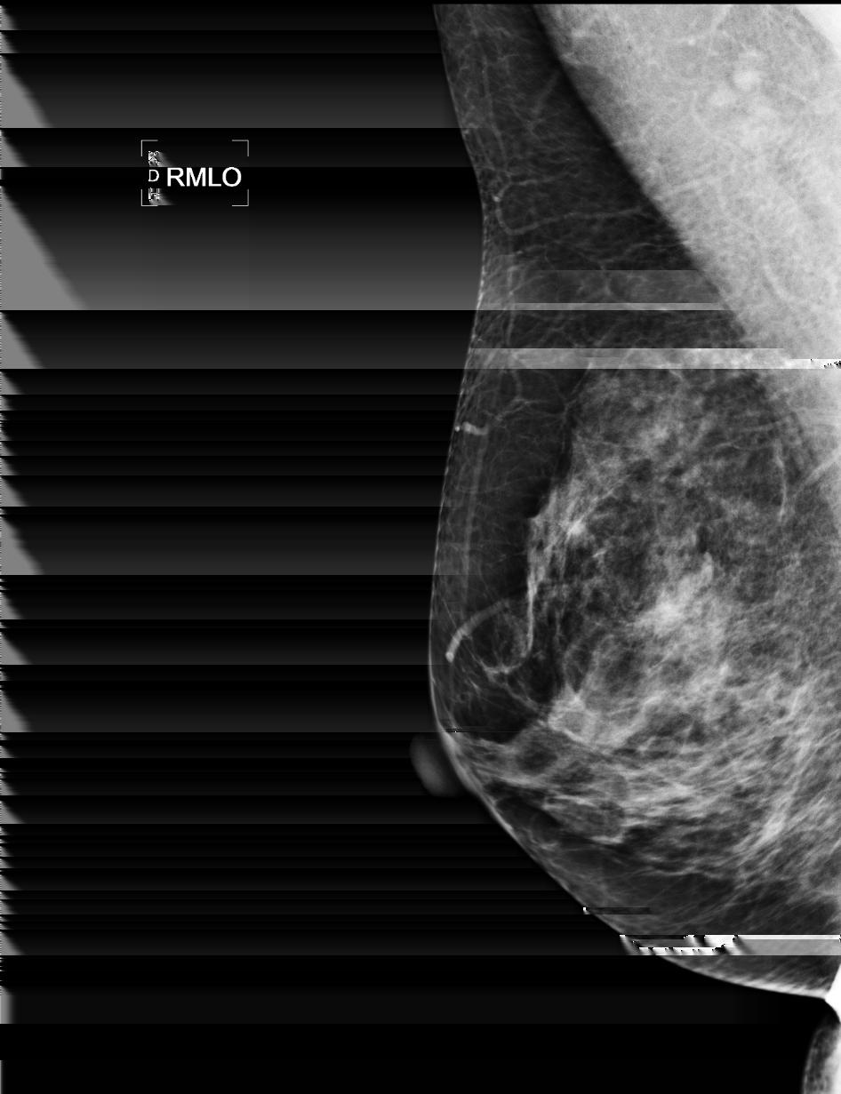
20 Heterogeneously Dense

21
22 Extremely Dense

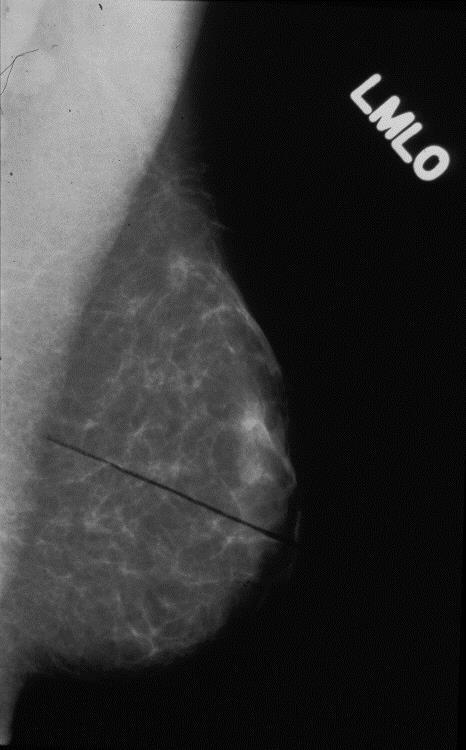
23
24 Breast Density Future We await publication of robust volume-based breast density data, using validated percentage cut points (not necessarily quartiles) that are readily and reproducibly determined at imaging, before again indicating percentage ranges for BI-RADS density categories. (ACR BI-RADS ATLAS, p 126, 2013)
25 Overall Clinical Evaluation Form ATTRIBUTE PROBLEM(S) NOTED POSSIBLE CAUSE(S) MLO: Poor visualization of posterior tissues Technologist technique MLO: Sagging breast Inappropriate mammographic projections MLO: Inadequate amount of pectoral muscle shown on image Wrong size recording system MLO: Inadequate inframammary fold (IMF) Uncertain CC: Poor visualization of posterior tissues CC: Excessive exaggeration A. Positioning Portion of breast cut off Skin folds Other body parts projected over breast Breast positioned too high on image receptor Posterior nipple line (PNL) on CC not within 1 cm of MLO PNL B. Compression Poor separation of parenchymal densities Non-uniform exposure levels Patient motion Under compression by technologist Unsuitable compression device Technologist positioning of compression device Uncertain C. Exposure Level D. Contrast Generalized underexposure Generalized overexposure Inadequate penetration of dense areas Excessive penetration of lucent areas Inadequate contrast Excessive contrast Film development Under compression with phototiming Radiologist preference Phototimer variability Uncertain Film development Improper kvp Excessive scatter Underexposure Digital: window width too wide Digital: window width too narrow Uncertain
26 Overall Clinical Evaluation Form ATTRIBUTE PROBLEM(S) NOTED POSSIBLE CAUSE(S) Poor delineation of linear structures Patient motion Poor delineation of feature margins Poor screen contact E. Sharpness Poor delineation of microcalcifications Film-screen selection Uncertain Visually striking mottle pattern Film development Noise limited visualization of detail Recording system speed Improper kvp F. Noise Digital: inadequate SNR Digital: window width too narrow Uncertain Punctate or lint Poor screen maintenance Scratches or pickoff Development related Roller marks Unsuitable grid or Bucky Grid related artifacts Film exposed to light Hair, deodorant, etc Lack of patient preparation G. Artifacts Poor screen-film alignment Digital: detector calibration (e.g. uniformity calibration) Digital: image receptor artifact Digital: foreign objects calibrated into calibration file Digital: laser printer artifact Digital: laser printer needs service Digital: laser printer scanning lines Uncertain Film handling Poor cassette closure Film fogging Damaged cassette H. Exam ID Patient name and additional patient identifier Facility name and location (city, state and zip) Date of examination View and laterality Unit identification (if more than one) Technologist identification Cassette-screen identification Technologist error Missing or non-standard labeling method Improper positioning of label Uncertain
27 Positioning: MLO Most Common Deficiencies Deficiency Frequency (%) Inadequate pectoral muscle 35 Sagging of the breast 22 Poor visualization posterior tissue 22 Skin folds overlying breast tissue 10 Breast positioned too high on image receptor 6 Portion of breast cut off 5


28 RMLO M LMLO N F FG IF FG = fibroglandular tissue; M = Pectoralis muscle; N = Nipple; IF = inframammary fold

29 RMLO Adequate Pectoral Muscle Optimal pectoral muscle on a digital image Length - To nipple line or below Shape - Convex anterior border - Wider superiorly - Gradually narrows inferiorly

30 RMLO Inadequate Pectoral Muscle When compared with a straight line, at a minimum the muscle should align with the line or go beyond Preferably, it will bulge beyond the edge Note difference in width of superior vs inferior aspect of muscle
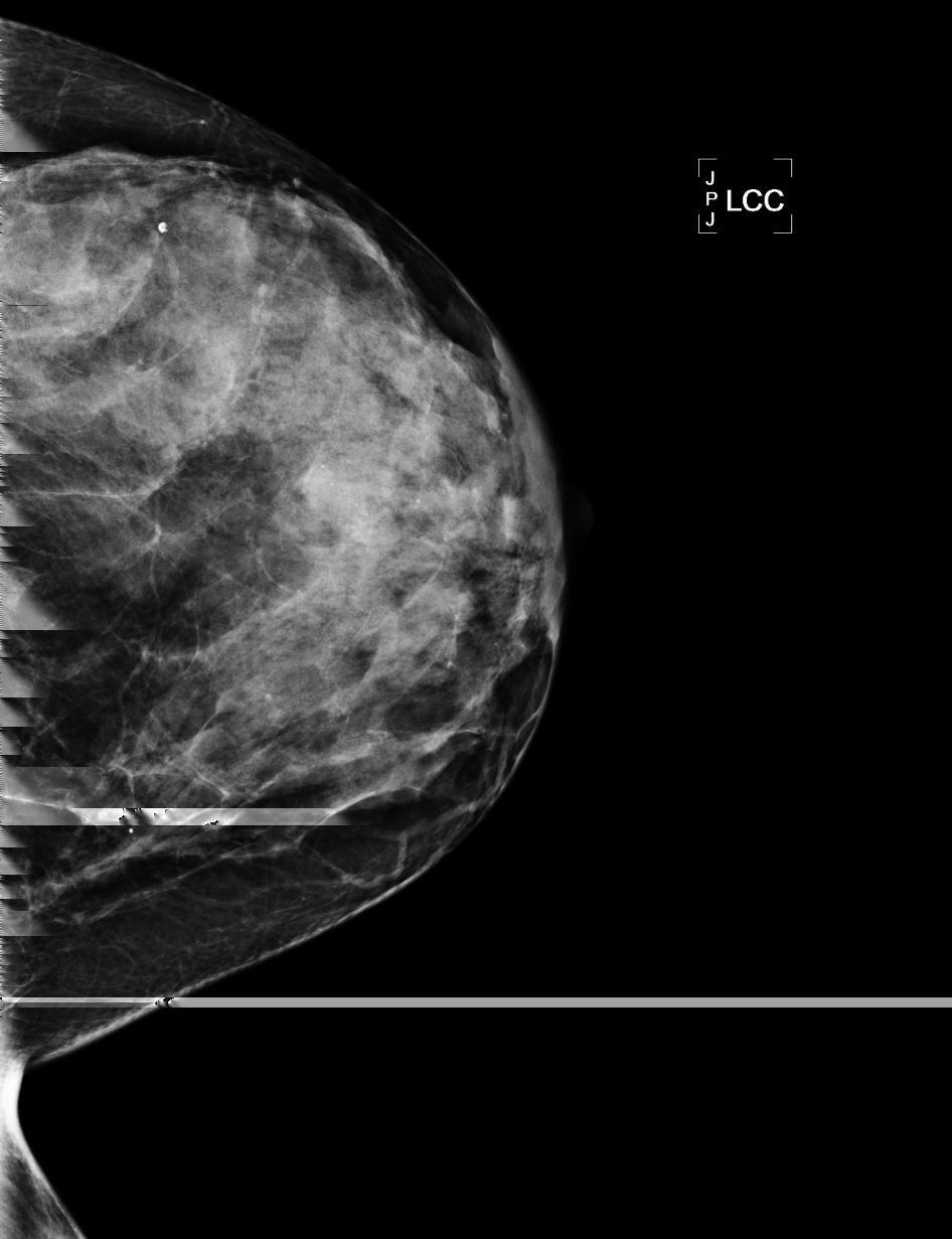
31 Sagging Breast: Poor Delineation of Structures Inadequate muscle Overlapping breast tissues N skin fold Fail


32 To prevent sagging: Out and Nipple-Up Maneuver

33 Sagging breast Inadequate muscle Repeated exam N skin fold N

34 Sagging Breasts with Inframammary Skin Folds Fail
35 Open IMF


36 Open Inframammary Fold MLO Skin overlying abdomen in front of edge of receptor Free of skin folds IMF
37 Positioning Criteria: CC Inclusion of medial tissue Nipple centered on film - No excessive exaggeration - Exaggeration may cause loss of medial or lateral posterior tissue Inclusion of posterior tissue Free of skin folds

38 Positioning: CC Most Common Deficiencies Deficiency Frequency (%) Poor visualization of posterior tissue on CC Posterior nipple line on CC not within 1 cm of MLO Excessive lateral/medial exaggeration on CC Skin folds overlying breast tissue
39 Visualization of Posterior Breast on CC PNL should measure within 1 cm of MLO - Depends on a well positioned MLO Pectoral muscle can be seen in 30% of women Requires proper positioning of CC: - Elevate inferior breast (freely movable tissue) - Pull superior and inferior tissue onto receptor - Lean patient s head forward to the side of tube Pectoral muscle in central and medial breast - Improper positioning if central and lateral
 on CC should be within")
40 RCC LCC LMLO PNL N FG F N PNL M N PNL (Posterior Nipple Line) on CC should be within 1cm of MLO
41 N PNL = 10 cm N PNL = 11.5 cm IMF was not elevated IMF properly elevated

42 Inclusion of Medial Tissue Skin reflection of cleavage visualized Retroglandular fat - Sternum flush with bucky - Contralateral breast draped Free of skin folds - IMF properly elevated - Check for gap - Must look from medial side
43 Nipple Centered on Film No excessive exaggeration Otherwise, nipple will point to the missing tissue - If points laterally RT should lift and pull out lateral tissue - If points medially RT should rotate to place sternum flush with bucky N RCC PNL N

44 Excessive Exaggeration Nipple will point to the laterality of missing tissue N - If it points laterally, lift and pull out lateral tissue - If points medially, rotate to place sternum flush with bucky Borderline


45 Skin Folds Overlying Breast Fail


46 Inframammary skin folds Borderline Pass, possibly a Fail, Depend on other images: Pec

47 Medial skin folds on CC Borderline
48 Affects sharpness Compression - Compression decreases unsharpness (blur) due to reduced time and motion Affects contrast - Good compression increases contrast due to less scattered radiation Affects exposure level - Compression increases exposure level due to reduced breast thickness
49 Compression: Most Common Deficiencies Deficiency Frequency (%) Poor separation of fibroglandular tissues 65 Non-uniform exposure of fibroglandular tissues 19 Patient motion 16

50 Inadequate Compression Poor separation of fibroglandular tissues Unequal exposure of fibroglandular tissues Motion unsharpness
 Subareolar area Inferior breast - Edges of vessels, calcifications, and Cooper s")
51 Assessing Sharpness on the MLO Anterior edge of pectoral muscle (blurred?) Subareolar area Inferior breast - Edges of vessels, calcifications, and Cooper s ligaments Fail


52 Compression Year 1 Year 2 Year 3 Fail Borderline Pass

53 Assessing Sharpness on CC Subareolar area Medial breast - Edges of vessels, calcification, ligaments
54 Exposure Level Minimum average optical density (OD) in fibroglandular tissue should be 1.0 when measured with a spot densitometer Fibroglandular tissue should be a shade of gray Pectoral muscle may have OD under 1.0; however, must be able to see underlying tissue Uniformly washed out look indicates under- exposure on screen film images
55 Exposure Level: Most Common Deficiencies Deficiency Generalized underexposure Inadequate exposure of dense tissues Generalized overexposure Overexposure of radiolucent tissues Frequency (%) (80) (20)


56 Inadequate exposure = inadequate contrast RMLO LMLO RMLO LMLO Initial Exam Fail 6 Months Later Pass


57 RCC LCC RCC LCC Initial Exam 6 Months Later
58 Contrast Differences in optical densities between different tissues. Fat should have high optical density (blacker) and fibro-glandular tissue should have much lower optical density (whiter).
59 Contrast: Most Common Deficiencies Deficiency Frequency (%) Inadequate contrast 90 Excessive contrast 10

60 Contrast Inadequate Good Excessive


61 Good Contrast Film-Screen and Digital: Same Patient LCC LCC
62 Sharpness Unsharpness (often referred to as blur by medical physicists) Blurring of margins of vessels and Cooper s ligaments Unclear margins of masses Unsharpness of calcifications
63 Assessing Sharpness on the MLO Inferior third of the breast Subareolar area Anterior edge of pectoral muscle Edges of vessels, calcification, and Cooper s ligaments

64 Inadequate Compression Motion unsharpness seen as poor delineation of linear structures This is usually most evident in inferior and subareolar aspect of MLO Also look at the edge of the pectoral muscle Fail Edge of pectoral muscle


65 Motion unsharpness Borderline
66 Assessing Sharpness on the CC Medial aspect of the breast Subareolar area Edges of vessels, calcifications, Cooper s ligament

67 Unsharpness related to inadequate compression and motion more likely to occur on MLO If question about sharpness, compare CC to MLO
68 Noise Mottled pattern of areas of relatively equal tissue density Noise limits visualization of details in the image
69 Mottled pattern of areas of relatively equal tissue density

70 If noise suspected, it helps to use a mag lens

71 Mottled pattern makes it difficult to see fine details in the images
72 Failing Score A substantial deficiency in any category can cause a failure Multiple borderline deficiencies in multiple categories can also cause a failure Category G: Artifacts Minor artifacts will not cause a fail prominent artifacts will be considered borderline Severe artifacts will cause a fail Category H: Examination ID If the patient s name, patient ID or R/L are missing, the examination will fail Other minor exam ID deficiencies will not fail
73 Case 1

74
75
76 Case 2

77

78
79 Case 3

80

81
82 Case 4

83


84
85 Case 5


86

87
88 Case 6

89

90
91 Case 7

92


93
94 Case 8


95


96
97 Case 9


98


99

100 ACR Stereotactic Breast Biopsy Accreditation Program

101
102 SBBAP: Physician Qualifications ACR and the American College of Surgeons agreed on and published guidelines for physician training, qualifications, and continuing experience* Collaborative setting: radiologists and surgeons work together Independent settings: radiologists or surgeons work independently * (ACR Bull, Jul 1998; Bull Am Coll Surg, May 1998 )
103 SBBAP: Physician Qualifications - Collaborative Setting Qualifications Radiologist* Other Physician Initial Performed 12 stereotactic breast Bx procedures or 3 hands-on stereotactic breast Bx procedures under a qualified physician AND 3 hours of Category 1 CME in stereotactic breast Bx AND Experienced in recommendations for Bx and lesion identification at time of Bx AND Qualified as an interpreting physician under MQSA AND 3 hours of Category 1 CME in stereotactic breast Bx (that includes image triangulation for lesion location) AND Experienced in post-bx patient management *radiologists must be currently qualified as an interpreting physician under MQSA
104 SBBAP: Physician Qualifications - Collaborative Setting Qualifications Radiologist Other Physician Continuing Experience Upon renewal, 36 image-guided breast Bx in prior 36 months; at least 9 of these must be stereotactic breast Bx Continuing Education Upon renewal: Currently meets MOC requirements for ABR, OR Completes 150 hours (including 75 Cat 1 CME) in prior 36 months pertinent to the physician s practice patterns, OR Completes 15 hours CME (half of which must be Category 1) in the prior 36 months specific to the imaging modality or organ system
105 Qualifications Radiologist* Other Physician Initial SBBAP: Physician Qualifications - Independent Setting Performed 12 stereotactic breast Bx procedures or 3 hands-on stereotactic breast Bx procedures under a qualified physician AND 3 hours of Category 1 CME in stereotactic breast Bx AND 15 hours of Category 1 CME in breast imaging including pathophysiology of benign and malignant breast disease as well as clinical breast examinations AND Qualified as an interpreting physician under MQSA AND 15 hours of Category 1 CME in stereotactic breast Bx or 3 years experience having performed 36 stereotactic breast Bx AND 4 hours of Category 1 CME in medical radiation physics AND Evaluated 480 mammograms every 2 years in consultation with MQSA-qualified physician *radiologists must be currently qualified as an interpreting physician under MQSA
106 SBBAP: Physician Qualifications - Independent Setting Qualifications Radiologist Other Physician Continuing Experience Upon renewal, 36 image-guided breast Bx in prior 36 months; at least 9 of these must be stereotactic breast Bx Upon renewal, 36 image-guided breast Bx in prior 36 months; at least 9 of these must be stereotactic breast Bx AND Evaluate 720 mammograms in the prior 36 months in consultation with MQSA-qualified physician Continuing Education Upon renewal: Currently meets MOC requirements for ABR, OR Completes 150 hours (including 75 Cat 1 CME) in prior 36 months pertinent to the physician s practice patterns, OR Completes 15 hours CME (half of which must be Category 1) in prior 36 months specific to the imaging modality or organ system
107 SBBAP: Radiological Technologist Qualifications Initial Qualifications Radiological Technologist Qualified to perform mammography under MQSA AND 3 Category A CEUs in stereotactic breast Bx AND Performed 5 stereotactic breast Bx procedures under supervision of a qualified physician or technologist Continuing Experience Upon renewal, 24 stereotactic breast Bx exams in prior 24 months
108 Qualifications SBBAP: Radiological Technologist Qualifications Radiological Technologist Continuing Education Registered technologists - In compliance with CE requirements of certifying organization for the imaging modality in which they perform services - CE includes credits pertinent to tech s accredited practice State licensed technologists - 24 hours of CE every 2 years - CE relevant to imaging and radiologic sciences, patient care - CE includes credits pertinent to tech s ACR accredited practice All others - 24 hours of CE every 2 years - CE relevant to imaging and radiologic sciences, patient care - CE includes credits pertinent to tech s accredited practice
109 SBBAP: Medical Physicist Qualifications Initial Qualifications Medical Physicist Qualified to perform mammography surveys under MQSA AND Performed 1 hands-on stereotactic breast Bx survey under a qualified medical physicist or at least 3 independent surveys prior to 6/1/97 Continuing Experience Continuing Education Upon renewal, 2 stereotactic breast Bx physics surveys over a 24-month period Upon renewal, 3 CEUs in stereotactic breast Bx every 3 years
110 SBBAP: Quality Assurance - Outcome Data 110 Total number of procedures Total number of cancers found Total number of benign lesions Total number of stereotactic Bx needing repeat Bx (open excisional or stereotactic Bx) Insufficient sample Non-concordance with imaging Ductal atypia, radial scar Other Total number of complications Hematomas requiring surgical attention Infections requiring treatment Other

111 SBBAP Pass Rate 82% of units passed on 1 st attempt (2011) All units at the site must pass evaluation for accreditation to be maintained A certificate and decal will be issued for each unit Accreditation is granted for three years
 much higher than in MAP After corrective action, less than 2% of total applicants fail")
112 SBBAP: Reasons for Failures 1 st attempt: Failures are primarily clinical, targeting issues Phantom failures also show image quality problems Dose failures (300mrad) much higher than in MAP After corrective action, less than 2% of total applicants fail again
113 Image Submission Requirements
114 Gun-needle biopsy probe required images a. A 2-view mammogram Circle the calcifications Print the images true size (i.e., without magnification or minification) or with a scale. Label the images Calc Mammo 1 and Calc Mammo 2 using the ACR barcode labels. b. A specimen radiograph Label the image Specimen Radiograph using the ACR barcode label. c. A Pre-Fire stereo Pair Label the images Calc Pre Fire Str Pair using the ACR barcode labels.
115 Vacuum-suction biopsy probe or other FDA-approved core biopsy device a. Pre-biopsy mammogram and specimen radiograph same as gunneedle biopsy probe. b. Pre-Biopsy (post fire) stereo pair Label the images Calc Pre Biopsy Str Pair using the ACR barcode labels. OR c. Pre-Fire Stereo Pair Label the images Calc Pre Fire Str Pair using the ACR barcode labels.


116

117

118 PRE FIRE

119 POST FIRE/PRE BIOPSY

120

121
122

123
124

125

126
127 Questions?
POSITIONING ACR REQUIREMENTS IMAGE REVIEW CATEGORIES DEFICIENCIES IN POSITIONING ACR REQUIREMENTS 3/28/2016 NUMBER 1 REASON FOR ACR FAILURE
 CERTIFYING AGENCIES MAMMOGRAPHY CLINICAL IMAGE EVALUATION Pam Fulmer, BA RT (R)(M)(QM) FDA approved certifying states States can only certify facilities within their state borders Illinois Iowa South Carolina
MAMMOGRAPHY QC: MORE THAN PHANTOM IMAGES. Stephanie Schofield RTR QC Technologist Nova Scotia Health Authority
 1 MAMMOGRAPHY QC: MORE THAN PHANTOM IMAGES Stephanie Schofield RTR QC Technologist Nova Scotia Health Authority Disclaimer 2 I have nothing to disclose. Outline 3 QC tests for the mammography technologist
6 Reasons Nipple Markers Add Value in Positioning and Image Critique:
 PROPER POSITIONING WITH THE USE OF NIPPLE MARKERS By Susan Sprinkle-Vincent, A.A.S., R.T. (R) (M) Mammography Program Manager and Consultant Advanced Health Education Center Houston, Texas Mammography
Breast Imaging Essentials
 Breast Imaging Essentials Module 8 Transcript 2017 ASRT. All rights reserved. Breast Imaging Essentials Module 8 Digital Procedures and Techniques 1. ASRT Animation 2. Welcome Welcome to Module 8 of Breast
The Radiology Aspects
 REQUIREMENTS FOR INTERNATIONAL ACCREDITATION OF BREAST CENTERS/UNITS The Radiology Aspects Miri Sklair-Levy, Israel RADIOLOGY GUIDELINES FOR QUALITY ASSURANCE IN BREAST CANCER SCREENING AND DIAGNOSIS Radiologists
Comparison Study: Impact of Thin Protective Coverlets on Positioning and Tissue Acquisition in Breast Imaging
 Comparison Study: Impact of Thin Protective Coverlets on Positioning and Tissue Acquisition in Breast Imaging Author: Steve K. Wagner Co-Author: Alex Merkulov, MD OBJECTIVE To determine whether the use
BICOE Breast Imaging Center of Excellence. What is it? - Requirements. National Mammography Database. What do you get? ACR Accreditation in:
 BICOE Breast Imaging Center of Excellence What is it? - Requirements William Geiser, MS DABR Senior Medical Physicist MD Anderson Cancer Center Houston, Texas wgeiser@mdanderson.org ACR Accreditation in:
ACCREDITATION DOCUMENT THE RADIOGRAPHER
 ACCREDITATION DOCUMENT THE RADIOGRAPHER Nijmegen, October 2012 1. Introduction An optimal quality of mammography is one of the fundamental requirements for successful breast cancer screening programmes.
BICOE Stereotactic Breast Biopsy and Breast Ultrasound Accreditation. Introduction. Educational Objectives
 BICOE Stereotactic Breast Biopsy and Breast Ultrasound Accreditation William Geiser, MS DABR Senior Medical Physicist MD Anderson Cancer Center Houston, Texas wgeiser@mdanderson.org 1 Introduction Objectives
Disclosure. Objectives 2/6/ Allina Health System. What Every Technologist Would Like Their Radiologists To Know
 What Every Technologist Would Like Their Radiologists To Know Louise C. Miller, RTRM Director of Education Mammography Educators San Diego, CA February 6, 2016 Disclosure There are no conflicts of interest
Q. Where can I find out if my state currently requires stereotactic breast biopsy accreditation?
 The American College of Radiology Stereotactic Breast Biopsy Accreditation Program: Frequently Asked Questions (Revised: September 7, 2017; updated questions in red) Table of Contents Application - General...
HISTORY OF MQSA AND ACR
 HISTORY OF MQSA AND ACR DEBORAH THAMES R.T. (R)(M)(QM) WHY MQSA? In the United States, there was a lack of standards in mammography imaging. Reporting Imaging Type of imaging screening/diagnostic Equipment
Mammography. What is Mammography? What are some common uses of the procedure?
 Mammography What is Mammography? Mammography is a specific type of imaging that uses a low-dose x-ray system to examine breasts. A mammography exam, called a mammogram, is used to aid in the early detection
BICOE Stereotactic Breast Biopsy and Breast Ultrasound Accreditation. Introduction. Educational Objectives
 BICOE Stereotactic Breast Biopsy and Breast Ultrasound Accreditation William Geiser, MS DABR Senior Medical Physicist MD Anderson Cancer Center Houston, Texas wgeiser@mdanderson.org 1 Introduction Objectives
Maintaining a quality mammography facility: Overview of ACR and MQSA Requirements and Update on BI-RADS 5 TH Edition
 Maintaining a quality mammography facility: Overview of ACR and MQSA Requirements and Update on BI-RADS 5 TH Edition Sandra S. Rao, M.D. Northwestern Medicine I have nothing to disclose. Objectives: Review
The American College of Radiology Digital Mammography QC Manual: Frequently Asked Questions (Revised 12/12/2018; new and updated items in red)
") The American College of Radiology Digital Mammography QC Manual: Frequently Asked Questions (Revised 12/12/2018; new and updated items in red) Table of Contents General... 1 Applicability... 3 Transitioning
The American College of Radiology Digital Mammography QC Manual: Frequently Asked Questions (Revised 12/12/2018; new and updated items in red) Table of Contents General... 1 Applicability... 3 Transitioning
RADIATION PROTECTION IN DIAGNOSTIC AND INTERVENTIONAL RADIOLOGY. L19: Optimization of Protection in Mammography
 IAEA Training Material on Radiation Protection in Diagnostic and Interventional Radiology RADIATION PROTECTION IN DIAGNOSTIC AND INTERVENTIONAL RADIOLOGY L19: Optimization of Protection in Mammography
Automating Quality Assurance Metrics to Assess Adequate Breast Positioning in Mammography
 Automating Quality Assurance Metrics to Assess Adequate Breast Positioning in Mammography Gerald R. Kolb, JD, The Breast Group, Sunriver, OR; Kaier Wang, PhD, VolparaSolutions, Wellington, NZ; Ariane Chan,
Session 2: The Role of Specialist Radiology Technologists
 Session 2: The Role of Specialist Radiology Technologists Louise M. Henderson, MSPH PhD Assistant Professor, Department of Radiology The University of North Carolina, Chapel Hill Overview Role of the technologist
Disclosures. Outline. Learning Objectives. Introduction. Introduction. Stereotactic Breast Biopsy vs Mammography: Image Quality and Dose.
 Disclosures Stereotactic Biopsy vs Mammography: and Dose None Vikas Patel, PhD, DABR Upstate Medical Physics 2014 Annual Meeting The American Association of Physicists in Medicine Austin, TX Learning Objectives
The American College of Radiology Breast Ultrasound Accreditation Program: Frequently Asked Questions (Revised: July 31, 2017)
") The American College of Radiology Breast Ultrasound Accreditation Program: Frequently Asked Questions (Revised: July 31, 2017) Table of Contents APPLICATION - GENERAL... 1 MOVED FACILITIES AND UNITS...
The American College of Radiology Breast Ultrasound Accreditation Program: Frequently Asked Questions (Revised: July 31, 2017) Table of Contents APPLICATION - GENERAL... 1 MOVED FACILITIES AND UNITS...
Mammography Quality Control: A Refresher
 Mammography Quality Control: A Refresher MATT WAIT, MS, DABR Objectives Attendees will re-familiarize themselves with the purpose of quality assurance and quality control Attendees will re-familiarize
Overview. ACR Update on FFDM Accreditation. New ACR Activities. QC Today. QC Tomorrow. ACR Breast Imaging Centers of Excellence BICOE
 ACR Update on FFDM Accreditation Overview New ACR Activities Eric Berns, PhD University of Colorado Hospital Denver Health Medical Center Denver, CO QC Today QC Tomorrow *No financial disclosures to report
Kish chakrabarti, Ph.D. Senior Physicist CDRH/FDA
 Facility Certification Extension Requirements, Quality Assurance and Medical Physicists role for Hologic Selenia Dimensions Digital Breast Tomosynthesis (DBT) System Kish chakrabarti, Ph.D. Senior Physicist
Mammography. Background and Perspective. Mammography Evolution. Background and Perspective. T.R. Nelson, Ph.D. x41433
 - 2015 Background and Perspective 2005 (in US) Women Men Mammography Invasive Breast Cancer Diagnosed 211,240 1,690 Noninvasive Breast Cancer Diagnosed 58,940 Deaths from Breast Cancer 40,410 460 T.R.
Update on MQSA and Mammography Accreditation
 MQSA - Who s Who Update on MQSA and Mammography Accreditation The Law: Mammography Quality Standards Act (MQSA) The Regulator: US Food and Drug Administration (FDA) Priscilla F. Butler, M.S. Senior Director,
THE DIAGNOSTIC WORKUP: THE TEAM APPROACH
 X-Ray Associates of New Mexico, P.C. THE DIAGNOSTIC WORKUP: THE TEAM APPROACH MICHAEL N. LINVER, MD, FACR DAWN DERENBURGER, RTRM Disclosure There are no conflicts of interest or relevant financial interests
Overview. ACR Accreditation Update in Mammography. New ACR Activities. Requirements Today. What s New For Tomorrow. ACR: Recognized by FDA and CMS
 ACR Accreditation Update in Mammography Eric Berns, PhD University of Colorado Hospital Denver Health Medical Center Denver, CO *No financial disclosures to report Overview New ACR Activities Requirements
Amammography report is a key component of the breast
 Review Article Writing a Mammography Report Amammography report is a key component of the breast cancer diagnostic process. Although mammographic findings were not clearly differentiated between benign
9/3/2015. New Mammographic Modality Training. Mammographer. Qualified Radiologic Technologist. Tomosynthesis Training
 FDA Required Breast Tomosynthesis Training New Mammographic Modality Training On any new mammography technology, such as breast tomosynthesis, the Mammography Quality Standards Act (MQSA) (http:fda.gov/radiation-emitting
Financial Disclosures
 Financial Disclosures 3D Mammography: The Latest Developments in the Breast Imaging Arena I have no financial disclosures Dr. Katharine Lampen-Sachar Breast and Body Radiologist Radiology Associates of
Stereotactic Breast Biopsy
 Scan for mobile link. Stereotactic Breast Biopsy Stereotactic breast biopsy uses mammography a specific type of breast imaging that uses low-dose x-rays to help locate a breast abnormality and remove a
Multicenter Trial, Phase I Assessment of 2-D FFDM Versus Combo of 2-D and 3-D Tomosynthesis in Breast Cancer Screening
 ACRIN PA 4006: Comparison of Full Field Digital Mammography with Digital Breast Tomosynthesis Image Acquisition in Relation to Screening Call-Back Rate Imaging Manual Multicenter Trial, Phase I Assessment
UW Radiology Review Course Breast Calcifications. BI-RADS 5 th Edition
 UW Radiology Review Course Breast Calcifications Grace Kalish, MD Vantage Radiology BI-RADS 5 th Edition Benign Skin Vascular Large rod like Coarse popcorn Suspicious Amorphous Coarse heterogenous Fine
Mammography. What is Mammography?
 Scan for mobile link. Mammography Mammography is a specific type of breast imaging that uses low-dose x-rays to detect cancer early before women experience symptoms when it is most treatable. Tell your
ACRIN 6666 IM Additional Evaluation: Additional Views/Targeted US
 Additional Evaluation: Additional Views/Targeted US For revised or corrected form check box and fax to 215-717-0936. Instructions: The form is completed based on recommendations (from ID form) for additional
Breast Tomosynthesis. What is breast tomosynthesis?
 Scan for mobile link. Breast Tomosynthesis Breast tomosynthesis is an advanced form of mammography, a specific type of breast imaging that uses low-dose x-rays to detect cancer early when it is most treatable.
The Mammography Examination
 JANUARY 2015 5 The Mammography Examination The purpose of The American Registry of Radiologic Technologists (ARRT ) Mammography Examination is to assess the knowledge and cognitive skills underlying the
Mammography: Analysis and Advanced Techniques $50. Special Price. 8 credit seminar October 28, 2017 Knoxville, TN. a division of Herzing University
 Mammography: Analysis and Advanced Techniques $50 Special Price 8 credit seminar October 28, 2017 Knoxville, TN a division of Herzing University Seminar Schedule 8:00 am Registration and Coffee 8:30 am
S. Murgo, MD. Chr St-Joseph, Mons Erasme Hospital, Brussels
 S. Murgo, MD Chr St-Joseph, Mons Erasme Hospital, Brussels? Introduction Mammography reports are sometimes ambiguous and indecisive. ACR has developped the BIRADS. BIRADS consists of a lexicon in order
EXAMINATION CONTENT SPECIFICATIONS ARRT BOARD APPROVED: JANUARY 2017 IMPLEMENTATION DATE: JULY 1, 2017
 EXAMINATION CONTENT SPECIFICATIONS Mammography The purpose of the mammography examination is to assess the knowledge and cognitive skills underlying the intelligent performance of the tasks typically required
ACCREDITATION CASE REVIEW 2: US AND US-GUIDED BIOPSY CRITERIA
 ACCREDITATION CASE REVIEW 2: US AND US-GUIDED BIOPSY CRITERIA Stamatia Destounis, MD, FSBI, FACR Elizabeth Wende Breast Care, LLC Clinical Professor University of Rochester School of Medicine and Dentistry
Contrast-Enhanced Spectral Mammography
 Contrast-Enhanced Spectral Mammography Illuminating Breast Cancer Detection SenoBright HD TM gehealthcare.com/senobright Mammography is the most reliable imaging technique for breasts, but limitations
FDA Executive Summary
 Meeting of the Radiological Devices Advisory Panel On October 24, 22, the panel will discuss, make recommendations, and vote on a premarket approval application supplement (P83/S) to expand the indications
BI-RADS Update. Martha B. Mainiero, MD, FACR, FSBI Brown University Rhode Island Hospital
 BI-RADS Update Martha B. Mainiero, MD, FACR, FSBI Brown University Rhode Island Hospital No Disclosures BI-RADS History 1980s Quality Issues ACR Accreditation BI-RADS 1994 2003 4 th Edition MRI, US January
8/31/2016 HIDING IN PLAIN SITE, ARCHITECTURAL DISTORTIONS AND BREAST ASYMMETRIES ARCHITECTURAL DISTORTIONS ARCHITECTURAL DISTORTIONS
 HIDING IN PLAIN SITE, ARCHITECTURAL DISTORTIONS AND BREAST ASYMMETRIES DEBORAH THAMES R.T. (R)(M)(QM) ARCHITECTURAL DISTORTIONS Definition is disruption of the natural flow of breast pattern towards the
TOMOSYNTHESIS: WORTH ALL THE HYPE?
 X-Ray Associates of New Mexico, P.C. TOMOSYNTHESIS: WORTH ALL THE HYPE? MICHAEL N. LINVER, MD, FACR MAMMOGRAPHY: THE GOOD, THE PRETTY GOOD, & THE NOT SO GOOD MAMMOGRAPHY: THE GOOD, THE PRETTY GOOD, & THE
UPMC 1 Delineation of Privileges Request Criteria Summary Sheet
 UPMC 1 Facility: Specialty: UPMC Altoona Radiology KNOWLEDGE TRAINING Successful Completion of an ACGME/AOA, accredited program. The successful completion of an approved (ACGME/AOA) post graduate residency
ACR MRI Accreditation: Medical Physicist Role in the Application Process
 ACR MRI Accreditation: Medical Physicist Role in the Application Process Donna M. Reeve, MS, DABR, DABMP Department of Imaging Physics University of Texas M.D. Anderson Cancer Center Educational Objectives
Challenges to Delivery of High Quality Mammography
 Challenges to Delivery of High Quality Mammography Overview of Current Challenges Barbara Monsees, Washington University Geographic Access, Equity and Impact on Quality Tracy Onega, Dartmouth Medical School
Hong Kong College of Radiologists Mammography Statement
 Hong Kong College of Radiologists Mammography Statement The Hong Kong College of Radiologists would like to give the following comments concerning mammography. Mammography screening: Breast cancer is the
ACR MRI Accreditation Program. ACR MRI Accreditation Program Update. Educational Objectives. ACR accreditation. History. New Modular Program
 ACR MRI Accreditation Program Update Donna M. Reeve, MS, DABR, DABMP Department of Imaging Physics University of Texas M.D. Anderson Cancer Center Educational Objectives Present requirements of the new
Performance and Practice Guidelines for Stereotactic Breast Procedures
 - Official Statement - Performance and Practice Guidelines for Stereotactic Breast Procedures The American Society of Breast Surgeons (the Society) was formed to encourage the study of breast surgery,
MRI FAQs. After reading these documents and checking your protocols, you can apply online here: https://acredit.acr.org.
 MRI FAQs Application - General Q. My facility plans to apply for ACR MRI Accreditation, where do I start? A. Start by reading the following documents, available on the ACR website: The Diagnostic Modality
CURRENTLY FDA APPROVED ARE FULL FIELD DIGITAL MAMMOGRAPHY SYSTEMS AND FILM SCREEN STILL BEING USED AT SOME INSTITUTIONS
 ABBY DUROJAYE,M.D CURRENTLY FDA APPROVED ARE FULL FIELD DIGITAL MAMMOGRAPHY SYSTEMS AND FILM SCREEN STILL BEING USED AT SOME INSTITUTIONS BOTH HAVE BEEN SHOWN TO BE EFFECTIVE TOOLS EARLY DETECTION OF BREAST
Hiding in Plain Sight / Site: Archictectural Distortions and Breast Asymmetries
 Hiding in Plain Sight / Site: Archictectural Distortions and Breast Asymmetries Dianne Georgian-Smith MD Associate Professor Harvard Med School Brigham and Women s Hospital Financial Disclosures Book Publication
AAPM Annual Meeting. ACR Accreditation Update in CT
 AAPM Annual Meeting ACR Accreditation Updates in CT, Ultrasound, Mammography and MRI: ACR Accreditation Update in CT Michael McNitt-Gray, PhD, DABR, FAAPM Professor, Department of Radiological Sciences
Since its introduction in 2000, digital mammography has become
 Review Article Smith A, PhD email : Andrew.smith@hologic.com Since its introduction in 2000, digital mammography has become an accepted standard of care in breast cancer screening and has paved the way
The American Society of Breast Surgeons
 The American Society of Breast Surgeons Performance and Practice Guidelines for Stereotactic Breast Procedures The American Society of Breast Surgeons (the Society) was formed to encourage the study of
Mammography: Analysis and Advanced Techniques
 Mammography: Analysis and Advanced Techniques March 25- Anaheim, CA & Simulcast June 3 - Indianapolis, IN & Simulcast Oct 7 - Montreal, QC Canada & Simulcast Dec 9 - Webinar only a division of Herzing
Imaging in breast cancer. Mammography and Ultrasound Donya Farrokh.MD Radiologist Mashhad University of Medical Since
 Imaging in breast cancer Mammography and Ultrasound Donya Farrokh.MD Radiologist Mashhad University of Medical Since A mammogram report is a key component of the breast cancer diagnostic process. A mammogram
Contrast Enhanced Spectral Mammography (CESM) Updates
 Updates") Contrast Enhanced Spectral Mammography (CESM) Updates Georgeta Mihai, PhD, DABR Medical Physicist, BIDMC, Boston Assistant Professor, Harvard Medical School, Boston Disclosures None Acknowledgments: Da
Contrast Enhanced Spectral Mammography (CESM) Updates Georgeta Mihai, PhD, DABR Medical Physicist, BIDMC, Boston Assistant Professor, Harvard Medical School, Boston Disclosures None Acknowledgments: Da
CASE STUDIES. Population. Compression
 CASE STUDIES > Critical Insight for Breast Imaging Quality and Workflow VolparaAnalytics provides key mammography metrics to support breast imaging centers in delivering high quality breast screening services
Breast Tomosynthesis An additional screening tool in the fight against breast cancer
 What to Expect Breast Tomosynthesis An additional screening tool in the fight against breast cancer Every woman over 40 should be examined for breast cancer once a year. American Cancer Society What to
WHAT TO EXPECT. Breast Tomosynthesis An additional screening tool in the fight against breast cancer HOLOGIC. The Women's Health Company
 WHAT TO EXPECT Breast Tomosynthesis An additional screening tool in the fight against breast cancer HOLOGIC The Women's Health Company ...,. Screening for breast cancer Doctors and scientists agree that
Breast Imaging Essentials
 Breast Imaging Essentials Module 1 Transcript 2016 ASRT. All rights reserved. Breast Imaging Essentials Module 1 - Fundamentals of Breast Imaging 1. ASRT Animation 2. Welcome Welcome to Module 1 of Breast
Digital Breast Tomosynthesis from a first idea to clinical routine
 International Master Programm Biomedical Engineering Digital Breast Tomosynthesis from a first idea to clinical routine Historical background 2D imaging of 3D objects has important limitations Jörg Barkhausen
Breast asymmetries in mammography: Management
 Breast asymmetries in mammography: Management Poster No.: C-1026 Congress: ECR 2015 Type: Educational Exhibit Authors: V. de Lara Bendahan 1, F. J. Hidalgo Ramos 2, J. L. Ortega Garcia 3, Keywords: DOI:
Armed Forces Institute of Pathology.
 Armed Forces Institute of Pathology www.radpath.com Armed Forces Institute of Pathology Breast Disease www.radpath.org Armed Forces Institute of Pathology Evaluation of Breast Calcifications Leonard M.
SenoBright Contrast Enhanced Spectral Mammography Technology. Ann-Katherine Carton Sylvie Saab-Puong Matt Suminski
 SenoBright Contrast Enhanced Spectral Mammography Technology Ann-Katherine Carton Sylvie Saab-Puong Matt Suminski White Paper October 2012 SenoBright Contrast Enhanced Spectral Mammography Technology Ann-Katherine
REGULATION: QUALITY ASSURANCE PROGRAMS FOR MEDICAL DIAGNOSTIC X-RAY INSTALLATIONS N.J.A.C. 7:28-22
 REGULATION: QUALITY ASSURANCE PROGRAMS FOR MEDICAL DIAGNOSTIC X-RAY INSTALLATIONS N.J.A.C. 7:28-22 New Jersey Department of Environmental Protection Bureau of X-ray Compliance PO Box 420, Mail Code 25-01
Ruud Pijnappel Professor of Radiology, UMC Utrecht. Chair Dutch Expert Centre for Screening Board EUSOBI
 Ruud Pijnappel Professor of Radiology, UMC Utrecht Best practice in Breast Imaging: what s new and what women need to know and Update on the Second Implementation Report of the 2003 Council Recommendation
Diagnostic Dilemmas of Breast Imaging
 Diagnostic Dilemmas of Breast Imaging Common Causes of Error in Breast Cancer Detection By: Jason Cord, M.D. Mammography: Initial Imaging The standard for detection of breast cancer Screening mammography
Implementation of a New Tomosynthesis Program: A Physicists Perspective
 Implementation of a New Tomosynthesis Program: A Physicists Perspective Bill Geiser, MS DABR Senior Medical Physicist wgeiser@mdanderson.org 1 Conflict of Interest None of the authors nor their immediate
1 Course Syllabus + Study Guide for Lecture and Laboratory
 1 Course Syllabus + Study Guide for Lecture and Laboratory /BERGEN COMMUNITY COLLEGE Division of Health Professions/Radiography Program Fall 2014 A. General Course Information Title: Radiography I Credits:
Mammography limitations. Clinical performance of digital breast tomosynthesis compared to digital mammography: blinded multi-reader study
 Clinical performance of digital breast tomosynthesis compared to digital mammography: blinded multi-reader study G. Gennaro (1), A. Toledano (2), E. Baldan (1), E. Bezzon (1), C. di Maggio (1), M. La Grassa
Epworth Healthcare Benign Breast Disease Symposium. Sat Nov 12 th 2016
 Epworth Healthcare Benign Breast Disease Symposium Breast cancer is common Sat Nov 12 th 2016 Benign breast disease is commoner, and anxiety about breast disease commoner still Breast Care Campaign UK
Computerized image analysis: Estimation of breast density on mammograms
 Computerized image analysis: Estimation of breast density on mammograms Chuan Zhou, Heang-Ping Chan, a) Nicholas Petrick, Mark A. Helvie, Mitchell M. Goodsitt, Berkman Sahiner, and Lubomir M. Hadjiiski
EARLY DETECTION: MAMMOGRAPHY AND SONOGRAPHY
 EARLY DETECTION: MAMMOGRAPHY AND SONOGRAPHY Elizabeth A. Rafferty, M.D. Avon Comprehensive Breast Center Massachusetts General Hospital Harvard Medical School Breast Cancer Screening Early detection of
Course Syllabus + Study Guide for Lecture and Laboratory
 1811 0916BERGEN COMMUNITY COLLEGE Division of Health Professions/Radiography Program Fall 2016 A. General Course Information Title: Radiography I Credits: 5 Semester: Fall (6 hrs. laboratory and 3 hrs.
Update on the ACR FFDM QC Manual
 Update on the ACR FFDM QC Manual Eric Berns, PhD University of Colorado Hospital Denver Health Medical Center Denver, CO AAPM 2011 Vancouver, Canada 9000 8500 8000 7500 7000 6500 6000 5500 5000 4500 4000
Ana Sofia Preto 19/06/2013
 Ana Sofia Preto 19/06/2013 Understanding the underlying pathophysiologic processes leading to the various types of calcifications Description and illustration of the several types of calcifications, according
History of MQSA ACR creates testing procedures Oct Mammography Quality Standards Act Dec 1993 Interim Regulations Oct All mammog
 15 Years of MQSA, What works, what doesn t Jerry Soen, MS DABR History of MQSA 1990-9191 ACR creates testing procedures Oct. 1992 Mammography Quality Standards Act Dec 1993 Interim Regulations Oct. 1994
Revisiting image quality assessment A revised version PGAI
 Revisiting image quality assessment A revised version PGAI Deputy Chief Radiographer DCR, CCPM, GCHPE Monash BreastScreen & BSV Radiographer Training Centre (Revised for online ) Assessing mammographic
NONE. Disclosures. Accreditation Update
 ACR Ultrasound Accreditation: Requirements and Pitfalls Presented to: American Association of Physicists in Medicine Presented by: Jennifer Walter RDMS,RVT, RT(R) ACR Quality & Safety August 03, 2016 Disclosures
Session 4: Test instruments to assess interpretive performance challenges and opportunities Overview of Test Set Design and Use
 Session 4: Test instruments to assess interpretive performance challenges and opportunities Overview of Test Set Design and Use Robert A. Smith, PhD American Cancer Society Test Sets vs. Audits Benefits
Leonard M. Glassman MD Analysis of Breast Calcifications
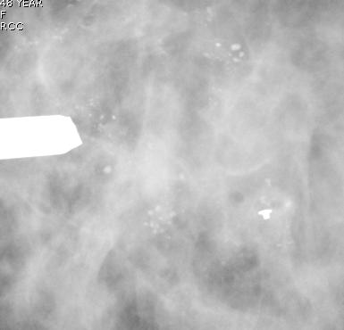
 Importance of Calcification Leonard M. Glassman MD FACR American Institute for Radiologic Pathology Washington Radiology Associates, PC Washington DC 45% of all breast cancers present as calcification
Pitfalls and Limitations of Breast MRI. Susan Orel Roth, MD Professor of Radiology University of Pennsylvania
 Pitfalls and Limitations of Breast MRI Susan Orel Roth, MD Professor of Radiology University of Pennsylvania Objectives Review the etiologies of false negative breast MRI examinations Discuss the limitations
EARLY DETECTION: MAMMOGRAPHY AND SONOGRAPHY
 EARLY DETECTION: MAMMOGRAPHY AND SONOGRAPHY Elizabeth A. Rafferty, M.D. Avon Comprehensive Breast Center Massachusetts General Hospital Harvard Medical School Breast Cancer Screening Early detection of
BREAST IMAGING ROTATION: RESIDENT GUIDE
 BREAST IMAGING ROTATION: RESIDENT GUIDE Rotation sessions minimum goal summary... 1 Identifying your rotations... 1 Curriculum... 2 Goals and Objectives rotation 1... 8 Goals and Objectives rotation 2...
Breast Imaging & You
 Breast Imaging & You What s Inside: Breast Imaging... 2 Digital Breast Tomosynthesis (DBT) mammograms... 4 Breast cancer screening... 6 Dense breast tissue... 8 Automated breast ultrasound (ABUS)... 9
Mammographic imaging of nonpalpable breast lesions. Malai Muttarak, MD Department of Radiology Chiang Mai University Chiang Mai, Thailand
 Mammographic imaging of nonpalpable breast lesions Malai Muttarak, MD Department of Radiology Chiang Mai University Chiang Mai, Thailand Introduction Contents Mammographic signs of nonpalpable breast cancer
Breast phantom with silicone implant for evaluation in conventional mammography
 JOURNAL OF APPLIED CLINICAL MEDICAL PHYSICS, VOLUME 12, NUMBER 1, winter 2011 Breast phantom with silicone implant for evaluation in conventional mammography Fábio A. R. Silva, 1a Luíza F Souza, 1 Carlos
MAMMOGRAPHIC IMAGING - A PRACTICAL GUIDE COURSE POST-TEST
 MAMMOGRAPHIC IMAGING - A PRACTICAL GUIDE COURSE POST-TEST RADUNITS.COM CHAPTER ONE: HISTORY OF MAMMOGRAPHY 1. It was not until the 1960s that, the father of mammography, began teaching his mammographic
Breast Imaging & You
 Breast Imaging & You What s Inside: Breast Imaging... 2 Digital Breast Tomosynthesis (DBT) mammograms... 4 Breast cancer screening... 6 Dense breast tissue... 8 Automated Breast Ultrasound (ABUS)... 9
Here are examples of bilateral analog mammograms from the same patient including CC and MLO projections.
 Good afternoon. It s my pleasure to be discussing Diagnostic Breast Imaging over the next half hour. I m Wei Yang, Professor of Diagnostic Radiology and Chief, the Section of Breast Imaging as well as
ORIGINAL ARTICLE EVALUATION OF BREAST LESIONS USING X-RAY MAMMOGRAM WITH HISTOPATHOLOGICAL CORRELATION
 Available online at www.journalijmrr.com INTERNATIONAL JOURNAL OF MODERN RESEARCH AND REVIEWS IJMRR ISSN: 2347-8314 Int. J. Modn. Res. Revs. Volume 3, Issue 10, pp 807-814, October, 2015 ORIGINAL ARTICLE
Mammography device use in Turkey, and quantity and quality analysis of mammography education
 Diagn Interv Radiol 2007; 13:129 133 Turkish Society of Radiology 2007 BREAST IMAGING ORIGINAL ARTICLE Mammography device use in Turkey, and quantity and quality analysis of mammography education Nuray
The role of quality assurance
 CE Quality Assurance and Ergonomics in the Mammography Department April Reynolds, MS, ELS Quality assurance (QA) in mammography is a system of checks that helps ensure the proper functioning of imaging
Implementation of the 2012 ACR CT QC Manual in a Community Hospital Setting BRUCE E. HASSELQUIST, PH.D., DABR, DABSNM ASPIRUS WAUSAU HOSPITAL
 Implementation of the 2012 ACR CT QC Manual in a Community Hospital Setting BRUCE E. HASSELQUIST, PH.D., DABR, DABSNM ASPIRUS WAUSAU HOSPITAL Conflict of Interest Disclaimer Employee of Aspirus Wausau