General Data. Age: 75y/o Sex: female Date of admission:
|
|
|
- Crystal Horn
- 5 years ago
- Views:
Transcription
1 General Data Age: 75y/o Sex: female Date of admission:
2 Chief complaint Poor oral intake, hunger pain for months and body weight loss about 10kg in 3 months
3 Present Illness Quit healthy before DM(-), HTN(-), CVD(-), smoking(-), drinking(-) 3 months ago, LLQ abdominal discomfort, poor appetite noted PES and colonoscopy done in Argentina -- Polyps noted -- Biopsy: unknown finding
4 Present Illness 1 month ago, intermittent LLQ abdominal pain, upper abdominal pain (hunger pain and relieved after meal) and vomiting noted Body weight loss: 10kg in 3 months
5 PE GA: fair Con s: clear Vital sign: TPR:36.4/90/17, BP:120/78mmHg HEENT: conjunctivae: pale LAP(+) about 1x1cm in size at left post-scm area Chest: breathing sounds: clear heart sounds: regular, no murmur Abdomen: flat and soft, LLQ tenderness bowel sounds: hypoactiv Extremities: freely movable, pitting edema(+)
6 Impression Peptic ulcer Poor oral intake and body weight loss suspect malignancy induced
7 Evaluation 11-1 Hb: 7.9, MCV: 79.1, Ret%:1.5% WBC: 8440, Seg: 86.8%, Plt: Albumin:2.1, GOT:28, GPT:34 BUN:8.6, Cr:0.7 Fe:1, TIBC: CEA: 1.39 (0~4.6), AFP: 1.13 (0~15), CA125: (10~35)
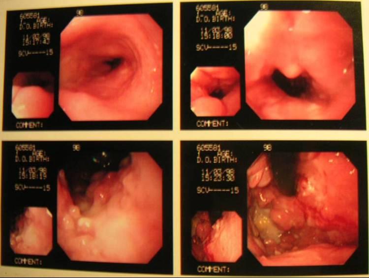
8 Evaluation 11-3 PES: A very big ulcerating tumor with infiltrating margin was found at cardia to angle. Biopsy x 4 IMP: carcinoma, advanced, stomach, with extension to esophagus
9
10 Evaluation 11-3 Abdominal Sonar: multiple hypoechoic nodule (1~2cm) were noted near pancreatic head & aorta. IMP: chronic parenchymal liver disease, liver sludge, thicken GB wall, hypoechoic lesions, nature? r/o LN enlargement
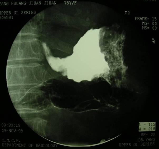
11 Evaluation 11-6 Hb:11.7 (PRBC:4U) 11-6 Abdomen CT 11-9 UGI series Small bowel series Patient transfer to 和信 Hospital for C/T

12 UGI
13 UGI

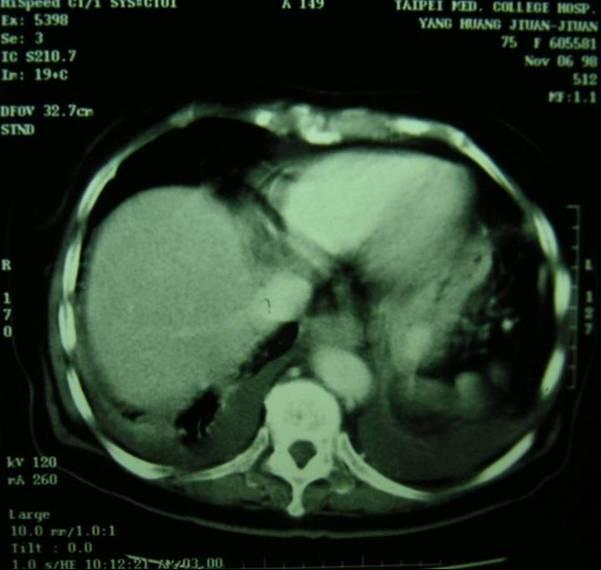
14 CT Pleura effusion Thickened esophageal wall
15 CT

16 CT Thickened gastric wall Soft tissue density at the lesser sac Liver intact

17 CT

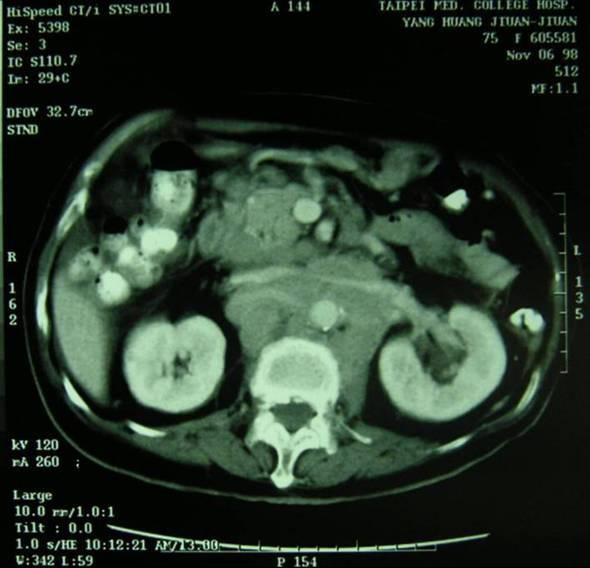
18 CT Soft tissue density at retroperitonium
19 CT

20 CT Soft tissue density at peri-aortic area near pelvic inlet
21 CT Impression: 1. Lymphoma 2. Gastric cancer with lymphatic metastasis
22 Pathology Stomach & Esophagus biopsy Malignant lymphoma made up of large-sized atypical lymphoid cells, B cell Diffuse infiltrating in the esophagus and gastric tissue
23 Diagnosis Gastric lymphoma, Diffuse large B cell type malignant lymphoma from cardia to angle
24 Differential Diagnosis
25 Gastric maliganancies The stomach is the site of variety of malignant neoplasms. They are primary carcinoma, gastric lymphoma, mesenchymal tumours and metastatic carcinomas. The most common of these are carcinoma and lymphoma.
26 Gastric carcinoma: Morphology Primary carcinoma of the stomach may be present as: 1. Superficial spreading carcinoma (Early gastric cancer) Type I lesions are elevated and protrude more than 5 mm into the lumen. Type II tumors are superficial lesions that are elevated (IIa), flat (IIb), or depressed (IIc). Type III early gastric cancers are shallow, irregular ulcers surrounded by nodular, clubbed mucosal folds.
27 Gastric carcinoma: Morphology 2. Polypoid or fungating carcinoma 3. Ulcerating or penetrating carcinoma (70%) 4. Advanced bulky carcinoma 5. Infiltrating or cirrhous carcinoma (5~15%) (linitis plastica)
28 Gastric carcinoma: UGI 1. Superficial spreading carcinoma a flat or slightly depressed lesion accompanied by converging folds 2. Polypoid or fungating carcinoma irregular protruding mass 3. Ulcerating or penetrating carcinoma ulcerated mass, amputation of folds surface may be highly irregular (cauliflower)
29 Gastric carcinoma: UGI 4. Advanced bulky carcinoma mass or bulk, one or more ulceration, irregular surface, margins may be a distinct angular demarcation (shelf) 5. Infiltrating or cirrhous carcinoma (linitis plastica) fibrotic reaction, contracted stomach lacking the normal rugal fold pattern with a smooth or a finely granular surface
30 Gastric carcinoma: CT thickening of the gastric wall, asymmetric thickening of folds, irregular nodular luminal surface, mass of uniform density
31 Gastric carcinoma: Spread 1. beyond the serosa, obliteration of the pre-gastric fat planes 2. direct extension to adjacent structures including the oesophagus, gastrocolic ligament, gastrohepatic ligament, gastrosplenic ligament and pancreas. 3. Further extension may involve any of the lymph node groups in the upper abdominal region. 4. The most common sites of distant metastases are the liver and peritoneal cavity
32 Gastric lymphoma Radiographic type: 1. Polypoid or nodular (47%) enlarged nodular folds 2. Ulcerative (42%) ulcerative lesions, may be complicated by perforation; aneurysmal configuration 3. Diffusely infiltrating(11%) diffuse hoselike thickening of bowel; decreased or absent peristalsis
33 Gastric lymphoma: UGI 1. Infiltrative, ulcerative, or nodular mass that often mimics the appearance of adenocarcinoma. Flexibility of gastric wall preserved. 2. The antrum and body are most commonly involved. Duodenum often affected when antrum involved. 3. Circumscribed mass with endogastric or exogastric growth 4. Large irregular ulcers
34 Gastric lymphoma: CT 1. diffuse involvement of entire stomach, typically more than half of gastric circumference 2. segmental involvement 3. thickened gastric wall 4. luminal irregularity, hyperrugosity 5. spread of the tumour: direct extension into pancreas, spleen, transverse colon and liver
35 Discussion Gastric Lymphoma
36 Lymphoma Hodgkin (1) non-hodgkin (10~15) Presentation Usually nodal Usually extranodal Spread Contiguous Hematogenous, Non-contiguous Mediastinum Common Uncommon(except lymphoblastic type) Spleen Common Uncommon Bone Marrow Uncommon Liver,GI,CNS Uncommon Common Common
37 Gastric Lymphoma Primary gastric lymphomas: less than 2% of all primary stomach malignancies Non Hodgkin's type and of B-cell lineage almost 75% of primary gastrointestinal lymphomas were of gastric origin. Gastric lymphomas are more prevalent in patients over the age of 50, and men are affected two to three times more frequently than women
38 Gastric Lymphoma Usually arise from MALT (mucosa associated lymphoid tissue)- also known as Marginal Zone B-cell lymphoma (Low and High grade). Diffuse large B-cell lymphoma include high grade lymphoma of MALT origin and non- MALT type and they are indistinguishable
39 MALToma Stomach, Mucosa associated lymphoid tissue Related to H. pylori Low grade, remission after H. pylori eradication Other sites: lung, breast, salivary gland, lacrimal gland
40 Gastric Lymphoma: Clinical Sign Most common: abdominal pain, nausea and vomiting, anorexia, weight loss, and bleeding. Early symptoms are vague. More advanced lesions may present with weakness, hemorrhage, pyloric stenosis, or signs of perforation. PE:Abdominal tenderness(35%), a palpable abdominal mass(20-30%), hepatomegaly(14%)
41 Gastric Lymphoma: Staging Ann Arbor staging system Stage IE: tumor confined to the GI tract Stage IIE: regional lymph node involvement Stage IIIE: spread to other organs within the abdomen Stage IV: spread beyond the abdomen
42 Gastric Lymphoma: Prognosis IPSS (International prognostic scoring system; for Diffuse large B cell lymphoma initially) 1. Age>60y/o 2. Ann Arbor stage ⅢorⅣ 3. Extranodal involvement >1 site 4. Poor performance status (ECOG 2~4) 5. High LDH Risk: low(<1), low-intermediate(2), intermediate-high(3), high(4~5)
43 Gastric Lymphoma: Treatment Surgery: 1. No decrease in survival for patients if adjuvant RT or C/T were given than total resection of all gross disease and involved lymph nodes. 3. In surgically treated patients, most recurrences are extraabdominal and local disease is well controlled. 4. Currently, splenectomy is only indicated in cases of direct tumor extension.
44 Gastric Lymphoma: Treatment Radiotherapy: Gastric lymphoma seems to be a more systemic disease with the majority of recurrences occurring at extraabdominal sites. So, in the absence of obvious persistent local disease, the need for additional local therapy with RT is put in question.
45 Gastric Lymphoma: Treatment Chemotherapy: 1. As stated earlier, most primary gastric lymphomas are of the diffuse histiocytic or the diffuse large cell type. They are quite responsive to current chemotherapy. 2. Stages IE & IIE treated with chemotherapy after surgery: excellent 5-year disease free survival
46 Gastric Lymphoma: Treatment C/T regimens: CHOP (cyclophosphamide, Adriamycin, vincristine, prednisone) CHOP-bleo (added bleomycin) COPP-bleo (cyclophosphamide, vincristine, procarbazine, prednisone, bleomycin) CVP (cyclophosphamide, vincristine, and prednisone)
47 Gastric Lymphoma: Treatment C/T High grade Aggressive course without tratment 80% complete remission rate by C/T 40% cure by C/T Low grade Indolent course even late stage Incurable by C/T
48 Gastric Lymphoma: Treatment Conclusion: Early stage: Surgery: for local control and preoperative staging Adjuvant C/T: for extraabdominal lesion
49 Gastric Lymphoma: Treatment Conclusion Invasion: C/T: mainstay of treatment with either surgery or radiation providing local control. In those patients with non-diagnostic biopsies, surgical exploration and resection are needed.
Birthday: 1952/07/31 Date of admission:1999/12/30 Age:48 y/o Past medication:esrd under regular HD for 5+ years; denied DM and HTN
 Birthday: 1952/07/31 Date of admission:1999/12/30 Age:48 y/o Past medication:esrd under regular HD for 5+ years; denied DM and HTN Chief Complaint : 1)intermittent LLQ cramping pain for 2 months 2) LGI
Patient. Male 76 year old C.C: abdominal pain
 Patient Male 76 year old C.C: abdominal pain Bowel stool retention Suspected pulmonary TB at right upper lung Infiltration in right lower lung Pleural thickening at the Right chest Localized dilated small
Gastrointestinal Tract Cancer
 Gastrointestinal Tract Cancer Tumors of the Stomach Gastric adenocarcinoma Incidence and Epidemiology Incidence mortality rates USA High incidence: Japan, China, Chile, Ireland risk lower socioeconomic
neral data Age: 66y/o Sex: male Date of admission:
 neral data Age: 66y/o Sex: male Date of admission: 94-04-12 Chief complaint High fever and body weight loss, cold sweating for 4 weeks Present Illness DM(+) for 20 yrs under OHA control, HTN(-), CVD(-),
Imaging in gastric cancer
 Imaging in gastric cancer Gastric cancer remains a deadly disease because of late diagnosis. Adenocarcinoma represents 90% of malignant tumors. Diagnosis is based on endoscopic examination with biopsies.
General Data. Gender : Male Birthday and age : 12/07/24,80 y/o Occupation : 無 Date of Admission :
 General Data Gender : Male Birthday and age : 12/07/24,80 y/o Occupation : 無 Date of Admission : 92-07-09 1 Chief complaint Upper abdominal fullness 30 minutes after having foods with sometimes epigastralgia
Case Scenario year-old white male presented to personal physician with dyspepsia with reflux.
 Case Scenario 1 57-year-old white male presented to personal physician with dyspepsia with reflux. 7/12 EGD: In the gastroesophageal junction we found an exophytic tumor. The tumor occupies approximately
Abstracting Upper GI Cancer Incidence and Treatment Data Quiz 1 Multiple Primary and Histologies Case 1 Final Pathology:
 Abstracting Upper GI Cancer Incidence and Treatment Data Quiz 1 Multiple Primary and Histologies Case 1 A 74 year old male with a history of GERD presents complaining of dysphagia. An esophagogastroduodenoscopy
Gastric Cancer Histopathology Reporting Proforma
 Gastric Cancer Histopathology Reporting Proforma Mandatory questions (i.e. protocol standards) are in bold (e.g. S1.01). S1.01 Identification Family name Given name(s) Date of birth Sex Male Female Intersex/indeterminate
By Prof. Mohamed Khaled Zaky, MB,BCh; MSc; MD; FRCSI (Gen. Surg.) Professor of Surgery, Taibah Univ.
 Professor of Surgery, Taibah Univ.") By Prof. Mohamed Khaled Zaky, MB,BCh; MSc; MD; FRCSI (Gen. Surg.) Professor of Surgery, Taibah Univ. Objectives Types Incidence Risk factors (& prevention) Pathology: Gross, microscopic, spread, staging,
By Prof. Mohamed Khaled Zaky, MB,BCh; MSc; MD; FRCSI (Gen. Surg.) Professor of Surgery, Taibah Univ. Objectives Types Incidence Risk factors (& prevention) Pathology: Gross, microscopic, spread, staging,
Case Scenario 1. The patient has now completed his neoadjuvant chemoradiation and has been cleared for surgery.
 Case Scenario 1 July 10, 2010 A 67-year-old male with squamous cell carcinoma of the mid thoracic esophagus presents for surgical resection. The patient has completed preoperative chemoradiation. This
Patient underwent hemicolectomy: 7 x 4.5 cm intusscepted segment of ileum in colon - mucosal
 Extranodal Lymphomas Rena Buckstein Odette Cancer Center Case: JT 69 yo male COO software company PMHx: basal cell back, cholesterol Presents to ER with severe abdominal pain, bloody diarrhea x 2d In ER
Oncology General Principles L A U R I E S I M A R D B R E A S T S U R G I C A L O N C O L O G Y F E L L O W D E C E M B E R
 Oncology General Principles L A U R I E S I M A R D B R E A S T S U R G I C A L O N C O L O G Y F E L L O W D E C E M B E R 2 0 1 2 Objectives Discuss Diagnostic and staging strategies in oncology Know
Gastrointestinal pathology 2018 lecture 4. Dr Heyam Awad FRCPath
 Gastrointestinal pathology 2018 lecture 4 Dr Heyam Awad FRCPath Topics to be covered Peptic ulcer disease Hiatal hernia Gastric neoplasms Peptic ulcer disease (PUD)= chronic gastric ulcer Causes H pylori
11/21/13 CEA: 1.7 WNL
 Case Scenario 1 A 70 year-old white male presented to his primary care physician with a recent history of rectal bleeding. He was referred for imaging and a colonoscopy and was found to have adenocarcinoma.
Sex: 女 Age: 51 Occupation: 無 Admission date:92/07/22
 Sex: 女 Age: 51 Occupation: 無 Admission date:92/07/22 Chief complaint Unknown fever for one month Hand tremor and left huge renal tumor was noted Present illness Suffered from fever for one month, hand
LYMPHOMA Joginder Singh, MD Medical Oncologist, Mercy Cancer Center
 LYMPHOMA Joginder Singh, MD Medical Oncologist, Mercy Cancer Center Lymphoma is cancer of the lymphatic system. The lymphatic system is made up of organs all over the body that make up and store cells
X-Ray Corner. Imaging of the Stomach. Pantongrag-Brown L
 THAI J 178 Imaging of the Stomach GASTROENTEROL 2014 X-Ray Corner Imaging of the Stomach Pantongrag-Brown L Imaging modalities used in stomach include plain radiographs, UGI study, US, CT, PET CT and MRI.
Indolent Lymphomas. Dr. Melissa Toupin The Ottawa Hospital
 Indolent Lymphomas Dr. Melissa Toupin The Ottawa Hospital What does indolent mean? Slow growth Often asymptomatic Chronic disease with periods of relapse (long natural history possible) Incurable with
Large cell immunoblastic Diffuse histiocytic (DHL) Lymphoblastic lymphoma Diffuse lymphoblastic Small non cleaved cell Burkitt s Non- Burkitt s
 Lymphoblastic lymphoma Diffuse lymphoblastic Small non cleaved cell Burkitt s Non- Burkitt s") Non Hodgkin s Lymphoma Introduction 6th most common cause of cancer death in United States. Increasing in incidence and mortality. Since 1970, the incidence of has almost doubled. Overview The types of
Non Hodgkin s Lymphoma Introduction 6th most common cause of cancer death in United States. Increasing in incidence and mortality. Since 1970, the incidence of has almost doubled. Overview The types of
Patient Information. Age: 8 y/o Sex: Female. Date of Admission: Date of Discharge:
 Patient Information Age: 8 y/o Sex: Female Date of Admission: 92-10-08 Date of Discharge: 92-10-18 Chief Complaint Severe admominal pain and vomiting with dysuria since last afternoon Present Illness Lower
CT EVALUATION OF GASTRIC LESIONS:
 CT EVALUATION OF GASTRIC LESIONS: Pictural essay Hasni Bouraoui I, Kahloun A, Jemni H, Elouni F, Moulahi H, Daadoucha A, Ben Ali A, Sriha B, Tlili Graies K Departments of Radiology, Gastro enterology,
PRIMARY GASTRIC LYMPHOMA: CASE REPORT WITH REVIEW OF LITERATURE
 PRIMARY GASTRIC LYMPHOMA: CASE REPORT WITH REVIEW OF LITERATURE Rana K. Sherwani, *Kafil Akhtar, Noorin Zaidi, Anjum Ara Department of Pathology, J.N. Medical College, Aligarh Muslim University, Aligarh,
A916: rectum: adenocarcinoma
 General facts of colorectal cancer The colon has cecum, ascending, transverse, descending and sigmoid colon sections. Cancer can start in any of the r sections or in the rectum. The wall of each of these
GIT RADIOLOGY. Water-soluble contrast media (e.g. gastrograffin) are the other available agents.which doesn t cause inflammatory peritonitis..
 are the other available agents.which doesn t cause inflammatory peritonitis..") GIT RADIOLOGY Imaging techniques-general principles: Contrast examinations: Barium sulphate is the best contrast for GIT (with good mucosal coating & excellent opacification & being inert); but is contraindicated
GIT RADIOLOGY Imaging techniques-general principles: Contrast examinations: Barium sulphate is the best contrast for GIT (with good mucosal coating & excellent opacification & being inert); but is contraindicated
performed to help sway the clinician in what the appropriate diagnosis is, which can substantially alter the treatment of management.
 Hello, I am Maura Polansky at the University of Texas MD Anderson Cancer Center. I am a Physician Assistant in the Department of Gastrointestinal Medical Oncology and the Program Director for Physician
Case conference. Welcome Dr. Lawrence Tierney
 Case conference Welcome Dr. Lawrence Tierney Case: 18 year-old male CC) hamatomesis, Fever and cough HPI) 1 st admission One month ago, he admitted to our hospital because of hematemesis. He had weight
Aggressive Lymphomas - Current. Dr Kevin Imrie Physician-in-Chief, Sunnybrook Health Sciences Centre
 Aggressive Lymphomas - Current Dr Kevin Imrie Physician-in-Chief, Sunnybrook Health Sciences Centre Conflicts of interest I have no conflicts of interest to declare Outline What does aggressive lymphoma
Efficacy of High Resolution Transabdominal Sonography of the Fluid Filled Stomach in the Evaluation of Gastric Carcinomas
 4-67 421 Efficacy of High Resolution Transabdominal Sonography of the Fluid Filled Stomach in the Evaluation of Gastric Carcinomas S SINGH, V CHOWDHURY ABSTRACT AIM: To evaluate the efficacy of high-resolution
Indolent Lymphomas: Current. Dr. Laurie Sehn
 Indolent Lymphomas: Current Dr. Laurie Sehn Why does indolent mean? Slow growth Often asymptomatic Chronic disease with periods of relapse (long natural history possible) Incurable with current standard
8. The polyp in the illustration can be described as (circle all that apply) a. Exophytic b. Pedunculated c. Sessile d. Frank
 a. Exophytic b. Pedunculated c. Sessile d. Frank") Quiz 1 Overview 1. Beginning with the cecum, which is the correct sequence of colon subsites? a. Cecum, ascending, splenic flexure, transverse, hepatic flexure, descending, sigmoid. b. Cecum, ascending,
Quiz 1 Overview 1. Beginning with the cecum, which is the correct sequence of colon subsites? a. Cecum, ascending, splenic flexure, transverse, hepatic flexure, descending, sigmoid. b. Cecum, ascending,
Chief complaint. A mass at right chest
 Chief complaint A mass at right chest Present illness This 1-year-5-month-old girl had a mass at right side chest since one month ago. flat and not tender at first In the recent 2 days, the mass enlarged
Gastroenterology Tutorial
 Gastroenterology Tutorial Gastritis Poorly defined term that refers to inflammation of the stomach. Infection with H. pylori is the most common cause of gastritis. Most patients remain asymptomatic Some
An Uncommon Presentation of Large B-cell Lymphoma of the kidney A Case Report and Literature Review
 An Uncommon Presentation of Large B-cell Lymphoma of the kidney A Case Report and Literature Review CHRISTOPHER ADILETTA M.D., AJAZ SHAWL M.D. ST. JOSEPH S HEALTH, SYRACUSE, NY Our Patient Case We present
METASTASES FROM GASTRIC CARCINOMA TO COLON LESIONS: A CASE REPORT IN THE FORM OF MULTIPLE FLAT ELEVATED CASE PRESENTATION
 H.C. Lee, M.T. Yang, K.Y. Lin, et al METASTASES FROM GASTRIC CARCINOMA TO COLON IN THE FORM OF MULTIPLE FLAT ELEVATED LESIONS: A CASE REPORT Hsi-Chang Lee, Min-Ta Yang, 1 Kuang-Yang Lin, 1 Hsing-Yang Tu,
Alison Douglass Gillian Lieberman, MD. November. Colon Cancer. Alison Douglass, Harvard Medical School Year III Gillian Lieberman, MD
 November Colon Cancer Alison Douglass, Harvard Medical School Year III Our Patient Mr. K. is a 67 year old man with no prior medical problems other than hemorrhoids which have caused occasional rectal
MECHANISMS OF HUMAN DISEASE: LABORATORY SESSION CYTOPATHOLOGY Monday, April 26, 2013 FACULTY COPY
 GOAL: MECHANISMS OF HUMAN DISEASE: LABORATORY SESSION CYTOPATHOLOGY Monday, April 26, 2013 FACULTY COPY 1. Understated the role of cytopathology in the clinical management of the patient and recognize
Case Presentation. Doron Boltin March, 2015
 Case Presentation Doron Boltin March, 2015 Case 68 year-old male Previously healthy Medication- vit D Family Hx father CRC (age 70) mother gastric ca (age 70) Screening colonoscopy 02.2011- diminutive
COLORECTAL CANCER FAISALGHANISIDDIQUI MBBS; FCPS; PGDIP-BIOETHICS; MCPS-HPE
 COLORECTAL CANCER FAISALGHANISIDDIQUI MBBS; FCPS; PGDIP-BIOETHICS; MCPS-HPE PROFESSOR OF SURGERY & DIRECTOR, PROFESSIONAL DEVELOPMENT CENTRE J I N N A H S I N D H M E D I C A L U N I V E R S I T Y faisal.siddiqui@jsmu.edu.pk
Title: Linitis plastica of the colon due to metastases of invasive lobular breast carcinoma
 Title: Linitis plastica of the colon due to metastases of invasive lobular breast carcinoma Authors: David Viso Vidal, Rafael Villanueva Pavón, Francisco Jorquera Plaza DOI: 10.17235/reed.2019.6065/2018
A CASE OF PRIMARY THYROID LYMPHOMA. Prof Dr.Dilek Gogas Yavuz Marmara University School of Medicine Endocrinology and Metabolism Istanbul, Turkey
 A CASE OF PRIMARY THYROID LYMPHOMA Prof Dr.Dilek Gogas Yavuz Marmara University School of Medicine Endocrinology and Metabolism Istanbul, Turkey 38 year old female She recognized a mass in her right neck
Gastric Tumors Dr. Taha
 Gastric Tumors Dr. Taha BENIGN TUMORS: Leiomyomas: smooth muscle tumors, equal in men /women, typically located in the middle &distal stomach. Can grow into the lumen with secondary ulceration & bleeding.
Case Study: #3: Gallbladder Carcinoma?
 Case Study: #3: Gallbladder Carcinoma? By: Megan Wyatt K. SON Wyatt 225 2B1 RDMS, RVT Patient: Male 85 YOA Caucasian Indication: Elevated Alkaline Phosphatase History Annual physical showed elevated alkaline
Non-Hodgkin lymphomas (NHLs) Hodgkin lymphoma )HL)
 Hodgkin lymphoma )HL)") Non-Hodgkin lymphomas (NHLs) Hodgkin lymphoma )HL) Lymphoid Neoplasms: 1- non-hodgkin lymphomas (NHLs) 2- Hodgkin lymphoma 3- plasma cell neoplasms Non-Hodgkin lymphomas (NHLs) Acute Lymphoblastic Leukemia/Lymphoma
Non-Hodgkin lymphomas (NHLs) Hodgkin lymphoma )HL) Lymphoid Neoplasms: 1- non-hodgkin lymphomas (NHLs) 2- Hodgkin lymphoma 3- plasma cell neoplasms Non-Hodgkin lymphomas (NHLs) Acute Lymphoblastic Leukemia/Lymphoma
objectives Pitfalls and Pearls in PET/CT imaging Kevin Robinson, DO Assistant Professor Department of Radiology Michigan State University
 objectives Pitfalls and Pearls in PET/CT imaging Kevin Robinson, DO Assistant Professor Department of Radiology Michigan State University To determine the regions of physiologic activity To understand
Done by: Dina Sawadha & Mohammad Abukabeer
 Done by: Dina Sawadha & Mohammad Abukabeer The stomach *the stomach is a dilated part of the gastro intestinal tract, it's "J" shape. *the lower surface of the stomach ( the greater curvature ) reaches
The surface mucous cells and the cardiac and pyloric glands secrete mucus which protects the stomach from self-digestion.
 PATHOLOGY OF THE STOMACH Stomach mucosa Gastric mucosa is covered by a layer of mucus. The mucosal glands comprise the cardiac glands, the fundic glands in the fundus and body of the stomach, and the pyloric
Case Scenario 1. Discharge Summary
 Case Scenario 1 Discharge Summary A 69-year-old woman was on vacation and noted that she was becoming jaundiced. Two months prior to leaving on that trip, she had had a workup that included an abdominal
Brief History. Identification : Past History : HTN without regular treatment.
 Brief History Identification : Name : 陳 x - Admission : 94/10/06 Gender : male Age : 75 y/o Chief Complaint : Urinary difficulty for months. Past History : HTN without regular treatment. Brief History
155.2 Malignant neoplasm of liver not specified as primary or secondary. C22.9 Malignant neoplasm of liver, not specified as primary or secondary
 ICD-9 TO ICD-10 Reference ICD-9 150.9 Malignant neoplasm of esophagus unspecified site C15.9 Malignant neoplasm of esophagus, unspecified 151.9 Malignant neoplasm of stomach unspecified site C16.9 Malignant
NON HODGKINS LYMPHOMA: INDOLENT Updated June 2015 by Dr. Manna (PGY-5 Medical Oncology Resident, University of Calgary)
") NON HODGKINS LYMPHOMA: INDOLENT Updated June 2015 by Dr. Manna (PGY-5 Medical Oncology Resident, University of Calgary) Reviewed by Dr. Michelle Geddes (Staff Hematologist, University of Calgary) and Dr.
NON HODGKINS LYMPHOMA: INDOLENT Updated June 2015 by Dr. Manna (PGY-5 Medical Oncology Resident, University of Calgary) Reviewed by Dr. Michelle Geddes (Staff Hematologist, University of Calgary) and Dr.
Perforation of a Duodenal Diverticulum. Elective Student S. C.
 Perforation of a Duodenal Diverticulum 2008 4 Elective Student S. C. Case History An elderly male presented to the Emergency Department with abdominal pain. Chief Complaint: Worsening, diffuse abdominal
Uncommon secondary tumour of the stomach
 Uncommon secondary tumour of the stomach B. Bancel, Hôpital CROIX ROUSSE LYON Bucharest Nov 2013 Case report 33-year old man Profound mental retardation and motor disturbances (sequelae of neonatal meningeal
Lymphoma (Lymphosarcoma) by Pamela A. Davol
 by Pamela A. Davol") Lymphoma (Lymphosarcoma) by Pamela A. Davol Cells derived from the bone marrow that mature and take part in cellular immune reactions are called lymphocytes. When lymphocytes undergo transformation and
Lymphoma (Lymphosarcoma) by Pamela A. Davol Cells derived from the bone marrow that mature and take part in cellular immune reactions are called lymphocytes. When lymphocytes undergo transformation and
LEUKAEMIA and LYMPHOMA. Dr Mubarak Abdelrahman Assistant Professor Jazan University
 LEUKAEMIA and LYMPHOMA Dr Mubarak Abdelrahman Assistant Professor Jazan University OBJECTIVES Identify etiology and epidemiology for leukemia and lymphoma. Discuss common types of leukemia. Distinguish
SEER Summary Stage Still Here!
 SEER Summary Stage Still Here! CCRA NORTHERN REGION STAGING SYMPOSIUM SEPTEMBER 20, 2017 SEER Summary Stage Timeframe: includes all information available through completion of surgery(ies) in the first
Pre-operative assessment of patients for cytoreduction and HIPEC
 Pre-operative assessment of patients for cytoreduction and HIPEC Washington Hospital Center Washington, DC, USA Ovarian Cancer Surgery New Strategies Bergamo, Italy May 5, 2011 Background Cytoreductive
A218 : Esophagus cancer tissues. (formalin fixed)
") (formalin fixed) For research use only Specifications: No. of cases: 40 Tissue type: Esophagus cancer tissues No. of spots: 2 spots from each cancer case (80 spots) 4 non-neoplastic spots (4 spots) Total
(formalin fixed) For research use only Specifications: No. of cases: 40 Tissue type: Esophagus cancer tissues No. of spots: 2 spots from each cancer case (80 spots) 4 non-neoplastic spots (4 spots) Total
DOI: /jemds/2014/1882 CASE REPORT
 NON HODGKIN'S LYMPHOMA OF STOMACH WITH SPONTANEOUS PERFORATION: A WITH REVIEW OF LITERATURE Rishikant Vashistha 1, Abhishek Kansal 2, S.M. Datey 3, Vikramaditya Singh 4 HOW TO CITE THIS ARTICLE: Rishikant
ד"ר דוד ירדני המכון לגסטרואנטרולוגיה ומחלות כבד מרכז רפואי סורוקה
 ד"ר דוד ירדני המכון לגסטרואנטרולוגיה ומחלות כבד מרכז רפואי סורוקה Presentaion: S.A is 38 years old. Referred for rectal bleeding investigation. Describes several occasions of bleeding and abdominal pain.
Polyps in general: is a descriptive term of forming a mass that is exophytic & polypoid.
 ميحرلا نمحرلا هللا مسب Gastric Tumors: Benign tumours & tumor-like conditions: -Mucosal: Gastric polyps (they are uncommon) -Mesenchymal tumours: Leiomyoma & Lipoma (can occur anywhere in the body) Malignant:
When to Refer for OGD and the Work Up of Upper GI Malignancies
 When to Refer for OGD and the Work Up of Upper GI Malignancies Dr Hong Qiantai Registrar, Department of Surgery GP Forum 27 May 2017 38 year old female, non-smoker, BMI 29 Works as investment banker Presents
Melanoma Case Scenario 1
 Melanoma Case Scenario 1 History and physical 11/5/16 Patient is a single, 48-year-old male in good health who presented to his primary physician for a yearly physical exam during which a 3.4 x 2.8 x 1.5
The many faces of extranodal lymphoma
 The many faces of extranodal lymphoma Frank Pameijer Departments of Radiology and Radiation Oncology University Medical Center Utrecht Special thanks to Ilona M Schmalfuss, MD University of Florida Gainesville,
References. GI Biopsies. What Should Pathologists Assistants Know About Gastrointestinal Histopathology? James M Crawford, MD, PhD
 What Should Pathologists Assistants Know About Gastrointestinal Histopathology? James M Crawford, MD, PhD jcrawford1@nshs.edu Executive Director and Senior Vice President for Laboratory Services North
Stomach Computerized Tomography indications, technique, examples. VUH SK Radiology and nuclear medicine center Radiologist Dileta Rutkauskaitė
 Stomach Computerized Tomography indications, technique, examples VUH SK Radiology and nuclear medicine center Radiologist Dileta Rutkauskaitė Stomach Computerized Tomography gastroente rologist Oncologist
58 year old male complaining of 3-week history of increasing epigastric pain
 Peptic Ulcer Disease 58 year old male complaining of 3-week history of increasing epigastric pain Has had dyspepsia in the past for which he took Tums, but this is much worse and only partially relieved
Greater Manchester & Cheshire Guidelines for Pathology Reporting for Oesophageal and Gastric Malignancy
 Greater Manchester & Cheshire Guidelines for Pathology Reporting for Oesophageal and Gastric Malignancy Authors: Dr Gordon Armstrong, Dr Sue Pritchard 1. General Comments 1.1 Cancer reporting: Biopsies
Disorders of Cell Growth & Neoplasia. Histopathology Lab
 Disorders of Cell Growth & Neoplasia Histopathology Lab Paul Hanna April 2010 Case #84 Clinical History: 5 yr-old, West Highland White terrier. skin mass from axillary region. has been present for the
Melanoma Case Scenario 1
 Melanoma Case Scenario 1 History and physical 11/5/16 Patient is a single, 48-year-old male in good health who presented to his primary physician for a yearly physical exam during which a 3.4 x 2.8 x 1.5
Lymphoma Read with the experts
 Lymphoma Read with the experts Marc Seltzer, MD Associate Professor of Radiology Geisel School of Medicine at Dartmouth Director, PET-CT Course American College of Radiology Learning Objectives Recognize
B. Environmental Factors. a. The major risk factor to papillary thyroid cancer is exposure to ionizing radiation, during the first 2 decades of life.
 B. Environmental Factors. a. The major risk factor to papillary thyroid cancer is exposure to ionizing radiation, during the first 2 decades of life. b. Deficiency of dietary iodine: - Is linked with a
Chapter 3. Neoplasms. Copyright 2015 Cengage Learning.
 Chapter 3 Neoplasms Terminology Related to Neoplasms and Tumors Neoplasm New growth Tumor Swelling or neoplasm Leukemia Malignant disease of bone marrow Hematoma Bruise or contusion Classification of Neoplasms
IMAGING GUIDELINES - COLORECTAL CANCER
 IMAGING GUIDELINES - COLORECTAL CANCER DIAGNOSIS The majority of colorectal cancers are diagnosed on colonoscopy, with some being diagnosed on Ba enema, ultrasound or CT. STAGING CT chest, abdomen and
Major Topic. Malignant Melanoma Plastic and Reconstructive Surgery R3 陸尊惠 /VS 吳瑞星
 Major Topic Malignant Melanoma Plastic and Reconstructive Surgery R3 陸尊惠 /VS 吳瑞星 Patient Data Name: OOO Age: 70 Gender: Male Date of admission: Day 1 Chief Complaint Black skin tumor at the back of the
Guidelines for Assigning Summary Stage 2000
 Guidelines for Assigning Summary Stage 2000 Mary Lewis, CTR National Program of Cancer Registries 2014 NCRA Annual Meeting May 17, 2014 National Center for Chronic Disease Prevention and Health Promotion
CT Evaluation of Bowel Wall Thickening. Dr: Adel El Badrawy; M.D. Lecturer of Radio Diagnosis Faculty of Medicine Mansoura University.
 CT Evaluation of Bowel Wall Thickening By Dr: Adel El Badrawy; M.D. Lecturer of Radio Diagnosis Faculty of Medicine Mansoura University. The CT findings of bowel wall thickening includes 1 Degree of thickening.
ACHIEVING EXCELLENCE IN ABSTRACTING: LYMPHOMA
 ACHIEVING EXCELLENCE IN ABSTRACTING: LYMPHOMA ACHIEVING EXCELLENCE IN ABSTRACTING LYMPHOMA Recoding Audit Performed in 2009 260 cases audited 17 data items audited per case 4420 possible discrepancies
Images In Gastroenterology
 Images In Gastroenterology Thong-Ngam D, et al. THAI J GASTROENTEROL 2005 Vol. 6 No. 2 May - Aug. 2005 105 Imaging of Gastrointestinal Stromal Tumors Pornpim Fuangtharnthip, M.D. Narumol Hargroove, M.D.
It is a malignancy originating from breast tissue
 59 Breast cancer 1 It is a malignancy originating from breast tissue including both early stages which are potentially curable, and metastatic breast cancer (MBC) which is usually incurable. Most breast
Boot Camp Case Scenarios
 Boot Camp Case Scenarios Case Scenario 1 Patient is a 69-year-old white female. She presents with dyspnea on exertion, cough, and right rib pain. Patient is a smoker. 9/21/12 CT Chest FINDINGS: There is
is time consuming and expensive. An intra-operative assessment is not going to be helpful if there is no more tissue that can be taken to improve the
 My name is Barry Feig. I am a Professor of Surgical Oncology at The University of Texas MD Anderson Cancer Center in Houston, Texas. I am going to talk to you today about the role for surgery in the treatment
Gastric (Stomach) Cancer
 Cancer") Gastric (Stomach) Cancer Gastric cancer is a disease in which malignant (cancer) cells form in the lining of the stomach. The stomach is a J-shaped organ in the upper abdomen. It is part of the digestive
Gastric (Stomach) Cancer Gastric cancer is a disease in which malignant (cancer) cells form in the lining of the stomach. The stomach is a J-shaped organ in the upper abdomen. It is part of the digestive
Esophageal Cancer Staging Essentials: The New TNM Staging System (7th edition) and Clinicoradiologic Implications
 and Clinicoradiologic Implications") Esophageal Cancer Staging Essentials: The New TNM Staging System (7th edition) and Clinicoradiologic Implications Poster No.: E-0060 Congress: ESTI 2012 Type: Scientific Exhibit Authors: K. Lee, T. J.
Esophageal Cancer Staging Essentials: The New TNM Staging System (7th edition) and Clinicoradiologic Implications Poster No.: E-0060 Congress: ESTI 2012 Type: Scientific Exhibit Authors: K. Lee, T. J.
Regression of Advanced Gastric MALT Lymphoma after the Eradication of Helicobacter pylori
 Gut and Liver, Vol. 6, No. 2, April 2012, pp. 270-274 CASE REPORT Regression of Advanced Gastric MALT Lymphoma after the Eradication of Helicobacter pylori Soo-Kyung Park, Hwoon-Yong Jung, Do Hoon Kim,
Neoplasia literally means "new growth.
 NEOPLASIA Neoplasia literally means "new growth. A neoplasm, defined as "an abnormal mass of tissue the growth of which exceeds and is uncoordinated with that of the normal tissues and persists in the
THE mainstay of the radiographic study of the upper gastrointestinal tract has
 BARIUM-SPRAY EXAMINATION OF THE STOMACH- PRELIMINARY REPORT OF A NEW ROENTGENOGRAPHIC TECHNIC EDWARD BUONOCORE, M.D., and THOMAS F. MEANEY, M.D. Department of Hospital Radiology THE mainstay of the radiographic
Abstracting Hematopoietic Neoplasms
 CASE 1: LYMPHOMA PHYSICAL EXAMINATION 43yo male with a history of lower gastrointestinal bleeding and melena undergoing colonoscopy and biopsy to rule out neoplasm versus inflammation. Patient had no other
Peptic ulcer disease. Nomin-Erdene. D SOM-531
 Peptic ulcer disease Nomin-Erdene. D SOM-531 Learning objectives Stomach gross anatomy PUD Epidemiology Pathogenesis Clinical manifestation Diagnosing Treatment Complicated ulcer disease Surgical procedures
Radiology Pathology Conference
 Radiology Pathology Conference Nadia F. Yusaf, M.D. PGY-3 1/29/2010 Presentation material is for education purposes only. All rights reserved. 2010 URMC Radiology Page 1 of 90 Case 1 60 year- old man presents
Clinical Radiological Pathological Conference
 Clinical Radiological Pathological Conference CASE 1: A 59-year-old female Housekeeper Live in Phuket, Thailand Progressive dyspnea for 1 year Present illness 1 year PTA : She developed dyspnea on exertion
Canine Histiocytic Disorders DR. MEREDITH GAUTHIER, DVM DACVIM (ONCOLOGY) OCTOBER 29, 2015
 OCTOBER 29, 2015") Canine Histiocytic Disorders DR. MEREDITH GAUTHIER, DVM DACVIM (ONCOLOGY) OCTOBER 29, 2015 Canine Histiocytes! Cells derived from CD34+ stem cells and blood monocytes! Macrophages! Dendritic cells (DC)!
Canine Histiocytic Disorders DR. MEREDITH GAUTHIER, DVM DACVIM (ONCOLOGY) OCTOBER 29, 2015 Canine Histiocytes! Cells derived from CD34+ stem cells and blood monocytes! Macrophages! Dendritic cells (DC)!
Endoscopic Corner CASE 1. Sirimontaporn N Klaikaew N Imraporn B Rerknimitr R
 Endoscopic Corner Sirimontaporn N, et al. THAI J GASTROENTEROL 2010 Vol. 11 No. 3 Sept. - Dec. 2010 171 Sirimontaporn N Klaikaew N Imraporn B Rerknimitr R CASE 1 A 47- year-old female presented to the
Malignant Melanoma Early Stage. A guide for patients
 This melanoma patient brochure is designed to help educate melanoma patients and their caregivers. It was developed under the guidance of Dr. Michael Smylie, Professor, Department of Oncology, University
CARCINOMA OF ESOPHAGUS PERFORATING THE AORTA*
 CARCINOMA OF ESOPHAGUS PERFORATING THE AORTA* HERBERT J. SCHATTENBERG AND JOSEPH ZISKIND From the Department of Pathology, Graduate School, Tulane University, and the Charity Hospital, New Orleans Perforation
Case Discussion Splenic Abscess
 Case Discussion Splenic Abscess Personal Data Gender: male Birth Date: 1928/Mar/06th Allergy: Mefenamic Smoking: 0.5 PPD for 55 years Alcohol: negative (?) 4 Months Ago Abdominal pain: epigastric area
Case: The patient is a 73 year old woman with vague complaints of dyspepsia and abdominal pain. Upper endoscopy showed features of gastritis and a
 Case: The patient is a 73 year old woman with vague complaints of dyspepsia and abdominal pain. Upper endoscopy showed features of gastritis and a nodular lesion in the body of the stomach. The patient
Gastrointestinal Tract. Anatomy of GI Tract. Anatomy of GI Tract. (Effective February 2007) (1%-5%)
 (1%-5%)") Gastrointestinal Tract (Effective February 2007) (1%-5%) Anatomy of GI Tract Esophagus bulls-eye or target EG junction seen on sagittal scan posterior to left lobe of liver and anterior to aorta Anatomy
Gastrointestinal Tract (Effective February 2007) (1%-5%) Anatomy of GI Tract Esophagus bulls-eye or target EG junction seen on sagittal scan posterior to left lobe of liver and anterior to aorta Anatomy
Imaging iconography of gallbladder cancer. Assessment by CT.
 1 REVISTA DE IMAGENOLOGIA- EII / Vol. XVI / Num. 2 Imaging iconography of gallbladder cancer. Assessment by CT. Doctors Crisci, Alejandro (1); Landó, Fernando.(2). CASMU CT Department Hospital of Tacuarembó
CODING PRIMARY SITE. Nadya Dimitrova
 CODING PRIMARY SITE Nadya Dimitrova OUTLINE What is coding and why do we need it? ICD-10 and ICD-O ICD-O-3 Topography coding rules ICD-O-3 online WHAT IS CODING AND WHY DO WE NEED IT? Coding: to assign