DR AISHA A UMAR CHIEF CONSULTANT RADIOLOGIST NATIONAL HOSPITAL ABUJA.
|
|
|
- Lynne Jackson
- 5 years ago
- Views:
Transcription

1 DR AISHA A UMAR CHIEF CONSULTANT RADIOLOGIST NATIONAL HOSPITAL ABUJA.
2 OUTLINE WHY DO WE IMAGE WHOM TO IMAGE WHEN TO IMAGE HOW TO IMAGE WHAT TO IMAGE WITH PERSONAL EXPERIENCE CONCLUSION/RECOMMENDATIONS
3 WHY DO WE IMAGE Breast cancer is life threatening Early detection remains the only defence.. Breast cancers that are smaller or non palpable are more treatable when detected Have a more favourable prognosis Stage I: 100% Stage IIA -IIB 81-92% Stage IIIA-B 54-67% Stage IV: 20% Based on 5 year survival, prognosis is as follows;
4 FINDINGS FROM STUDIES In a study by Ntekim et al, in 221 young women of 40 years and below in Ibadan, Nigeria Stage I disease was diagnosed in 5 (2%) stage II disease 29(13%) Stages III 102 (46%) Stage IV disease 85(39%) African Health Sciences Vol 9 No 4 December 2009
5 Ihekwaba found in his study of Breast cancer in Nigerian women. Stage I and Stage II- 17.3% Stage III % Stage IV % Br J Surg Aug;79(8):771-5.
6 OUTCOME OF SCREENING Howell et al in his review of Risk determination and prevention of breast cancer As a result of the introduction of screening and optimizing treatments, deaths from breast cancer have decreased by approximately one third over the past 20 years. Howell et al. Breast Cancer Research 2014, 16:446
7 WHOM TO IMAGE HIGH RISK PATIENTS Screening is recommended before 40 years In some cases 10 years earlier. Not before 25 years Hereditary breast cancer mutation gene Lower age of menarche, Late age of first pregnancy, Fewer pregnancies, Shorter or no periods of breastfeeding, and A later menopause. Other risk factors Increase in obesity, Alcohol consumption, Inactivity, or low physical activity Hormone replacement therapy (HRT).

8 Average risk patients Under 40 years Clinical breast examination Limited benefit for screening

9 Average risk patients Over 40 years of age Screening is recommended
10 WHEN TO IMAGE ASYMPTOMATIC SYMPTOMATIC
11 Indications for mammography in Ilorin by Akande et al 2015 Percent Screening Symptomatic Breast lump Breast pain Nipple discharge Breast swelling Axillary swelling Known cancer Niger Med J May- Jun; 56(3):
12 Guidelines for screening Various expert groups have different recommendations. A careful consideration must be given to the risks of developing breast cancer The benefits and harms of the screening intervention, The cost involved.
13 Expert groups in United states American Cancer Society (ACS), The American College of Obstetricians and Gynaecologists (ACOG), American College of Physicians (ACP), American College of Radiology (ACR), American Medical Association (AMA), The National Cancer Institute (NCI), National Comprehensive Cancer Network (NCCN), United States Preventive Services Task Force (USPSTF).
14 Expert groups guidelines Agreement Benefits of screening Disagreement When to start and end screening How often to screen What imaging modality to use for screening
15 US Preventive Services Task Force (USPSTF) breast cancer screening guidelines, 2009 No requirement for routine screening mammography in women aged years The decision to start regular, biennial screening mammography before age 50 years is individual Biennial screening mammography for women between age 50 and 74 years Insufficient current evidence to assess the additional benefits and harms of screening mammography in women aged 75 years or older
16 American college of Obstetrics and gynaecology guidelines ACOG recommend adherence to its current guidelines, Screening mammography every 1-2 years for women aged years Screening mammography every year for women aged 50 years or older
17 American college of Radiology (ACR) guidelines- High-risk women Women with a breast cancer (BRCA) gene mutation Their untested first- degree relatives, Women with a history of chest irradiation between the ages of 10-30, Women with 20% or greater lifetime risk of breast cancer
18 ACR guidelines for high risk women (1) Mammography screening Beginning at age years Or 10 years before age of first-degree relative with breast cancer Or 8 years after radiation therapy, Not before age of 25. (2)Breast MRI with and without contrast (3) Ultrasonography, where MRI is not available, or when patient cannot have MRI. Mammography and MRI are complementary examinations, both should be performed
19 ACR guidelines for intermediate risk women Women with personal history of breast cancer, Lobular neoplasia, Atypical ductal hyperplasia, Or 15%-20% lifetime risk of breast cancer. Mammography screening MRI breast without and with contrast Ultrasonography, where MRI is not available or where patient cannot have MRI.
20 ACR guide lines for average risk women Women with <15% lifetime risk of breast cancer, Breasts not dense. Screening from age 40 years, Mammography screening ONLY is appropriate MRI and Ultrasonography are not usually appropriate except in dense breast.
21 American Cancer society guidelines High risk women, similar to ACR guidelines, with complementary Mammography and Pre and post contrast MRI Intermediate risk women, only Mammography is indicated Average risk women, clinical breast examination with screening mammography.
22 HOW TO IMAGE By use of various radiological imaging modalities Mammography Ultrasound Magnetic resonance imaging Computed tomography X ray Used alone or in combination.


23 Mammography Film screen or digital Mammography. Patient is examined in the equipment room. Breast compressed Both breast are imaged in two standard views Cranio-caudal view Medio lateral oblique views Focal magnification views maybe required Images can be reviewed on either a PACS database Printed on films, to be viewed on a monitor


24 Ultrasonography Patient supine on a couch Use of high frequency linear probe >5.5Mhz Non ionising sound waves are introduced Ultrasound gel is used. Images acquired are displayed on the monitor Real time imaging Images obtained, can be stored, transferred and printed

25 MRI Using a high field MR Magnet of at least 1,0T With a additional appropriately sized surface breast coil, Patient is put in a prone position Both breast are examined at the same time Pre and post contrast studies are performed
26 Role of imaging Detection Follow up Post treatment Detection Mammography Ultrasound Magnetic resonance imaging
27 Follow up Follow up To assess size Number Extent Spread of lesion Ultrasound Mammography Computed tomography Magnetic resonance imaging
28 Post treatment Imaging To assess residual Recurrent disease Spread of disease Cure Mammography Ultrasound Computed tomography Magnetic resonance imaging
29 Mammography reports The goal of the radiologist is to determine whether the findings are normal, benign, or suspicious enough to warrant tissue sampling. First, breast symmetry, size, general density, and glandular distribution are observed. Next, a search for masses, densities, calcifications, architectural distortions, and associated findings is performed. For masses, the shape, margins, and density are analysed. The features of benign and malignant masses can be similar
30 WHAT TO IMAGE WITH ASYMPTOMATIC Screening based on high or average risk Age of patient SYMPTOMATIC Diagnostic imaging Mammography Ultrasound Magnetic resonance imaging Computed tomography

31 Mammography Mammography is a special type of low-dose x-ray imaging used to create detailed images of the breast. The best available population-based method to detect breast cancer. Mammography often reveals a lesion before it is palpable by CBE
32 Breast density Mammographic breast density pattern is a reflection of the relative proportion of radiolucent fat to the radiodense glandular epithelium and connective tissue. It is a known independent risk factor for developing breast cancer and can be used to predict who will develop breast cancer
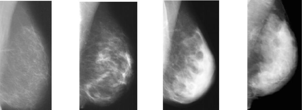
33 Mammographic densities
34 Cost of imaging An estimated 48 million mammograms are performed each year in the United States. With a female population of about 162 million, this is equivalent to about 29% of female population. In Nigeria, with a female population of about 6.5million above 55 years (7.3% female population) At least 6.5million mammograms are required, with an estimated cost at N10,000/exam, equivalent to 65 billion naira/per year
35 Mammography outcomes Of all of the screening mammograms approximately 90% show no evidence of cancer. Only 2% of all screening mammograms are shown to be abnormal and require biopsy. Among cases referred for biopsy, approximately 80% of the abnormalities are benign, 20% are shown to be cancerous.
36 Limitations of Mammography Mammography sensitivity (67.8%) and specificity (75%) are not ideal. Mammography combined with clinical breast examination (CBE) slightly improves sensitivity (77.4%), with a modest reduction in specificity (72%). Sensitivity for breast cancer declines significantly with increasing breast density Mammography uses low-dose ionizing radiation, which may be harmful to the patient. Nevertheless, the benefits of mammography far outweigh the risks.
37 False positive and negative mammogram False positive, due to micro calcifications, some benign lesions, and summation of parenchyma 8-10% false negative Possible causes for missed breast cancers include ; Dense parenchyma obscuring a lesion, Poor positioning or technique, Perception error, Incorrect interpretation of a suspect finding, Subtle features of malignancy, and Slow growth of a lesion


38 Mammographic features Benign lesions Benign masses are often round or oval with circumscribed margins. Benign calcifications are usually large The presence of very low density fat in a lesion often indicates benign findings such as oil cysts, lipomas, galactoceles, and hamartomas.


39 Malignant features Malignant lesions tend to have irregular, indistinct, or spiculated margins. Malignancies tend to have density greater than that of the normal breast tissue. Micro calcifications
40 BI-RADS- Breast imaging reporting and data system The American College of Radiology (ACR) has established the Breast Imaging Reporting and Data System (BI-RADS) to guide the breast cancer diagnostic routine Lexicon of descriptors, A recommended reporting structure including final assessment categories with accompanying management recommendations, and A framework for data collection and auditing
41 BI-RADS CLASSIFICATION Inconclusive study BIRADS 0, Normal study - BIRADS 1 Benign findings BIRADS 2, Probably Benign findings BIRADS 3 Suspicious findings BIRADS 4 Highly suspicious findings BIRADS 5 Known Cancer BIRADS 6

42 Digital breast tomosynthesis DBT, also known as 3D mammography, uses the same compression views as 2D mammography and adds a 3D volume acquisition. Requires only a few additional seconds for each view, DBT has shown to be an advance over digital mammography, with higher cancer detection rates and fewer patient recalls for additional testing. (ACR statement)
43 ULTRASONOGRAPHY Ultrasonography uses non ionising radiation in imaging. A widely available and useful adjunct to mammography in the clinical setting. As a screening device, ultrasound is limited, notably by the failure to detect micro calcifications Poor specificity (34%). Differentiating solid and cystic masses. Also useful in the guidance of biopsies and therapeutic procedures

44 Role in cancer screening Kolb et al and Buchberger et al found that when performed carefully, ultrasonography may be useful in detecting occult breast cancer in dense breasts.
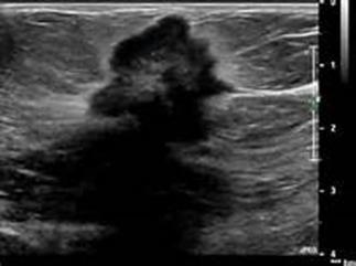

45 LESIONS ON ULTRASONOGRAPHY Suspicious lesion Benign lesion
46 Magnetic resonance imaging Complementary to mammography in high risk women The many advantages of breast MRI over conventional breast imaging for the detection of malignancy have become apparent with increasing clinical experience. No ionizing radiation All imaging planes possible Capability of imaging the entire breast volume and chest wall Superb 3-D lesion mapping with techniques such as maximum intensity projection (MIP) slab 3-D reconstruction Greater than 90% sensitivity to invasive carcinoma Detection of occult, multifocal, or residual malignancy Accurate size estimation for invasive carcinoma Good spatial resolution Ability to image regional lymph nodes (although accurate staging remains problematic)

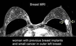
47 MRI
48 Disadvantages of MRI High equipment and examination costs Limited scanner availability Need for the injection of a contrast agent No standard technique Poor throughput compared with that of ultrasonography or mammography Large number of images Long learning curve for interpretation False-positive enhancement in some benign tissues (limited specificity) Variable enhancement of in situ carcinoma A 5% incidence of slowly or poorly enhancing invasive carcinomas
49 Contraindications of MRI Contraindication to gadolinium-based contrast media ( allergy, pregnancy) Patient's inability to lie prone Marked kyphosis or kyphoscoliosis Marked obesity Extremely large breasts Severe claustrophobia
50 MRI sensitivity In a high-risk population, MRI and mammography combined have a higher sensitivity (92.7%) than ultrasound (US) and mammography combined (52%) Screening high-risk women with breast MRI is costeffective and the cost-effectiveness of screening MRI increases with increasing breast cancer risk. The American Cancer Society recommends screening breast MRI in certain high-risk women, and the ACR and Society of Breast Imaging endorse those recommendations
51 Personal Experience with breast screening colleague and friend diagnosed with breast cancer Radiology department, National hospital Abuja in collaboration with some NGO carries out cancer screening Zamfara well women, cancer screeing programme in collaboration with medical women association of Nigeria (MWAN) at Garki hospital, Abuja ( 100 women brought from Zamfara state) MWAN in collaboration with the Ministry of women affairs, organised a breast cancer awareness lecture at the National women centre, Abuja ( Over 300 women from across Nigeria) Radiology department, National hospital Abuja, organised a breast awareness symposium, with 155 participants, and delivered a free 10 units CME, for doctors using a multidisciplinary faculty Thereafter commenced Management approved free mammography screening for all female staff, of 40 years and above from March 2015 to date.
52 Mammography at National hospital Abuja Months J 38 0 strike F 6 5 M 28 0 fault A 31 0 fault M 14 4 J J 59 0 NMA A 15 7 NMA S O 22 0 JOHESU N 30 0 JOHESU D 10 0 JOHESU Total Challenges Frequents strikes Equipment faults Cost of examination No NHIS coverage for screening Lack of awareness Poor communication across teams Poor record keeping Lack of subspecialisation No dedicated breast management team
53 Mammography screening challenges in developing countries The World Health Organization has suggested that for a mammography screening program to be effective in the reduction of mortality, it needs to cover at least 70% of the population at risk. The Breast Health Global Initiative Guidelines recommend that a population-based screening mammography program should not be implemented until access to the basic cancer diagnosis and treatment resources is guaranteed
54 Cost effective strategy for LMIC Early diagnosis Promotion of the awareness of early signs and symptoms among the public, Education of first line health professionals Improved referral procedures to facilitate the prompt and adequate diagnosis and treatment of breast cancer in early stages
55 Down staging programme Training first-line health personnel in hospitals and rural clinics Raising public awareness through visual information and sensitization by trained health personnel. In Malaysia after 4 years of implementation, late-stage (Ⅲ and Ⅳ) breast cancer cases were reduced from 60% to 35%[
56 Conclusion Mammography is the only method of screening for breast cancer shown to decrease mortality. Annual screening mammography is recommended starting at: 1) age 40 for general population; 2) Age in some intermediate and high risk patients. OR 10 years earlier than the age of the affected relative at diagnosis in some other high risk patients. Mammography plus supplemental screening is recommended in selected high-risk populations and those with dense breast.
57 Recommendations Adoption and implementation of appropriate mammography screening guidelines, for Nigerian women in both high risk and average risk groups should be carried out. Adequate education of patients and health workers of the guidelines as well as early signs and syptoms Provision of appropriate framework for implementation of the guidelines through government support and private sector participation. Institution of an interdisciplinary breast cancer screening and management programme, with adequate provision of infrastructure, equipment, manpower, trained personnel and resources. Ensuring appropriate diagnosis and treatment of all patients, identified from the screening programme through health insurance and not fee for service.

58 THANK YOU
Amammography report is a key component of the breast
 Review Article Writing a Mammography Report Amammography report is a key component of the breast cancer diagnostic process. Although mammographic findings were not clearly differentiated between benign
Mammographic imaging of nonpalpable breast lesions. Malai Muttarak, MD Department of Radiology Chiang Mai University Chiang Mai, Thailand
 Mammographic imaging of nonpalpable breast lesions Malai Muttarak, MD Department of Radiology Chiang Mai University Chiang Mai, Thailand Introduction Contents Mammographic signs of nonpalpable breast cancer
Melissa Hartman, DO Women s Health Orlando VA Medical Center
 Melissa Hartman, DO Women s Health Orlando VA Medical Center Most common non-skin cancer and Second deadliest cancer in women Majority are diagnosed by abnormal screening study An approach to breast cancer
Breast Cancer Imaging
 Breast Cancer Imaging I. Policy University Health Alliance (UHA) will cover breast imaging when such services meet the medical criteria guidelines (subject to limitations and exclusions) indicated below.
Standard Breast Imaging Modalities. Lilian Wang, M.D. Breast Imaging Section Department of Radiology Northwestern Medicine
 Standard Breast Imaging Modalities Lilian Wang, M.D. Breast Imaging Section Department of Radiology Northwestern Medicine Overview Standard breast imaging modalities Mammography Ultrasound MRI Imaging
Imaging in breast cancer. Mammography and Ultrasound Donya Farrokh.MD Radiologist Mashhad University of Medical Since
 Imaging in breast cancer Mammography and Ultrasound Donya Farrokh.MD Radiologist Mashhad University of Medical Since A mammogram report is a key component of the breast cancer diagnostic process. A mammogram
Ge elastography cpt codes
 Ge elastography cpt codes Aetna considers digital mammography a medically necessary acceptable alternative to film mammography. Currently, there are no guideline recommendations from leading medical professional
EARLY DETECTION: MAMMOGRAPHY AND SONOGRAPHY
 EARLY DETECTION: MAMMOGRAPHY AND SONOGRAPHY Elizabeth A. Rafferty, M.D. Avon Comprehensive Breast Center Massachusetts General Hospital Harvard Medical School Breast Cancer Screening Early detection of
Armed Forces Institute of Pathology.
 Armed Forces Institute of Pathology www.radpath.com Armed Forces Institute of Pathology Breast Disease www.radpath.org Armed Forces Institute of Pathology Interpretation of Breast MRI Leonard M. Glassman
Disclosures. Breast Cancer. Breast Imaging Modalities. Breast Cancer Screening. Breast Cancer 6/4/2014
 : Information for the Primary Care Physician Disclosures No financial relationships with commercial entities producing health care products/services. Roxsann Roberts, MD Section Chief, MRI Erlanger/EmCare
Breast Cancer Screening
 Scan for mobile link. Breast Cancer Screening What is breast cancer screening? Screening examinations are tests performed to find disease before symptoms begin. The goal of screening is to detect disease
Diagnostic Dilemmas of Breast Imaging
 Diagnostic Dilemmas of Breast Imaging Common Causes of Error in Breast Cancer Detection By: Jason Cord, M.D. Mammography: Initial Imaging The standard for detection of breast cancer Screening mammography
BREAST DENSITY WHAT IS IT? WHY IS IT IMPORTANT? & What IOWA SF250 Means to Patients and Providers
 BREAST DENSITY WHAT IS IT? WHY IS IT IMPORTANT? & What IOWA SF250 Means to Patients and Providers Arnold Honick, MD Radiology Consultants of Iowa, PLC ahonick@rciowa.com BREAST DENSITY LEGISLATION Nancy
BREAST IMAGING and NEW IMAGING MODALITIES- A Surgeons view
 BREAST IMAGING and NEW IMAGING MODALITIES- A Surgeons view DR CHANTEL THORNTON SPECIALIST BREAST CANCER SURGEON BMSc (hons) MBBS (hons) FRACS Epworth Hospital, Richmond- Agora Centre for Women s Health
8/31/2016 HIDING IN PLAIN SITE, ARCHITECTURAL DISTORTIONS AND BREAST ASYMMETRIES ARCHITECTURAL DISTORTIONS ARCHITECTURAL DISTORTIONS
 HIDING IN PLAIN SITE, ARCHITECTURAL DISTORTIONS AND BREAST ASYMMETRIES DEBORAH THAMES R.T. (R)(M)(QM) ARCHITECTURAL DISTORTIONS Definition is disruption of the natural flow of breast pattern towards the
EARLY DETECTION: MAMMOGRAPHY AND SONOGRAPHY
 EARLY DETECTION: MAMMOGRAPHY AND SONOGRAPHY Elizabeth A. Rafferty, M.D. Avon Comprehensive Breast Center Massachusetts General Hospital Harvard Medical School Breast Cancer Screening Early detection of
MANAGEMENT OF DENSE BREASTS. Nichole K Ingalls, MD, MPH NW Surgical Specialists September 25, 2015
 MANAGEMENT OF DENSE BREASTS Nichole K Ingalls, MD, MPH NW Surgical Specialists September 25, 2015 No financial disclosures National Cancer Institute National Cancer Institute Increased Cancer Risk... DENSITY
Imaging Guidelines for Breast Cancer Screening
 Imaging Guidelines for Breast Cancer Screening Sarah Colwick, MD Dr. Sarah Colwick was born and raised in Sikeston, MO. She attended college and medical school at the University of Missouri-Kansas City
S. Murgo, MD. Chr St-Joseph, Mons Erasme Hospital, Brussels
 S. Murgo, MD Chr St-Joseph, Mons Erasme Hospital, Brussels? Introduction Mammography reports are sometimes ambiguous and indecisive. ACR has developped the BIRADS. BIRADS consists of a lexicon in order
Breast Imaging! Ravi Adhikary, MD!
 Breast Imaging! Ravi Adhikary, MD! ACS Estimated Cancers Statistics 2014! Breast! New Cases in Women! 232,670 (+67,570 in situ)! Deaths in Women! 40,000! Colon! 48,380! 24,040! Cervical! 12,360! 4,020!
Breast Cancer Screening Clinical Practice Guideline. Kaiser Permanente National Breast Cancer Screening Guideline Development Team
 NATIONAL CLINICAL PRACTICE GUIDELINE Breast Cancer Screening Clinical Practice Guideline Kaiser Permanente National Breast Cancer Screening Guideline Development Team This guideline is informational only.
Breast Health and Imaging Glossary
 Contact: Lorna Vaughan HerSpace Breast Imaging & Biopsy Associates 300 State Route 35 South W. Long Branch, NJ 07764 732-571-9100, ext. 104 lorna@breast-imaging.com Breast Health and Imaging Glossary Women
The Radiology Aspects
 REQUIREMENTS FOR INTERNATIONAL ACCREDITATION OF BREAST CENTERS/UNITS The Radiology Aspects Miri Sklair-Levy, Israel RADIOLOGY GUIDELINES FOR QUALITY ASSURANCE IN BREAST CANCER SCREENING AND DIAGNOSIS Radiologists
Breast Imaging Update: Old Dog New Tricks
 Breast Imaging Update: Old Dog New Tricks Claire McKay, DO M&S Imaging Assoc. San Antonio, TX cmckayhart@juno.com Goals Describe modalities available, old and new Provide understanding of pros and cons
BI-RADS Update. Martha B. Mainiero, MD, FACR, FSBI Brown University Rhode Island Hospital
 BI-RADS Update Martha B. Mainiero, MD, FACR, FSBI Brown University Rhode Island Hospital No Disclosures BI-RADS History 1980s Quality Issues ACR Accreditation BI-RADS 1994 2003 4 th Edition MRI, US January
Mammography and Other Screening Tests. for Breast Problems
 301.681.3400 OBGYNCWC.COM Mammography and Other Screening Tests What is a screening test? for Breast Problems A screening test is used to find diseases, such as cancer, in people who do not have signs
Leonard M. Glassman MD
 BI-RADS The New BI-RADS Leonard M. Glassman MD FACR Former Chief of Breast Imaging American Institute for Radiologic Pathology Washington Radiology Associates, PC Breast Imaging Reporting and Data System
Policy Library Clinical Advantages of Digital Breast Tomosynthesis in Symptomatic Patients
 Policy Library Clinical Advantages of Digital Breast Tomosynthesis in Symptomatic Patients Version: 1 Approved by: Faculty of Clinical Radiology Council Date of approval: Click and type: day month and
ROLE OF MRI IN SCREENING, DIAGNOSIS AND MANAGEMENT OF BREAST CANCER. B.Zandi Professor of Radiology
 ROLE OF MRI IN SCREENING, DIAGNOSIS AND MANAGEMENT OF BREAST CANCER B.Zandi Professor of Radiology Introduction In the USA, Breast Cancer is : The Most Common Non-Skin Cancer The Second Leading cause of
ANNEX 1 OBJECTIVES. At the completion of the training period, the fellow should be able to:
 1 ANNEX 1 OBJECTIVES At the completion of the training period, the fellow should be able to: 1. Breast Surgery Evaluate and manage common benign and malignant breast conditions. Assess the indications
Current Status of Supplementary Screening With Breast Ultrasound
 Current Status of Supplementary Screening With Breast Ultrasound Stephen A. Feig, M.D., FACR Fong and Jean Tsai Professor of Women s Imaging Department of Radiologic Sciences University of California,
Breast Imaging Lexicon
 9//201 200 BI RADS th Edition 201 BI RADS th Edition Breast Imaging Lexicon Mammographic Pathology and Assessment Categories Deborah Thames, R.T.(R)(M)(QM) The Advanced Health Education Center Nonmember:
Epworth Healthcare Benign Breast Disease Symposium. Sat Nov 12 th 2016
 Epworth Healthcare Benign Breast Disease Symposium Breast cancer is common Sat Nov 12 th 2016 Benign breast disease is commoner, and anxiety about breast disease commoner still Breast Care Campaign UK
ORIGINAL ARTICLE EVALUATION OF BREAST LESIONS USING X-RAY MAMMOGRAM WITH HISTOPATHOLOGICAL CORRELATION
 Available online at www.journalijmrr.com INTERNATIONAL JOURNAL OF MODERN RESEARCH AND REVIEWS IJMRR ISSN: 2347-8314 Int. J. Modn. Res. Revs. Volume 3, Issue 10, pp 807-814, October, 2015 ORIGINAL ARTICLE
The radiologic workup of a palpable breast mass
 Imaging in Practice CME CREDIT EDUCTIONL OJECTIVE: The reader will consider which breast masses require further workup and which imaging study is most appropriate Lauren Stein, MD Imaging Institute, Cleveland
Mammography. What is Mammography? What are some common uses of the procedure?
 Mammography What is Mammography? Mammography is a specific type of imaging that uses a low-dose x-ray system to examine breasts. A mammography exam, called a mammogram, is used to aid in the early detection
BR 1 Palpable breast lump
 BR 1 Palpable breast lump Palpable breast lump in patient 40 years of age or above MMG +/- spot compression or digital breast tomosynthesis over palpable findings Suspicious or malignant findings (BIRADS
Breast Cancer. American Cancer Society
 Breast Cancer American Cancer Society Reviewed February 2017 What we ll be talking about How common is breast cancer? What is breast cancer? What causes it? What are the risk factors? Can breast cancer
National Diagnostic Imaging Symposium 2013 SAM - Breast MRI 1
 National Diagnostic Imaging Symposium 2013 December 8-12, 2013 Disney s Yacht Club Resort Lake Buena Vista, Florida Self Assessment Module Questions, Answers and References Day SAM Title - Each SAM title
Detailed Program of the second BREAST IMAGING AND INTERVENTIONS PROGRAM am am : Clinician s requirements from breast imaging
 Detailed Program of the second BREAST IMAGING AND INTERVENTIONS PROGRAM 2012 Day one, 2 nd November BREAST IMAGING AND INTERVENTIONS PROGRAM 2012 9.00 AM 9.10 am Introduction 9.10 am - 9.30 am : Clinician
Screening Mammograms: Questions and Answers
 CANCER FACTS N a t i o n a l C a n c e r I n s t i t u t e N a t i o n a l I n s t i t u t e s o f H e a l t h D e p a r t m e n t o f H e a l t h a n d H u m a n S e r v i c e s Screening Mammograms:
Screening for Breast Cancer
 Understanding Task Force Recommendations Screening for Breast Cancer U.S. Preventive Services Task Force (Task Force) has issued a final recommendation statement on Screening for Breast Cancer. se final
Here are examples of bilateral analog mammograms from the same patient including CC and MLO projections.
 Good afternoon. It s my pleasure to be discussing Diagnostic Breast Imaging over the next half hour. I m Wei Yang, Professor of Diagnostic Radiology and Chief, the Section of Breast Imaging as well as
CONTENTS NOTE TO THE READER...1 LIST OF PARTICIPANTS...3
 CONTENTS NOTE TO THE READER....1 LIST OF PARTICIPANTS....3 WORKING PROCEDURES...7 A. GENERAL PRINCIPLES AND PROCEDURES...7 1. Background....7 2. Scope....7 3. Objectives....8 4. Meeting participants...8
Emerging Techniques in Breast Imaging: Contrast-Enhanced Mammography and Fast MRI
 Emerging Techniques in Breast Imaging: Contrast-Enhanced Mammography and Fast MRI Lilian Wang, M.D. Breast Imaging Section Department of Radiology Northwestern Medicine Overview Rationale for new imaging
Breast Cancer Screening and Diagnosis
 Breast Cancer Screening and Diagnosis Priya Thomas, MD Assistant Professor Clinical Cancer Prevention and Breast Medical Oncology University of Texas MD Anderson Cancer Center Disclosures Dr. Thomas has
Ultrasonography. Methods. Brief Description. Indications. Device-related Prerequisites. Technical Requirements. Evaluation Criteria
 1 Ultrasonography Brief Description Imaging modality using sound waves Tissue-specific wave reflection. Indications Evaluation of palpable breast nodules Evaluation of clinically occult mammographic findings
International Day of Radiology 2016 Interview on Breast Imaging Australia / Dr. Michelle Reintals. Breast imaging in Australia
 International Day of Radiology 2016 Interview on Breast Imaging Australia / Dr. Michelle Reintals Breast imaging in Australia An interview with Dr. Michelle Reintals, Director of Breast at IMED Queensland
Presented by: Lillian Erdahl, MD
 Presented by: Lillian Erdahl, MD Learning Objectives What is Breast Cancer Types of Breast Cancer Risk Factors Warning Signs Diagnosis Treatment Options Prognosis What is Breast Cancer? A disease that
Tissue Breast Density
 Tissue Breast Density Reporting breast density within the letter to the patient is now mandated by VA law. Therefore, this website has been established by Peninsula Radiological Associates (PRA), the radiologists
Breast Cancer Screening and High Risk
 Breast Cancer Screening and High Risk Mary Freyvogel, DO Breast Surgeon Clinical Assistant Professor of Surgery University Hospitals Case Medical Center St. John Medical Center / Elyria Medical Center
Breast density: imaging, risks and recommendations
 Breast density: imaging, risks and recommendations Maureen Baxter, MD Radiologist Director of Ruth J. Spear Breast Center Providence St. Vincent Medical Center Alison Conlin, MD/MPH Medical Oncologist
EARLY DETECTION. Screening Mammography Programs. Knowledge Summary
 EARLY DETECTION Screening Mammography Programs Knowledge Summary EARLY DETECTION Screening Mammography Programs INTRODUCTION Early detection is an important component of a comprehensive breast cancer care
NATIONAL GUIDELINE CLEARINGHOUSE (NGC) GUIDELINE SYNTHESIS SCREENING FOR BREAST CANCER
 GUIDELINE SYNTHESIS SCREENING FOR BREAST CANCER") NATIONAL GUIDELINE CLEARINGHOUSE (NGC) GUIDELINE SYNTHESIS SCREENING FOR BREAST CANCER Guidelines 1. American Cancer Society (ACS). (1) ACS guidelines for breast cancer screening: update 2003. (2) American
NATIONAL GUIDELINE CLEARINGHOUSE (NGC) GUIDELINE SYNTHESIS SCREENING FOR BREAST CANCER Guidelines 1. American Cancer Society (ACS). (1) ACS guidelines for breast cancer screening: update 2003. (2) American
Contrast-enhanced Breast MRI RSSA 2013
 Contrast-enhanced Breast MRI RSSA 2013 Prof. dr. Maurice van den Bosch University Medical Center Utrecht, the Netherlands Index 1) Breast cancer 2) Why MRI of the breast 3) Technique 4) Interpretation
Supplemental Screening for Dense Breasts. Reagan Leverett, MD, MS
 Supplemental Screening for Dense Breasts Reagan Leverett, MD, MS Outline Anatomy and Density Risk of dense breasts Theory of Supplemental Screening Options for supplemental screening Tomosynthesis Ultrasound
Cairo/EG, Khartoum/SD, London/UK Biological effects, Diagnostic procedure, Ultrasound, Mammography, Breast /ecr2015/C-0107
 Role of sono-mammography in the evaluation of clinically palapble breast masses during pregnancy & lactation with differentaition between true patholgical & false physiological lobular hyperlpasia.sudanese
Mammography. What is Mammography?
 Scan for mobile link. Mammography Mammography is a specific type of breast imaging that uses low-dose x-rays to detect cancer early before women experience symptoms when it is most treatable. Tell your
Updates In Cancer Screening: Navigating a Changing Landscape
 Updates In Cancer Screening: Navigating a Changing Landscape Niharika Dixit, MD I have no conflict of interest. 1 Why Should You Care Trends in Cancer Incidence by Site United States. Siegal Et al: CA
Breast Cancer Update 2018 The Latest in Diagnosis and Treatment SARATH K, PALAKODETI, DO, FAACS GENERAL, BREAST, AND COSMETIC SURGEON TOLEDO CLINIC
 Breast Cancer Update 2018 The Latest in Diagnosis and Treatment SARATH K, PALAKODETI, DO, FAACS GENERAL, BREAST, AND COSMETIC SURGEON TOLEDO CLINIC Objectives Identify breast lesions and masses, and know
Breast and Ovarian Cancer
 Patient Education Breast and Ovarian Cancer Screening and detection The goal of screening for cancer is to find it as early as possible, when it is easiest to cure. This handout describes the symptoms
BI-RADS and Breast MRI. Kathy Borovicka, M.D. Thursday February 15, 2018
 BI-RADS and Breast MRI Kathy Borovicka, M.D. Thursday February 15, 2018 Learning Objectives Be familiar with the Breast Imaging Reporting and Data System (BI-RADS) Understand the components of a breast
Current Strategies in the Detection of Breast Cancer. Karla Kerlikowske, M.D. Professor of Medicine & Epidemiology and Biostatistics, UCSF
 Current Strategies in the Detection of Breast Cancer Karla Kerlikowske, M.D. Professor of Medicine & Epidemiology and Biostatistics, UCSF Outline ν Screening Film Mammography ν Film ν Digital ν Screening
Breast Health. Learning Objectives. Breast Anatomy. Poll Question. Breast Anatomy
 Learning Objectives Describe breast anatomy to a patient Breast Health Answer questions about causes of breast pain and masses Explain breast cancer screening/diagnostic modalities Appropriately triage
Page 1. Cancer Screening for Women I have no conflicts of interest. Overview. Breast, Colon, and Lung Cancer. Jeffrey A.
 Cancer Screening for Women 2017 Breast, Colon, and Lung Cancer Jeffrey A. Tice, MD Professor of Medicine Division of General Internal Medicine University of California, San Francisco I have no conflicts
Breast Cancer. What is breast cancer?
 Scan for mobile link. Breast Cancer Breast cancer is a malignant tumor in or around breast tissue. It usually begins as a lump or calcium deposit that develops from abnormal cell growth. Most breast lumps
FACT: FACT: Breast Cancer Staging. For cancer to occur, something must damage nucleus of the cell. Stage I. Stage II 10/9/2018
 Digital Breast Tomosynthesis (DBT) Pathology Findings: Case Studies and Beyond For cancer to occur, something must damage nucleus of the cell. Advanced Health Education Center www.aheconline.com Copyright
Breast Cancer Screening
 Breast Cancer Screening Eileen Rakovitch MD MSc FRCPC Sunnybrook Health Sciences Centre Medical Director, Louise Temerty Breast Cancer Centre LC Campbell Chair in Breast Cancer Research Associate Professor,
Breast Tomosynthesis. What is breast tomosynthesis?
 Scan for mobile link. Breast Tomosynthesis Breast tomosynthesis is an advanced form of mammography, a specific type of breast imaging that uses low-dose x-rays to detect cancer early when it is most treatable.
Diagnostic Breast Evaluation: When to Refer and What the Results Really Mean. Julie Dreadin RN, MS, WHNP-BC. Peggy Mancuso PhD, CNM, RN
 Diagnostic Breast Evaluation 1 Running head: DIAGNOSTIC BREAST EVALUATION Diagnostic Breast Evaluation: When to Refer and What the Results Really Mean Julie Dreadin RN, MS, WHNP-BC Peggy Mancuso PhD, CNM,
AMSER Case of the Month: September 2018
 AMSER Case of the Month: September 2018 60-year-old woman with a left breast mass noted on screening mammography. Catherine McNulty, MS4 Tulane University School of Medicine Dr. Robin Sobolewski Breast
SCREENING FOR BREAST CANCER BREAST IMAGING
 SCREENING FOR BREAST CANCER BREAST IMAGING Liane Philpotts, MD, FSBI, FACR Professor, Radiology and Biomedical Imaging Division Chief, Breast Imaging Dec. 5, 2017 Warner, E. NEJM 2011 Screening for
What s New in Breast Imaging. Jennifer A. Harvey, M.D., FACR Professor of Radiology University of Virginia
 What s New in Breast Imaging Jennifer A. Harvey, M.D., FACR Professor of Radiology University of Virginia Disclosure Hologic, Inc. Shareholder and research agreement. Volpara Solutions, Ltd. Shareholder
CHAPTER 2 MAMMOGRAMS AND COMPUTER AIDED DETECTION
 9 CHAPTER 2 MAMMOGRAMS AND COMPUTER AIDED DETECTION 2.1 INTRODUCTION This chapter provides an introduction to mammogram and a description of the computer aided detection methods of mammography. This discussion
Breast Cancer. What is breast cancer?
 Scan for mobile link. Breast Cancer Breast cancer is a malignant tumor in or around breast tissue. It usually begins as a lump or calcium deposit that develops from abnormal cell growth. Most breast lumps
BREAST MRI. VASILIKI FILIPPI RADIOLOGIST CT MRI & PET/CT Departments Hygeia Hospital, Athens, Greece
 BREAST MRI VASILIKI FILIPPI RADIOLOGIST CT MRI & PET/CT Departments Hygeia Hospital, Athens, Greece Breast ΜR Imaging (MRM) Breast MR imaging is an extremely powerful diagnostic tool, that when used in
Non-mass Enhancement on Breast MRI. Aditi A. Desai, MD Margaret Ann Mays, MD
 Non-mass Enhancement on Breast MRI Aditi A. Desai, MD Margaret Ann Mays, MD Breast MRI Important screening and diagnostic tool, given its high sensitivity for breast cancer detection Breast MRI - Indications
BIRADS 3 and 4 lesions viewed by ultrasound and not seen in digital mammograms and tomosynthesis.
 Original article Anales de Radiología México 2016 Jul;15(3):205-213. BIRADS 3 and 4 lesions viewed by ultrasound and not seen in digital mammograms and tomosynthesis. García-Quintanilla JF 1, González-Coronado
Angela Gilliam, MD University of Colorado Surgical Grand Rounds November 3, 2008
 Angela Gilliam, MD University of Colorado Surgical Grand Rounds November 3, 2008 Breast Cancer Most common cancer in American women 180,000 new cases per year Second most common cause of cancer death 44,000
Current issues and controversies in breast imaging. Kate Brown, South GP CME 2015
 Current issues and controversies in breast imaging Kate Brown, South GP CME 2015 JUDICIOUS USE OF RESOURCES IN REFERRALS FOR BREAST IMAGING THE DILEMMA How do target referrals for breast imaging? Want
MRI in breast cancer: diagnosis and intervention. Dr Sue Barter Addenbrookes Hospital, Cambridge UK
 MRI in breast cancer: diagnosis and intervention Dr Sue Barter Addenbrookes Hospital, Cambridge UK Intervention will be discussed in High Risk Screening! Indications UK and Europe: Breast MRI is well established
Lesion Imaging Characteristics Mass, Favoring Benign Circumscribed Margins Intramammary Lymph Node
 Lesion Imaging Characteristics Mass, Favoring Benign Circumscribed Margins Intramammary Lymph Node Oil Cyst Mass, Intermediate Concern Microlobulated Margins Obscured Margins Mass, Favoring Malignant Indistinct
RADIOLOGIC EVALUATION OF BREAST CANCER
 RADIOLOGIC EVALUATION OF BREAST CANCER Orsolya Farkas, Gabriella Bodrogi and Gábor Szalai Department of Radiology, Pécs University Orsifarkas@yahoo.com Complex evaluation of the breast Patient history
Updates in Mammography. Dr. Yang Faridah A. Aziz Department of Biomedical Imaging University Malaya Medical Centre
 Updates in Mammography Dr. Yang Faridah A. Aziz Department of Biomedical Imaging University Malaya Medical Centre Updates in Mammography Breast Imaging Dr. Yang Faridah A. Aziz Department of Biomedical
Case Report Tubular Carcinoma of the Breast: Advantages and Limitations of Breast Tomosynthesis
 Case Reports in Radiology Volume 2016, Article ID 3906195, 4 pages http://dx.doi.org/10.1155/2016/3906195 Case Report Tubular Carcinoma of the Breast: Advantages and Limitations of Breast Tomosynthesis
New Imaging Modalities for better Screening and Diagnosis
 New Imaging Modalities for better Screening and Diagnosis Miri Sklair-Levy, MD Department of Diagnostic Imaging Sheba Medical Center, Sackler School of Medicine, Tel Aviv University Department of Diagnostic
Breast Imaging & You
 Breast Imaging & You What s Inside: Breast Imaging... 2 Digital Breast Tomosynthesis (DBT) mammograms... 4 Breast cancer screening... 6 Dense breast tissue... 8 Automated Breast Ultrasound (ABUS)... 9
ACRIN 6666 IM Additional Evaluation: Additional Views/Targeted US
 Additional Evaluation: Additional Views/Targeted US For revised or corrected form check box and fax to 215-717-0936. Instructions: The form is completed based on recommendations (from ID form) for additional
MEDICAL IMAGING AND BREAST DISEASE HOW CAN WE HELP YOU?
 MEDICAL IMAGING AND BREAST DISEASE HOW CAN WE HELP YOU? Barbara M. Preston, M.D. SCREENING MAMMOGRAPHY AVERAGE RISK PATIENTS KAISER RECOMMENDATION: ALL WOMEN (INCLUDING TRANSGENDER FEMALES) Every 1-21
MRI BI-RADS: How to make it out?
 MRI BI-RADS: How to make it out? Poster No.: C-1850 Congress: ECR 2016 Type: Educational Exhibit Authors: M. Ben Ammar, A. Ben Miled, O. Ghdes, S. Harguem, A. Gaja, N. Mnif; Tunis/TN Keywords: Breast,
MR sin plass i brystkreftdiagnostikk, dagens anbefalinger og fremtidsperspektiver
 MR sin plass i brystkreftdiagnostikk, dagens anbefalinger og fremtidsperspektiver Kathinka Kurz, MD, PhD, seksjonsoverlege SUS, kathinka.dehli.kurz@sus.no Technique - Subtraction Without contrast agent
Screening Mammography: Who, what, where, when, why and how?
 Screening Mammography: Who, what, where, when, why and how? Jillian Lloyd, MD, MPH Breast Surgical Oncologist University Surgical Oncology Department of Surgery University of Tennessee Medical Center Disclosures
Dense Breasts. A Breast Cancer Risk Factor and Imaging Challenge
 Dense Breasts A Breast Cancer Risk Factor and Imaging Challenge Renee Pinsky, MD University of Michigan Department of Radiology Division of Breast Imaging No Disclosures QUIZ: ARE YOU DENSE? a. Breast
Breast Cancer Screening: Changing Philosophies in Educating Women and Teens
 Breast Cancer Screening: Changing Philosophies in Educating Women and Teens Courtney Benedict CNM MSN Disclosures Merck Nexplanon trainer Session Objectives Explain the rationale for initiation and frequency
Breast Imaging & You
 Breast Imaging & You What s Inside: Breast Imaging... 2 Digital Breast Tomosynthesis (DBT) mammograms... 4 Breast cancer screening... 6 Dense breast tissue... 8 Automated breast ultrasound (ABUS)... 9
Breast Ultrasound: Improving Your Skills & Patient Care
 Breast Ultrasound: Improving Your Skills & Patient Care Objectives Discuss US techniques available for image optimization. Review & compare the US appearances of benign & malignant masses. Cherie M. Kuzmiak,
Population Prospective. Big Picture
 Mai Elezaby, MD? Population Prospective Big Picture Breast Cancer Most common cancer in women 2 nd leading cause of death U.S. 2016 estimates 246,660 new cases 40,450 deaths from breast cancer https://seer.cancer.gov/statfacts/html/breast.html
Ana Sofia Preto 19/06/2013
 Ana Sofia Preto 19/06/2013 Understanding the underlying pathophysiologic processes leading to the various types of calcifications Description and illustration of the several types of calcifications, according
Breast Cancer Risk Factors 8/3/2014
 Breast Cancer Screening: Changing Philosophies in Educating Women and Teens Courtney Benedict CNM MSN Session Objectives Explain the rationale for initiation and frequency of clinical breast exams to clients
Breast Cancer Task Force of the Greater Miami Valley A collaborative effort of health care professionals and breast cancer survivors in the Greater
 Breast Cancer Task Force of the Greater Miami Valley A collaborative effort of health care professionals and breast cancer survivors in the Greater Dayton Area Last Updated Fall 2014 TABLE OF CONTENTS