Identification of HETE isoforms produced by activated platelets and study of their roles within the vasculature
|
|
|
- Henry Norman
- 5 years ago
- Views:
Transcription
1 ` Francesca Rauzi Supervisors: Prof. Prof. Timothy D. Warner Amrita Ahluwalia Ph.D Identification of HETE isoforms produced by activated platelets and study of their roles within the vasculature Translational Medicine and Therapeutics (TMT) Cardiac, Vascular and Inflammation Research William Harvey Research Institute, Barts and The London, Queen Mary's School of Medicine and Dentistry
2 Abstract Abstract Background: Prostaglandins (PGs) and hydroxyeicosatetraeinoic acids (HETEs) are synthesized from arachidonic acid (AA) released from the membrane phospholipids of cells through the activity of cytosolic phospholipase A 2α (cpla 2α ). In platelets AA is metabolised through cyclooxygenase-1 (COX-1) and 12-lipoxygenase (12-LOX) leading to the production of a range of eicosanoids. This project determined the enzymes responsible for the production of the major prostanoid and HETE metabolites released by activated platelets and investigated the roles of these within the vasculature. Methods: The responses of whole blood and platelets collected from healthy volunteers or patients lacking cpla 2α were examined in the absence and presence of anti-platelet drugs (aspirin and/or prasugrel). The effects of selective platelet agonists and global stimulants of blood cells were tested to provide multiple functional responses against which to compare the ability of platelets and blood to produce eicosanoid molecules, as measured by liquid chromatography-tandem mass spectrometry (LC-MS/MS). Results: Stimulation of blood and PRP produced large increases in the levels of TXA 2, PGE 2, PGD 2, 11- and 15-HETE which were found to be produced by platelets via the activity of cpla 2α and COX-1 enzymes. 12-HETE was dependent upon cpla 2α but not COX and 15-HETE have not previously been reported as major platelet products. A series of experiments indicated that 11-, 12- and 15-HETE had very weak influences on platelet aggregation/adhesion responses. Notably, 12(S)-HETE, which represents the enantiomer released by platelets, strongly promoted neutrophil chemotactic responses. Furthermore, 15(S)-HETE was identified as the bioactive enantiomer produced by activated platelets and was able to induce tube formation and cell migration in cultures of human microvascular endothelial cells (HMEC-1) and promote the formation of sprouts from rat aortic rings. Conclusions: Platelets are major producers of prostanoids and HETEs in activated blood. Notably, aspirin abolished the production of not only prostanoids, but also 11-HETE and 15-HETE, identifying these as major COX-1 products. Moreover, 12(S)- I
3 Abstract HETE robustly induced neutrophil chemotaxis, while 15(S)-HETE promoted angiogenic responses both in vitro and ex vivo. These data identified 15(S)-HETE as a potential target for the development of therapies able to limit angiogenic processes in conditions such as tumour progression and metastasis. II
4 Acknowledgements Acknowledgements Although it has been a tough ride, only now that it is all coming to an end can I acknowledge the many positive aspects of pursuing a PhD; a goal that I couldn t have achieved without the help of many people towards whom I feel the need to sincerely express my gratitude. To begin I would like to thank all the members of my group of research who guided me during this long journey and contributed to my scientific and personal development. Among these, I am deeply grateful to my supervisor, Tim, for his support, availability, intelligence and humanity. I also thank him for always showing a high interest in my personal and professional growth and, thus, for being a mentor in both life and science. A particular thanks to Ivana, one of the most warm-hearted and helpful people I had the fortune to come across, who has always been available to truly listen to me whenever I had an issue. During the years I received support and training from Nick, who I look up to for his success and remarkable intelligence, and Paul, who I admire for his maturity and helpfulness. I also want to thank Thomas with whom I shared the difficulties and rewards of a PhD and all those people that joined the group throughout these years. I am thankful to Slobodan and Marija, both under care at the Barts hospital, for allowing me to test their blood and for their generosity towards research. I am also grateful to all the collaborations I had the opportunity to establish, from those within my institute to those with the Barts Hospital and the National Institutes of Environmental Health (NIEH). I want to deeply thank Elisa, Maria and Jale with whom I shared many meaningful moments. A big THANK YOU to all my close friends outside the world of research, who contributed to make these years an unforgettable and exciting experience. Most importantly, I want to dedicate my thesis to my parents and my older brother, Matteo, as a symbolic gesture of gratitude for their love, education and support. III
5 Contents Contents Abstract... I Acknowledgements... III List of figures... X Abbreviations..XIV Chapter 1: Introduction Human phospholipase A 2 isoforms Eicosanoids Cyclo-oxygenases and thromboxane A 2 /prostaglandins... 8 Thromboxane A Prostacyclin Prostaglandin E Prostaglandin D Lipoxygenases and HpETEs/HETEs lipoxygenase and leukotrienes/5-hete lipoxygenase and 12(S)-HETE lipoxygenase1 and 12(S)-HETE/15(S)-HETE Transcellular biosynthesis of eicosanoids P45 epoxygenase and EETs Non-enzymatic metabolism and isoprostanes Phospholipase A 2 inhibitors Pyrrolidine inhibitors COX inhibitors IV
6 Contents Non-steroidal anti-inflammatory drugs (NSAIDs) Aspirin LOX inhibitors Baicalein Nordihydroguaiaretic acid (NDGA) Other LOX inhibitors Platelets Thrombopoiesis Mature platelets Platelets and atherogenesis Platelets and thrombus formation Adhesion... 3 vwf Collagen Aggregation Thrombin TXA ADP Secretion Coagulation Thrombotic vascular events Antiplatelet treatment Aspirin Thienopyridine P2Y 12 blockers Clopidogrel V
7 Contents Prasugrel Integrin α IIb β 3 inhibitors Abciximab HETEs and haemostasis/thrombosis HETEs and platelet aggregation HETEs and vascular tone regulation Platelet-leukocyte interplay HETEs and leukocytes in inflammation HETEs and leukocytes in coagulation Platelets and angiogenesis COX-derived 15(S)-HETE (S)-HETE and angiogenesis Chapter 2: Aims of the research Aims of the research Chapter 3: Material and Methods Materials Methods Ethics and consent Screening of healthy blood donors Venepuncture Preparation of PRP and PPP from whole blood Isolation of platelets Treatment of PRP or whole blood with antiplatelet drugs Treatment of PRP or whole blood with LOX or cpla 2α inhibitors Preparation of platelet agonists VI
8 Contents 3.9 Preparation of HETE isoforms Preparation of human blood samples for LC-MS/MS analysis LC-MS/MS analysis well plate light transmission aggregometry Light transmission aggregometry (LTA) Platelet adhesion under flow conditions Platelet adhesion under static conditions (acid phosphate assay) Statistical analysis Chapter 4: Eicosanoid formation of blood from cpla 2α -deficient patients Phospholipase A 2 (cpla 2α )-deficient patients Introduction Methods Results Conclusions Chapter 5: Effect of lack of COX-1 or cpla 2α activity on platelet aggregation Effect of COX or cpla 2α inhibitor on platelet aggregation Introduction Methods Results Conclusions Chapter 6: Effect of AA-derived HETE isoforms on platelet aggregation Effect of platelet COX-1 or LOX inhibition and prostanoids or HETE isoforms on platelet aggregation Introduction VII
9 Contents Methods Results Conclusions Chapter 7: Effect of anti-platelet therapy on eicosanoid formation Effect of anti-platelet treatments on eicosanoid production in whole blood and PRP Introduction Methods Results Effect of platelet COX-1-derived prostanoids and 11(R), 15(S,R)HETE on aggregation Introduction Methods Results Conclusions Chapter 8: Effect of HETE isoforms on neutrophil chemotaxis Effect of platelet-derived HETEs on neutrophil chemotaxis Introduction Methods Results Effect of platelet-derived HETEs on neutrophil activation Introduction Methods Results Conclusions Chapter 9: Effect of 15(S)HETE on HMEC-1 angiogenic responses VIII
10 Contents 9.1 Production of the 15(S)HETE stereoisomer in whole blood and PRP Introduction Methods Results Effect of 15(S)-HETE on HMEC-1 angiogenic responses Introduction Methods Results Conclusions Chapter 1: General discussion Future work and limitations Chapter 11: References IX
11 List of figures List of figures Figure 1.1 Structures of cpla isoforms 4 Figure 1.2 Regulatory mechanisms for cpla 2α - and spla 2 -mediated AA release 5 Figure 1.3 Scheme of AA-derived eicosanoid products 7 Figure 1.4 Structures of COX isoforms 8 Figure 1.5 Biosynthesis of AA-derived prostanoids via COX activity 9 Figure 1.6 Lipoxygenase pathways of AA metabolism 14 Figure 1.7 Structures of the cpla 2α inhibitors pyrrolidine-1 and pyrrophenone 21 Figure 1.8 Structures of the COX inhibitor, aspirin 22 Figure 1.9 Structure of the 12 lipoxygenase inhibitor, baicalein 24 Figure 1.1 Structure of the lipoxygenase inhibitor, NDGA 25 Figure 1.11 Figure 1.12 Figure 1.13 Figure 1.14 Schematic representation of key ligands and receptors involved in primary platelet adhesion 32 Schematic representation of the signalling events following receptor ligation during primary platelet adhesion 33 Schematic representation of platelet receptors activated by soluble agonists essential for platelet aggregation 38 a) Extrinsic coagulation pathway; b) Intrinsic coagulation pathway 4 Figure 1.15 Stages of plug formation 42 Figure 1.16 Figure 3.1 A schematic representation of the multiple bidirectional interactions between platelets and tumour cells 61 Standard layout of 96-well plate light transmission aggregometry 75 Figure 4.1 LC-MS/MS analysis of eicosanoid levels in whole blood from cpla 2α - deficient patients and healthy volunteers 82 X
12 List of figures Figure 4.2 LC-MS/MS analysis of TXB 2, PGE2, PGD 2, 11-HETE, 12-HETE, 15-HETE and 5-HETE in stimulated blood and PRP obtained from healthy volunteers 84 Figure 5.1 Effect of aspirin and pyrrophenone on platelet aggregation 92 Figure 5.2 Adhesion/aggregation to collagen (1 µg/ml) under shear (1 s -1 ) of mepacrine-labelled platelets from cpla 2α -deficient patients and from healthy volunteers treated with abciximab (1 µg/ml), aspirin (1 µm), pyrrophenone (4 µm) or vehicle 93 Figure 5.3 Figure 6.1 Figure 6.2 Figure 6.3 Images taken by epifluorescent microscopy (2X) of mepacrinelabelled platelets aggregating to collagen (1 µg/ml) under shear (1 s -1 shear rate) in lepirudinized-blood from the cpla 2a -deficient patients and blood from healthy volunteers treated with abciximab (1 µm), aspirin (1 µm), pyrrophenone (4 µm), or vehicle 94 Adhesion of mepacrine-labelled platelets to collagen (1 µg/ml) under shear (1 s -1 shear rate) after treatment with prostacyclin (PGI 2 ; 2 µg/ml), aspirin (3 µm), baicalein (1 µm) or vehicle 13 Images taken by epifluorescent microscopy (2X) of mepacrinelabelled platelets aggregating to collagen (1 µg/ml) under shear (1 s -1 shear rate) of citrated whole blood from healthy volunteers treated with PGI 2 (2 µg/ml), aspirin (3 µm), baicalein (1 µm), or vehicle 14 Effect of 12-HETE or 12-LOX inhibition on platelet adhesion/ aggregation in static conditions 15 Figure 6.4 Effects of PGI 2 on platelet aggregation 16 Figure 6.5 Effect of aspirin on platelet aggregation 17 Figure 6.6 Effect of baicalein on platelet aggregation 18 Figure 6.7 Effect of 12(S)HETE on platelet aggregation 19 Figure 6.8 Effect of PGD 2 on platelet aggregation 11 Figure 6.9 Effect of PGE 2 on platelet aggregation 111 Figure 6.1 Effect of 5-HETE, 11-HETE, 12-HETE and 15-HETE on platelet aggregation 115 Figure 6.11 Effect of NDGA on platelet aggregation 116 XI
13 List of figures Figure 7.1 Figure 7.2 Figure 7.3 Figure 7.4 Figure 7.5 Figure 7.6 Figure 7.7 Figure 7.8 Figure 8.1 Figure 8.2 LC-MS/MS analysis of TXA 2, PGD 2 and PGE 2 levels in TRAP-6 (3µM)- stimulated PRP and whole blood of healthy volunteers in the presence of aspirin (1µM), prasugrel (3µM) or the combination of aspirin and prasugrel 123 LC-MS/MS analysis of 11-HETE and 15-HETE levels in TRAP-6 (3µM)- stimulated PRP and whole blood of healthy volunteers in the presence of aspirin (1µM), prasugrel (3µM) or the combination of aspirin and prasugrel 124 LC-MS/MS analysis of 5-HETE and 12-HETE levels in TRAP-6 (3µM)- stimulated PRP and whole blood of healthy volunteers in the presence of aspirin (1µM), prasugrel (3µM) or the combination of aspirin and prasugrel 125 LC-MS/MS analysis of TXA 2, PGD 2 and PGE 2 levels in collagen (3µg/ml)-stimulated PRP and whole blood of healthy volunteers in the presence of aspirin (1µM), prasugrel (3µM) or the combination of aspirin and prasugrel 127 LC-MS/MS analysis of 11-HETE and 15-HETE levels in collagen (3µg/ml)-stimulated PRP and whole blood of healthy volunteers in the presence of aspirin (1µM), prasugrel (3µM) or the combination of aspirin and prasugrel 128 LC-MS/MS analysis of 5-HETE and 12-HETE levels in collagen (3µg/ml)- stimulated PRP and whole blood of healthy volunteers in the presence of aspirin (1µM), prasugrel (3µM) or the combination of aspirin and prasugrel 129 Scheme of the aggregation study using LTA to test the effects of the combination of COX-1-derived prostanoids and HETEs on platelet aggregation 131 Effects of the combination of U46619, PGD 2, PGE 2, 11(R)-HETE, 15(S)- HETE and 15(R)-HETE on platelet aggregation 133 Effects of LTB 4 (1-1-1nM), 11-HETE, 15-HETE, 5-HETE and the 12(S)- HETE stereoisomer (.3 3µM) on neutrophil chemotaxis 142 Effects on neutrophil chemotaxis of 12(S)-HETE or 5-HETE (1µM) added singularly on top of increasing concentrations of 5-HETE and 12(S)- HETE (.25 2µM) respectivey 143 Figure 8.3 Effect of HETEs on neutrophil activation 148 Figure 9.1 EIA assay of 15(S)-HETE levels in collagen- and TRAP-6 stimulated lepirudinized PRP and whole blood of healthy volunteers in the XII
14 List of figures presence of aspirin (1µM), prasugrel (3µM) or the combination of aspirin and prasugrel 154 Figure 9.2 Figure 9.3 Figure 9.4 Figure 9.5 Figure 9.6 Figure 9.7 Images of HMEC-1 tube formation in the presence of releasates from TRAP-6 stimulated platelets 163 Images of HMEC-1 in the presence of releasates fromtrap-6 stimulated and aspirin-treated platelets 164 HMEC-1 tube formation in the presence of releasates from TRAP-6 stimulated platelets 165 Images of HMEC-1 tube formation in the presence of releasates from collagen-stimulated platelets 166 Images of HMEC-1 in the presence of releasates from collagenstimulated and aspirin-treated platelets 167 HMEC-1 tube formation in the presence of releasates from collagenstimulated platelets 168 Figure 9.8 Effect of 15(S)-HETE on HMEC-1 migration 169 Figure 9.9 Figure 9.1 Effect of 15(S)-HETE on formation of angiogenic sprouts from rat aortic rings in the presence of relesates from TRAP-6-stimulated platelets 17 Images of the effect of 15(S)-HETE on formation of angiogenic sprouts from rat aortic rings in the presence of releasates from TRAP-6- stimulated platelets 171 Figure 9.11 Figure 9.12 Figure 1.1 Effect of 15(S)-HETE on formation of angiogenic sprouts from rat aortic rings in the presence of releasates from collagen-stimulated platelets 172 Images pf the effect of 15(S)-HETE on formation of angiogenic sprouts from rat aortic rings in the presence of releasates from collagenstimulated platelets 173 Scheme of the enzymatic pathways involved in the production of prostanoids and HETEs by activated platelets together with their biological function within the vasculature 187 XIII
15 Abbreviations Abbreviations AA Arachidonic acid AACOCF3 Arachidonyl Trifluoromethyl Ketone ACS Acute Coronary Syndrome ADP Adenosine Diphosphate ANOVA Analysis of Variance APC Activated Protein C ASA Aspirin; acetylsalycilic acid AT Antithrombin ATP Adenosine Triphosphate AUC Area Under the Curve BSA Bovine Serum Albumin C Celcius camp cyclic Adenosine Monophosphate COX Cyclo-oxygenase CRP Collagen-related peptide CYP45 Cytochrome P 45 DAG Diacylglycerol DMSO Dimethyl Sulfoxide EC Endothelial Cell EIA Enzyme-linked Immunoassay ER Endoplasmic Reticulum FGF Fibroblast Growing Factor fmlp Formyl-Methionyl-Leucyl-Phenylalanine FLAP 5-Lipoxygenase-Activating Protein GCRP G-Protein Coupled Receptor GP Glycoprotein GST Glutathione S-Transferase HETE Hydroxyeicosatetraenoic Acid HpETE Hydroperoxyeicosatetraeinoic Acid XIV
16 Abbreviations HT HMEC HUVEC IC IL-8 IP isop LC/MS LDL LM LOX LTA LT LX MAC-1 MAPK MK NDGA NSAID PAF PAM PAR PBMC PBS PDGF PG PK PL PMNL PPP PRP PUFA Hydroxytryptamine Human Microvascular Endothelial Cell Human Umbilical Vein Endothelial Cell Inhibitory Concentration Interleukin-8 Inositol Phosphate isoprostane Liquid Chromatography/Mass Spectrometry Low Density Lipopolysaccaride Lipid Mediator Lipoxygenase Light Transmission Aggregometry Leukotrienes Lipoxins Macrophage-1 Antigen Mitogen-activated Protein Kinase Megakaryocytes Nordihydroguaiaretic Acid Non-Steroidal Anti-Inflammatory Drug Platelet Activating Factor Prasugrel Active Metabolite Protease Activated Receptor Peripheral Blood Mononuclear Cell Phosphate Buffered Saline Platelet Derived Growth Factor Prostaglandin Protein Kinase Phospholipase Polymorphonuclear Leukocyte Platelet Poor Plasma Platelet Rich Plasma Poly Unsaturated Fatty Acids XV
17 Abbreviations rpm SMC TF TP TPO TRAP TX VEGF vwf Revolution per minute Smooth Muscle Cell Tissue Factor Thromboxane receptor Thrombopoietin Thrombin Receptor Activating Peptide Thromboxane Vascular Endothelial Growth Factor von Willebrand Factor XVI
18 Introduction Chapter 1: Introduction 1
19 Introduction Within the cardiovascular system platelets represent the blood component assigned to regulate and control haemostasis. Disruption of the vascular wall due to injury induces platelets to become activated and aggregate leading to thrombus formation and, thus, to pathological conditions which manifest clinically as acute cardiovascular events (e.g. heart attack). Anti-platelet therapies are currently used to control platelet activation and prevent the occurrence of acute cardiovascular events. However, chronic use of these therapies is associated with a high risk of gastrointestinal and intracranial bleeding, side effects that counteract the benefits produced by the treatment. Research is currently focused on identifying novel targets and elucidating platelet pathways in order to develop therapeutic approaches able to overcome these side effects and, thus, improve cardiovascular therapy. The aim of this project was to better understand the platelet LOX pathway, the role of which is still unclear, and investigate the interplay between COX and LOX pathways in the regulation of haemostasis/thrombosis. Ultimately, these studies may lead to the identification of novel therapeutic targets to support and ameliorate the therapies currently used in cardiovascular disease. 1.1 Human phospholipase A 2 isoforms The phospholipase A 2 (PLA 2 ) enzyme catalyses the hydrolysis at sn-2 position of the cell s membrane phospholipids releasing arachidonic acid (AA), a free fatty acid from which active metabolites, eicosanoids, are subsequently synthesized. 1,2 The release of AA is indeed the rate limiting step in the generation of prostanoids and leukotrienes. 2 At least 19 PLA 2 isoenzymes have been identified and classified depending on their chemical structure, enzymatic characteristics, cellular distribution and cellular functions. 1,3 The PLA 2 s have been classified in different families, the cytosolic calcium dependent PLA 2 (cpla), the secretory PLA 2 (spla 2 ) and the cytosolic calcium independent PLA 2 (ipla 2 ). 3 These enzymes play a fundamental role in the production of lipid mediators in the presence of certain stimuli (e.g. calcium 2
20 Introduction ionophore) and in phospholipid remodelling. 3-5 The cpla 2 family is represented by three different isoenzymes, cpla 2α, cpla 2β and cpla 2ϒ which are classified into group IVA, ICB and IVC, respectively. 6-8 The 85-kDa cytosolic PLA 2, cpla 2α, shows a significant preference for AA over other fatty acids and is activated by submicromolar concentrations of Ca 2+ and associated with inflammatory responses, platelet activation and cell proliferation. 3,9-11 The important mechanisms by which cpla 2 activation and consequent AA release are regulated involve agonist-induced MAP kinase phosphorylation of the enzyme and Ca ++ -dependent translocation of cpla 2 from the soluble to the membrane fraction of cells where the enzyme is able to elicit its catalytic function. 6,12 3
21 Introduction Figure 1.1 Structures of cpla 2 isoforms. a) The N-terminal region of cpla 2α and cpla 2β is constituted by a C2 domain responsible for Ca 2+ -dependent membrane association. All three cpla 2 isoforms possess two catalytic domains, A and B. The catalytic domain A of the three isoforms has lipase consensus motifs. Only in the cpla 2 α isoform two serine residues, Ser55 and Ser727, are present and phosphorylated by protein kinases in the MAPK cascade, while the C-terminal portion of cpla 2ϒ is isoprenylated. b) Schematic tertiary structure of cpla 2α. The twoca 2+ ions which leads to the exposure of hydrophobic surfaces on the top of the domain. The vimentin-binding site in the C2 domain is separated from the hydrophobic, membrane-binding region. The catalytic centre in the globular catalytic domain is located in the active site hole, into which the AA residue enters. PI(4,5)P 2 interacts with the cationic residues (+) in both the C2 and catalytic domains inducing conformational change of the enzyme that brings its active site closer to the membrane. This allows a substrate molecule to enter the active site for catalytic turnover. The two critical phosphorylation sites are located in the interspaced region. Figures a) and b) were adapted from those in the review of Makoto Murakami. 3 4
22 Introduction The secretory PLA 2 s, spla 2 s, constitute the largest family of PLA 2 in mammals of which 1 closely related isoforms have been identified, i.e. spla 2 IB, IIA, IIC, V, X, IID, IIE, IIF, III and XII. 3,13,14 This class of enzymes is characterised by a low molecular weight (14-19 KDa), histidine in the catalytic site and a Ca 2+ -binding loop in the active site. 3,15 The hydrolysis requires abstraction of a proton from a water molecule and a nucleophilic attack on the sn-2 bond. 15 SPLA 2 IIA is the most wildly distributed subtype in humans and its production is robustly induced by pro-inflammatory factors. 16,17 Figure 1.2 Regulatory mechanisms for cpla 2 α and spla 2 -mediated AA release. Upon receptor activation, phospholipase C (PLC) enzymes produce diacylglycerol (DAG) and inositol triphosphate (IP 3 ), inducing an increase in intracellular Ca 2+ levels. In response to this Ca 2+ elevation, cpla 2α translocates from the cytosol to the endoplasmic reticulum (ER) and perinuclear membranes, where COX isozymes are situated (1). The cpla 2α is activated also via the MAPK cascade, where MAPKs and MAPKAPKs phosphorylate Ser55 and Ser727 on the enzyme, respectively (2). This dual phosphorylation is required for full activation of cpla 2 α. In the HSPG-shuttling pathway (3), the heparin-binding group II subfamily spla 2 s (IIA, IID, IIE, and V) are associated with the GPI-anchored HSPG glypican and are sorted into rafts. As a consequence these spla 2 s are internalized and release AA adjacent to COXs. The 5
23 Introduction PC-hydrolyzing spla 2 s (IIF, III, V and X) act on the PC-rich outer leaflet of the plasma membrane, the external plasma membrane pathway (4). The AA released by spla 2 s from the external plasma membrane is incorporated into cells, diffuses across the cytosol, and reaches the perinuclear COXs. The spla 2 enzymes can also act on neighbouring cells to induce transcellular AA metabolism (5). Figure adapted from image in the review of Makoto Murakami. 3 6
24 Introduction 1.2 Eicosanoids Eicosanoids (eicosa: twenty) are bioactive lipids derived from carbon-2 unsaturated fatty acids. Arachidonic acid (AA) is an essential fatty acid in humans and a ubiquitous component of the cell membrane from which it is hydrolysed through the activity of phospholipase A 2 (PLA 2 ). Once released in response to certain stimuli, AA becomes the precursor of a broad range of bioactive metabolites, known as eicosanoids, via the action of constitutive or inducible enzymes, non-enzymatic pathways or by transcellular metabolism 18 The main enzymatic pathways involved in eicosanoid biosynthesis are a) PG endoperoxide H synthases-1 and -2 (PGHS-1 and PGHS-2) also known as COX-1 and COX-2, 19 b) 5-, 12-, 15-lypoxygenases (LOX), 2 c) P45 epoxygenases. 21 Eicosanoids play a crucial role in both physiological and pathophysiological scenarios within the cardiovascular system. Figure 1.3 Scheme of AA-derived eicosanoid products. 7
25 Introduction Cyclo-oxygenases and thromboxane A 2 /prostaglandins Prostaglandins and thromboxane A 2 are synthesized from AA through the activity of COX-1 and COX-2, which catalyse both cyclooxygenase and hydroperoxidase reactions. 22 In particular, the cyclooxygenase activity of COX includes the incorporation of two oxygen molecules into AA leading to the formation of an unstable intermediate, prostaglandin endoperoxide G 2 (PGG 2 ), which is then reduced to prostaglandin endoperoxide H 2 (PGH 2 ) via the hydroperoxidase (HOX) activity of the enzyme. 22 PGH 2 is subsequently converted into PGD 2, PGE 2 PGF 2α, PGI 2 and TXA 2 via specific synthases. 23,24,25 It has been shown that the cpla 2 is coupled with both COX-1 and COX-2 enzymes. Figure 1.4 Structures of COX isoforms. Both COX isoforms have an EGF-like domain, a MBD, and a catalytic domain. The C-terminal PTEL and STEL sequences in COX-1 and COX-2, respectively, represent an ER retention signal. Close to the C-terminus of the COX-2 isoform an 18 amino acid insertion is present. The residues that are crucial for the enzyme catalysis and for aspirin binding are indicated. Humans express two COX enzymes, COX-1 and COX-2, which are both located in the endoplasmic reticulum (ER). 26,27 COX-1 is a constitutive enzyme which produced prostaglandins mainly involved in housekeeping functions, e.g. gastric protection, haemostasis and renal water balance. 28 COX-2 is the inducible isoform activated in 8
26 Introduction response to inflammatory stimuli, although can be constitutively expressed in kidney, brain, trachea, epithelium and endothelial cells. 28 Figure 1.5 Biosynthesis of AA-derived prostanoids via COX activity. COX-1 is characterised by a smaller catalytic site compared to COX-2, providing an explanation for COX-1 being fully inhibited when the active site is acetylated by the COX inhibitor, aspirin, whereas COX-2 is still able to produce 15(R)-HETE. 29 Therefore, these differences in the structure lead to biological and pharmacological differences. Despite these structural differences both these enzymes present the same kinetic properties (K m ). 22 These two COX isoforms after catalysing the cyclooxygenase and peroxidase reactions undergo suicide inactivation within 1-2 min even in the presence of sufficient substrate. 3 This phenomenon causes a fall of the cyclooxygenase and peroxidase activities within 2 min. 9
27 Introduction Thromboxane A 2 Thromboxane A 2 (TXA 2 ) is mainly a platelet-derived COX-1 product and a potent pro-thrombotic mediator that induces platelet aggregation, vasoconstriction and smooth muscle cell (SMC) proliferation. TXA 2 is synthesized through the activity of COX-1 and thromboxane synthase (TXAS), which uses PGH 2 as a precursor. Platelets express high levels of TXAS and, thereby, are the major source of TXA 2. 31,32 This prostanoid is highly unstable with a half-life of only 3 s and is rapidly metabolised into its inactive form TXB TXA 2 elicits its effects by activating G-protein coupled receptors known as thromboxane/prostanoid receptors (TP receptors) of which two different isoforms have been identified: TP α which is highly expressed in platelets and TP β mainly expressed in endothelial cells (ECs). Both these receptors have the same ligand binding site but differ in the C-terminal ends suggesting they are coupled with different G proteins. 32 TP receptors are coupled with G q and G 13 families of G proteins which trigger activation of PLC and RhoGEF, respectively. 32,34 Although TXA 2 is the main agonist of TPs, also other PGs and isoprostanes can act through these receptors at higher concentrations. Prostacyclin Prostacyclin (PGI 2 ) was discovered in 1976 as a substance inhibiting platelet aggregation and vasoconstriction and is generated by ECs through the activity of COX-1 or COX-2 and prostacyclin synthase. 35,36,37 It has been suggested that in normal conditions the expression of COX-2 by ECs might occur as a consequence of physiological shear stress within the normal vasculature. 38 The use of selective COX- 2 inhibitors, e.g. celecoxib and rofecoxib, has shown to be associated with a reduction of PGI 2 levels which might suggest that COX-2, not COX-1, is involved in the synthesis of this product. 39 Conversely, recent studies conducted on cultured endothelial cells not only do not support this theory but show that these cells mainly express the COX-1 isoform. 4,41 PGI 2 is a potent vasodilator and platelet inhibitor and, therefore, the interplay between PGI 2 and TXA 2 is crucial for the regulation of vascular haemostasis. Indeed, the balance between these opposing effects maintains platelets in a quiescent state in physiological conditions. PGI 2 acts 1
28 Introduction on IP receptors (GCPR) which are expressed on platelets and SMCs leading to platelet inhibition and vascular relaxation, respectively. IP receptors couple with protein G s and activate adenylyl cyclase leading to an increase of intracellular camp levels. 42,43 Moreover, this prostanoid has a half-life of 2-3 min in the circulation and is subsequently metabolised to 6-keto-PGF 1α which can be measured. Prostaglandin E 2 Prostaglandin E 2 (PGE 2 ) is involved in several housekeeping and pathological processes which include important roles in cardiovascular settings. Different prostaglandin E synthases (PGES) generating PGE 2 from PGH 2 have been identified. In particular, the membrane-bound isoforms, PGE synthase-1 and -2 (mpges-1 and - 2), the cytosolic PGES (cpges) and the µ form of the cytosolic glutathione S- transferase (GST) family. 3 The cpges enzyme has been recognised as the only isoform able to use COX-1-derived, not COX-2-derived, PGH 2 as a precursor for PGE 2 synthesis. 3 Conversely, the mpges is associated with COX-2 synthesis of PGE 2. 3 In the cardiovascular system PGE 2 is produced and released by several cell types, such as endothelial cells, vascular smooth muscle cells and platelets. 44 Once released PGE 2 acts on GPCRs, EP receptors, of which 4 different isoforms have been identified: EP1, EP2, EP3 and EP4. Studies have shown that PGE 2 affects platelet aggregation by acting on both EP3 and EP4 receptors. 45 In particular, low concentrations of PGE 2 act on EP3 causing a decrease in platelets camp levels and, therefore, promoting platelet aggregation. In contrast, higher concentrations of PGE 2 produce an increase of camp levels inhibiting platelet aggregation by acting on EP2 and EP4 receptors. 44 Thus, the net effects of PGE 2 on platelet function reflect the balance between opposing effects at EP3 and EP4 and any perturbation of this balance may lead to a shift from inhibition to potentiation of platelet function accounting for the variability of the data collected in different studies. 45 Furthermore, on vascular smooth muscle cells PGE 2 induces vasoconstriction or vasodilatation when acting on EP1/EP3 or EP2/EP4, respectively
29 Introduction Prostaglandin D 2 The prostaglandin D 2 synthase (PGD 2 ), lipocalin-like PGD synthase or haemopoietic synthase (lpgds or hpgds), 46 generates PGD 2 which can act on two different isoforms of DP receptor, i.e. DP1 and DP2. Mast cells are the main source of PGD 2, with lower amounts being generated by other cell types such as platelets, leukocytes and lymphocytes. It has been shown that platelet-derived PGD 2 acts on DP1 expressed on platelets stimulating adenylyl cyclase and thus causing platelet inhibition. 47,48 12
30 Introduction Lipoxygenases and HpETEs/HETEs The lipoxygenases (LOXs) are non-heme iron-containing enzymes present in both plants and animals. 49 The LOXs metabolise AA to generate bioactive metabolites, hydroperoxy-eicosatetraeinoic acids (HpETEs), then reduced to the more stable form, hydroxy-eicosatetraeinoic acids (HETEs). 5 The major products of the mammalian LOXs are 5-, 8-, 12- and 15-HETE. 51 The human LOX genes are localised in a joint gene cluster mapped to chromosome 17, with the only exception of the 5- LOX gene which is localised on chromosome The LOXs catalyse the dioxygenation of 1,4-cis, cis-pentadiene containing polyunsaturated fatty acids (PUFA) via hydrogen abstraction. 53,54,49 The LOXs are classified with respect to their positional specificity of AA oxygenation 55 and are, furthermore, differentiated in subtypes according to tissue distribution: the leukocyte 5-LOX which induces formation of pro-inflammatory products, i.e. 5(S)- HETE and leukotrienes (e.g. LTB4) 56 ; the 12-LOX which is expressed in different isoforms i) the platelet-type, (ii) the leukocyte-type and (iii) the epidermal-type. 57 This enzyme is abundant in platelets and synthesizes the formation of 12(S)- HpETE/12(S)-HETE. 54,58 Both the 5-LOX and the platelet-type 12-LOX are absent in normal epithelia, although can be often expressed in several epithelial cancers, such as colon cancer 5 ; the 15-LOX of which two different subtypes are expressed in mammalian cells, i.e. the 15-LOX1 and 15-LOX2. 55,59 The 15-LOX1, reticulocyte-type and leukocyte-type, is able to metabolise linoleic acid in addition to AA and is mainly expressed in reticulocytes, eosinophils, macrophages and airway epithelial cells 55. The 15-LOX2, epidermis type, is expressed in the prostate, lung and skin and is far less distributed than 15-LOX1. In humans the 15-LOX1 and leukocyte 12-LOX have a similar structure and are referred to as 12/15-LOXs since both are able to synthesise 12(S)-HETE and 15(S)-HETE 55,59 Human 12/15-LOX exhibits predominant 15-LOX activity and, thereby, AA-derived 15HpETE formation. 6 13
31 Introduction Figure 1.6 Lipoxygenase pathways of AA metabolism. 1) Leukotriene A4 hydrolase; 2) Leukotriene C4 synthase. There are significant species-species differences in the products generated through the different 12- and 15-LOX isoforms. 52,55 Mice for example express the leukocytetype 12-LOX but not the 15-LOX isoform, whereas rabbits express both the reticulocyte 15-LOX and the leukocyte 12-LOX. 61,57 These differences have to be taken into account when choosing an animal model and when translating data from these models of disease to humans. Both 12- and 15-LOX are cytosolic enzymes and once activated by an agonist (e.g. calcium ionophore) are able to translocate to the cell membrane and generate esterified eicosanoids, phosphatidyl-ethanolamine (PE-HETEs) or phosphatidyl-choline (PC-HETEs). 55,62 The esterified 12-HETEs and 15- HETE are abundant in human platelets and peripheral monocytes, respectively. 62,63 These esterified eicosanoids have been shown to play an important role in prothrombotic and inflammatory responses
32 Introduction 5-lipoxygenase and leukotrienes/5-hete It is already established that 5-LOX is expressed in polymorphonuclear leukocytes (PMN) and in association with the 5-LOX-activating protein (FLAP) 64 is able to 65 transform AA into 5,6-epoxy-7,9-11,14-eicosatetraeinoic acid or LTA 4. In inflammatory cells such as neutrophils, macrophages and monocytes LTA 4 is metabolised by LTA 4 hydrolase (LTAH 4 ) to form 5,12-dihydroxy-6,8,1,14- eicosatetraenoic acid (LTB 4 ) 66 and 5S-hydroxy-6-trans,8,11,14-cis-eicosatetraeinoic acid (5(S)-HETE). 67 Basophils, eosinophils, mast cells and alveolar macrophages, instead, use LTA 4 as a substrate for the synthesis of cysteinyl leukotrienes and, in particular, the formation of 5-hydroxy-7,9,11,14-eicosatetraeinoic acid (LTC 4 ) through the activity of the LTC 4 synthase (LTC4S) 68,69 which can be further metabolised into LTD 4 and then into LTE 4. 7 Leukotrienes play an important role in inflammation and hypersensitivity. 66,67 Indeed, LTC 4, LTD 4 and LTE 4 cause airway obstruction 71 and LTB 4 exhibits a potent leukocyte chemoattractant effect acting through BLT1 and BLT2 receptors. 72 LTB 4 has high affinity for BLT1 and is able to drive potent chemotactic effects on leukocytes. 73,74 12-lipoxygenase and 12(S)-HETE The platelet-derived 12-LOX is the most abundant subtype in mammalian cells. There is evidence that collagen and collagen-activated peptide (CRP) signalling via glycoprotein VI (GPVI) acutely activate the 12-LOX and promote secretion of 12- HpETE in a dose and time dependent manner. 75 Activating GPVI triggers PI 3 -kinase turnover leading to activation of phospholipase C ϒ2 (PLC ϒ2 ) and further PKC activation and Ca 2+ mobilization. 75 PI 3 -kinase activation and intracellular Ca 2+ mobilization precede 12-LOX activation which involves 1) oxidation of the ferrous iron and 2) translocation of the cytosolic enzyme to the plasma or nuclear membrane. 75 The platelet 12-LOX is slowly activated and remains active beyond the time required for platelets to aggregate, very differently from the COX enzyme which is rapidly inactivated after a short burst of activity. 76 There are several conflicting reports regarding platelet-derived 12-LOX products, 12(S)-HpETE/12(S)- HETE, and their ability to display pro- or anti-aggregatory effects. Studies which 15
33 Introduction report an anti-thrombotic effect suggest that 12-HETE inhibits endoperoxideinduced aggregation and TXA 2 formation with consequent reduction of AAstimulated platelet aggregation. 77 In addition, in vivo studies on 12-LOX knockout mice showed a selective hypersensitivity of the platelets of these animals to ADPinduced aggregation. 78 In contrast, there are studies reporting that the 12-LOX pathway induces platelet aggregation, 79,8 and dense granule secretion through activation of protein kinase C (PKC). 81 Pro-aggregatory effects of 12-HETE/12-HpETE (1-2 µm) co-incubated with sub-threshold concentrations of AA have been reported to be associated with an increase of TXB 2 levels and potentiation of platelet aggregation through enhancement of COX metabolism. 79 Furthermore, there are conflicting reports on the role 12(S)-HETE plays within the vasculature. It has been shown that the 12-LOX product exhibits vasoactive effects, for example vasoconstriction by enhancing angiotensin II-induced signalling 82, or vasodilatation through membrane hyperpolarization. 83 In vivo studies reported that the plateletderived 12(S)-HETE levels increase in both spontaneously hypertensive rats and subjects with hypertension. 84,85 15-lipoxygenase1 and 12(S)-HETE/15(S)-HETE The reticulocyte-type and leukocyte-type 15-lipoxygenase1 (15-LOX1) enzyme (murine ortholog 12/15-LOX) synthesizes 12-HpETE and 15-HpETE which are then reduced to 12-HETE and 15-HETE. 54 It has been shown that both 12(S)-HETE and 15(S)-HETE promote expression of adhesion molecules (e.g. CAM) and monocyte adhesion to the vasculature though PKC activation. 55 These products are also involved in cell proliferation acting through various mitogen-activated protein kinase (MAPKs) LOX is also able to metabolise linoleic acid and generate 13(S)- hydroxyoctadecadienoic acid (13(S)-HODE), a pro-inflammatory molecule which acts on transcriptional factors (e.g. NF-kB). 87 Foam cells originate from monocyte/macrophages or specialised smooth muscle cells (SMCs), and under certain circumstances have been shown to express 12/15- LOX enzymes. 88 In humans the 15-LOX1 is expressed in macrophages developed from peripheral blood monocytes (PBMCs) in the presence of interleukin-4 (IL-4) 16
34 Introduction suggesting a role of the enzyme in monocyte/macrophage transitions and/or in macrophage function. 89 This enzyme has been identified in atherosclerotic lesions, although there is debate on whether it plays a pro- or anti-atherogenic role. It has been suggested that 15-LOX1 may act as a lipid-catabolizing enzyme at early stages of foam cell formation, counteracting cytosolic deposition of cholesterol esters. 88 In contrast, during advanced stages of plaque formation, the LOX enzyme may contribute to the extracellular LDL oxidation promoting pro-atherogenic processes. 9,91 The mechanistic reasons for the contrasting effects of the enzyme remain unclear and need further investigation. In vivo studies using 12/15-LOX knockout mice crossbred with pro-atherogenic mice (e.g ApoE and LDL-receptordeficient strains) demonstrated a pro-atherogenic effect of the enzyme. 92 In contrast with these results, an anti-atherosclerotic effect of the enzymes was shown in in vivo studies employing transgenic rabbits overexpressing 15-LOX1. 93 It has been already demonstrated that 12/15-LOX enzyme plays a role in the pathogenesis of both atherosclerosis and restenosis by oxidising low-density lipoprotein (LDL). However, this enzyme appears to be implicated also in other vascular pathologies that require further understanding. Indeed, both in vitro and in vivo studies showed that the conversion of AA to 15(S)-HETE is crucial for the induction of angiogenic processes opening the door for a whole area of investigation which sees 15(S)-HETE as an important mediator of both vascular pathologies and cancer. 94 Transcellular biosynthesis of eicosanoids This process relies on the cross-talk between cells for the synthesis of eicosanoids. In particular, a donor cell transforms AA into an intermediate that is then used by other cell types as a substrate to generate an active metabolite. An example of eicosanoid synthesis through a transcellular pathway is the formation of proinflammatory LTs by ECs or platelets from neutrophil-derived LTA Indeed, it has been reported that co-incubation of human umbilical endothelial cells (HUVEC) or platelets with neutrophils in the presence of calcium ionophore (A23187) promotes LTC 4 production. 96,97 Trancellular biosynthesis is also involved in the formation of 17
35 Introduction lipoxins (LXs), which play a role in haemostasis, resolution of inflammation and modulation of the immune system. 98,99 Among the anti-inflammatory effects, these metabolites inhibit PMNs and eosinophil chemotaxis, 1 inhibit PMNs transmigration 11 and modulate PMN adhesion to ECs. 12 Also, in the microcirculation lipoxins elicit vasodilatory responses and inhibit the inflammatory effects of LTB Lipoxins are the product of two distinct lipoxygenations of AA through either the 5-LOX and 12-LOX pathways or the 15-LOX and 5-LOX pathways. 99 In the former scenario the leukocyte 5-LOX metabolises AA to LTA 4 which then is converted to LXs through the platelet-type 12-LOX. 14,95 In the latter case the 15-LOX produces 15(S)-HETE from AA which is then converted to LXs through the PMN 5-LOX. 53 Interestingly, aspirin inhibition of the COX-2 enzyme in ECs or epithelial cells induces the formation of aspirin-triggered lipoxins (ATLs). 12 Under pro-inflammatory conditions the COX-2 enzyme is upregulated in ECs cells and in the presence of aspirin, although the formation of prostanoids is inhibited, the enzyme is able to form 15(R)-HETE which might be used as a substrate by the 5- LOX of PMNs for the synthesis of 15epimeric-LXA4 (or ATL). 12 Other antiinflammatory lipid mediators (LMs), such as resolvins and protectins, have been identified and are generated from omega-3 and omega-6 essential polyunsaturated fatty acids, respectively EPA and DHA, though the activity of 5- and 15-LOX P45 epoxygenase and EETs AA can be further metabolised by the P45 (CYP) monooxygenases and two particular isoforms, CYP2C 16 and CYP2J 17, have been identified in human ECs. From this enzymatic pathway derive several region- and stereospecific cis-epoxides, in particular 14,15-, 11,12-, 8,9-, and 5,6-epoxyeicosatetraeinoic acids (EETs). 18 Although it is still unclear through which receptors these products act, studies have suggested a role in smooth muscle cells hyperpolarization and relaxation via activation of K + channels. 19,11 Moreover, CYP4A and 4F are two isoforms of the enzyme able to generate 2-HETE from AA in organs such as kidney, liver and lung. 111,112,113 It has been proposed that 2-HETE acts through ion channels 114 such 18
36 Introduction as the TRPV1 channels 115, or through TP receptors inducing cerebral vasoconstriction in both humans and rats Non-enzymatic metabolism and isoprostanes Oxidative stress and the consequent generation of radical oxygen species (ROS) contribute to the development of cardiovascular disease. Free radicals indeed can attack the membrane phospholipids releasing AA which can be modified nonenzymatically and generate isoprostanes (IsoPs). 117 These products are a family of PGs isomers (e.g. F 2 -isops) and in vivo studies have shown they can act through TP receptors. 118 Indeed, blockade of the TP receptor produced more potent antiinflammatory and anti-atherogenic effects than inhibition of TXA 2 generation alone. 119 Also, isops can be used as biomarkers of lipid peroxidation and therefore of oxidative stress. 19
37 Introduction 1.3 Phospholipase A 2 inhibitors Pyrrolidine inhibitors The AA-derived eicosanoids described above, such as PGs, HETEs, LTs and lipoxins, are involved in a vast array of physiological and pathophysiological processes. The release of AA is the rate-limiting factor in the production of these lipid mediators and, therefore, the development of PLA 2 inhibitors to better understand the biosynthesis and functionality of these metabolites has been a matter of interest. Initially, mepacrine and p-bromophenacyl were used for this purpose, although their lack of specificity for the enzyme and their high IC 5 values limited their application. 12 Subsequently, these molecules were substituted by inhibitors, such as arachidonyl trifluoromethyl ketone (AACOCF3), which more recent studies have found to also be unspecific for the PLA 2 enzyme. 121 In vivo studies on mice have demonstrated that cpla 2α is the major isoform implicated in the formation of LTs, PGs and PAF. 122 Therefore, the identification of specific inhibitors for the cpla 2α isoform was thought to be crucial for the research of these lipid mediators. Pyrrolidine inhibitors, which include pyrrolidine-1 and pyrrophenone, represent a new class of cpla 2α inhibitors recently developed. 12,123 These inhibitors displayed greater potency and specificity compared to the inhibitors previously used (e.g. AACOCF3) when tested in vitro. 12,124 The unspecific effects of pyrrophenone were found only at concentrations 1-fold greater than the ones required to inhibit synthesis of LTs in A23187-activated PMNs, suggesting that this inhibitor is a more reliable pharmacological tool for the investigation of eicosanoid biosynthesis and function in cells and tissues. 12 The inhibitory effect of pyrrophenone has been thoroughly characterised in studies using enzyme and cell-based assays in which the production of PGE 2 and LTC 4 in THP-1 cells was inhibited at levels comparable to those of the cyclooxygenase (COX) inhibitor, indomethacin, and the 5-lipoxygenase (5-LOX) inhibitor, AA-861 (LTC 4 ), respectively. 123 Studies conducted in A stimulated whole blood revealed that pyrrophenone strongly blocked AA release with consequent decrease in eicosanoid production and without causing cytotoxicity. 123 Ultimately, these studies suggest that pyrrophenone is suitable for in 2
38 Introduction vivo experiments differently from AACOCF3 which did not inhibit cpla 2α enzyme in assays performed in whole blood. 123 Figure 1.7 Structures of the cpla 2 α inhibitors pyrrolidine-1 and pyrrophenone. 1.4 COX inhibitors Non-steroidal anti-inflammatory drugs (NSAIDs) Non-steroidal anti-inflammatory drugs (NSAIDs) share the same mechanism of action which consists of the inhibition of the COX enzymes with consequent reduction of prostaglandin formation. 125 NSAIDs are commonly used for their antiinflammatory, analgesic and antipyretic properties and large amounts of these drugs are sold over the counter. 126 The mechanisms by which NSAIDs inhibit the COX enzymes vary between the different chemical classes of these drugs according to their chemical structure which determines the pharmacology of the interaction between drug and enzyme. 19 The anti-inflammatory effects of NSAIDs are a consequence of the inhibition of the inducible COX-2, the isoform involved in inflammatory responses. 126 Aspirin, one of the most common COX inhibitors, has a higher affinity for COX-1 compared to COX-2 which accounts for its strong antithrombotic effects. 127 It has been well established that chronic use of NSAIDs is 21
39 Introduction associated with severe gastrointestinal toxicity and nephrotoxicity. 128 Indeed, the constitutive COX-1 enzyme is involved in the synthesis of gastric mucosal prostaglandins (e.g. PGE 2 ) and, thus, inhibition of their production by NSAIDs contributes to the development of gastroduodenal complications which may lead to bleeding disorders and, in the worst case, death. 129 Also, inhibition of platelet COX-1 produces an impairment of physiological wound healing processes increasing the risk of gastrointestinal bleeding. 129 Clinical trials published since 1997 indicate that there is around a 2% mortality in patients who have a severe gastrointestinal bleed or perforation resulting from exposure to NSAIDs or aspirin Aspirin In the 196s aspirin was found to exhibit anti-platelet effects and in 197 Vane discovered that the mechanism of action by which NSAIDs, including aspirin, reduced the production of prostaglandins was inhibition of the COX enzyme. 125 Aspirin irreversibly inactivates both isoforms of the COX enzyme (COX-1 and -2) via acetylation of the Ser529 molecule within the active site. 131,132 The catalytic site of the COX-1 enzyme undergoes pharmacological modifications creating a stearic impediment for the binding of AA to COX-1 with consequent full inhibition of the enzyme s activity. 19,132 Differently, acetylation of COX-2 by aspirin, although still preventing the formation of PGH 2 -derived prostanoids, allows the conversion of AA to 15(R)-HETE. 19,133,134 This phenomenon has been suggested to be a consequence of the bigger size of the COX-2 catalytic site compared to COX-1 which enables AA to enter the active site even in the presence of aspirin acetylation. 135 Figure 1.8 Structures of the COX inhibitor, aspirin. 22
40 Introduction 1.5 LOX inhibitors Lipoxygenases have been shown to be involved in several diseases (e.g. cancer, asthma, atherosclerosis) and, therefore, in order to gain a better understanding of their physiopathological effects, it has been of crucial importance to develop inhibitors against the specific LOX isoenzymes through the investigation of the mechanism of inhibition of these molecules. 49 It had been suggested that LOX inhibitors have beneficial neuroprotective properties in several conditions 136,137, although their mechanisms of action are still unclear. LOX inhibitors are commonly used in basic research to investigate the role of the different LOX isoenzymes in cell physiology/pathology, in inflammation, ischaemia/reperfusion, Alzheimer s and Parkinson s diseases. 138 Data indicated that most of the LOX inhibitors have strong antioxidant properties by both inactivating the LOX enzymes and by directly scavenging free radicals Baicalein In the literature the use of numerous 5-LOX and 15-LOX1 specific inhibitors have been reported, but not platelet-derived 12-LOX. Due to the involvement of 12-LOX products in several pathological conditions, the identification of an inhibitor specific for this enzyme has the subject of research effort. Flavonoids are polyphenolic compounds and natural products of several fruits, vegetables and vascular plants which have shown to be non-toxic and potent LOX inhibitors ,142 Studies have reported that flavonoids displayed beneficial effects due to their antioxidant 143, anti-inflammatory 144, antitumor 145, antimicrobial 146 and antiviral properties. 147 Baicalein (5,6,7-trihydroxy-2-phenyl-4H-1-benzopyran-4-one), a major component of the root of Scutellaria baicalensis (Chinese name, Huangqin), has been identified as a specific flavonoid LOX inhibitor. 148 This drug exhibited anticancer and chemopreventive activities by inducing apoptosis of several human cancer cell lines 142 and, in particular, breast, prostate, colon and pancreatic cancer cells. 149,15 Baicalein is also a strong antioxidant and a direct free radical scavenger, although the IC 5 for its antioxidant activity is over 1 times higher than IC 5 for catalytic 23
41 Introduction activity. 49 Sekiya et al. reported that the potency of this drug is associated with its ability to selectively inhibit the platelet 12-LOX isoform and not other LOX isoenzymes. 151 The mechanism of action that has been suggested for baicalein is chelation and consequent reduction of the active site iron of the enzyme leading to the oxidation of the drug to its quinone form. 152 However, recent steady-state kinetics studies established that baicalein is not selective for the 12-LOX enzyme in vitro. 49 Thus, further studies might be requires to establish whether baicalein is selective for 12-LOX in cell culture and animal models. 49 Furthermore, high clinical doses of baicalein have been reported to be multi-therapeutic, health-promoting and disease-preventing. 153 Figure 1.9 Structure of the 12 lipoxygenase inhibitor, baicalein Nordihydroguaiaretic acid (NDGA) Nordihydroguaiaretic acid (NDGA) is a natural compound which derives from Larrea tridentate and is used in traditional medicine with several biological properties in cardiovascular disease, neurological disorders and cancers. 138,154 NDGA is a nonselective LOX inhibitor with powerful antioxidant properties and the IC 5 for its inhibitory effects on the LOX catalytic activity is.91-7 µm. Data have reported that low concentrations of this inhibitor showed strong radical scavenger properties. 138 Currently, this drug is undergoing phase II clinical study in men with non-metastatic hormone-sensitive prostatic cancer. 155 Despite the numerous beneficial effects of NDGA, several side effects have been detected, such as cognitive disturbance, nausea, syncope and impaired liver function

42 Introduction Figure 1.1 Structure of the lipoxygenase inhibitor, NDGA Other LOX inhibitors Zileuton is a selective iron-chelating inhibitor of the 5-LOX enzyme which exhibits much weaker antioxidant effects compared to other LOX inhibitors, including those described previously. 138 Data have reported that the IC 5 values for the antioxidant properties of this drug are significantly higher compared to those for the catalytic activity. 156 Properties in lipid peroxidation of the drug were observed at concentrations of 5 µm and only at very high concentrations it prevented carbonyl group formation. 157 Zileuton was approved by the USA FDA in 1996 for the prophylaxis and chronic treatment of asthma in both adults and children. 158 However, the use of this drug caused side effects which include hepatotoxicity and neuropsychiatric events. 158,159 BW87C, a selective iron-chelating redox-type inhibitor of the 5-LOX enzyme, has been identified as a very strong antioxidant able to significantly reduce lipid and protein oxidation at concentrations between 2.5 µm and 5 µm, respectively. 138 This compound was also effective as a radical scavenger independently of the ironchelating mechanism. 138 A therapeutic agent could not be developed as serious side effects were observed in rats treated with this inhibitor. AA-861 is a selective redox-active 5-LOX inhibitor and showed weak antioxidant effects against lipid peroxidation and no properties as a free radical scavenger. 138 The pharmacology and toxicology of this inhibitor require further investigation. In vitro studies have reported that treatment of human tumour cells with this 25
43 Introduction compound produced cytotoxic and antiproliferative effects through mechanisms that were not associated with 5-LOX inhibition
44 Introduction 1.6 Platelets Platelets are non-nucleated blood cells present only in mammalians and circulating in the blood stream. The main function of platelets within the vasculature is wound repair upon vessel injury and they play a crucial role in both haemostasis and thrombosis Thrombopoiesis Megakaryocytes (MKs) are polyploidy hematopietic cells that reside in the bone marrow and develop from pluripotent haematopoietic stem cells upon the stimulation of the cytokine thrombopoietin (TPO) The human body produces about 1 billion platelets per day and each MK, under the stimulation of TPO, is able to produce 1-2 platelets over a 4-hour time course. 164 In 196 Wright established that platelets are fragments originating from pseudopodia of megakaryocytes 58 which were subsequently described in more detail by Thiery & Bessis and Behnke. 166,167 The proplatelet theory of platelet formation was then proposed by Becker and Debruyn suggesting that megakaryocytes develop cytoplasmic pseudopodia-like processes, proplatelets, which represent the intermediate structures in the megakaryocyte-to-platelet transition. 168 Within these processes proteins and granules necessary for platelet function are accumulated. More recent models of platelet formation suggest that proplatelets extend from megakaryocytes in the bone marrow through junctions in the endothelial lining of blood sinuses where they are then released into the circulation undergoing further fragmentation into individual platelets Mature platelets Mature platelets are non-nucleated products of MK fragmentation characterised by a discoid shape provided and supported by the cytoskeleton and have a diameter and thickness of 2 5 µm and.5 µm, respectively. 172,173 Normal platelet count is around x 1 9 per litre. 174,175 The life-span of platelets is 7 1 days and they are ultimately sequestered by the spleen. 174 The platelet cytoplasm contains 27
45 Introduction various organelle and, in particular, α-granules, dense granules and lysosomes. α- granules are greater in number, 4 to 8 per platelet, and in dimension compared to the others. A vast range of proteins are stored in α-granules such as P-selectin, portions of the main platelet adherence receptors (i.e. GPIb-IX-V and integrin α IIb β 3 ), adhesive components (e.g. von Willebrand factor), fibronectin, fibrinogen, vitronectin, thrombospondine, coagulation factor V 176,177,178 and factors involved in the regulation of angiogenic processes (e.g. VEGF and PDGF). 179 Dense granules are smaller and fewer compared to α-granules and contain ATP and ADP, 5-HT, pyrophosphate, calcium and magnesium. Platelets also contain a surface-connected canalicular system characterised by invaginations of the surface membrane which facilitate granule release. 174 Although platelets are non-nucleated fragment of cells and, hence, are not in possession of a DNA, the legacy from MKs of low levels of mrna, ribosomes and endoplasmic reticulum allow platelets to synthesize proteins. This ability is limited and accounts for their very short life-span (7-1 days). 174 The role that platelet mrna plays is currently an area of interest that requires further investigation. 18 Platelets express a great number of 7-transmembrane G protein-coupled receptors (GPCRs) and integrins on their surface. Integrins are formed by two subunits, a big subunit α and a smaller subunit β, and in normal conditions are present in a low affinity state. 181 In order to be in a high affinity state integrins undergo a conformational change induced by intracellular signalling pathways upon platelet activation. 181,182 This process is referred to as outside-in signalling and integrin α IIb β 3 is an example Platelets and atherogenesis Atherogenesis is a progressive and inflammatory disease of the arterial vascular wall characterised by vascular dysfunction, deposition of oxidised LDL, neutrophil adhesion, monocyte and macrophage adhesion and transformation to foam cells, proliferation of SMCs, and accumulation of prothrombotic extracellular matrix 28
46 Introduction proteins. 183 Activated platelets in addition to playing a major role in thrombus formation are also involved in the progression of atherogenesis via the release of inflammatory and mitogenic mediators (e.g. chemokines and growth factors). 184,185 The interaction of these mediators with the vascular wall alters the chemotactic and adhesive properties of ECs leading to leukocyte chemotaxis, adhesion, transmigration and, thereby, the development of atherosclerotic plaques. 186 Also, reactive oxygen species (ROS) released by both ECs and activated platelets in these conditions contribute to the progression of this vascular disease via the oxidation of circulating LDL leading to the production of pro-inflammatory isoprostanes, also identified in atherosclerotic lesions Platelets and thrombus formation Physiologically platelets play a fundamental role in haemostasis, the defence mechanism that prevents blood loss following from disruption of the vessel by traumatic injury. Within the arterial laminar blood flow, where shear rate values range between 5 5, s -1, platelets circulate very closely to the vascular wall due to their small size compared to other blood cells. 188 Platelets under shear are inhibited by active metabolites released from the vascular wall, such as PGI 2 and NO, which maintain the cells in a quiescent state. 189 At sites of atherosclerosis shear rates can increase significantly up to 2, 4, s -1 just upstream of a severe stenosis. 19 Atherosclerotic plaques are characterised by lesions rich in thrombogenic components, such as collagen and tissue factor, and are formed by a fibrous cap and a lipid-rich core. 183 Therefore, these plaques are prone to rupture leading to the exposure of thrombogenic components to circulating platelets. 191 In these pathological scenarios platelets adhere at the site of injury and become activated playing a central role in the development of arterial thrombi. 19 Moreover, it has been demonstrated that changes in the blood haemodynamics play crucial roles in the development of a thrombus downstream from the site of vascular injury also in the absence of prothrombotic mediators. 192 In particular, vessel stenosis or thrombus formation have been shown to produce local shear 29
47 Introduction microgradients and the exposure of platelets to these haemodynamic changes is sufficient for them to become activated and form aggregates Adhesion Vascular wall damage leads to the exposure of extracellular matrix adhesive proteins and in particular laminin, fibronectin, von Willebrand factor (vwf) and collagens. Platelets adhere to these exposed proteins, thus, initiating thrombus formation. 193 vwf It is well established that vwf plays a crucial role in initiating platelet adhesion and in supporting platelet aggregation under high shear conditions. 194 VWF is both a plasma component and a constitutive component of the extracellular matrix of endothelial cells where it is associated with collagen type IV. 195 Indeed, vascular injury leads to the immobilization of plasma vwf onto the subendothelial surface through the binding of extracellular proteins and through self-association with other vwf multimers. 19 Particularly, vwf binds to collagen type I and III in deeper levels of the vessel wall and collagen IV in the subendothelial matrix. 19 The A1 and A3 domains of vwf are involved in the interaction with collagen and the respective roles depend on the type of collagen and on the fluid dynamic conditions. 196,197 In conditions of high shear which can be encountered in arteries and arterioles, platelets are captured at the site of injury via binding of the platelet GPIb-XI-V receptor to vwf immobilised to collagen fibrils. 19,193 This particular process is defined tethering where the A1 domain of vwf forms a catch bond with the ligand-binding domain GPIbα of the platelet GPIb-IX-V receptor. 19,198 However, this interaction is rapidly reversible with a fast dissociation rate and, thus, unable to produce stable adhesion on its own. 199 The interaction of platelets with vwf does not require activation of GPIb which facilitates the capture of platelets and decrease their velocity allowing the formation of more stable adhesive bonds. 161,19 3
48 Introduction Collagen In order to fully adhere and become activated platelets are required to bind directly to exposed collagen through specific platelet receptors, GPVI and integrin α 2 β There are several types of collagen within the extracellular matrix, types I-II-III-IV, and the conformation of these proteins influences the mechanism of interaction with platelets. 2 The binding of platelets with these components produces a sequence of events that lead to platelet shape change, spreading, integrin expression, firm adhesion and activation. Integrin α 2 β 1 is associated to Src family kinases and ultimately induces activation of PLA 2 and PLC ϒ. 21 The grade of contribution of α 2 β 1 to collagen-dependent platelet responses is still under debate, whereas GPVI is established to be indispensable for full platelet adhesion and activation. 19 Indeed, in vivo studies have shown that platelets depleted of the GPVI receptor under arterial blood flow display severe impairment of adhesion and aggregation at sites of vascular injury. 22 GPVI is a member of the immunoglobulin superfamily composed of two IgG domains, a transmembrane and an intracytoplasmic domain. 19 GPVI specifically recognizes the sequence Gly-Pro-Hyp, also known as collagen-related peptide (CRP), which is able to induce platelet activation as efficiently as collagen. 23 This receptor is associated with the adapter protein FcRϒ and its binding to collagen leads to a signalling cascade of events characterised by tyrosine phosphorylation of PLC ϒ2 and consequent activation of integrin α IIb β 3, increase of intracellular calcium levels, secretion of α and dense granules contents, synthesis and release of bioactive metabolites. 181,24 Horm collagen is the most common collagen preparation experimentally used which consists of a fibril suspension formed by equine type I collagen and acts through both α 2 β 1 and GPVI, whereas collagen-related peptide (CRP) activates only GPVI. 181,21 31
49 Introduction Figure 1.11 Schematic representation of key ligands and receptors involved in primary platelet adhesion. Circulating platelets are captured at site of vascular injury through the binding of GPIb-IX-V to subendothelial vwf resulting in platelet translocation along the subendothelium. Stable adhesion requires the binding of GPVI to collagen fibrils and of different β 1 integrins to their respective ligands exposed in the subendothelium (e.g. α 2 β 1 integrin to collagen) as well as the binding of integrin α IIb β 3 to vwf. 32
50 Introduction Figure 1.12 Schematic representation of the signalling events following receptor ligation during primary platelet adhesion. Platelet adhesion leads to signalling cascade of events including Scr family inase (Scr), Syk and PI3-kinase (PI3K) leads to the activation of phospholipase C ϒ (PLC ϒ ) which hydrolyses the membrane phospholipids to generate IP 3. The binding of IP 3 with its receptor, IP 3 R, on the dense tubular system (DTS) stimulates Ca 2+ mobilization leading to granule secretion, TXA 2 generation, and the conversion of integrin α IIb β 3 to a high-affinity receptor for vwf and fibrinogen. The binding of integrin to subendothelium vwf contributes to stabilise platelet adhesion to the vessel wall, while its binding to fibrinogen promotes platelet aggregation. Once platelets become activated through binding to collagen, rearrangements in the platelet cytoskeleton occur resulting in shape change. This process involves reorganization of the actin-based cytoskeleton which leads platelets to lose their characteristic discoid shape, formation of filopodia which allow platelet spreading at the site of injury and centralization of α and dense granules within platelets prior to their release. 25 It has been reported that G q and G 13 linked receptors are 33
51 Introduction involved in platelet shape change. 34 Both thrombin and TXA 2 are able to induce shape change by acting on such receptors, preferentially on G 13, while ADP acts solely through G q -linked processes Aggregation In order to aggregate and develop a stable thrombus, activated platelets secrete, synthesize and release a vast range of metabolites able to recruit additional platelets and amplify the signals which sustain platelet activation and promote platelet-platelet interactions. 161 Amongst platelet agonists, thrombin, collagen, ADP and TXA 2 are able to activate integrin α IIb β 3, or GPIIb-IIIa, which plays a crucial role in the formation of platelet aggregates. 182 Upon activation integrin α IIb β 3 undergoes a conformational change, referred to as inside-out signalling, which increases its affinity to fibrinogen. 182 Integrin α IIb β 3 is the most abundant integrin in platelets with 6, 8, receptors on the platelet surface and further copies stored within the α-granules and expressed on the platelet surface upon activation. 182,193,19 This integrin strongly binds proteins containing an RGD (arginine-glycine-aspartic acid) sequence, such as VWF, fibronectin and fibrinogen, and outside-in signalling leads to activation of the platelet through PLC ϒ. 161,193 Fibrinogen and vwf are able to bridge integrin α IIb β 3 on the surface of adjacent platelets promoting thrombus growth. 161 Fibrinogen in particular is responsible for the development of stable thrombi through the formation of strong bonds between platelets decreasing the risk of embolism under flow conditions. Thrombin Thrombin activation is mediated by tissue factor which becomes exposed upon vascular injury and induces activation of a cascade of coagulation factors. 26 As a result of this cascade of events, activated factor X, Xa, and Va are responsible for the cleavage of prothrombin to form thrombin which occurs on the platelet surface. 27 Thrombin is the most potent platelet agonist and is involved in both aggregation and coagulation processes. At very low concentrations it is able to drive 34
52 Introduction strong aggregation responses inducing a rapid rise of intracellular calcium levels, cytoskeleton rearrangements, shape change, activation of integrin α IIb β 3, platelet activation, aggregation and also decreases the platelet camp levels directly via Gicoupled receptor or indirectly through the release of ADP. 28,29 Thrombin acts on two protease activating receptors (PAR), human PAR-1 and PAR-4, which are members of the GPCRs. 27 In particular, these receptors are activated by thrombin proteolytic cleavage of the N-terminal. 21 Both these receptors are required for platelet aggregation responses. It has been shown that thrombin acts first on PAR-1 and subsequently on PAR-4 where higher concentrations of the agonist are required. 211,212 PAR-1 and PAR-4 are coupled to G q and G 12/ G q -mediated signalling leads to PLC β activation and generation of IP 3 and DAG with the consequent rise in intracellular calcium levels which promote the activation of cytosolic phospholipases and, thus, the formation of TXA 2. 34,27 TXA 2 Thromboxane A 2 is a short-lived (3 s) lipid mediator synthesized using AA as a substrate which is released from the membrane phospholipids through the activity of PLA 2. 4,32 The 85-kDa cytosolic PLA 2 (cpla 2 ) is the main isoform express in platelets and the main source of AA. 213 COX-1 and TXA synthase are the platelet enzymes involved in the synthesis of TXA It has been reported that platelets expressed also the COX-2 isoform which was found to increase in patients with a high platelet turnover (e.g. immune thrombocytopenia or peripheral blood stem cell transplantation) 214 This prostanoid belongs to the class of platelet mediators, such as ADP, that promote second wave aggregation characterised by the amplification of platelet activation and recruitment of additional platelets in order to promote thrombus growth and stabilization. In particular TXA 2 acts as a positive feedback regulator and leads to platelet activation, aggregation, integrin α IIb β 3 activation, secretion, and membrane lipid phosphorylation but affects only slightly platelet camp levels. Due to the chemical instability of this mediator, a TXA 2 mimetic, U46619, is used experimentally. 32,33 Both these agonists act on TP receptors of which two isoforms have been identified, TP α and TP β. Platelets express only the TP α isoform which couples with G q and G 12/13, although platelets present both 35
53 Introduction transcripts. 32,215,216 G q stimulates PLC β to increase the platelet intracellular calcium levels and activate PKC promoting platelet aggregation, while the G 12/13 -mediated Rho/Rho-kinase-dependent regulation of myosin light chain phosphorylation participates in receptor-induced platelet shape change. 217 Lack of TP receptors in mouse platelets has been shown to cause a bleeding tendency which illustrates the importance of TXA 2 in supporting platelet aggregation. 33 ADP Along with TXA 2, ADP represents a positive-feedback mediator and is secreted from the dense granules upon platelet activation. 34,218 Damaged erythrocytes at the site of injury are also a source of ADP. This platelet agonist induces a cascade of events including TXA 2 synthesis, increase of intracellular Ca 2+ levels, integrin α IIb β 3 insideout signalling, protein phosphorylation, platelet shape change, secretion and aggregation. ADP also decreases the platelet camp levels, which promotes platelet activation and aggregation. This platelet agonist acts on two different purinergic GPCRs, P2Y 1 and P2Y 12, both of which are required for a full platelet aggregation response. 218 The P2Y 1 receptor is coupled with G qα which leads to the activation of PLC pathway, while the P2Y 12 receptor is coupled with G i inducing a decrease in camp levels. 218 Although full aggregation responses require the activation of both these receptors, the lack of P2Y 12 strongly impairs platelet aggregation and causes severe bleeding. Conversely, aggregation responses to different agonist are unaffected by the lack of P2Y 1. Moreover, in the presence of serotonin, a weak -/- agonist which activates PLC, ADP responses were preserved in P2Y 1 mice. Therefore, the anti-platelet therapy currently used for the prevention of secondary thrombotic events focuses upon P2Y 12 as a therapeutic target. Also, the P2Y 1 receptor becomes selectively desensitized following ADP stimulation. 218 P2X 1 is a third platelet P2 receptor which acts on gated cation channels triggering increases in intracellular Ca 2+ levels upon ATP stimulation. Activation of this receptor alone does not induce platelet aggregation, although it has been suggested it might support collagen-induced aggregation
54 Introduction Activated platelets are able to release a vast range of other agonists which indicate quite a high level of system redundancy. Although these mediators exhibit weak effects on platelet aggregation they are able to potentiate the effects induced by strong agonists (e.g. thrombin, TXA 2, ADP). Epinephrine represents an example of mediator able to potentiate platelet aggregation through the activation of α 2 - adrenergic receptors which couple to G z (G i type of G-protein). 34 Moreover, 5-TH (serotonin) is released from dense granules by activated platelets and acts on 5-34,219 HT 2A receptors which couple with G q Secretion As described above, platelet α-granules and dense granules store a vast range of mediators that play a crucial role in supporting thrombus formation. In particular, α-granules contain factors such as fibrinogen, vwf, integrin α IIb β 3, coagulation factors, and P-selectin, whereas dense granules store other agonists, such as ADP and serotonin. Therefore, secretion of these mediators represents a fundamental step of platelet activation and consequent aggregation. Both G q and G 13 signalling pathways are crucial for platelet secretion. Indeed, G q stimulates secretion by increasing the intracellular calcium levels and PKC activation, whereas G 13 acts by inducing the centralization of granules prior to their release
55 Introduction Figure 1.13 Schematic representation of platelet receptors activated by soluble agonists essential for platelet aggregation. Following primary adhesion platelets generate or release several agonists, in particular ADP, TXA 2 and thrombin which induce biochemical and functional platelet responses such as intracellular Ca 2+ mobilization, platelet shape change, and granule secretion which ultimately lead to the formation of stable platelet aggregates. ADP, TXA 2 and thrombin bind to specific G-protein coupled receptors (ADP, P2Y 1 and P2Y 12 ; TXA 2, TP; thrombin, PAR1 and PAR4) to amplify platelet activation. Receptors for ADP, TXA 2 and thrombin are coupled to Gα q leading to the activation of PLC β, and the hydrolysis of membrane phospholipids to generate IP 3 and DAG which induce Ca 2+ mobilization and protein kinase C (PKC) activation, respectively. The P2Y 12 receptor couples to G αi leading to the inhibition of adenylyl cyclase (AC) and activation of PI3K. All these signalling pathways induce the integrin high-affinity state which is able to bind both fibrinogen and vwf, thus promoting thrombus growth. 38
56 Introduction Coagulation Upon vascular injury the key haemostatic processes induced are represented by the coagulation pathway involved in fibrin formation and, as previously described, by platelet activation, adhesion at the site of injury and thrombus growth. In normal conditions the endothelium provides several anti-coagulatory and anti-aggregatory factors (e.g. tissue factor pathway inhibitor, anti-thrombin, NO, PGI 2 ) within the vascular system. Within the vascular wall tissue factor (TF) is highly expressed and represents the initiator of the coagulation pathway, also known as extrinsic pathway, when exposed to the blood stream. 22 Coagulation can be also initiated through an intrinsic pathway which requires the activation of FX by the FVIIIa-FIXa complex. 221 Once the vascular wall presents an injury, TF becomes exposed to the blood triggering the activation of a cascade of coagulation factors. 22,222 The membrane protein TF binds factor VII and the active factor VIIa. 22 Binding of TF to factor VIIa leads to the activation of factor IX and factor X. 22,223 Factor VIIa, IXa and Xa are able to activate TF when bound to factor VII, thus promoting a positivefeedback amplification of the system. 22 Factor VII circulates in the blood stream bound to vwf and, once activated, it dissociates from the complex and binds factor IX to form the tenase complex on the negatively charged membrane which then activates factor X. 22,223 Factor V is activated to Va by both the negatively charged membrane bound to factor Xa and thrombin. 22 Factor Xa and factor Va form a prothrombinase complex which activates prothrombin to thrombin. 223 Ultimately, thrombin activates fibrinogen to fibrin which plays a crucial role in stabilising the thrombus. 22 Also, thrombin via a positive-feedback amplification mechanism activates factors V, VIII and XI, where factor VIII has been shown to stabilise the plug by crosslinking fibrin. 22 The amplification of the system produces a notable increase in the plasma levels of these coagulation factors, such as factor VII and fibrinogen. 22 As described before thrombin is a potent platelet agonist and plays a key role in both pro-/anti-coagulatory and pro-aggregatory processes. Once activated, both platelets and platelet microparticles express TF and exhibit negatively charged phospholipids providing a pro-thrombogenic surface where both tenase-complexes and prothombinase-complexes can be formed. 22,222,224 39
57 Introduction Figure 1.14 a) Extrinsic coagulation pathway; b) Intrinsic coagulation pathway. PL: phospholipids; HMWK: high molecular weight kininogen; PK: prekallikrein. A number of circulating endogenous coagulation inhibitors physiologically circulate within the vasculature with the purpose of counteracting the pro-coagulatory processes during the development of the thrombus. Amongst these inhibitors, tissue factor pathway inhibitor (TFPI) prevents the amplification of the coagulation cascade by inhibiting the production of both FXa and FIXa through the TF-FVII/FVIIa complex. 221 The optimal conditions for TFPI to elicit its inhibitory effects occur when the enzyme is in the prothrombinase complex which includes FXa, FV and calcium. 221,223 In vivo 8% of the plasma TFPI is bound to plasma lipoproteins (e.g. LDL, HDL and VLDL) within the circulation, whereas free TFPI represent only the 5 2%. 221 The lipoprotein-bound TFPI exhibits weaker anti-coagulant effects compared to the unbound. 221 Another crucial inhibitor of the coagulation cascade is represented by antithrombin (AT), a serine-protease inhibitor, which preferentially 4
58 Introduction inhibits unbound coagulation enzymes. 22 The function of the enzyme is to limit coagulation processes at site of vascular damage by reducing the levels of the procoagulatory enzymes released in the circulation. 22 Circulating AT does not exhibit efficient protease activity and requires stimulation by heparin and heparine-derived molecules expressed on the surface of endothelial cells which accounts for the use of heparin as a therapeutic anticoagulant. 22,225 Ultimately, the protein C system inhibits the coagulation pathway through a key component which is represented by protein C, a vitamin K-dependent zymogen (proenzyme) to an anticoagulant protease. 22 Protein C is activated by thrombin bound to the membrane protein thrombomodulin I on the surface of intact endothelial cells which accounts for both pro- and anti-coagulatory role of thrombin. 22,226 Activated protein C (APC) together with a cofactor, protein S, inhibits the pro-coagulatory activities of the cofactors involved in the tenase- and prothrombinase-complexes by cleaving peptide bonds of FVa and FVIIIa. 226,227 41
, nitric oxide (NO) and CD39, an ADPase expressed on the surface of endothelial cells.")
59 Introduction Figure 1.15 Stages of plug formation. a) In physiological conditions platelets are maintained in a resting state by inhibitory factors released from vascular endothelial cells. These factors include prostacyclin (PGI 2 ), nitric oxide (NO) and CD39, an ADPase expressed on the surface of endothelial cells. b) As a consequence of vascular injury extracellular matrix proteins, collagen-von Willebrand factor (vwf) complex, are exposed to the blood stream. These proteins together with thrombin capture and activate circulating platelets which then adhere and spread at the site of injury. c) Activated platelets release a range of agonists, such as TXA 2 and ADP, which recruit and activate further platelets contributing to the development of the plug. Platelet-platelet interactions are mediated by bridges via the binding of fibrinogen, fibrin or vwf to activated integrin α IIb β 3. d) Platelet contacts together with fibrin formation contribute to stabilise the forming plug. 42
60 Introduction 1.7 Thrombotic vascular events The development of a thrombus within the arterial vasculature, as previously described, can lead to partial or total occlusion of the vessel lumen with consequent depletion of oxygen supplies to organs and tissues. This pathological condition can manifest clinically as coronary artery syndrome, myocardial infarction or ischaemic stroke which are associated with a high risk of morbidity and mortality. 184 In order to avoid the occurrence of such events prompt intervention is required Antiplatelet treatment Anti-platelet drugs are currently used as a therapeutical approach in the prevention of thrombotic events such as myocardial infarction and stroke. Currently the classes of anti-platelet drugs that are most wildly in use are aspirin, thienopyridines and GPIIbIIIa antagonists. Aspirin is considered the gold standard anti-platelet treatment because of the its history of use and the greater cost:benefit ratio compared to other drugs. Several clinical trials showed that that double antiplatelet therapy where aspirin (75 1 mg daily) and ticlopidine, e.g. clopidogrel, are used in combination reduces the risk of myocardial infarction in patients with peripheral artery disease, produces beneficial effects in patients with unstable angina or MI without ST-segment elevation and significantly reduced the incidence of death, reinfarction or stroke. 228,229 Also, double-dose clopidogrel significantly reduced secondary outcome of stent thrombosis in patients who underwent percutaneous coronary intervention (PCI). The ISAR study showed that treatment with ticlopidine plus aspirin, compared with oral anticoagulants plus aspirin, robustly decreased the incidence of death from cardiac causes or the occurrence of MI, aorto-coronary bypass surgery, or repeat angioplasty 3 days after the intervention. 23,87,231 Among the ticlopidine prasugrel at 6 mg loading dose and 1 mg maintenance dose was proven in the TRITON TIMI-38 clinical trial to significantly reduced the incidence of cardiovascular events, particularly stent thrombosis as compared with clopidogrel. 232 Moreover, phosphodiesterase (PDE) inhibitors increase the intracellular levels of cyclic guanine monophosphate (cgmp) and/or camp by 43
61 Introduction blocking the degradation of cyclic nucleotides with pleiotropic effects on platelet function and vasoreactivity. 233 In particular, dipyridamole acts on both PDE3 and PDE5 and in combination with aspirin has been proven more effective than aspirin alone in secondary prevention of stroke and equally protective as compared with clopidogrel although with an increased risk of bleeding. 234, Aspirin Aspirin is established as a default anti-platelet treatment in the prevention of cardiovascular disease. 236 Indeed, a reduction of about 3% in the occurrence of thrombotic events can be achieved by the use of low doses of aspirin (75-1 mg). 127,236 Aspirin irreversibly inactivates the cyclooxygenase activity of both COX-1 and -2 isoforms through the acetylation of the Ser529 and Ser516 residue, respectively, in the active site. 127,237 It is well established that platelets only express the constitutive COX-1 isoform and, thus, the presence of aspirin leads to the inability of the enzyme to synthetize AA-derived pro-thrombotic prostanoids, such as TXA Conversely from COX-1, inhibition of the COX-2 enzyme requires higher concentrations of aspirin within the circulation since acetylation is determined by the oxidative state of the enzyme and is inhibited in cells with high peroxide tone. 238 Also, it requires a shorter dosing interval due to the fact that nucleated cells, differently from platelets, are able to rapidly resynthesize the enzyme. Aspirin is administered orally and rapidly absorbed in the stomach and upper intestine with a bioavailability of 5%. 127 The plasma levels peak 3-4 min following ingestion of the uncoated formulation of the drug and the inhibition of TXA 2 -dependent platelet responses is evident by 1 hour. 239 Aspirin has a half-life of 15-2 min and is characterised by rapid clearance from the circulation as it undergoes gastric and mucosal hydrolysis. 239 As already mentioned, platelets are non-nucleated and, therefore, the effects of aspirin via irreversible inactivation of the COX-1 enzyme last for the life of the platelet. 127 For this reason the only way to reverse these effects is normal platelet turnover. In patients affected by certain conditions that cause increased platelet turnover (e.g. diabetics) more frequent doses might be required to obtain the anti-thrombotic effects desired. 24 Despite the rapid 44
62 Introduction clearance, platelets are inhibited by concentrated levels of aspirin within the hepatic portal circulation which accounts for the long lasting effects of once-a-day regimen of the drug. 127 Meta-analysis recently established that administration of higher doses of aspirin, up to 5-15 mg per day, are no more effective than low dose aspirin (75-1 mg per day) in the prevention of severe vascular events. 241 The Antithrombotic Trialists Collaboration meta-analysis studies conducted in patients that already presented with ischaemic vascular events or at high risk of arterial thrombosis have demonstrated that aspirin treatment was able to reduce the occurrence of cardiovascular events (i.e. myocardial infarction and stroke) of about 25%. 241,242 It has been established that daily doses of aspirin (75-1 mg) lead to side effects such as gastrointestinal complications and increased risk of haemorrhagic stroke and extracranial bleeding which can be aggravated with higher dosage of the drug. 127,243 Therefore, a safer but yet beneficial anti-platelet therapy would be desirable and investigations continue Thienopyridine P2Y 12 blockers Thienopyridines are a class of drugs which selectively and irreversibly block the platelet P2Y 12 receptor inhibiting the ADP-induced amplification of platelet aggregation and thrombus formation. 244 Initially, these compounds were used as molecular tools to characterise platelet responses to ADP and the pro-thrombotic effects of this agonist. 244 Currently, these drugs are used in combination with aspirin as a dual antiplatelet therapy in patients at risk of cardiovascular events. 244 Thienopyridines are pro-drugs and, hence, require hepatic metabolism in order to become pharmacologically active metabolites. By irreversibly blocking the P2Y 12 receptor, as in the case of aspirin, platelets are inhibited for all their remaining life. 44 P2Y 12 blockade leads to several intracellular effects and, in particular, inhibition of the ADP-mediated reduction of camp levels while preserving the calcium signalling pathways. 236,245 The mechanisms by which these drugs pharmacologically inhibit platelet aggregation are 1) blockade of the P2Y 12 receptor, 2) inhibition of the TXA 2 pathway of platelet activation and 3) sensitization to the 45
63 Introduction inhibitory effects of PGI 2 and adenosine against platelet aggregation through disinhibition of the effects of P2Y 12 activation on camp 236,246 4) conversion of ADP to adenosine which inhibits platelet aggregation in PRP and in whole blood when its uptake into red blood cells is inhibited, for instance in the presence of dipyridamole 247 5) potentiation of the effects of nitric oxide (NO) through a synergistic mechanism. 248 Recently the interest in introducing oral drugs that reversibly inhibited the P2Y 12 receptor led to the development of ticagrelor (or AZD614), member of the new cyclopentyl-triazolo-pyridines class of drugs. 236 Several reviews have emphasised the clinical relevance of the P2Y 12 receptor as a target for antiplatelet drugs and have investigated blockers of the P2Y 12 receptor. 218 Largescale clinical trials have demonstrated the beneficial effects of thienopyridines in the prevention of major cardiac events after coronary artery stent insertion and in the secondary prevention of major vascular events in patients with a history of cerebrovascular, coronary or peripheral artery disease. 218 Clopidogrel Clopidogrel is an oral pro-drug metabolised in the liver through cytochrome P45 (CYP45), although recent studies have suggested that paraoxonase-1 might be involved in the generation of the active metabolite. 249 Clopidogrel treatment inhibits ADP-induced platelet aggregation in a dose-dependent manner, although preserving shape change and transient week aggregation driven by P2Y The clopidogrel active metabolite covalently binds cysteine residues of the P2Y 12 receptor, hence preventing binding with ADP. 245 Administration of the drug at standard doses leads to partial and inter-individual variability (resistance to clopidogrel). 245 Clinical studies have suggested that patients that present resistance to the drug would perhaps benefit from a more personalised treatment
64 Introduction Prasugrel The variability of the effects following treatment with clopidogrel led to the development of new thienopyridines, and in particular prasugrel. Prasugrel is characterised by rapid and consistent inhibition of platelet aggregation. 25 Differently from clopidogrel, the new modifications introduced in the chemical structure of prasugrel allow this pro-drug to be metabolised to the active compound with less dependence on the CYP enzymes, therefore reducing the interindividual variability of the drug. In particular, a thiolactone is initially formed in the intestine through the activity of carboxylesterase (hce)-2, then oxidised by intestinal and hepatic CYP3A and CYP2B6, with less contribution of CYP2C9 and CYP2C Prasugrel, an oral pro-drug, peaks in the plasma after 3 min following ingestion and shows 1- to 1-fold more potency in inhibition of platelet aggregation ex vivo and in thrombus formation in vivo compared to other tienopyridines. 25 Also, phase I clinical studies showed greater potency of prasugrel at 6 mg loading dose against the 3 mg of clopidogrel. 254 In a phase III randomized, double-blind, parallel-group, multinational clinical trial (TRITON TIMI- 38) the effects of prasugrel (6 mg loading dose and 1 mg daily maintenance dose) were compared to those of clopidogrel (3 mg loading dose and 75 mg daily maintenance dose) on patients with moderate-to-high risk of acute coronary syndrome (ACS) who also required percutaneous coronary intervention (PCI). 255,232 As an outcome, prasugrel was associated with reduced ischaemic events, including stent thrombosis, although with an increased incidence of major and fatal bleeding Integrin α IIb β 3 inhibitors Integrin α IIb β 3 is a platelet-specific receptor and has been identified as a therapeutic target in the prevention or treatment of ischaemic cardiovascular disease because of the crucial role it plays in thrombus formation. 256,257 As previously described, integrin α IIb β 3, represents a fundamental and common element of the many pathways of platelet activation. 256 Therefore, integrin α IIb β 3 has been targeted using monoclonal antibodies (e.g. abciximab) and peptides or peptidomimetics, based on 47
65 Introduction the arginine glycine-aspartic acid cell recognition sequence (RGD). 256,257 The doseresponse curves of these drugs is very steep indicating the need to closely monitor the dose. 257 As patients affected by Glanzmann thrombasthenia, where the expression of integrin α IIb β 3 is 5-6% of the normal number, rarely present with haemorrhagic diathesis, it appears most likely that pharmacological inhibition of 4-5% of the receptor would not produce significant major bleeding. 257 The prothrombotic effects of integrin α IIb β 3 are mostly associated with platelet aggregation and thrombus formation, although this receptor is involved in other processes which may account for its beneficial effects. 256 Indeed, it has been demonstrated that inhibition of this receptor reduces thrombin formation both quantitatively and qualitatively by decreasing the number of platelets in the thrombus and by decreasing platelet activation and release of FVa, respectively. 256,258,259 Abciximab Abciximab is a mouse/human chimeric monoclonal antibody 7E3 Fab fragment, which irreversibly blocks the integrin α IIb β 3 receptor with a high affininity (1 5 nm) Administration of a bolus (.25 mg/kg) of abciximab produced 8% inhibition of both integrin α IIb β 3 and platelet aggregation to ADP in the majority of patients following percutaneous transluminal coronary angioplasty (PCI). 258 The EPIC clinical trial also reported the beneficial effects of the drug in the prevention of ischaemic complications following PCI. 26 The EPILOG study supported these data and showed that major bleeding was not increased by treatment with heparin and abciximab in combination during PCI. However, interindividual variations in the receptor blockade have been detected and suggested to be associated with differences in platelet count, integrin α IIb β 3 expression and presence of hepatic or kidney diseases. 257 Human studies have reported that treatment with abciximab elicited a much more robust inhibitory effect on platelet function compared to treatment with aspirin. 256 Also, phase III clinical trials in humans showed that administration of a bolus followed by 12h infusion of abciximab in combination with aspirin and heparin just before PCI reduced the risk of myocardial infarction, although major bleeding significantly increased
66 Introduction Low-molecular weight integrin α IIb β 3 inhibitors have been developed with the aim of overcoming the immunogenic complications and the irreversible inhibition of abciximab. Eptifibatide, tirofiban, and lamifiban are members of this class of drugs which are commonly administrated intravenously as adjuvant therapy in situations of high risk of thrombosis where PCI is required These molecules have been shown to have shorter durations of action and less affinity to integrin α IIb β ,262 Several trials have shown that integrin α IIb β 3 are effective in patents with high risk of acute coronary syndrome (e.g. abciximab, tirofiban) and in those undergoing percutaneous coronary intervention (eptifibatide, tirofiban). 233 However, their clinical use is limited as P2Y 12 antagonists and new anticoagulants are the therapeutical strategies more broadly adopted. 233,263 Generally, these drugs are now used only in refractory ischemia and high-risk PCI
67 Introduction 1.8 HETEs and haemostasis/thrombosis HETEs and platelet aggregation Platelets possess different PLA 2α isoforms including the cytosolic phospholipase A 2 (cpla 2α ), the calcium-independent phospholipase A 2 (ipla 2 ) and the inducible group II secretory phospholipase A 2 (spla 2 ), although the release of AA in platelets is dependent on cpla 2α, not spla 2. 75,264 Upon platelet activation a link between cpla 2α and the integrin α IIb β 3 outside-in signalling was found in studies in which patients with a functional deficiency of cpla 2α demonstrated reduced platelet aggregatory and secretory responses when stimulated with ADP and collagen. 75 Also, in cpla 2α - deficient mice and human platelets treated with the selective cpla 2α inhibitor, pyrrophenone, no increase in integrin-associated cpla 2α activity in normal platelets occurred. 75 Alongside the platelet COX-1 enzyme, acute activation of the platelet-type 12-LOX has been reported in collagen- and thrombin-stimulated platelets leading to the production of 12(S)-HpETE/12(S)-HETE. However, the target receptor involved and intracellular signalling pathways of these metabolites are still not clear. There is evidence that collagen and collagen-related peptide (CRP) signalling via GPVI acutely promote the production of 12-HpETE in a dose and time dependent manner. 75 Activating GPVI triggers P13-kinase turnover, leading to activation of phospholipase C ϒ2 (PLC ϒ2 ). PLC ϒ2 forms DAG and IP 3, which then activate PKC and Ca 2+ mobilization, respectively. Phosphorylation of PI 3 -kinase and stimulation of intracellular Ca 2+ pools precedes activation of p12-lox which involves 1) oxidation of the ferrous iron (Fe ++ ) and 2) translocation of the cytosolic enzyme to plasma or nuclear membranes. It has been reported that in intact platelets the 12-LOX isoform is slowly activated and it remains active beyond the time required for platelets to aggregate, differently from the COX enzyme which is rapidly inactivated after a short burst of activity. 76,265 Multiple studies aiming at investigating the role these products played in regulating platelet responses have been reported. These studies have often used 12-LOX 5
68 Introduction inhibitors, but unfortunately their specificity is still debatable (as mentioned earlier) which may explain the lack of any consensus. It has been suggested that due to the high capacity of 12-LOX to oxygenate AA, the consequent oxygen depletion caused by its activity in a microenvironment represented by a mass of aggregated platelets during plug formation might render the system anaerobic. The reduction of the unstable 12-HpETE form involves a series of redox reactions and, ultimately, the oxidation of the gluthatione (GSH), the depletion of which was shown to cause inhibition of platelet aggregation and secretion. 266 Thus, it has been hypothesized that 12-HpETE and other hydroperoxides might act as inhibitors of platelet activation. These findings are supported by studies in which both 12-HpETE and 15- HpETE exhibited anti-aggregatory effects on human platelets at concentrations of 2-6 µm. 77 These inhibitory effects on platelet responses were suggested to be the consequence of the inactivation of free AA preventing the formation of proaggregatory metabolites. Further studies that support an antiaggregatory effect of the 12-LOX pathway were conducted in 12-LOX -/- mice, which lacked the ability to synthetize 12-HETE from exogenous and endogenous AA, whereas TXA 2 production remained unchanged. 78 Platelets of these animals manifested a selective hypersensitivity to ADP ex vivo by both increasing the slope and the percentage of aggregation and by increasing the mortality in ADP-induced mouse model of thromboembolism. 78 Therefore, these results suggest that the platelet-derived 12- LOX pathway may play a role in inhibiting ADP-induced aggregation. Moreover, the cell membranes of inactivated platelets are rich in esterified AA and when platelet activation occurs the membrane structure is altered undergoing shape change, adhesion and secretion. Thus, it has been hypothesized that 12-LOX metabolises AA and consequently either promotes changes in the platelet activation processes and/or plays part in the structural changes the membrane undergoes upon activation. Conversely, Calzada et al. reported that physiologically relevant concentrations of lipid peroxides (e.g 12-HpETE), but not the reduced forms, potentiate platelet function when co-incubated with sub-threshold concentrations of AA. 79 The inhibitory effect of HpETEs on platelet aggregation described in several studies were suggested to be linked to the high concentrations tested, whereas Calzada et al. 51
69 Introduction showed that hydroperoxides are able to potentiate platelet aggregation when used at concentrations closer to the physiological (i.e. 1-2 µm) and co-incubated with sub-threshold concentrations of AA. 77,79 In particular, the addition of hydroperoxides, such as H 2 O 2 and 12(S)-HETE (1-2 µm), to platelets primed with a non-aggregating concentration of AA resulted in an increased formation of TXB 2 supporting the hypothesis that the 12-LOX pathway might potentiate platelet aggregation through the enhancement of COX metabolism. Also nanomolar concentrations of 12(S)-HpETE were reported to potentiate aggregation of platelets co-incubated with sub-threshold concentrations of collagen and this was ascribed to activation of cpla 2α. 267 Platelet 12-LOX inhibitors, such as 5,8,11-eicosatriynoic acid (ETI) and baicalein (1µM), attenuated thrombin- and U46619-induced increases of platelet intracellular calcium and aggregation in washed human platelets. 268 These results suggest that the platelet-derived LOX products are regulators of platelet intracellular calcium mobilization. The exogenous addition of 12-HETE was without effect on platelet intracellular calcium, whereas stimulation of 12-LOX with AA in ibuprofen-treated platelets did increase platelet calcium levels in response to thrombin suggesting that an intermediate of the LOX pathway, and not 12-HETE, is responsible for this effect. 268 LOX inhibition had no effect on platelet IP 3 production suggesting that it may attenuate IP 3 -induced release of calcium from intracellular compartments or decrease the amount of calcium stored in IP 3 -sensitive compartments in platelets. 268 Sekiya and al. reported that 12-HETE enhances thrombin-stimulated aggregation of bovine platelets and is able to prevent the elevation of camp levels induced by prostaglandin E 1 (or prostacyclin in vivo) counteracting its inhibitory effect on platelet aggregation. 8 The potentiating activity was also found in saturated hydroxyfatty acid, whereas polyunsaturated fatty acids without a hydroxyl group (e.g. linoleic acid) were not potentiating but rather inhibitory. Similarly to 12(S)-HETE, other naturally occurring HETEs, including 12(R)-HETE, 5-HETE, 15-HETE and leukotriene B 4, were also effective suggesting that the activity of 12-HETE might be attributable to the hydroxyl group within the molecule. 8 This may raise the question on whether a potential interplay between all hydroxyl fatty acids might exist in acute conditions. 52
70 Introduction The divergent outcomes of the studies described might be linked to the nonspecificity of the inhibitors used which might have actions in addition to those caused by the inhibition of the platelet-type 12-LOX. Another possibility might lie in the differences between humans and mice in the p-12-lox signalling pathway HETEs and vascular tone regulation Both the 12- and 15-LOX isoforms have been shown to play a role in modulating vascular tone and remodelling by acting on the vascular endothelium, smooth muscle cells, or both. 55 In vitro studies reported that LOX and NO signalling pathways interact one with the other and that the former is involved in progression of vascular diseases. 55 Indeed, LOX expressing cells were able to reduce NO bioavailability compared to LOX non-expressing cells and these data were supported by in vivo studies where 12/15-LOX knockout mice presented high levels of NO which prevented angiotensin II (AngII) signalling. 269 Several reports suggested that inhibition of the 12-LOX enzyme was able to reduce both in vivo and in vitro responses to Ang II by altering intracellular calcium levels. 82 Furthermore, the production of 12(S)-HETE from patients affected by essential hypertension was suggested to be greater than in normotensive control subjects and excreted higher amounts of 12(S)-HETE and lower amounts of PGI 2 in the urine. 84 Animal experiments support the results obtained in humans showing that spontaneously hypertensive rats generated higher amounts of 12(S)-HETE than control Wistar- Kyoto rats. 85 However, it has to be taken into account that in many studies the inhibitors used are not specific for LOXs and, therefore, that LOX-independent biological responses may be involved in these effects on blood pressure. However, vasodilatory effects of HETEs on the coronary bed have been reported suggesting a role of these products in limiting or modulating the responses produced by the powerful vasoconstrictive action of TXA 2 that could be released by aggregating platelets. In vitro and in vivo studies showed that both 12(S)-HETE and its precursor, 12(S)-HpETE, produced potent hyperpolarizing vasodilator effects in porcine coronary microvessels and smooth muscle cells, particularly in settings of oxidative stress. In these studies 12(S)-HETE was shown to potentially activate 53
71 Introduction large-conductance Ca 2+ -activate K + smooth muscle cell. 83 currents in porcine coronary microvascular 54
72 Introduction 1.9 Platelet-leukocyte interplay Leukocytes and platelets mutually interact and contribute to the development of thrombotic ischaemic events at sites of inflammation and vascular injury (e.g. rupture of an atherosclerotic plaque) where the release of local pro-inflammatory stimuli attracts both platelets and leukocytes to the luminal vascular endothelial surface. 27 It has been suggested that platelets arrive early at sites of inflammation contributing to both coagulation and immune responses, partly by facilitating leukocyte-endothelium interactions. 27 Platelets activated at the site of vascular damage play a key role in the polymorphonuclear leukocyte (PMNL) accumulation in a growing thrombus and the interaction between these two blood cell types is mediated by platelet P-selectin and leukocyte P-selectin glycoprotein ligand-1 (PSGL-1). 271 Both endothelial cells and platelets express P-selectin which is stored in the Weibel-Palade bodies (WPBs) and α-granules, respectively. 27 Once activated, the P-selectin released by both platelets and endothelial cells supports adhesion with several leukocyte types and induces their activation. 272,273,274 It has been suggested that activated platelets at the site of vascular injury are crucial for initiating migration/activation of leukocytes, although there is also evidence that platelet-pmnl recruitment might be induced and/or amplified by PMN activation. 27 Indeed, the inflammatory factors released by the injured vessel may activate PMNL causing further damage of endothelial cells and, thus, aggravating vascular injury. 275 These events might trigger the activation of integrin integrin α IIb β 3 promoting platelet binding to fibrinogen and, therefore, aggregation. 276,277 Another study has shown that stimulated platelets are able to activate PMNL in a contactdependent manner which requires the exposure of fibrinogen on the platelet surface. 278 Independent of the cell type that initiates haemostatic processes, it is evident that both platelets and leukocytes are involved in sustaining their mutual recruitment and activation in both physiological and pathological conditions. 55
73 Introduction HETEs and leukocytes in inflammation LOX metabolites have been shown to be crucial in inflammatory processes. In vitro experiments showed that HETEs possess chemotactic activity for human eosinophils and neutrophils, which was lost after treatment of these cells with the non-selective lipoxygenase inhibitor, NDGA. 279,28,281 Several LOX metabolites were identified in supernatants from homogenates of human-derived neutrophils and, in particular, 12-HETE, 11-HETE and 5-HETE. 28 The human neutrophil chemotactic activity of these HETE products exhibited a rank-order of potency with 5-HETE>>11-HETE=12- HETE. 281 Therefore, these results suggest that the HETEs might influence the mobility of human neutrophils. It is already held that biosynthesis of prostaglandins, leukotrienes, and lipoxins occur during cell-cell interactions and there is evidence for AA serving as a transcellular biosynthetic intermediate. 282 The interaction between 12-LOX and 5-LOX has been shown to promote the formation of leukotrienes (LT) or lipoxins (LXs) suggesting a cooperation between platelets and leukocytes in inflammatory responses. 283 Indeed, co-incubation of leukocytes and platelets promotes the formation of fatty acids, such as 5-HETE, LTB 4, 5,12-diHETE, known to be neutrophil chemotactic factors, via transcellular biosynthesis. 283,282 In support of the idea of cell-cell interaction between platelets and neutrophils being important in the production of bioactive metabolites, studies have also shown that the great amounts of both arachidonate and of the 12-LOX metabolite, 12(S)-HETE, released by activated platelets are then used as a substrate by neutrophils for the synthesis of LTB4/5-HETE and DHETE, respectively HETEs and leukocytes in coagulation Tissue factor (TF) is a membrane glycoprotein associated with phospholipids, which triggers blood coagulation leading to fibrin formation. 285 TF is constitutively expressed in extravascular cells, whereas vascular cells can express it when exposed to inflammatory-derived mediators, which can induce its synthesis and expression on their membrane. Indeed, activated blood monocyte highly express TF on their surface when exposed to ECs stimulated with inflammatory factors (e.g. TNF). 286 It has been reported that 12-HETE released by AA-activated platelets is able to 56
74 Introduction enhance the expression of TF by endotoxin-stimulated leukocytes and that this enhancement is more robust when platelets are treated with aspirin due to diversion of AA to the production of HETEs via the LOX pathway. 287,71,76 Also, this effect was 12-HETE specific whereas leukocyte-derived 5-HETE and 5-LOX-derived leukotrienes were inactive, suggesting that platelets play a crucial role in this process
75 Introduction 1.1 Platelets and angiogenesis Platelets, in addition of playing a fundamental role in haemostasis, are also known to be involved in the regulation of inflammation and neovascularization. 288 In the late 196s platelets were found to influence endothelial cells in the development of new vessels, a process referred to as angiogenesis. Although, physiologically revascularization, characterised by recruitment of endothelial cells and release of angiogenic regulators, is crucial in restoring the function of damaged and regenerating organs, angiogenesis also plays part in tumour growth and metastases allowing the invasion of other tissues and organs. 179,289,29 Several in vitro and in vivo studies in animal models and patients have shown that platelets are a relevant source of factors with pro-angiogenic potential in both haemostatic and pathological conditions (e.g. tumour progression) and used for tissue regenerative purposes in clinical practice. 291,288 Indeed, both platelets and megakaryocytes regulate angiogenesis by storing in α-granules both pro-angiogenic factors, such as vascular endothelial growth factor (VEGF), platelet derived growth factor (PDGF) and fibroblast growth factor (FGF), and anti-angiogenic factors, such as endostatin and thrombospondin (TPO). VEGF it is known to play a key role in increasing vessel wall permeability and as a chemoattractant in endothelial cell sprouting. Recently, in vitro and in vivo studies have shown that the platelet-derived deoyribose-1- phoaphate (drp) stimulates HUVEC migration and angiogenesis. 288 Upon activation platelets release these factors in a differential manner depending on the receptor and, therefore, the pathway involved.179,289,292 By storing pro-/antiinflammatory and pro-/anti-angiogenic factors together with the high expression of integrins on their surface platelets may be considered as an optimal intermediary between resident endothelial cells and recruited cells. There is evidence that platelets influence all three of the steps of angiogenesis: a) the initial inflammatory/haemostatic phase; b) the proliferative phase where fibroblast and progenitor cells are recruited; c) the remodelling phase consisting of the reorganization of the collagen fibres. 58
76 Introduction Several studies have reported that TXA 2, ADP or thrombin, through PAR1 and PAR4 receptors, can differentially promote the release of both pro- and anti- angiogenic factors. In particular, TXA 2 stimulates the release of α-granules containing endostatin, and ADP those containing VEGF. Moreover, PAR-1 and PAR-4 mediate, respectively, pro-angiogenic responses by promoting the release of alpha-granules containing VEGF and anti-angiogenic responses by promoting the release of those containing endostatin. Upon activation the net effect of platelets on endothelial cell angiogenic responses has been shown to be pro-angiogenic. 179,293 Physiologically thrombin is one of the most potent platelet agonists enhancing the release of both TXA 2 and ADP, although the mechanisms by which it modulates angiogenesis are still unclear. 179 Also, the levels of thrombin are significantly elevated in pathological conditions, such as inflammation and cancer. In this respect animal models of metastasis have identified a link between fibrin formation by platelets through the proteolytic activity of thrombin and tumour spreading. 294,295,296 This is suggestive of thrombin being a strong promoter of angiogenesis. Therefore, a better understanding of how this agonist can affect platelet function in the regulation of angiogenic responses might give further insight into the identification of new targets for the development of anti-cancer therapy. 46 Interestingly, in several studies the irreversible inactivation of platelets by aspirin led to a reduction in angiogenic processes via the inhibition of the release of VEGF from α-granules resulting in anti-metastatic effects. 292 Also other anti-platelet treatments, such as P2Y 12 blockers and integrin α IIb β 3 antagonists, produced the same outcome. 292 Recently a study performed on human microvascular endothelial cells (HMEC-1) by Etulain s group reported that the presence of aspirin in platelet releasates stimulated with thrombin strongly inhibited HMEC-1 angiogenic processes (e.g. migration and tube formation). 297 Based on these outcomes aspirin has been suggested as a potential adjuvant therapy for cancer due to its inhibitory effects on the proangiogenic activity of thrombin-stimulated platelets. 297 Data from randomized trials reported that daily aspirin is able to delay (5 years) the deaths of different cancers, such oesophageal, pancreatic, brain and lung cancer which were even more delayed for colonrectal, stomach and prostate cancer. 298,299 Moreover, 59
77 Introduction analysis of five large randomised trials of daily aspirin have shown that patients diagnosed with cancer taking low doses of aspirin (75 mg) used for secondary prevention of vascular events, had a reduced risk of metastatic adenocarcinoma. 3 In particular, aspirin has been recognised to be beneficial against the development of colorectal cancer 289,297,31,3 Therefore, aspirin in addition to its already known beneficial effects in cardiovascular disease can also be considered as an adjuvant to conventional chemotherapeutic and hormonal therapies
 by promoting arrest of")
78 Introduction Figure 1.16 A schematic representation of the multiple bidirectional interactions between platelets and tumor cells. Platelets and tumour cells express many of the same receptors, such as GPIb and integrin α IIb β 3, which may participate in tumour cell induced platelet aggregation (TCIPA) by promoting arrest of tumour cells in the vasculature and by promoting interactions with bridging proteins such as VWF, fibronectin and fibrinogen. Tumour cells are able to both up-regulate the expression of TF leading to thrombin formation and to directly secrete platelet agonists, such as ADP and thrombin, which then activate platelets in the tumour microenvironment. In turn, activated platelets secrete growth factors and proteinases that can regulate tumour growth and invasion. Activated platelets also shed microparticles, which promote cell invasion and angiogenesis. 61
79 Introduction COX-derived 15(S)-HETE Studies have reported that 15-HETE, in addition to being a 15-LOX product, can also be generated via the lipoxygenase activity of the COX enzyme in several cell types within the vascular system. Cultured human umbilical endothelial cells (HUVEC) generate prostaglandins and mono-/di-hetes with 15-HETE being the major lipoxygenase metabolite but also a minor cyclooxygenase product (2%), together with 11-HETE. 32,33 Moreover, experiments conducted on rat smooth muscle cells stimulated with thrombin released great amounts of PGI 2 together with 11-HETE and 15-HETE via the cyclooxygenase pathway. 34 Smith et al. demonstrated that isolated and purified native forms of ovine COX-1 enzyme exhibit lipoxygenase activity by producing, in addition to PGG 2, also 11(R)-HETE, 15(S)-HETE and 15(R)- HETE; this follows from the ability of AA to assume three catalytically productive rearrangements within the cyclooxygenase site of the enzyme. 26 Aspirin acetylation of the COX-2 enzyme, although causing the loss of the cyclooxygenase activity, yet permits lipoxygenase activity from which 15(R)-HETE is produced. 35 Abnormalities in platelet-vascular AA metabolism that may contribute to the development of atherosclerotic or vascular disease have been observed in both humans with diabetes mellitus and animal models. 32 These include enhanced platelet production of proaggregatory TXA 2 and concomitant decrease in vascular PGI 2 generation. 15-HETE caused both inhibition of PGI 2 biosynthesis and formation of other COX metabolites in isolated microsomes from human umbilical arteries and in endothelial cells. 32 Moreover, studies performed on the vasculature from infants of diabetic mothers produce significantly more 15-HETE and less 6-keto-PGF 1α compared to control tissues HETE enhanced endothelial cell migration in vitro and elicited neovascularization in an in vivo angiogenic system using the rabbit corneal pocket assay. 36 Therefore, an increase of 15-HETE production in diabetes mellitus could both modulate endogenous vascular PGI 2 production and play a role in the abnormal neovascularization which occurs in pathological states associated with vascular changes. 32 The studies of Vanderhoek et al. recently suggested a possible regulatory role of 15-HETE in the lipoxygenation of AA-derived metabolites in platelets, PMNs and T lymphocytes and in particular this fatty acid has been shown to act as a selective inhibitor of platelet lipoxygenase
80 Introduction (S)-HETE and angiogenesis Interestingly, AA-derived 15-LOX metabolites, 15(S)-HpETE and 15(S)-HETE, have been shown to elicit opposing angiogenic effects. 37 In particular, 15(S)-HETE stimulates angiogenesis in both HDMVECs and HUVECs by up-regulating VEGF through the PI3K/Akt and p38mapk signalling pathways. 38,94,37,39 In contrast, 15(S)-HpETE elicits anti-angiogenic effects by down-regulating the expression of VEGF and FGF, although the mechanism through which this is caused is not understood. 37 The relative rate of conversion of 15(S)-HPETE to 15(S)-HETE during inflammation might affect angiogenesis depending on the cellular levels of peroxides which catalyse the reduction of 15(S)-HpETE to 15(S)-HETE. Similar opposing effects have also been identified for COX metabolites. For instance, PGE 2 showed pro-angiogenic effects while PGD 2 displayed anti-angiogenic ones. 31 Anti-platelet levels of aspirin are clinically used with the aim of controlling the state of activation of platelets in order to prevent the occurrence of cardiovascular events (e.g. heart attack and stroke). Aspirin elicits its therapeutic effect by irreversibly inhibiting the platelet COX-1 enzyme and, therefore, by reducing the production of pro-thrombotic mediators, such as TXA 2. However, chronic use of this treatment may lead to side effects, such as gastrointestinal and intracranial bleeding, which are harmful for patients. Thus, new strategies able to improve current therapies are desirable. Furthermore, anti-platelet levels of aspirin have been shown to have benefits beyond inhibition of acute thrombosis and notably to be associated with a reduction in risk for cancer, in particular colorectal cancer, which is consistent with the drug having anti-angiogenic properties. As described previously, AA-derived prostanoids and HETE isoforms have been shown to be involved in different vascular processes, such as haemostasis, inflammation and angiogenesis and, upon activation, platelets, along with producing TXA 2 and prostaglandins through the COX-1 enzyme, generate high amounts of 12(S)-HETE, the role of which is still unclear and matter of controversy, as elucidated previously. Based on this knowledge the aim was to study the roles these AA-derived eicosanoids of platelets play within the vasculature and their potential associations with the protective effects of aspirin in both thrombosis and cancer with the 63
81 Introduction ultimate goal of identifying potential targets for the development of novel therapeutics. 64
82 Aims of the research Chapter 2: Aims of the research 65
83 Aims of the research Aims of the research Based on the evidence that aspirin has protective effects in both thrombosis and cancer, I was interested in studying the AA-derived products generated by activated platelets and their sensitivity to anti-platelet levels of aspirin with the ultimate goal of identifying potential targets for the development of treatments able to ameliorate current anti-platelet or anti-cancer treatments. In particular, attention was addressed to those AA-derived products generated through the platelet COX-1 (prostanoids) and LOX enzymes (HETEs) in order to study the interplay between these products, their function within the vasculature and their potential association with the protective properties of aspirin in both thrombosis and cancer. Hence, the aims of the project were the following: 1) To qualitatively and quantitatively determine via LC-MS/MS analysis the production of AA-derived eicosanoids in blood stimulated in vitro, with particular attention to prostanoids and HETEs, respectively COX and LOX products. 2) To establish whether platelets or platelets in association with other blood cells represent the source of AA-derived prostanoids and HETEs in blood stimulated in vitro. 3) To elucidate the enzymatic pathways involved in the production of eicosanoids by using blood samples from patients affected by a homozygous deletion for the cpla 2α isoform. 4) To evaluate how the COX inhibitor, aspirin, the P2Y 12 blocker, prasugrel, or the combination of aspirin and prasugrel, which represents the anti-platelet therapy used for secondary prevention of cardiovascular events, might affect the production of eicosanoids in stimulated whole blood and platelet- 66
84 Aims of the research rich-plasma (PRP) in vitro using LC-MS/MS analysis. These results might elucidate the enzymatic pathways downstream of cpla 2α involved in the production of prostanoids and HETEs. 5) To establish the biological/physiological roles the different HETE isoforms play within the vasculature by investigating whether these produced autocrine effects on platelet responses, hence regulating haemostasis/thrombosis. I furthermore aimed at determining whether an interplay between platelet-derived prostanoids and HETEs in regulating platelet responses existed. 6) To study whether the different HETE isoforms identified via LC-MS/MS analysis elicited paracrine effects on leukocytes, in particular PMNLs, in regulating or modulating inflammatory processes, with particular focus on neutrophil chemotaxis and expression of adhesion molecules. 7) To investigate whether the 15(S)-HETE stereoisomer affected angiogenic processes of human microvascular endothelial cells (HMEC-1) through the assessment of tube formation and cell migration in vitro. Angiogenesis was also studied ex vivo through the measurement of sprouts produced by sections of rat aorta. 67
85 Materials and methods Chapter 3: Material and Methods 68
86 Material and methods Materials 12(S)-HETE 5-HETE 11-HETE 15-HETE 11(R)-HETE 15(S)-HETE 15(R)-HETE Abciximab Acetylsalicilic acid (aspirin) Adenosine Diphosphate Arachidonic acid Ascorbic Acid Baicalein BME reduced growth factor Calcein Calcium ionophore (A23187) CD11b/isoCD11b CD62L/isoCD62L Dimethyl Sulfoxide Epinephrine Ethanol FACS Lysis Buffer Solution Fetal Bovine Serum (FBS) ϒ-Globulins from human blood Horm collagen Hirudin (Refludan ) HMEC-1 Leukotriene B 4 Mepacrine Cayman Chemicals Company, USA Cayman Chemicals Company, USA Cayman Chemicals Company, USA Cayman Chemicals Company, USA Cayman Chemicals Company, USA Cayman Chemicals Company, USA Cayman Chemicals Company, USA Lilly, USA Sigma, UK Labmedics, UK Sigma, UK Sigma, UK Sigma, UK Amsbio Invitrogen, USA Sigma, UK ebioscence ebioscence VWR, UK Labmedics, UK VWR, UK BD Bioscence Life Technologies, UK Sigma, UK Nycomed, Switzerland Celgene, UK CDC, Atlanta, USA Cayman Chemicals Company, USA Sigma, UK 69
87 Material and methods N-Formyl-Met-Leu-Phe Nordihydroguaiaretic acid Penicillin/Streptomycin PGD 2 PGE 2 Phosphate buffer saline Prasugrel active metabolite Prostacyclin Recombinant Human CXCL8/IL-8 RPMI164 medium + L-glutamine TRAP-6 amide Trypsin/EDTA solution Tri-sodium citrate U46619 VEGF 164 (murine) R&D Systems, USA Sigma, UK Sigma, UK Cayman Chemicals Company, USA Cayman Chemicals Company, USA Sigma, UK AstraZeneca, Sweden R&D Systems, USA R&D Systems, USA APP, UK Bachem, UK Sigma, UK Sigma, UK Cayman Chemicals Company, USA R&D Systems, USA 7
88 Material and methods Methods In this chapter are detailed the common experiments and analytical methods used in the following chapters of the thesis. 3.1 Ethics and consent For the two patients affected by a homozygous deletion of the cpla 2α enzyme blood samples were taken as part of clinical analysis and care following patient consent. 3.2 Screening of healthy blood donors After proving written consent potential blood donors were screened based on answers to a structured questionnaire, previous and current medical history, current medication and physical examination of temperature, pulse rate, heart rhythm and blood pressure to determine health status. Donors were aged 18 4 and both female and male donors were included in the studies of my thesis. Approval for these studies was obtained from St. Thomas s Hospital Research Ethics Committee (reference 7/Q72/24). 3.3 Venepuncture Venepuncture was performed in the ante-cubital fossa using a 19 gauge butterfly needle. Blood was drawn into a syringe containing an anticoagulant to prevent blood clotting. The anticoagulants used were either 3.2% of the dehydrate form of trisodium citrate or 25 µg/ml lepirudin, as specified in each chapter, in order to obtain as final concentrations at the end of the venepuncture.19 M and 25 µg/ml, respectively. Lepirudin was generally used as the anticoagulant of choice for all the experiments. Blood was taken into lepirudin or trisodium citrate solution to determine whether the use of different anticoagulants might influence the effect of 12-LOX inhibition on platelet aggregation. 71
89 Material and methods 3.4 Preparation of PRP and PPP from whole blood Platelet rich plasma (PRP) was obtained by the centrifugation of whole blood at 175 x g for 15 minutes. Subsequently, the PRP was removed paying attention not to extract the buffy coat. Platelet poor plasma (PPP) was obtained by the centrifugation of PRP or whole blood at 13 x g for 2 minutes. 3.5 Isolation of platelets Human blood from healthy volunteers was collected by venepuncture into trisodium citrate (3.2% w/v final) and centrifuged (175 g, 15 min) to obtain plateletrich plasma (PRP). The PRP was centrifuged (1 g, 1 min), following addition of apyrase (.2 U/ml) and PGI 2 (2 µg/ml) in order to inhibit platelet adhesion and aggregation. Platelets were gently suspended in Modified Tyrodes Hepes buffer ph 7.4 (glucose 5mM; BSA 1 mg/ml; NaCl 134 mm; HEPES 2 mm; KCl 2.9 mm; Na 2 HPO 4.34 mm; MgCl 1 mm; apyrase.2 U/ml) and PGI 2 (2 µg/ml) was added. Platelets were then centrifuged (1 g, 1 min) and resuspended in MTH buffer. 311 Washed platelets were diluted to the concentration of 1x1 8 plt/ml. 3.6 Treatment of PRP or whole blood with antiplatelet drugs Aspirin was prepared at a concentration of 1 mm in 1% ethanol and then diluted in PBS to a concentration 1-2 times the required final concentration before addition at 1:1 or 1:2 to PRP or whole blood. Prasugrel active metabolite (PAM) was prepared to a concentration of 1 mm in DMSO and then diluted to a concentration of 1-2 times the required final concentration before being added 1:1 or 1:2 into PRP. Abciximab was provided as a solution (2 mg/ml in dh 2 O) and added to PRP or whole blood in order to obtain 1 µg/ml as a final concentration. After addition of the treatments, PRP was incubated in a water bath at 37 C for 3 minutes before use in platelet assays. The concentrations of the drugs used in vitro represent the anti-platelet levels achieved clinically. 72
90 Material and methods 3.7 Treatment of PRP or whole blood with LOX or cpla 2α inhibitors Baicalein was prepared at a concentration of 1 mm in 25% DMSO and 75% PBS and then diluted in PBS to a concentration 2 times the required final concentration (1 µm) before addition at 1:2 to PRP. NDGA was prepared at a concentration of 1 mm in ethanol and diluted to a concentration 1 times the required in PBS before addition at 1:1 to PRP (final 1 µm). Pyrrophenone was prepared at a concentration of 25 mm in DMSO and directly added to PRP or whole blood in order to obtain a final concentration of 4 µm, minimizing the % of DMSO. The therapeutic concentrations of both baicalein and NDGA were chosen based on previous reports. 268,312 The inhibitory concentration of pyrrophenone was identified by performing a dose-response curve using collagen at aspirin-sensitive concentrations as a stimulus. After addition of the treatments, PRP was incubated in a water bath at 37 C for 3 minutes before use in platelet assays. 3.8 Preparation of platelet agonists ADP, TRAP-6 and U46619 were all prepared from 1 mm stocks diluted in PBS. Horm collagen 1 mg/ml was diluted in isotonic glucose buffer supplied with the agonist. Arachidonic acid was prepared at 1 mm stock in 1% ethanol and epinephrine at 1 mm stock and both diluted in.1% ascorbic acid in PBS. Calcium ionophore (A23187) was prepared from 5 mm stock and diluted in PBS. All agonists were prepared at a concentration 1 times that required and added 1:1 to PRP or whole blood in platelet assays. 3.9 Preparation of HETE isoforms All HETE (25 µg) isoforms were provided dissolved in 25 µl ethanol and stored at -2 C. HETEs were all prepared diluted in PBS and kept on ice prior to use. All isoforms were prepared at a concentration 1 times that required and added 1:1 to PRP or whole blood in platelet assays. 73
91 Material and methods 3.1 Preparation of human blood samples for LC-MS/MS analysis Whole blood or PRP from cpla 2α -deficient patients and healthy volunteers was incubated in a 24-well plate for 3 min at 37 C in the presence of collagen (3 µg/ml), TRAP-6 (3 µm), calcium ionophore (A23187; 5 µm), or vehicle. The high concentrations of collagen and TRAP-6 were used to induce strong platelet activation and thus to mimic in vitro an acute scenario in vivo. The stimulatory concentration of A2387 was chosen based on results obtained from previous work. 313 Plasmas were separated from the samples by centrifuging the plates at 3, rpm for 2 min at 4 C. Supernatants (25 µl) were transferred into prelabelled eppendorfs kept on dry ice and immediately stored at -8 C. A lipidomic analysis of these samples was then performed by our collaborator, Dr. Darryl Zeldin (National Institute of Environmental Health Sciences, Research Triangle Park, NC2779, USA) LC-MS/MS analysis This is an analytical chemistry technique which employs negative mode electrospray ionization and HPLC with tandem quadrupole mass spectrometry (LC-MS/MS), as described previously. 314 his method was used by Dr. Darryl Zeldin (National Institute of Environmental Health Sciences, Research Triangle Park, NC2779, USA), with whom we established a collaboration, in order to identify AA-derived eicosanoids in plasmas obtained from stimulated blood or PRP in vitro of patients or healthy volunteers well plate light transmission aggregometry This method is a modification of standard light transmission aggregometry to allow a high throughput assessment of platelet aggregation. 315 Samples of PRP, 1 µl, were placed into the individual wells of a 96-well plate containing 1 µl volumes of agonists at 1X final concentration. On the top row of the plate four wells were reserved for PRP and other four wells for PPP as controls. The plate was then placed in a Tecan Sunrise 96-well plate reader, pre-warmed to 37 C, and the absorbance of 595 nm light was measured every 15 seconds for 16 minutes for a total of 64 74
92 Material and methods measurements, with lateral shacking at 738 rpm for 7 seconds between readings. Percentage aggregation was calculated with reference to the absorbance of PPP as a surrogate for 1% aggregation. An example of standard layout of the 96-well plate light transmission aggregometry is represented below: PPP PRP PBS ADP Arachidonic acid Collagen Epinephrine TRAP-6 amide U46619 Calcium ionophore Treatment 1 Treatment 2 Increasing concentrations of the agonist Figure 3.1 Standard layout of 96-well plate light transmission aggregometry. Measurement of platelet aggregation to increasing concentrations of seven different agonists Light transmission aggregometry (LTA) Platelet aggregation was measured using PAP-8E aggregometry (BioData Corporation, USA). A blank reference reading was made using a 25 µl sample of PPP obtained from each donor. When performing the aggregation test, 225 µl samples of PRP were placed into individual cuvettes containing siliconised magnetic stir bars (Alpha Laboratories, UK) and incubated for 2 min at 37 C with continuous stirring at 12 rpm. The platelet agonists were prepared 1 times final concentration and 25 µl aliquots added to the PRP samples. Aggregation responses followed for 5 min at 37 C with continuous stirring. The percentage aggregation 75
93 Material and methods within each cuvette was calculated internally by the instrument, using the absorbance of the blank PPP or buffer as a surrogate for 1% aggregation Platelet adhesion under flow conditions Platelet labelling Mepacrine is a fluorescent dye uptaken by the dense granules of platelets and was dissolved in dh 2 O at a concentration 1 times that required and then added to whole blood samples (1µM final concentration) and incubated for 3 min at 37 C. Flow chamber and perfusion system The flow system consisted of a chamber provided with six channels (IBIDI chamber; height.1 cm; width.1 cm), five of which were pre-coated overnight (37 C; 1% humidity) with 1 µg/ml Horm collagen (Type I) and one with PBS as control. The following day BSA 2% was added to each channel (1 µl/channel), incubated for 2 hours (37 C; 1% humidity) and washed with PBS. The chamber was positioned on a fluorescent microscope (2X) connected to a camera. The software used to capture images was spot advance software (green emission wavelength; gain 4). A 2 ml syringe was mounted on a Harvard pump and used to withdraw blood at 1 s -1 shear rate (flow rate (Q):.1 ml/min). The sample tubes and the syringes allocated on the pump for blood withdrawal were connected to the chamber using VWR-Tygon tubing (S-5-HL). Platelet adhesion was measured by fluorescent videomicroscopy. After 5 min of blood flow 8 images/channel were taken from different fields of the channel, stacked and consequent mean fluorescent intensity quantified using Image J software Platelet adhesion under static conditions (acid phosphate assay) 96-well plate collagen-coating Collagen plays a fundamental role in the first phases of plug formation. In order to validate this assay method a range of collagen concentrations able to stimulate a concentration dependent response of platelet adhesion was assessed. Thus, a flat 76
94 Material and methods bottomed 96-well plate was coated with increasing concentrations of collagen type I (.1 μg/ml 2 μg/ml) and kept overnight at +4 C. The 96-well plates were then washed two times with PBS and incubated for 2 hours with 2% BSA. After a further two washes with PBS, plates were stored at +4 C to avoid evaporation. 311 Acid phosphate assay Washed platelets were added to the 96-well plate, leaving the wells for the controls, and incubated for 3 min in a humidified thermostat at 37 C. After incubation, wells were washed with saline and rapidly supplemented with 14 µl of.1 M citrate buffer, ph 5.4, containing 5 mm p-nitrophenyl phosphate (acid phosphate substrate) and.1 Triton X-1 for 4 min at room temperature. Triton X-1 causes lysis of platelets without affecting the acid phosphatase activity. The number of platelets adhered was assessed by measuring the activity of acid phosphatase. The percentage of adhesion was calculated by reference to platelet standards for each experiment. There is a linear relationship between platelet number, with the top number of 1x1 8 plt/ml representing 1% adhesion, and absorbance at 45 nm. As a result in each experiment 1% of 1x1 8 platelet standard was used, chosen due to the expected level of platelet adhesion, so that the adhesion percentages per well were calculated from this known standard Statistical analysis Statistical analysis were conducted using GraphPad Prism v5 (GraphPad Software Inc, CA, USA). Concentration-response curves were compared by two-way ANOVA. Differences were taken to be significant at P values of less than.5. Different statistical analyses will be specified in each chapter. 77
95 Results Chapter 4: Eicosanoid formation of blood from cpla 2α -deficient patients 78
96 Results 4.1 Phospholipase A 2 (cpla 2α )-deficient patients Introduction Two siblings affected by a homozygous deletion of the cytosolic phospholipase A 2 (cpla 2α ), which supplies AA to the COX and LOX enzymes, were identified at the Department of Clinical Immunology, Barts Health NHS Trust of London, where these patients were under care. 316 These patients were affected by cryptogenic multifocal ulcerous stenosing enteritis (CMUSE), a disease that presents symptoms such as bowel ulceration and stenosis, which closely resemble the side effects associated with chronic use of NSAIDs. Whole exome sequencing studies were performed and the deletion of the gene encoding for the constitutive phospholipase A 2α (cpla 2α), which underpins much eicosanoid production (see Introduction) was found. Functional platelet studies showed a drastic impairment of the platelet responses of these patients due to their inability to synthetize pro-thrombotic AA-derived eicosanoids (e.g. TXA 2 ). 316 The purpose of the experiments reported here was to analyse how the lack of the cpla 2α enzyme could affect the broader range of eiscosanoid production within the blood. The LC-MS/MS analysis established that stimulated blood generates great amounts of both COX- and LOX-derived eicosanoids, the production of which depends on the activity of the constitutive cpla 2α isoform. Of particular interest was the significant increase in the levels of both prostanoids and HETEs in blood stimulated with specific platelet stimuli, i.e. collagen and TRAP-6. The aim was, therefore, to qualitatively and quantitatively using LC-MS/MS whether the source of eicosanoids in acute conditions might be platelets or platelets in association with other blood cells, such as leukocytes. 79
97 Results Methods LC-MS/MS analysis of human blood samples The preparation of blood and PRP samples from the cpla 2α -deficient patients and healthy volunteers along with the LC-MS/MS analysis were performed as described in the Methods section of Chapter Results LC-MS/MS analysis of stimulated-blood from cpla 2α -deficient patients and healthy volunteers The LC-LC/MS analysis was performed on blood samples of the cpla 2α -deficient patients and healthy volunteers using high concentrations of stimuli in order to determine eicosanoid production in vitro in conditions that mimicked those during an acute event. The results showed that blood stimulated in vitro with A2387, collagen or TRAP-6, but not vehicle, produced a vast range of eicosanoids. Among these, AA-derived prostanoids and HETEs were found in particularly high concentrations and were selected for further analyses as they represented the focus of my research (Figure 4.1). Interestingly, no increase in the levels of prostanoids and HETEs was found in blood from the patients, whereas stimulated blood from healthy volunteers showed a robust production of both. Interestingly, high levels of 11-, 12- and 15-HETE were found when blood was stimulated with platelet selective agonists or A23187, whereas 5-HETE was strongly produced only when A21387 was used as a stimulus. This is consistent with 5-HETE being a neutrophil product through the activity of the 5-LOX enzyme.. Overall these are very interesting findings which establish that AA-derived prostanoids and HETEs produced in blood cells mainly derive from the activity of the constitutive cpla 2α enzyme. Thus, blood of these unique human knockouts for the cpla 2α isoform represented a useful human model to show that this specific enzyme is essential for the production of prostanoids and HETEs from blood cells (Figure 4.1). 8
98 Results LC-MS/MS analysis of eicosanoid formation from stimulated-whole blood and PRP The LC-MS/MS data showed that TXA 2, PGs and HETEs were highly produced in both stimulated-whole blood and PRP indicating that all these AA-derived products are mainly generated by platelets. Among these eicosanoids, 5-HETE was the only product uniquely detected in whole blood after stimulation with A Thereby, these data established that stimulated platelets are the main source of COX and LOX products in these experimental conditions, with the exception of 5-HETE which was only produced in whole blood stimulated with A23187, consistent with it being a neutrophil product (Figure 4.2). The levels of prostanoids and HETEs identified differ from the previous analysis (Figure 4.1) due to the variability among individuals. However, the results clearly show that both prostanoids and HETEs are the major products of platelets activated with collagen or TRAP-6 as the levels produced do not differ significantly between blood or PRP. 81
99 [eicosanoid] (pg/ml) [eicosanoid] (pg/ml) [eicosanoid] (pg/ml) [eicosanoid] (pg/ml) Results a) b) vehicle A ketoPGF1a TXB2 PGF2a PGE2 PGD2 8isoPGF2a 15-HETE 12-HETE 11-HETE 5-HETE c) d) ketoPGF1a TXB2 PGF2a PGE2 PGD2 8isoPGF2a 15-HETE 12-HETE 11-HETE 5-HETE collagen TRAP ketoPGF1a TXB2 PGF2a PGE2 PGD2 8isoPGF2a 15-HETE 12-HETE 11-HETE 5-HETE ketoPGF1a TXB2 PGF2a PGE2 PGD2 8isoPGF2a 15-HETE 12-HETE 11-HETE 5-HETE Figure 4.1 LC-MS/MS analysis of eicosanoid levels in whole blood from cpla 2α - deficient patients and healthy volunteers. LC-MS/MS analysis of plasmas obtained from whole blood of cpla 2α -deficient patients and healthy volunteers (n=4) incubated for 3 min at 37 C in the presence of collagen (3 µg/ml), TRAP-6 (3 µm), calcium ionophore (A23187; 5 µm), or vehicle. Blood from healthy volunteers showed no increase of eicosanoid levels in the presence of a) vehicle; a robust increase of TXB 2, PGD 2, 5-, 11-, 12-, 15-HETE in the presence of b) A23187; an increase of TXB 2, PGD 2, 11-, 12-, 15-HETE levels in the presence of c) collagen or d) TRAP-6. Blood from the patients did not show any increase in the levels of eicosanoids. 82
100 [5-HETE] (pg/ml) [15-HETE] (pg/ml) [12-HETE] (pg/ml) [PGD2] (pg/ml) [11-HETE] (pg/ml) [TXB2] (pg/ml) [PGE2] (pg/ml) Results WB PRP WB PRP vehicle A23187 collagen TRAP-6 vehicle A23187 collagen TRAP WB PRP WB PRP vehicle A23187 collagen TRAP-6 vehicle A23187 collagen TRAP WB PRP WB PRP vehicle A23187 collagen TRAP-6 vehicle A23187 collagen TRAP *** WB PRP vehicle A23187 collagen TRAP-6 83
101 Results Figure 4.2 LC-MS/MS analysis of TXB 2, PGE 2, PGD 2, 11-HETE, 15-HETE, 12-HETE and 5-HETE in stimulated blood and PRP obtained from healthy volunteers. LC-MS/MS analysis of plasmas obtained from whole blood and PRP of healthy volunteers incubated for 3 min at 37 C in the presence of calcium ionophore (A23187; 5 µm), collagen (3 µg/ml), TRAP-6 (3 µm), or vehicle. A significant increase of the levels of TXB 2, PGE 2, PGD 2, 11-HETE, 15-HETE and 12-HETE was found in both PRP and whole blood stimulated with A23187, collagen or TRAP-6 compared to vehicle. No significant difference between PRP and whole blood in the production of these metabolites was detected. The levels of 5-HETE increased exclusively in whole blood stimulated with A Differences were taken to be significant at P values of less than.5 (***p.1) by two-way ANOVA; n=5. 84
102 Results Conclusions The opportunity to test blood samples from two human knockouts for the cpla 2α enzyme allowed me to acquire very useful information via LC-MS/MS regarding the eicosanoids that are qualitatively and quantitatively produced in stimulated whole blood. Compared to healthy volunteers blood of cpla 2α -deficient patients did not generate any AA-derived prostaglandins (PGs) or HETEs when stimulated in vitro with very high concentrations of platelets agonists, collagen and TRAP-6, or calcium ionophore. Conversely, blood from healthy volunteers produced high levels of both prostaglandins and HETEs. The levels of prostaglandins measured by LC-MS/MS, in particular TXB 2 and PGE 2, are consistent with those obtained in our laboratory using enzymatic-immune assay (EIA) which gave further validation to the lipidomic analysis. Among the AA-metabolites, 12-HETE was the most abundant product, almost one order greater than TXB 2. Platelets express the 12-LOX enzyme isoform and the production of very high levels of 12(S)-HETE is already established. Curiously, great amounts of 15-HETE and 11-HETE were also found and I was, therefore, interested in further investigating whether these products derived from platelets alone or in association with other blood cells. To address these questions a LC-MS/MS analysis was performed through a collaboration with Dr. Darryl Zeldin (NIEH, USA) on the releasates obtained from whole blood and platelet-rich plasma (PRP) of healthy volunteers incubated with collagen, TRAP-6 or A Interestingly, the production of prostaglandins, 11-HETE, 12-HETE and 15-HETE didn t differ significantly between blood and PRP suggesting that platelets are the main source of these products in human stimulated blood. It is already known that 5-HETE is a leukocyte-derived product through the activity of the 5-LOX enzyme. 56 Indeed, 5-HETE represented an optimal control for my analysis since it is produced only in whole blood in the presence of calcium ionophore but not collagen or TRAP- 6 confirming that it is not a platelet metabolite. The differences between the two eicoisanomic analyses were adressed to the variability among the individuals from which we obtained the blood samples. In conclusion, the LC-MS/MS data established that the 85-kD constitutive cpla 2α isoform is the enzyme in charge of initiating the synthesis of AA-derived fatty acids, and platelets represent important contributors in the production of both 85
103 Results prostaglandins and HETEs in stimulated human blood. Moreover, these results revealed that platelets generate great amounts of 11-HETE and 15-HETE in addition to 12-HETE. Therefore, my next aim was to determine the enzymatic pathways that support the synthesis of these platelet metabolites and whether they play a role in regulating haemostasis/thrombosis. 86
104 Results Chapter 5: Effect of lack of COX-1 or cpla 2α activity on platelet aggregation 87
105 Results 5.1 Effect of COX or cpla 2α inhibitor on platelet aggregation Introduction The data of the previous chapter indicate that the cytosolic cpla 2α isoform is crucial for eicosanoid production by catalysing the release of AA from the membrane phospholipids and platelets are the principal source of these hormones in stimulated blood. I was, therefore, interested in better understanding whether platelet aggregation is mainly driven by the COX-1-derived TXA 2 with its already known prothrombotic and vasocontrictive effects, or whether other pathways might be involved. In particular, the aim was to determine whether a window of aggregation that was COX-independent could be identified by comparing platelet aggregation responses of blood from the cpla 2α -deficient patients and blood from healthy volunteers treated in vitro with the selective cpla 2α inhibitor, pyrrophenone (4 µm), with those of blood from healthy volunteers treated with aspirin (1 µm). These experiments would indicate whether there was an eicosanoid pathway, other than that dependent upon COX-1, involved in the regulation of platelet activation and aggregation. The effects of the lack of cpla 2α or COX activity on platelet aggregation were next investigated under shear conditions. Flow chambers allow studies at shear rates throughout the physiological and pathophysiological ranges and also the precise control of rheological parameters, such as shear rate and shear stress. These chambers are coated with agents such as collagen and fibronectin to which platelets adhere, and consequently platelet adhesion under shear can be assessed. The system allows one to flow whole blood, reconstituted blood or PRP and to take under consideration physiological elements (e.g. convective and diffusive forces) that play a fundamental role in platelet adhesion and activation. Another important advantage of using whole blood in flowing systems is the possibility to take into account the eventual influence/interference of other blood cells and plasma factors while assessing platelet reactivity. Here these studies were performed using whole blood of the patients and healthy volunteers in which platelets were labelled with mepacrine (quinacrine dihydrochloride), a fluorescent dye up-taken by the platelet 88
106 Results dense granules and leukocyte granules, which has no effect on normal platelet function at the concentration of 1 µm. 317,318 Platelets and leukocytes are clearly distinguished by epifluorescence microscopy. It has been established that any fluorescence from within erythrocytes is quenched by haemoglobin and that leukocytes do not adhere to collagen-coated surfaces at the relatively high shear rates used in the studies here reported (i.e. 1 s -1 ). 317,319 Platelet secretion from the dense granules following adhesion is sufficiently limited in rate and extent that platelet fluorescence does not decrease detectably during adhesion and aggregation under the experimental conditions used Methods 96-well plate aggregometry PRP was treated with the COX inhibitor, aspirin (1µM), the PLA 2α inhibitor, pyrrophenone (4µM), or vehicle. All treatments were incubated for 3 min (37 C). Platelets were activated by a range of agonists, in particular by collagen (.1 3 µg/ml), TRAP-6 amide (.1 3 µm), ADP (.1 3 µm), U46619 (.1 3 µm), epinephrine (.1 1 µm) or A23187 ( µm). Platelet aggregation was determined by 96-well plate light transmission aggregometry (Tecan Sunrise) as described in the methods section of Chapter 3. Statistical analysis Statistical analysis was conducted using GraphPad Prism v5 (GraphPad Software Inc, CA, USA). Agonist concentration-response curves were plotted and analysed by the equation: Y=Bottom + (Top-Bottom)/(1+1^(LogEC5-X)*HillSlope)). Concentrationresponse curves were compared by two-way ANOVA. Differences were taken to be significant at P values of less than.5. Platelet adhesion under flow condition Blood from both the cpla 2α -deficient patients and healthy volunteers was collected into lepirudin. Whole blood from healthy volunteers was treated with abciximab (1 µg/ml), aspirin (1 µm), pyrrophenone (4 µm) or vehicle (3 min; 37 C). 89
107 Results In order to distinguish platelet adhesion from platelet aggregation, blood samples was treated with abciximab (1 µg/ml), an inhibitor of integrin α IIb β 3. The fluorescent signal given by blood treated with abciximab was associated with platelet adhesion only and used as the threshold to separate the two processes, adhesion from aggregation. Therefore, platelet aggregate size was measured by subtracting the signal given by blood in the presence of abciximab from that given by blood of the patients and that by blood of healthy volunteers treated with aspirin, pyrrophenone or vehicle. Platelet aggregation was determined using a perfusion system as described in the methods section of Chapter 3. Statistical analysis Statistical analysis was conducted using GraphPad Prism v5 (GraphPad Software Inc, CA, USA). Data shown as mean ± SEM; ANOVA with Dunnett post-test was used for comparison of all columns to vehicle column. Differences were taken to be significant at P values of less than.5; n=4. 9
108 Results Results Effect of aspirin and pyrrophenone on platelet aggregation Aspirin and pyrrophenone inhibited platelet aggregations to collagen and A23187, but not those to TRAP-6, ADP, U46619 and epinephrine (Figure 5.1). Platelet aggregation under shear from blood of cpla 2α -deficient patients and healthy volunteers treated with aspirin or pyrrophenone Platelet aggregation under shear to collagen was strongly impaired in blood of the cpla 2α -deficient patients compared to control. During the first minutes of flow platelets formed a thin layer by weakly adhering to collagen, but over time were carried away by the shear unable to stick one to the other and generate stable aggregates. Blood from healthy volunteers, instead, formed dense and stable aggregates which were significantly reduced by pre-treatment with aspirin or pyrrophenone. The pattern of aggregating platelets of both the patients and volunteers treated with pyrrophenone differed from that of volunteers treated with aspirin. Indeed, pyrrophenone showed a pattern similar to that described for the patients, whereas platelets treated with aspirin were able to form small islands of aggregates suggesting that their ability to aggregate was not fully impaired. These data showed that the lack of cpla 2α enzyme leads to a fuller impairment of platelet aggregation in this particular system compared to aspirin alone, although the responses are probably mainly driven by TXA 2. Treatment with abciximab allowed analysis of the basal level of signal seen in the absence of platelet aggregation (Figure 5.2). 91
109 Aggregation % Aggregation % Aggregation % Aggregation % Aggregation % Aggregation % Results a) vehicle aspirin (1 M) pyrrophenone (4 M) b) ** [collagen] (log g/ml) [TRAP6] (log M) c) d) [ADP] (log M) [U46619] (log M) e) f) *** ** [EPI] (log M) [A23187] (log M) Figure 5.1 Effect of aspirin and pyrrophenone on platelet aggregation. Concentration-aggregation curves to a) collagen (.1 3 µg/ml), b) TRAP-6 (.1 3 µm), c) ADP (.1 3 µm), d) U46619 (.1 3 µm), e) epinephrine (.1-1 µm), f) A23187 ( µm) in platelet-rich plasma (PRP) in the presence of aspirin (1 µm), pyrrophenone (4 µm), or vehicle. Treatment with pyrrophenone did not show greater inhibitory effects on platelet aggregation to collagen and A23187 compared to aspirin. This suggests that platelet responses in this particular system are mainly COX-1 driven. Data shown as mean± SEM. Differences were taken to be significant at P values of less than.5 (**p.1, ***p.1) by two-way ANOVA; n=4. 92
110 Average aggregates size ( mm 2 ) Average aggregates size ( mm 2 ) Results a) 1 vehicle aspirin pyrrophenone PLA 2 patient abciximab time (sec) 1 b) *** *** *** *** abciximab vehicle aspirin pyrrophenone PLA 2 patient Figure 5.2 Adhesion/aggregation to collagen (1 µg/ml) under shear (1 s -1 ) of mepacrine-labelled platelets from cpla 2α -deficient patients and from healthy volunteers treated with abciximab (1 µg/ml), aspirin (1 µm), pyrrophenone (4 µm) or vehicle. Time-lapse epifluorescent microscopy (2 X) was used to measure a) platelet aggregate size during 5 min of blood flow and b) platelet aggregate size of stacks of 8 images per sample from different fields of each channel following the 5 min aggregation. The aggregation response of cpla 2α -deficient patients was significantly impaired and similar to that elicited by blood from healthy volunteers treated with pyrrophenone or aspirin. Data shown as mean ± SEM; ANOVA with Dunnett test was used for comparison of all columns to vehicle column. Differences were taken to be significant at P values of less than.5 (***p.1); n=4. 93
 cpla 2α patient Figure 5.")
")

, or vehicle.")

111 Results abciximab (1 µg/ml) vehicle aspirin (1 µm) pyrrophenone (4 µm) cpla 2α patient Figure 5.3 Images taken by epifluorescent microscopy (2 X) of mepacrine-labelled platelets aggregating to collagen (1 µg/ml) under shear (1 s -1 shear rate) in lepirudinized-blood from the cpla 2a -deficient patients and blood from healthy volunteers treated with abciximab (1 µm), aspirin (1 µm), pyrrophenone (4 µm), or vehicle. After 5 min of blood flow, in blood treated with vehicle, collagen produced a robust stimulation of platelet aggregation which was strongly impaired in the patients and inhibited by both aspirin and pyrrophenone. 94
112 Results Conclusions The results obtained from these functional studies showed that platelets of the cpla 2α -deficient patients and those of healthy volunteers treated with the selective cpla 2α inhibitor, pyrrophenone (4 µm), were fully inhibited and unable to form platelet-platelet interactions under shear, whilst untreated platelets from healthy volunteers developed dense and stable aggregates. Interestingly, both blood from the patients and blood from volunteers treated with pyrrophenone showed the same pattern of aggregation in which platelets weakly adhered to the collagencoated surface forming a thin layer and further circulating platelets were carried away by the blood stream unable to form aggregates. Similarly, aspirin treatment of blood from healthy volunteers strongly inhibited platelet aggregation in these conditions, although the pattern slightly differed from that of blood where the activity of cpla 2α was inhibited or absent. In particular, platelets in the presence of aspirin were able to form islands of aggregation, although highly unstable. This was visually apparent but difficult to capture for statistical analysis. Therefore, the lack of cpla 2α or COX-1 activity produced slightly different patterns of platelet aggregation, but the overall inhibitory effect on platelet responses did not differ significantly. This outcome suggested that platelet aggregation in these systems is mainly driven by the COX-1 product, TXA 2, and that inhibition of the more upstream cpla 2α enzyme does not produce further significant inhibition on platelet aggregation. My next aim was to investigate whether downstream pathways of AA metabolism, particularly the COX and LOX pathways, might underpin these pattern differences and whether an interplay between the products of these enzymes in modulating platelet responses existed. The high levels of HETEs, in addition to prostanoids, found via LC-MS/MS analysis suggested that these might represent bioactive products that could play roles in these responses. 95
113 Results Chapter 6: Effect of AA-derived HETE isoforms on platelet aggregation 96
114 Results 6.1 Effect of platelet COX-1 or LOX inhibition and prostanoids or HETE isoforms on platelet aggregation Introduction The results obtained up to this point established that platelets release a range of AA-derived eicosanoids and, in particular, produce large amounts of TXA 2 and 12(S)- HPETE/12(S)-HETE, as described in the Introduction. It is also well established that platelets express the 12-LOX enzyme which, upon activation, synthetizes large amounts of 12(S)-HETE. Platelet COX-1 underpins the generation of prostanoids other than TXA 2, such as PGD 2 and PGE 2, which have been shown to play an important role in modulating platelet activation and function. Indeed, studies have reported that PGD 2 elicits a strong inhibitory effect on platelet activation, whereas PGE 2 has both pro- and anti-thrombotic effects depending on the receptors through which this acts. Here in vitro platelet function testing was performed to investigate whether the 12-LOX product, 12(S)-HETE, or 12-LOX inhibition affect platelet function by modulating the already established pro-thrombotic effect of the COX product, TXA 2. Initially, platelet function was investigated under shear comparing citrated- or lepirudinized-blood as it is established that citrate enhances TXA 2 -driven adhesion/aggregation and, therefore, the effects of aspirin on platelet function are overestimated. 32 Citrate produces its anticoagulant effect by chelating extracellular calcium, while lepirudin directly inhibits thrombin. Use of lepirudin may render a more physiological system and allow one to study better potential effects on ion mobilization (e.g. calcium mobilization). Alongside, platelet adhesion was assessed also under static conditions using an assay designed to mimic the exposure of collagen but to highly discourage aggregation, although these two phases coexist in physiological conditions. Moreover, platelet aggregation tests were performed to study the effects of COX-1 or 12-LOX inhibition and of TXA 2, PGD 2, PGE 2 or 12-LOX metabolites in order to predict the net effect produced by the interplay between prostanoids and HETEs on platelet aggregation in acute conditions. The LC-MS/MS data showed that activated platelets are the main source of different HETE isoforms in stimulated blood in vitro. In addition to 12-HETE, 97
115 Results established as a major platelet product, also 11-HETE and 15-HETE were produced at high concentrations by platelets. I was, therefore, interested in investigating whether exogenous addition of 11-HETE, 15-HETE and 12(S)-HETE, which represents the enantiomer produced by activated platelets through 12-LOX, or inhibition of HETE production with the non-selective LOX inhibitor, NDGA (1µM), affected platelet aggregation in vitro. The LC-MS/MS analysis identified also 5-HETE as a product of stimulated blood and, although it is known to be a neutrophil product, its effect on platelet aggregation was assessed along with the other HETE isoforms. 98
116 Results Methods Platelet adhesion under flow conditions Citrated- or lepirudinized-blood was taken from healthy volunteers and treated with prostacyclin PGI 2 (2 µg/ml), aspirin (3 µm), baicalein (1 µm), or vehicle and incubated for 3 min at 37 C. Because of their short t 1/2, PGI 2 (2 µg/ml), positive control was added to whole blood immediately before starting the flow. Platelet adhesion was assessed using the flow system described in the Methods section of Chapter 3. Statistical analysis Statistical analysis was conducted using GraphPad Prism v5 (GraphPad Software Inc, CA, USA). Data shown as mean ± SEM; ANOVA with Dunnett test was used for comparison of all columns to vehicle column. Differences were taken to be significant at P values of less than.5; n=4. Static adhesion/aggregation of isolated platelets to collagen-coated 96-well plate Washed platelets were treated with prostacyclin, PGI 2 (2 µg/ml), 12(S)-HETE (1 μm), 12(S)-HpETE (1.5 μm), the COX inhibitor, aspirin (3 µm), the 12-LOX inhibitor, baicalein (1 µm), or vehicle. Platelet adhesion was assessed by acid phosphate assay as described in the Methods section of Chapter well plate aggregometry PRP was treated with prostacyclin, PGI 2 (2 µg/ml), the COX inhibitor, aspirin (3 µm), the 12-LOX inhibitor, baicalein (1 µm), or vehicle. All treatments were incubated for 3 min (37 C) except for PGI 2, which was added right before aggregation measurement (t 1/2 =2-3 min). 12(S)-HETE (.1 µm - 1µM) was kept on ice and added right before aggregation measurement. Platelets were activated by a range of agonists, in particular by adenosine diphosphate (ADP,.1 3 µm), collagen (.1 3 µg/ml), TRAP-6 amide (.1 3 µm), U46619 (.1 3 µm), epinephrine (.1 1 µm), arachidonic acid (AA,.3 1 mm). PGD 2 (.1nM 3 µm) was added to PRP stimulated with ADP, collagen, TRAP-6 amide, U46619, epinephrine and AA at both.3 µm and 3 µm, while PGE 2 (.1 1 µm) was added 99
117 Results to PRP stimulated with ADP (.3 3µM), collagen (.3 3µg/ml), TRAP-6 amide (.3 3µM), U46619 (.3 3µM), epinephrine (.1 1 µm) and AA (.3.3mM). Platelet aggregation was determined by 96-well plate light transmission aggregometry (Tecan Sunrise) as described in the methods section of Chapter 3. Statistical analysis Statistical analysis was conducted using GraphPad Prism v5 (GraphPad Software Inc, CA, USA). Agonist concentration-response curves were plotted and analysed by the equation: Y=Bottom + (Top-Bottom)/(1+1^(LogEC5-X)*HillSlope)). Concentrationresponse curves were compared by two-way ANOVA. Differences were taken to be significant at P values of less than well plate aggregometry PRP was treated with NDGA (1 µm) for 3min at room temperature. Platelets were then activated by a range of agonists, in particular by adenosine diphosphate (ADP,.1 3 µm), collagen (.1 3 µg/ml), TRAP-6 amide (.1 3 µm), U46619 (.1 3 µm), epinephrine (.1 1 µm), arachidonic acid (AA,.3 1 mm), calcium ionophore (A23187; µm). Aggregation was then measured with a 96-well plate reader (Tecan Sunrise) as previously described in the Methods section of Chapter 3. Statistical analysis Statistical analysis were conducted using GraphPad Prism v5 (GraphPad Software Inc, CA, USA). Agonist concentration-response curves were plotted and analysed by the equation: Y=Bottom + (Top-Bottom)/(1+1^(LogEC5-X)*HillSlope)). Concentration-response curves were compared by two-way ANOVA. Differences were taken to be significant at P values of less than.5. 1
118 Results Results Effect of COX-1 and 12-LOX inhibition on platelet adhesion under shear Platelet adhesion to collagen under shear after 5 min of blood flow was strongly inhibited by PGI 2 in both citrated- and lepirudinized-whole blood and partly inhibited (5%) by aspirin only in citrated blood, which is consistent with citrate enhancing TXA 2 -driven aggregation. In lepirudinized-blood aspirin reduced platelet adhesion but not significantly. In contrast, baicalein did not alter platelet adhesion compared to control in the presence of either anti-coagulant. The aggregation and adhesion assays failed to demonstrate any effect of 12-LOX inhibition on platelet function (Figure 6.1) (Figure 6.2). Effect of COX-1 and 12-LOX inhibition and 12-LOX metabolites on platelet adhesion/aggregation under static conditions The effect of aspirin, baicalen, 12(S)-HETE and PGI 2 (positive control) on platelet adhesion was determined under static conditions using an acid phosphate assay. Collagen at the different concentrations assessed induced platelet adhesion/aggregation in a concentration-dependent manner which was robustly inhibited by PGI 2, as expected, and little inhibited by aspirin, whereas no effect was shown by either baicalein or the exogenous addition of 12(S)-HETE. (Figure 6.3). Effect of COX-1 and 12-LOX inhibition on platelet aggregation PGI 2 produced a strong inhibitory effect on platelet aggregation to all agonists (Figure 6.4), whereas aspirin inhibited only those to collagen, ADP, epinephrine and AA (Figure 6.5). In contrast the 12-LOX inhibitor, baicalein, did not affect aggregation in response to any agonists (Figure 6.6). Effect of TXA 2 and 12(S)HETE on platelet aggregation There is a much controversy in the literature as to whether 12(S)-HETE exhibits a pro- or anti-aggregatory effect on activated platelets. My aim was to determine whether the exogenous addition of the 12-LOX product, 12(S)-HETE, could modulate the pro-aggregating effects induced by TXA 2. Platelet aggregation to both AA (.3 1 mm) and U46619 (.1 3 µm) was assessed in the presence of 12(S)- 11
119 Results HETE (.1 µm 1 µm). 12(S)-HETE at the concentrations tested did not elicit any effect on platelet aggregation(figure 6.7). Effect of PGD 2 on platelet aggregation PGD 2 produces strong inhibitory effects on platelet aggregation through DP receptors, activation of which stimulates adenylyl cyclase within the cell. All agonists at the highest concentrations caused full aggregation responses that were robustly reduced by PGD 2 at nanomolar concentrations (Figure 6.8). Effect of PGE 2 on platelet aggregation High concentrations of PGE 2 inhibit platelet aggregation through non-specific activation of IP receptor, whereas low concentrations stimulate platelet aggregation through EP3 receptors. Indeed, the results showed that PGE 2 at low concentrations was able to induce platelet aggregation, whereas at higher concentrations these responses were inhibited (Figure 6.9). Effect of exogenous HETEs on aggregation of activated-platelet in PRP The exogenous addition of 11-HETE, 12(S)-HETE, 15-HETE, 5-HETE (Figure 6.1) or non-selective LOX-inhibition by NDGA did not affect platelet aggregation compared to control (Figure 6.11). 12
120 Mean fluorescence intensity Mean fluorescence intensity Results Citrated-blood Lepirudinized-blood * ** 2 * vehicle aspirin baicalein PGI 2 vehicle aspirin baicalein PGI 2 Figure 6.1 Adhesion of mepacrine-labelled platelets to collagen (1 µg/ml) under shear (1 s -1 shear rate) after treatment with prostacyclin, PGI 2 (2µg/ml), aspirin (3 µm), baicalein (1 µm) or vehicle. Data shown as mean ± SEM; ANOVA with Dunnett test used for comparison of all columns to vehicle column. Differences were taken to be significant at P values of less than.5 (*p.5, **p.1); n=4. 13
 of mepacrine-labelled platelets aggregating to")
, PGI 2 (2 µg/ml), baicalein (1 µm) or vehicle.")

121 Results vehicle aspirin PGI 2 baicalein Figure 6.2 Images taken by epifluorescent microscopy (2 X) of mepacrine-labelled platelets aggregating to collagen (1 µg/ml) under shear (1 s -1 shear rate) of citrated whole blood from healthy volunteers treated with aspirin (3 µm), PGI 2 (2 µg/ml), baicalein (1 µm) or vehicle. After 5 min of blood flow, collagen produced a robust stimulation of platelet aggregation which was strongly impaired in the presence of PGI 2 and reduced with aspirin. In contrast, neither baicalein or 12(S)- HETE altered the platelet responses compared to the vehicle control (n=4). 14
122 Adhesion% Adhesion% Adhesion% Adhesion% Results 2 vehicle PGI 2 2 vehicle 12(S)-HETE * ** log[collagen] (g/ml) log[collagen] (g/ml) 2 vehicle aspirin 2 vehicle baicalein 15 ** log[collagen] (g/ml) log[collagen] (g/ml) Figure 6.3 Effect of 12-LOX products or 12-LOX inhibition on platelet adhesion/aggregation in static conditions. Concentration-adhesion curves to collagen (.1 μg/ml 2 μg/ml) in the presence of vehicle, PGI 2 (2 µg/ml), 12(S)- HETE (1 µm), aspirin (3 µm) or baicalein (1 µm). Differences were taken to be significant at P values of less than.5 (*p.5; **p.1) by two-way ANOVA; n=4 15
123 Aggregation% Aggregation% Aggreagation% Aggreagation% Aggreagation% Aggreagation% Results a) b) 1 vehicle PGI vehicle PGI *** 4 *** log[adp] (M) log[trap6] (M) c) d) 1 vehicle PGI vehicle PGI *** 6 4 *** log[collagen] (g/ml) log[u46619] (M) e) f) 1 vehicle PGI vehicle PGI *** 4 2 *** log[epi] (M) log[aa] (M) Figure 6.4 Effects of PGI 2 on platelet aggregation. Concentration-aggregation curves to a) ADP (.1 3 µm), b) TRAP-6 (.1 3 µm), c) collagen (.1 3 µg/ml) d) U46619 (.1 3 µm), e) epinephrine (.1-1 µm), f) AA (.3 1 mm), in platelet-rich plasma (PRP) treated with prostacyclin (PGI 2 ; 2 µg/ml). Aggregation to all agonists was strongly inhibited by PGI 2 compared to vehicle. Differences were taken to be significant at P values of less than.5 (***p.1) by two-way ANOVA; n=4. 16
124 Aggreagation% Aggreagation% Aggregation% Aggregation% Aggregation % Aggregation % Results a) b) 1 vehicle aspirin 8 1 vehicle aspirin *** log[adp] (M) log[trap6] (M) c) d) 1 aspirin vehicle 8 12 vehicle aspirin ** * log[collagen] (g/ml) e) f) log[u46619] (M) 1 vehicle aspirin 8 1 vehicle aspirin * 4 2 ** 2 *** *** log[epi] (M) log[aa] (M) Figure 6.5 Effect of aspirin on platelet aggregation. Concentration-aggregation curves to a) ADP (.1 3 µm), b) TRAP-6 (.1 3 µm), c) collagen (.1 3 µg/ml) d) U46619 (.1 3 µm), e) epinephrine (.1-1 µm), f) AA (.3 1mM) in platelet-rich plasma (PRP) treated with aspirin (3 µm). Aggregation to AA was fully inhibited and aggregation to collagen, ADP and epinephrine partly inhibited by aspirin. Differences were taken to be significant at P values of less than.5 (*p.5; **p.1; ***p.1) by two-way ANOVA; n=4. 17
125 Aggregation% Aggregation% Aggregation% Aggregation% Aggregation % Aggregation% Results a) b) 1 vehicle baicalein 8 1 vehicle baicalein log[adp] (M) log[trap6] (M) c) d) 1 vehicle baicalein 8 1 vehicle baicalein log[collagen] (g/ml) log[u46619] (M) e) f) 1 vehicle baicalein 8 1 vehicle baicalein log[epi] (M) log[aa] (M) Figure 6.6 Effect of baicalein on platelet aggregation. Concentration-aggregation curves to a) ADP (.1 3 µm), b) TRAP-6 (.1 3 µm), c) collagen (.1 3 µg/ml) d) U46619 (.1 3 µm), e) epinephrine (.1-1 µm), f) AA (.3 1 mm) in platelet-rich plasma (PRP) treated with baicalein (1 µm). None of the aggregation responses were affected by baicalein (1 µm). Differences were taken to be significant at P values of less than.5 by two-way ANOVA; n=4. 18
126 Aggregation % Aggregation % Aggregation % Results a) b) 12 vehicle 12-HETE (.1 M) 12-HETE (1 M) 12 vehicle 12-HETE (.1 M) 12-HETE (1 M) log[aa] (M) log[u46619] (M) 12 Figure 6.7 Effect of 12(S)-HETE on platelet aggregation. Concentration-aggregation 1 curves to a) AA (.3 1 mm) and b) U46619 (.1 3 µm) in platelet-rich plasma 8 (PRP) treated with 12(S)-HETE (.1 1 µm) or vehicle. Aggregations to both agonists 6 were not affected by the addition of 12(S)-HETE. Differences were taken to be 4 significant at P values of less than.5 by two-way ANOVA; n=4. 2 vehicle 12-HETE (.1 M) 12-HETE (1 M) log[u46619] (M) 19
127 Aggregation % Aggregation % Aggregation % Aggregation % Aggregation % Aggregation % Results 1 a) b) ADP (.3 M) ADP (3 M) 1 TRAP-6 (.3 M) TRAP-6 (3 M) *** 4 *** [PGD2] log M [PGD2] log M 1 c) d) collagen (.3 g/ml) collagen (3 g/ml) 1 U46619 (.3 M) U46619 (3 M) *** *** [PGD2] log M [PGD2] log M 1 e) e) epinephrine (.1 M) epinephrine (1 M) 1 AA (.2 mm) AA (.6 mm) *** 6 4 *** [PGD2] log M [PGD2] log M Figure 6.8 Effect of PGD 2 on platelet aggregation. Concentration-aggregation curves to a) ADP (3 3 µm), b) TRAP-6 (3 3 µm), c) collagen (3 3 µg/ml), d) U46619 (3 3 µm), e) epinephrine (.1 1 µm), f) AA (.2.6 mm) in platelet-rich plasma (PRP) in the presence of PGD 2 (.1 nm 3 µm). All agonists at their higher concentrations elicited platelet aggregations which were strongly inhibited by PGD 2 in the nanomolar range of concentrations. Differences were taken to be significant at P values of less than.5 (***p.1) by two-way ANOVA; n=4. 11
128 Aggregation% Aggregation% Aggregation% Aggregation% Aggregation% Aggregation% Results a) b) ADP (.1 M) ADP ( 3 M) ADP (1 M) ADP (3 M) TRAP-6 (.1 M) TRAP-6 (.3 M) TRAP-6 (1 M) TRAP-6 (3 M) PGE2 [ log M] [PGE2] (log M) c) 1 Collagen (.1 g/ml) Collagen (.3 g/ml) Collagen (1 g/ml) Collagen (3 g/ml) d) 1 U46619 (.1 M) U46619 (.3 M) U46619 (1 M) U46619 (3 M) [PGE2] (log M) [PGE2] (log M) e) 1 8 Epinephrine (.1 M) Epinephrine (.1 M) Epinephrine (.1 M) Epinephrine (1 M) f) 1 8 AA (.3mM) AA (.1mM) AA (.2mM) AA (.3mM) [PGE2] (log M) [PGE2] (log M) Figure 6.9 Effect of PGE 2 on platelet aggregation. Concentration-aggregation curves to a) ADP (3 3 µm), b) TRAP-6 (3 3 µm), c) collagen (3 3 µg/ml), d) U46619 (3 3 µm), e) epinephrine (.1 1 µm), f) AA (.2.6 mm) in platelet-rich plasma (PRP) in the presence of PGE 2 (.1 nm 3 µm). Low concentrations of PGE 2 promoted platelet aggregation to all agonist, whereas high concentrations of PGE 2 inhibited platelet aggregation to all agonists. Differences were taken to be significant at P values of less than.5 by two-way ANOVA; n=4. 111
129 Aggregation% Aggregation% Aggregation% Aggregation% Aggregation% Aggregation% Aggregation% Results a) 1 vehicle 5-HETE (1 M) 8 b) 1 vehicle 5-HETE (1 M) log[adp] (M) c) d) log[trap6] (M) 1 vehicle 5-HETE (1 M) 8 1 vehicle 5-HETE (1 M) log[collagen] (g/ml) e) f) 1 vehicle 5-HETE (1 M) log[u46619] (M) 1 vehicle 5-HETE (1 M) log[epi] (M) log[aa] (M) g) 1 vehicle 5-HETE (1 M) log[a23187] (M) 112
130 Aggregation% Aggregation% Aggregation% Aggregation% Aggregation% Aggregation % Aggregation% Results a) 1 vehicle 11-HETE (1 M) 8 b) 1 vehicle 11-HETE (1 M) c) log[adp] (M) d) log[trap6] (M) 1 vehicle 11-HETE (1 M) 8 1 vehicle 11-HETE (1 M) log[collagen] (g/ml) e) f) log[u46619] (M) 1 vehicle 11-HETE (1 M) 8 1 vehicle 11-HETE (1 M) g) log[epi] (M) log[aa] (M) 1 vehicle 11-HETE (1 M) log[a23187] (M) 113
131 Aggregation% Aggregation% Aggregation% Aggregation% Aggregation% Aggregation % Aggregation% Results a) 1 vehicle 12-HETE (1 M) 8 b) 1 vehicle 12-HETE (1 M) c) log[adp] (M) d) log[trap6] (M) 1 vehicle 12-HETE (1 M) 8 1 vehicle 12-HETE (1 M) e) log[collagen] (g/ml) 1 vehicle 12-HETE (1 M) 8 f) log[u46619] (M) 1 vehicle 12-HETE (1 M) log[epi] (M) log[aa] (M) g) 1 vehicle 12-HETE (1 M) log[a23187] (M) 114
132 Aggregation% Aggregation% Aggregation% Aggregation% Aggregation% Aggregation% Aggregation% Results a) 1 vehicle 15-HETE (1 M) 8 b) 1 vehicle 15-HETE (1 M) log[adp] (M) c) d) log[trap6] (M) 1 vehicle 15-HETE (1 M) 8 1 vehicle 15-HETE (1 M) e) log[collagen] (g/ml) 1 vehicle 15-HETE (1 M) 8 f) log[u46619] (M) 1 vehicle 12-HETE (1 M) log[epi] (M) log[aa] (M) g) 1 vehicle 12-HETE (1 M) log[a23187] (M) Figure 6.1 Effect of 5-HETE, 11-HETE, 12-HETE and 15-HETE on platelet aggregation. Concentration-aggregation curves to a) ADP (3 3 µm), b) TRAP-6 (3 3 µm), c) collagen (3 3 µg/ml), d) U46619 (3 3 µm), e) epinephrine (.1 1 µm), f) AA (.2.6 mm), g) A23187 ( µm) in platelet-rich plasma (PRP) in the presence of 5-HETE, 11-HETE, 12-HETE and 15-HETE (1 µm) or vehicle. Aggregations to all agonists were unaffected by the addition of HETEs. Differences were taken to be significant at P values of less than.5 by two-way ANOVA; n=4. 115
133 Aggregation% Aggregation% Aggregation% Aggregation% Aggregation% Aggregation % Aggregation% Results a) 1 vehicle NDGA (1 M) 8 b) 1 vehicle NDGA (1 M) log[adp] (M) c) d) log[trap6] (M) 1 vehicle NDGA (1 M) 8 1 vehicle NDGA (1 M) log[collagen] (g/ml) log[u46619] (M) e) f) 1 vehicle NDGA (1 M) 8 1 vehicle NDGA (1 M) log[epi] (M) log[aa] (M) g) 1 vehicle NDGA (1 M) log[a23187] (M) Figure 6.11 Effect of NDGA on platelet aggregation. Concentration-aggregation curves to a) ADP (3 3 µm), b) TRAP-6 (3 3 µm), c) collagen (3 3 µg/ml), d) U46619 (3 3 µm), e) epinephrine (.1 1 µm), f) AA (.2.6 mm, g) A µm) in platelet-rich plasma (PRP) in the presence of NDGA (1µM) or vehicle. Treatment with the LOX inhibitor did not alter platelet responses. Differences were taken to be significant at P values of less than.5 by two-way ANOVA; n=4. 116
134 Results Conclusions With this set of data I wanted to determine whether 12(S)-HETE, known to be a major platelet product following activation, or 12-LOX inhibition modulated platelet function by assessing platelet aggregation and adhesion, the two characteristic processes of plug formation. In the literature there is a lot of controversy regarding this topic and therefore the role of 12(S)-HETE is still unclear. Ultimately, by comparing these effects with those of prostanoids (i.e TXA 2, PGD 2 and PGE 2 ) my goal was to estimate the effect all these metabolites combined might produce on platelet aggregation. Initially, I established whether 12-LOX metabolites or treatment with baicalein affected the initial processes of plug formation, i.e. platelet adhesion to collagen, both under static and shear conditions. PGI 2 is one of the most potent physiological platelet inhibitors released by the vascular endothelium to maintain platelets in a resting state in normal conditions and was, thus, used as a positive control for functional testing studies. Experiments under shear were performed in both citrated- and lepirudinized-whole blood as citrate binds extracellular calcium, hence promoting TXA 2 -driven aggregation which leads to an over-estimation of the inhibitory effects of aspirin. 32 By comparing the two anticoagulants the aim was to investigate whether an effect of the 12-LOX products could be detected in a system where aggregation was not exacerbated by the effects of TXA 2. The results obtained in both static and shear conditions showed full inhibition of platelet adherence to collagen by PGI 2 and a 5% inhibition in the presence of aspirin, whereas 12-LOX inhibition with baicalein did not produce any effect. Successively, 96-well plate aggregometry (light transmission aggregometry) was used to measure platelet aggregation to different agonists at a range of concentrations in order to examine the effect of 12-LOX inhibition with baicalein (1 µm) compared to that of COX inhibition with aspirin (3 µm). As expected, PGI 2 fully inhibited platelet aggregation responses to all agonists, while aspirin inhibited those to collagen, ADP and epinephrine at the concentrations in which aggregation is mainly TXA 2 -driven. Baicalein, instead, did not show any effect on platelet aggregation compared to control. I failed to find any effect of exogenously added 12-LOX products on platelet aggregation in vitro. My results indicate that 12(S)-HETE, although a major platelet 117
135 Results metabolite, does not elicit autocrine effects on platelet function in terms of modulating platelet aggregation or adhesion in either static or shear conditions. Focus was then addressed in examining whether the other HETE isoforms identified as platelet products by my lipidomic analysis influenced platelet function. In particular, with this set of experiments I aimed to investigate whether the different platelet-derived HETEs, 11-HETE and 15-HETE, and the neutrophil-derived 5-HETE could exhibit an effect on platelet aggregation. Experiments were performed in lepirudinized-blood in order to limit the artefactual enhancement of TXA 2 -driven aggregation, which can be encountered in studies performed using citrated-blood. Platelets were activated with a range of concentrations of different agonists, in particular collagen, ADP, U46619, TRAP-6, A23187, AA or epinephrine, and aggregation measured using 96-well plate aggregometry. None of the HETE isoforms tested in vitro affected platelet aggregation in these conditions. Prior to investigating the functional role that platelet LOX products play within the vasculature, I was interested in determining whether anti-platelet therapy, represented by the combination of aspirin and prasugrel, affected the production of COX and LOX metabolites in in vitro stimulated whole blood and PRP. These results from these studies, performed by LC-MS/MS in collaboration with Dr. Darryl Zeldin (NIEH, USA), would allow me to clarify the pathways involved in the production of eicosanoids by platelets and how this production might be affected by currently used anti-platelet therapies. 118
136 Results Chapter 7: Effect of anti-platelet therapy on eicosanoid formation 119
137 Results 7.1 Effect of anti-platelet treatments on eicosanoid production in whole blood and PRP Introduction LC-MS/MS analysis reported in earlier chapters established that the formation of PGs and HETEs, with the only exception of 5-HETE, depends on platelet activation, not other blood cells. My aim was then to study how anti-platelet therapy might affect eicosanoid production in both PRP and whole blood in order to gain more insight on the pathways involved in their synthesis. For this study eicosanoid production was assessed in both PRP and whole blood in the presence of aspirin, prasugrel, and the combination of aspirin with prasugrel, which matches the therapy used clinically for patients at risk of secondary thrombotic events. This assay allowed me to investigate how the interplay between platelets and other blood cells and the presence of clinically relevant anti-platelet therapy might affect the production of eicosanoids in acute conditions Methods Sample preparation Platelets in both PRP and whole blood, prepared as previously described, were activated under stirring conditions at 37 C in a PAP-8E aggregometer in the presence of collagen (3 µg/ml) or TRAP-6 (3 µm) for 5 min. Plasma was then separated from the samples by centrifugation (12, rpm; -4 C) and analysed by LC-MS/MS as described in the Methods section in Chapter 3. Statistical analysis Statistical analysis were conducted using GraphPad Prism v5 (GraphPad Software Inc, CA, USA). ANOVA and Dunnett post-tests. Differences were taken to be significant at P values of less than.5. 12
138 Results Results LC-MS/MS analysis of eicosanoid levels in whole blood and PRP stimulated with TRAP-6 (3 µm) or collagen (3 µm) and treated with aspirin, prasugrel or the combination of aspirin and prasugrel As expected the productions of TXA 2, PGD 2 and PGE 2 increased in the presence of TRAP-6 (3 µm) or collagen (3 µg/ml) under stirring conditions, which were significantly reduced by aspirin (1 µm), confirming that these are COX-derived products (Figure 7.1) (Figure 7.4). Alongside, the levels of 12-HETE strongly increased in the presence of both the platelet agonists, but were not affected by the presence of aspirin, establishing that it is not a COX product, consistent with what reported in the literature. Nevertheless, 12-HETE levels decreased in the presence of aspirin and prasugrel in combination. Particularly this reduction was significant when TRAP-6 was used as a stimulus (Figure 7.3) (Figure 7.6). This might suggest 12- HETE could be a potential marker of platelet activation in acute conditions. Most interestingly, both11-hete and 15-HETE reached high levels upon stimulation with TRAP-6 or collagen and were fully inhibited by aspirin, suggesting that both 11-HETE and 15-HETE are COX, not LOX, products (Figure 7.2) (Figure 7.5). The levels of 5- HETE were not affected by the addition of platelet stimuli or by the addition of antiplatelet treatments supporting the evidence that this represents a 5-LOX-derived neutrophil product (Figure 7.3) (Figure 7.6). In this set of data the levels of eicosanoids produced by activated platelets were quantitatively differ from those obtained in the previous LC-MS/MS analyses since for this study platelets were stimulated with the agonists under stirring conditions and, therefore, are also driven by platelet-patelet mechanical interactions which play an important role on platelet activation. 121
139 [PGD2] (pg/ml) [PGD2] (pg/ml) [PGE2] (pg/ml) [PGE2] (pg/ml) [TXB2] (pg/ml) [TXB2] (pg/ml) Results LC-MS/MS analysis of TBX 2, PGD 2 and PGE 2 in whole blood and PRP stimulated with TRAP-6 (3 µm) PRP whole blood *** *** *** *** vehicle aspirin prasugrel aspirin+prasugrel vehicle aspirin prasugrel aspirin+prasugrel PRP whole blood *** *** *** *** vehicle aspirin prasugrel aspirin+prasugrel vehicle aspirin prasugrel aspirin+prasugrel PRP whole blood *** *** *** *** vehicle aspirin prasugrel aspirin+prasugrel vehicle aspirin prasugrel aspirin+prasugrel 122
140 Results Figure 7.1 LC-MS/MS analysis of TXA 2, PGD 2 and PGE 2 levels in TRAP-6 (3 µm)- stimulated PRP and whole blood of healthy volunteers in the presence of aspirin (1 µm), prasugrel (3 µm) or the combination of aspirin and prasugrel. The production of TXA 2, PGD 2 and PGE 2 was fully inhibited by aspirin in both PRP and whole blood. Data shown as mean ± SEM; ANOVA with Dunnett test used for comparison of all columns to vehicle column. Differences were taken to be significant at P values of less than.5 (***p.1); n=4. 123
141 [15-HETE] (pg/ml) [15-HETE] (pg/ml) [11-HETE] (pg/ml) [11-HETE] (pg/ml) Results PRP whole blood *** *** *** *** vehicle aspirin prasugrel aspirin+prasugrel vehicle aspirin prasugrel aspirin+prasugrel PRP whole blood *** *** *** *** vehicle aspirin prasugrel aspirin+prasugrel vehicle aspirin prasugrel aspirin+prasugrel Figure 7.2 LC-MS/MS analysis of 11-HETE and 15-HETE levels in TRAP-6 (3 µm)- stimulated PRP and whole blood of healthy volunteers in the presence of aspirin (1 µm), prasugrel (3 µm) or the combination of aspirin and prasugrel. The production of 11-HETE and 15-HETE was fully inhibited by aspirin in both PRP and whole blood. Data shown as mean ± SEM; ANOVA with Dunnett test used for comparison of all columns to vehicle column. Differences were taken to be significant at P values of less than.5 (***p.1); n=4. 124
142 [12-HETE] (pg/ml) [12-HETE] (pg/ml) [5-HETE] (pg/ml) [5-HETE] (pg/ml) Results PRP whole blood * vehicle aspirin prasugrel aspirin+prasugrel vehicle aspirin prasugrel aspirin+prasugrel PRP whole blood * vehicle aspirin prasugrel aspirin+prasugrel vehicle aspirin prasugrel aspirin+prasugrel Figure 7.3 LC-MS/MS analysis of 5-HETE and 12-HETE levels in TRAP-6 (3 µm)- stimulated PRP and whole blood of healthy volunteers in the presence of aspirin (1 µm), prasugrel (3 µm) or the combination of aspirin and prasugrel. Aspirin and prasugrel in combination inhibited the production of 12-HETE in PRP. However, the levels of 5-HETE were not affected by any treatment in either PRP or whole blood. Data shown as mean ± SEM; ANOVA with Dunnett test was used for comparison of all columns to vehicle column. Differences were taken to be significant at P values of less than.5 (*p.5); n=4. 125
143 [PGD2] (pg/ml) [PGD2] (pg/ml) [PGE2] (pg/ml) [PGE2] (pg/ml) [TXB2] (pg/ml) [TXB2] (pg/ml) Results LC-MS/MS analysis of whole blood and PRP stimulated with collagen (3 µg/ml) PRP whole blood *** *** *** *** vehicle aspirin prasugrel aspirin+prasugrel vehicle aspirin prasugrel aspirin+prasugrel PRP whole blood *** *** *** *** vehicle aspirin prasugrel aspirin+prasugrel vehicle aspirin prasugrel aspirin+prasugrel PRP whole blood *** *** *** *** vehicle aspirin prasugrel aspirin+prasugrel vehicle aspirin prasugrel aspirin+prasugrel 126
144 Results Figure 7.4 LC-MS/MS analysis of TXA 2, PGD 2 and PGE 2 levels in collagen (3 µg/ml)- stimulated PRP and whole blood of healthy volunteers in the presence of aspirin (1 µm), prasugrel (3 µm) or the combination of aspirin and prasugrel. The production of TXA 2, PGD 2 and PGE 2 was fully inhibited by aspirin in both PRP and whole blood. Data shown as mean ± SEM; ANOVA with Dunnett test used for comparison of all columns to vehicle column. Differences were taken to be significant at P values of less than.5 (***p.1); n=4. 127
145 [15-HETE] (pg/ml) [15-HETE] (pg/ml) [11-HETE] (pg/ml) [11-HETE] (pg/ml) Results PRP whole blood * *** *** vehicle aspirin prasugrel aspirin+prasugrel vehicle aspirin prasugrel aspirin+prasugrel PRP whole blood * *** *** vehicle aspirin prasugrel aspirin+prasugrel vehicle aspirin prasugrel aspirin+prasugrel Figure 7.5 LC-MS/MS analysis of 11-HETE and 15-HETE levels in collagen (3 µg/ml)- stimulated PRP and whole blood of healthy volunteers in the presence of aspirin (1 µm), prasugrel (3 µm) or the combination of aspirin and prasugrel. The production of 11-HETE and 15-HETE was fully inhibited by aspirin in both PRP and whole blood. Data shown as mean ± SEM; ANOVA with Dunnett test used for comparison of all columns to vehicle column. Differences were taken to be significant at P values of less than.5 (*p.5, ***p.1); n=4. 128
146 [12-HETE] (pg/ml) [12-HETE] (pg/ml) [5-HETE] (pg/ml) [5-HETE] (pg/ml) Results PRP whole blood vehicle aspirin prasugrel aspirin+prasugrel vehicle aspirin prasugrel aspirin+prasugrel PRP whole blood vehicle aspirin prasugrel aspirin+prasugrel vehicle aspirin prasugrel aspirin+prasugrel Figure 7.6 LC-MS/MS analysis of 5-HETE and 12-HETE levels in collagen (3 µg/ml)- stimulated PRP and whole blood of healthy volunteers in the presence of aspirin (1 µm), prasugrel (3 µm) or the combination of aspirin and prasugrel. Aspirin and prasugrel in combination inhibited the production of 12-HETE in PRP and whole blood. However, the levels of 5-HETE were not affected by any treatment in either PRP or whole blood. Data shown as mean ± SEM; ANOVA with Dunnett test used for comparison of all columns to vehicle column. Differences were taken to be significant at P values of less than.5; n=4. 129
147 Results 7.2 Effect of platelet COX-1-derived prostanoids and 11(R), 15(S,R)HETE on aggregation Introduction The LC-MS/MS analysis (Chapter 7) shows that platelet COX-1 is responsible for the synthesis of not only prostanoids but also 15-HETE and 11-HETE. Interestingly, the COX-1 derived 11-HETE, 15-HETE and TXA 2 were produced in stimulated blood and PRP in the proportions of (1:1:2) suggesting that all three are major platelet bioactive metabolites and not COX-1 side products. The aim was then to investigate whether platelets in the presence of COX-derived prostanoids and HETEs in combination, using the concentrations and proportions identified via LC/MS/MS, might modulate platelet aggregation. Smith s biochemical studies, have shown that 11(R)-HETE, 15(R)-HETE and 15(S)-HETE are the major HETE enantiomers produced by semi-purified ovine COX-1 (ocox-1) in the presence of AA. 26 Thus, in the following set of experiments I examined the effects elicited by these three specific enantiomers. To assess whether the exogenous addition of COX-1 derived prostanoids and HETEs affected platelet responses, PRP was treated with aspirin in combination with prasugrel in order to increase the window of aggregation in which the effects of the metabolites could be tested and then stimulated with aspirinsensitive concentrations of collagen (1 3 µg/ml) Methods Incubation of PRP For this study PRP was prepared from lepirudinized-blood of healthy volunteers and treated with the combination of aspirin (1 µm) and prasugrel (3 µm) (3 min at 37 C) in order to increase the aspirin-sensitivity in the system. Light transmission aggregometry Samples (6 per donor) were incubated under stirring (12 rpm; 1 min) using light transmission aggregometry and stimulated for 1 min with collagen (1 µg/ml) in order to generate a wide window of aggregation. All eicosanoids were added to the 13
148 Results samples at the concentrations and proportions identified by LC-MS/MS analyses. U46619 (2 µm) was added after 1 min of collagen stimulation which represents the lag time required for TXA 2 levels to increase and induce aggregation. The effects of PGD 2 /PGE 2 (1 µm) in the presence or absence of the 11(R)-HETE, 15(S)-HETE and 15(R)-HETE (1 µm) enantiomers on U46619-induced aggregation were studied. HETEs were prepared in EtOH since they are more stable in organic solvents and the final percentage of EtOH per sample was.96%, which was compared to a vehicle sample. Aggregation was then assessed over 7 minutes using light transmission aggregometry (LTA) as described in the Methods section of Chapter 3 and the area under the curve (AUC) was calculated using PAP-8E V2.1. software. Statistical analysis Statistical analysis was conducted using GraphPad Prism v5 (GraphPad Software Inc, CA, USA). Repeated measures ANOVA and Bonferroni post-tests. Differences were taken to be significant at P values of less than.5. Figure 7.7 Scheme of the aggregation study using LTA to test the effects of the combination of COX-1-derived prostanoids and HETEs on platelet aggregation 131
149 Results Results Aspirin and prasugrel in combination significantly inhibited aggregation in collagen stimulated PRP which was restored by the addition of U46619 (2 µm) alone. The addition of PGD 2 and PGE 2 on top of U46619 reduced platelet aggregation consistent with PGD 2 being an inhibitory mediator of platelet activation. HETEs in combination with PGE 2 /PGD 2 produced a small although significant inhibitory effect on platelet aggregation compared to the response elicited by platelets stimulated with U46619 alone. This might be suggestive of COX-1 derived HETEs being weak modulators of platelet aggregation. Based on the evidence that the HETE isoforms alone or in combination on platelet responses have weak effects on platelet responses I hypothesized that the effects of these metabolites need to be sought on neighbouring cells within the vasculature (e.g leukocytes or endothelial cells) (Figure 7.8). 132
150 AUC Results 8 vehicle aspirin+prasugrel 6 4 * 2 vehicle vehicle U46619 U46619+PGs U46619+PGs+.96%EtOH U46619+PGs+HETEs Figure 7.8 Effects of the combination of U46619, PGD 2, PGE 2, 11(R)-HETE, 15(S)- HETE and 15(R)-HETE on platelet aggregation. PRP obtained from healthy volunteers was treated with the combination of aspirin (1 µm) and prasugrel (3 µm) and stimulated with collagen (1 µg/ml). Samples were then also treated with U46619, PGD 2, PGE 2 (2:1:1) alone or in combination with 11(R)-HETE and 15(S,R)- HETE (1:1:1). HETE stereoisomers in combination with PGE 2 /PGD 2 produced a small although significant inhibitory effect on platelet aggregation compared to the response elicited by platelets stimulated with U46619 alone. Differences were assessed repeated measures ANOVA and taken to be significant at P values of less than.5 (*p.5); n=4. 133
151 Results Conclusions This third LC-MS/MS analysis was performed on releasates obtained from both PRP and whole blood of healthy volunteers where platelets not only were stimulated with collagen or TRAP-6 but also kept under stirring conditions (12 rpm; 37 C) using a standard aggregometer and aggregation measured over 5 min. Once again these results showed that platelets are the main source of prostaglandins and HETEs when blood is exposed to collagen and TRAP-6. Additionally, these data showed that 11-HETE, 12-HETE and also 15-HETE are produced in significant amounts and, thus, are consistent with the previous lipidomic analyses. To investigate which pathways might be involved in the production of platelet-derived eicosanoids and how these might be affected by the presence of anti-platelet therapy, both PRP and whole blood were treated in vitro with aspirin and prasugrel (3 µm), or aspirin and prasugrel in combination, which represents the anti-platelet therapy currently used for secondary prevention of vascular events (e.g. heart attack). Interestingly, treatment with aspirin alone or in combination with prasugrel fully inhibited the production of both 11-HETE and 15-HETE in addition to prostaglandins, i.e. TXA 2, PGE 2, PGD 2. Hence, this finding established that 11-HETE and 15-HETE are produced by activated platelets through the activity of COX-1 and not through a LOX activity. This is consistent with Smith s biochemical studies performed on solubilized and partly purified ovine COX-1 which showed that the enzyme was able to display a lipoxygenase activity depending on the arrangements assumed by AA within the catalytic site. 26,321 Conversely, the production of 12-HETE was not inhibited by aspirin or prasugrel alone, but was reduced when these were added in combination. This outcome suggested that the platelet-derived 12-HETE is a 12-LOX product and potentially represents a useful marker of platelet activation as the reduction of its levels correlates with the overall inhibitory state of the platelet. These results established that both 11-HETE and 15-HETE, in addition to prostanoids, are platelet COX-1 metabolites. Based on this knowledge, I was interested in assessing whether the combination of 11-HETE and 15-HETE was able to modulate the net aggregation responses induced by prostaglandins, in particular by TXA 2 in combination with PGE 2 and PGD 2, using the concentrations and 134
152 Results proportion found in the lipidomic analysis. I used as a reference for my experiments biochemical studies conducted by Smith s group in which isolated ovine COX-1 metabolised AA into 15(S)-HETE, 15(R)-HETE and 11(R)-HETE enantiomers. In order to determine the effects of the exogenous addition of prostanoids together with 15(S)-HETE, 15(R)-HETE and 11(R)-HETE on platelet responses, PRP was treated with aspirin in combination with prasugrel in order to obtain a bigger window of aggregation in which these effects could be assessed. Initially, the aim was to establish that the addition of the TXA 2 -mimetic, U46619, alone was able to restore platelet aggregation responses of aspirin-treated platelets and how these responses might be affected by further addition of PGE 2 and PGD 2 in combination. Ultimately, the three HETE enantiomers were added on top of prostanoids and the area under the curve (AUC) measured and used as the parameter to evaluate whether HETEs were able to modulate platelet responses over 7 min aggregation. This recapitulated the conditions seen in platelets responding to activation in the absence of aspirin. As expected, U46619 restored platelet aggregation which was partly inhibited by the presence of both PGE 2 and PGD 2 (TXA 2 :PGE 2 :PGD 2 2:1:1), consistent with PGD 2 being a strong platelet inhibitor. Further addition of the HETE enantiomers (TXA 2 :PGE 2 :PGD 2 :15(S)-HETE:15(R)-HETE:11(R)-HETE 2:1:1:1:1:1) did not alter the AUC measured in the presence of prostaglandins alone. These results proved once more that platelet-derived HETEs do not elicit autocrine effects on platelet function. The results also emphasise that it is not only TXA 2 produced by platelet COX-1 that regulates platelet responses. The data collected up to this point provided important information regarding the qualitative and quantitative production of eicosanoids by activated platelets and clarified the enzymatic pathways involved. In particular, the cpla 2α enzyme has been identified as the isoform involved in the production of eicosanoids by platelets. Furthermore, both 11-HETE and 15-HETE were identified as platelet COX- 1, not LOX, major metabolites and produced in amounts comparable to those of prostaglandins, which is strongly suggestive of these being bioactive metabolites and not side products. These data also established that platelet-derived HETEs do not regulate or modulate platelet aggregation responses and, thus, my research 135
153 Results aimed at identifying the functional role of these metabolites by investigating their paracrine effects on neighbouring cells, such as leukocytes. 136
154 Results Chapter 8: Effect of HETE isoforms on neutrophil chemotaxis 137
155 Results 8.1 Effect of platelet-derived HETEs on neutrophil chemotaxis Introduction In my studies reported in earlier chapters I had detected no significant effects of LOX products on platelet adhesion/aggregation. Therefore, I aimed to investigate whether these metabolites elicited paracrine effects on neighbouring blood cells other than platelets, particularly leukocytes, and so affected inflammatory responses or modulated leukocyte trafficking, recruitment and/or activation. It is already accepted that platelets and leukocytes mutually influence one another in both haemostatic and inflammatory responses. 27 The presence of polymorphonuclear leukocytes (PMNL) and macrophages in platelet thrombi together with their ability to develop prothrombotic as well as thrombolytic activities suggests that leukocytes may be involved in localized modulation of both thrombotic and haemostatic processes in addition to the role they play in inflammation. 27,271 Thus, the first step was to study whether the different HETE isoforms alone or in combination, generated by platelets at high concentrations in acute conditions, were involved in the recruitment of PMNL, in particular on neutrophil chemotaxis. LTB 4 is a neutrophil 5-LOX product, along with 5-HETE, which I used as the positive control for my studies due to its potent chemotactic properties Methods Isolation of neutrophils from human blood Polymorphonuclear leukocytes (PMNL) were obtained by centrifugation (4 g; 3 min) of citrated blood/rpmi (1:1) from healthy volunteers using a double density gradient media (Hystopaque 119 Hystopaque 17). Following collection of the PMN layer, an equal volume of RPMI 164 was added and cells were then centrifuged (3 g; 15 min). The supernatant was discarded and the pellet resuspended by gently flicking the tubes. Then, ice cold distilled water was added to induce lysis of contaminating erythrocytes and 3.6% of sodium chloride solution 138
156 Results was subsequently added to preserve PMNs. Cells were finally centrifuged (3 g; 1 min). Cell counting For neutrophil counting, a 1 µl aliquot of cell pellet was diluted in 99 µl of Turk s [.1% (w/v) crystal violet in 3% (v/v) acetic acid]. This method allows one to distinguish between different cells (PMNs, monocytes, and lymphocytes based on nuclear morphology). Cells were finally resuspended in RPMI medium supplemented with.1% BSA to count (4x1 6 cells/ml). Preparation of LTB 4 and HETEs LTB 4 and HETEs were diluted in RPMI 164 in the presence of.1% BSA. LTB 4 was used as the positive control for this assay at final concentrations of 1 nm, 1 nm, and 1 nm. 5-HETE, 11-HETE, 12(S)-HETE and 15-HETE were prepared at the final concentrations of.3 µm and 3 µm. The vehicle containing.96% EtOH was also prepared and tested on neutrophil chemotaxis. 5-HETE and 12(S)-HETE were prepared at 1µM and singularly added on top of increasing concentrations (.25 2 µm) of 5-HETE, 11-HETE, 12(S)-HETE or 15-HETE. Chemotaxis assay For this assay I used commercially available chemotaxis plates (Neuroprobe; ChemoTx ; Disposable Chemotaxis System 3.2 mm dia. sites, 3 μl 96-well plate, 3 μm), which consist of a 96-well microplate format with an upper filter (3 µm pore size) and lid. The chemoattractant (positive control and HETEs) and the medium alone (negative control) are pipetted (3 µl) into the bottom wells of the chamber while avoiding the development of air bubbles. The upper filter was then properly fixed in place at each corner with the membrane of the filter in contact with the fluid in the bottom compartment of every well. Care was taken to ensure that there were no bubbles between the filter and media in the bottom wells in order to avoid any false results. The cell suspension (25 µl/well) was then added onto the top wells and the lid put on the plate. The plate was then placed in a cell incubator in 5% CO 2 at 37 C for 1.5 hour. At the end of this time, the remaining cells/media were 139
157 Results removed from the top membrane using a cotton bud. Each well was then washed with 25 µl/well of PBS to remove any remaining cells. The plate and membrane were then centrifuged (312 g for 5 min) to pellet cells in the bottom well. The filter was then removed and the cell pellet resuspended. Neutrophil chemotaxis quantitation AlamarBlue is a fluorometric/coloumetric growth indicator used to detect metabolic activity. From each sample/well 2 µl was taken and mixed with 3 µl of AlamarBlue, pre-diluted (1/1) in PBS, in a 96-well plate. Similarly, a standard curve was prepared using 2 µl of cell suspensions at different densities (4x1 6 cells/ml downwards in doubling dilution) and mixed with 3 µl of pre-diluted AlamarBlue. The indicator gives a blue colour to each well. The plate was incubated overnight in the incubator (5% CO 2 at 37 C). After this time the colour should shift from blue to pink in the presence of cells. The plate was then read by fluorescent detection (excitation nm, emission nm). Unknown values were converted using the standard curve constructed with known PMN number. Statistical analysis Statistical analysis were conducted using GraphPad Prism v5 (GraphPad Software Inc, CA, USA). ANOVA and Bonferroni post-tests. Differences were taken to be significant at P values of less than.5. 14
158 Results Results Effect of 5-, 11-, 12(S)-, 15-HETE on neutrophil chemotaxis LTB 4 is a potent neutrophil chemoattractant synthesized through the activity of the 5-LOX enzyme which is expressed in these cells. As expected LTB 4 at the low concentration of 1 nm produced a strong chemoattractant effect on neutrophils. 5-HETE, similarly a product of the 5-LOX enzyme, at the highest concentration used produced a significant chemoattractant effect. Interestingly, 12(S)-HETE (3 µm) had a very robust effect on neutrophil chemotaxis comparable to that of LTB 4. contrast, 11-HETE and 15-HETE were without effect (Figure 8.1). In Effect of the combination of fixed concentrations of 5-HETE or 12HETE and 5-, 12(S),- 11-, 15- HETEs on neutrophil chemotaxis The addition of 12(S)-HETE on top of a fixed concentration of 5-HETE increased neutrophil responses, whereas this did not occur in the reverse case. The addition of increasing concentrations of 11-HETE and 15-HETE to 5-HETE or 12(S)-HETE both at 1µM did not produce any alteration in responses (data not shown). This suggested that 5-HETE and 12(S)-HETE, but not the other HETEs tested, are able to produce chemoattractant effects on neutrophils (Figure 8.2). 141
159 Neutrophils Results 1 *** LTB4 5-HETE 12(S)HETE 8 *** *** *** * RPMI LTB4 (1nM) LTB4 (1nM) LTB4 (1nM) 5-HETE (.3 M) 5-HETE (3 M) 11-HETE (.3 M) 11-HETE (3 M) 12(S)HETE (.3 M) 12(S)HETE (3 M) 15-HETE (.3 M) 15-HETE (3 M) vehicle Figure 8.1 Effects of LTB 4 (1-1-1 nm), 11-HETE, 15-HETE, 5-HETE and the 12(S)- HETE stereoisomer (.3 3 µm) on neutrophil chemotaxis. The chemotactic responses of isolated neutrophils obtained from human blood and stimulated with 11-HETE, 15-HETE, 5-HETE or 12(S)-HETE (.3 3 µm) were studied using a disposable Chemotaxis System and compared to those produced by LTB 4. LTB 4 ( nm) significantly induced neutrophil chemotaxis, as did 5-HETE. 12(S)-HETE (3 µm) produced strong chemoattractant effect on neutrophils comparable to that of LTB 4. In contrast, 11- and 15-HETE had no chemoattractant effects on isolated neutrophils. Data shown as mean ± SEM; ANOVA with Dunnett post-test was used for comparison of all columns to vehicle column. Differences were taken to be significant at P values of less than.5 (*p.5, ***p.1); n=4 142
160 Neutrophils Neutrophils Results 5-HETE 12(S)HETE *** *** *** 8 *** *** *** *** 6 ** ** ** 6 *** *** * * RPMI LTB4 (1nM) LTB4 (1nM) 12(S)HETE (1 M) 12(S)HETE (1 M)+5HETE(.25 M) 12(S)HETE (1 M)+5HETE (.5 M) 12(S)HETE (1 M)+5HETE (1 M) 12(S)HETE (1 M)+5HETE (2 M) vehicle (.96% EtOH) RPMI LTB4 (1nM) LTB4 (1nM) 5HETE 5HETE (1 M)+12(S)HETE(.25 M) 5HETE (.5 M) 5HETE (1 M)+12(S)HETE (1 M) 5HETE (1 M)+12(S)HETE (2 M) vehicle (.96% EtOH) Figure 8.2 Effects on neutrophil chemotaxis of 12(S)-HETE or 5-HETE (1 µm) added singularly on top of increasing concentrations of 5-HETE and 12(S)-HETE (.25 2 µm) respectively. LTB 4 robustly elicited chemoattractant effects on neutrophils. 12(S)-HETE (1 µm) alone produced a strong chemoattractant effect on neutrophil migration comparable to that of LTB 4, while 5-HETE (1 µm) alone produced a weaker response. The addition of 12(S)-HETE (1 µm) to increasing concentrations of 5-HETE enhanced neutrophil chemotaxis, while in the reversed case this occurred only when 5-HETE was added at high concentrations (2 µm). Data shown as mean ± SEM; ANOVA with Dunnett post-test was used for comparison of all columns to vehicle column. Differences were taken to be significant at P values of less than.5 (*p.5, **p.1; ***p.1); n=4. 143
161 Results 8.2 Effect of platelet-derived HETEs on neutrophil activation Introduction After assessing the role the HETE products played on neutrophil chemotaxis and identifying 12(S)-HETE as a potent neutrophil cheoattractant, I wanted to study the effects these eicosanoids might exhibit on neutrophil activation. Neutrophils are captured at site of inflammation, followed by their arrest and migration between the endothelial junctions. This sequence of events is mediated by three different families of adhesive receptors: integrins, selectins and Ig gene superfamily. 323 It is thought that selectins, such as L-selectin (CD62L) on neutrophils, E-selectin or P- selectin on endothelial cells, mediate neutrophil capture and rolling. 323 Neutrophil arrest and transmigration requires the expression of adhesive molecules on the neutrophil surface represented by the family of CD18 integrins, in particular CD11a/CD18 (LFA-1) and CD11b/CD18 (Mac-1), which constitute useful markers of neutrophil activation. 324 The expression of these integrins requires fluid shear, selectin-mediated signalling and stimulation by chemoattractants (e.g. fmlp, IL-8 and LTB4) The integrin CD11b/CD18 (Mac-1) is an adhesion molecule stored in the secondary granules of neutrophils which translocates from the cytoplasm to the membrane upon activation. 327 This integrin modulates neutrophil adhesion and migration on endothelial cells 328 but is also involved in the immobilization of activated neutrophils to platelets or proteins such as albumin and fibrinogen. 325,329 There is evidence that neutrophil activation requires the expression of L-selectin (CD62L) which is also useful as a marker of neutrophil activation. Indeed, crosslinking of L-selectin has been shown to up-regulate Mac-1 expression and, therefore, enhance neutrophil adhesion and transmigration across endothelial cells (e.g HUVECs) and adhesion to albumin and fibrinogen. 323 Ultimately, this family of integrins is highly expressed by neutrophils following stimulation with potent chemoattractants such as fmlp, IL-8 released by endothelial cells, and LTB 4 which is generated by neutrophils through the 5-LOX enzyme. I was, therefore, interested in establishing whether the platelet COX-1 dependent products (11(R)-HETE, 15(S)- HETE and 15(R)-HETE and also the COX independent 12(S)-HETE, which I have shown to be involved in neutrophil chemoattractant responses, elicited effects on 144
162 Results neutrophil activation. In order to answer these questions human blood was stimulated with the different HETE isoforms and both CD11b and CD62L used as markers of neutrophil activation, the expression of which was then measured by flow cytometry. The effects of HETEs were compared to those produced by known strong neutrophil chemoattractants and activators, i.e. fmlp, IL-8 and LTB4. 322,33, Methods Treatment of human lepirudinized-blood Lepirudinized-blood from healthy volunteers (n=6) was treated with vehicle (.32% EtOH+.1% DMSO), fmlp (1 µm), IL8 (1 nm), LTB4 (1 nm), 5-HETE, 12(S)-HETE, 11-HETE or 15-HETE HETE (1 µm). FACS analysis of CD11b and CD62L expression as markers of neutrophil activation To study the expression of the neutrophil markers of activation Mac-1 (CD11b) and L-selectin (CD62L) mouse monoclonal antibodies reacting with human Mac-1 (Antihuman CD11b APC; 1 µg/ml) and with human L-selectin (Anti-human CD62-L PEcyanine5; 1 µg/ml) were used. To discard a non-specific background signal the isotypes CD11b and CD62L were used as controls. All blood samples containing the different tretaments were labelled with CD11b+CD62L in combination. Blood samples (vehicle) were labelled with isocd11b+iso CD62L, CD11b or CD62L as controls for the assay. Monoclonal antibodies for CD11b and CD62L were diluted (2/1) in 1:1 ratio of PBS with.1% BSA and IgG blocker (1 mg/ml stock in HETEs buffer) to limit unspecific binding. Neutrophil isolation and labelling Samples were incubated for 1-15 min at 37 C, then spun (12 rpm-; 5 min) and washed in ice cold PBS (12 rpm; 5min). Red blood cells were lysed using cell lysis buffer (BD FACS lysis solution, 34922). Samples were spun at 15 rpm and each pellet resuspended in 3 µl of the antibody solutions prepared and transferred in a round-bottomed 96-well plate, then incubated on ice for 3 min. After the incubation period, PBS (1 µl) was added to each well/sample to resuspend the 145
163 Results pellets. Each sample was then transferred into FACS tubes and the pellets were resuspended with 1% PFA (5µL) to fix the samples for flow cytometry analysis (FACS). Statistical analysis Statistical analysis were conducted using GraphPad Prism v5 (GraphPad Software Inc, CA, USA). ANOVA and Bonferroni post-tests. Differences were taken to be significant at P values of less than
164 Results Results Effect of the combination 12(S)-, 11(R)-, 15(S,R) - HETEs on neutrophil activation. The positive controls, fmlp and IL-8, induced neutrophil activation as shown by increased expression of the adhesive protein MAC-1 (CD11b) and L-selectin (CD62L) activation, which requires shedding and, thus, a conformational change in order to be in the activate state. Therefore, the expression of the CD62L marker of the inactive selectin is inversely proportional to neutrophil activation. LTB 4 also promoted neutrophil activation although not to the same level as fmlp and IL-8. 12(S)-HETE slightly promoted neutrophil activation by increasing the expression of CD11b similarly to LTB 4. All other HETEs were without effect on neutrophil activation (Figure 8.3). 147
165 CD62L (MFI) CD11b (MFI) Results 6 CD62L 14 CD11b Figure 8.3 Effect of HETEs on neutrophil activation. Neutrophil activation was assessed by flow cytometry using double staining with CD11b (MAC-1) and CD62L (L-selectin), as markers of neutrophil activation. fmlp, IL-8 and LTB4, potent neutrophil activators, were used as positive controls to stimulate human whole blood and their effects compared to those elicited by the different HETE isoforms. Both fmlp and IL-8 stimulated neutrophil activation as noted by increased expression of CD11b and decreased expression of CD62L. LTB 4 produced a weak stimulatory effect on both markers of activation. 12(S)-HETE weakly increased the expression of the CD11b marker, whereas the other HETEs were without effect. These results did not show any significance due to the variability among subjects. One-way ANOVA; n=6. iso+iso cd62l ss CD11b ss vehicle fmlp IL-8 LTB4 12(S)HETE 5HETE 15(S)HETE 15(R)HETE 11(R)HETE 4 2 iso+iso cd62l ss CD11b ss vehicle fmlp IL-8 LTB4 12(S)HETE 5HETE 15(S)HETE 15(R)HETE 11(R)HETE 148
166 Results Conclusions This set of data allowed me to determine whether the different isoforms of HETEs, that I had identified to be mainly platelet products in stimulated blood, played a role in regulating chemotactic responses on neutrophils. Indeed, there are studies suggesting that these products may interact with PMNL promoting their recruitment. 281 Based on this knowledge I initially studied cell migration to establish whether HETEs elicited chemoattractant effects on neutrophils isolated from human blood. The effects of HETEs were compared to those of LTB 4, a potent neutrophil chemoattractant, which is formed by neutrophils via the 5-LOX pathway (S)-HETE elicited strong chemoattractant effects similar to those elicited by LTB 4. Also, it is held that there is a cross-talk between platelets and neutrophils underpinning the production of LTs and lipoxins through the platelet 12-LOX and the neutrophil 5-LOX. Thus, it can be suggested that 12(S)-HETE might be an intermediate substrate for the synthesis of inflammatory mediators or perhaps that the similar structures of 12(S)-HETE and LTB 4 might be reflected in the ability of both these products to elicit potent chemoattractant effects on neutrophils. 5-HETE, a neutrophil 5-LOX product, also significantly increased neutrophil chemotaxis, while all the other HETE isoforms did not show any effect on neutrophil migration. Based on the previous results I wanted to investigate whether HETEs were also involved in neutrophil activation by evaluating the expression of adhesive protein, MAC-1, and L-selectin. The responses were compared to those of strong neutrophil activators, in particular fmlp and IL The results did not show any significant effect or trend of the different HETE isoforms on neutrophil activation. This may suggest that 12(S)-HETE and 5-HETE might be released by platelets and neutrophils, respectively, increasing the cross-talk between these two cell types and promoting neutrophil recruitment, but not neutrophil activation. Ultimately, based on the results reported so far the roles of 15-HETE and 11-HETE require further investigation as no convincing effects of these products on platelet or leukocyte function have been found. The role of these platelet COX-1 products might be in processes other than haemostasis/thrombosis and inflammation in which platelets 149
167 Results are major contributors, such as angiogenesis. Indeed, studies have shown (that 15(S)-HETE promotes neovascularization. 37 Therefore, I wanted to assess whether 15(S)-HETE could influence/modulate angiogenesis. 15
168 Results Chapter 9: Effect of 15(S)HETE on HMEC-1 angiogenic responses 151
169 Results 9.1 Production of the 15(S)HETE stereoisomer in whole blood and PRP Introduction The data obtained so far clearly established that 15-HETE is a major platelet COX-1 metabolite, however I was interested in measuring the levels of the 15(S)-HETE enantiomer via enzymatic immune assay (EIA) in the same conditions used for the LC-MS/MS analysis. The aim was to confirm the results obtained by LC-MS/MS and determine the levels of this particular enantiomer in stimulated blood and PRP. In particular, it was important to establish the enantiomer produced by platelets as studies have shown that 15(S)-HETE represents the bioactive metabolite and able to elicit pro-angiogenic effects on human vascular endothelial cells Methods Incubation of PRP and whole blood Blood from healthy volunteers was taken into lepirudin. ¾ of the blood taken from each heathy volunteer was centrifuged (175 g; 15 min) in order to obtain PRP. Both PRP and whole blood were treated with aspirin (1 µm), prasugrel (3 µm) or the combination of aspirin and prasugrel and stimulated with collagen or TRAP-6 as previously described for the LC-MS/MS sample preparation. 15(S)-HETE EIA assay The EIA assay was performed in accordance with the kit booklet. PRP and whole blood samples were diluted 1:1 and assayed in parallel to known 15(S)-HETE standards, a maximum binding control, non-specific binding control and blank on a 96-well plate coated with goat anti-igg antibodies. The plate was incubated for 18 hours after the addition of 15(S)-HETE-acetylcholinesterase conjugate and 15(S)- HETE rabbit antiserum. After washing, Elmann reagent was added, the plate was covered and allowed to develop for 6 minutes on an orbital shacking platform. Once developed, the absorbance of each well was determined at 45 nm. From the absorbance data, the percentage binding of known standards was calculated in 152
170 Results reference to the maximum binding control wells, plotted against the logarithm of concentration and analysed by non-linear regression using a four-parameter logistical fit model. Unknown samples were expressed in a similar fashion, interpolated from this standard curve and corrected for dilution. Statistical analysis Statistical analysis were conducted using GraphPad Prism v5 (GraphPad Software Inc, CA, USA). ANOVA and Bonferroni post-tests. Differences were taken to be significant at P values of less than Results Production of 15(S)-HETE in TRAP-6 or collagen stimulated whole blood and PRP treated with aspirin, prasugrel or the combination of aspirin and prasugrel The results showed that the levels of 15(S)-HETE were comparable to those obtained from the LC-MS/MS analysis and were also in agreement with those reported in Smith s study. Also, as shown from the LC-MS/MS data, the production of 15(S)-HETE was strongly inhibited in samples treated with aspirin which establishes once again that this eicosanoid is a COX-1 product. Further studies were then aimed at investigating the functional role that the platelet-derived 15(S)-HETE plays in angiogenesis (Figure 9.1). 153
171 [15HETE] (pg/ml) [15HETE] (pg/ml) [15HETE] (pg/ml) [15HETE] (pg/ml) Results PRP+collagen whole blood+collagen ** *** *** ** ** vehicle aspirin prasugrel aspirin+prasugrel vehicle aspirin prasugrel aspirin+prasugrel PRP+TRAP whole blood+trap ** ** *** *** vehicle aspirin prasugrel aspirin+prasugrel vehicle aspirin prasugrel aspirin+prasugrel Figure 9.1 EIA assay of 15(S)-HETE levels in collagen- and TRAP-6 stimulated lepirudinized PRP and whole blood of healthy volunteers in the presence of aspirin (1µM), prasugrel (3 µm) or the combination of aspirin and prasugrel. The levels of 15(S)-HETE are similar to those identified by LC-MS/MS analysis. Aspirin alone and aspirin and prasugrel in combination significantly inhibited the production of 15(S)- HETE in both PRP and whole blood consistent with it being a COX-1 product. Data shown as mean ± SEM; ANOVA with Dunnett post-test. Differences were taken to be significant at P values of less than.5 (**p.1; *** p.1); n=4. 154
172 Results 9.2 Effect of 15(S)-HETE on HMEC-1 angiogenic responses Introduction Both my LC-MS/MS and EIA data established that upon stimulation 15(S)-HETE is a major platelet COX-1 product, together with TXA 2. Studies have shown that 15(S)- HETE produces pro-angiogenic effects on both HUVECs and HDMVECs by increasing endothelial cell migration and formation of tubular network-like structures and also promotes rat aortic ring sprouting. 37,38 It is already held that platelets play a crucial role in modulating angiogenesis in both physiological and pathological conditions (e.g. tumour growth and metastases) through the release of pro- and anti-angiogenic mediators stored in α-granules. 179 The differential release of these mediators is dependent on the platelet receptor and pathway involved and, thus, on the stage of the angiogenic process. Interestingly, several in vitro studies have reported that releasates from platelets stimulated with thrombin increased angiogenic responses in both HUVECs and HMEC-1, but that this effect was reduced when platelets were pre-treated with aspirin. 297 These data were supported by clinical studies in which low doses of aspirin appear beneficial in limiting tumour progression in patients suffering from cancer. I was, therefore, interested in investigating whether the great amounts of 15(S)- HETE that were produced by platelets through the activity of the COX-1 enzyme were able to affect endothelial cell angiogenic responses. To do this I determined the effects of platelet releasates on in vitro tubular network-like structure formation on Matrigel, and measured migration of human microvascular endothelial cells (HMEC-1). For these angiogenic experiments HMEC-1 (CDC, Atlanta) were my model of choice as they were used by the Etulain et al. group. Thus my aim was to reproduce this in vitro model and evaluate whether addition of 15(S)-HETE to aspirin-treated releasates would restore the angiogenic responses. Moreover, experiments were conducted ex vivo by measuring the formation of sprouts from rat aortic rings in order to support and give further relevance to the results obtained in vitro. 155
173 Results Methods Tube formation assay Human microvascular endothelial cells (HMEC-1 cells) HMEC-1 is a cell line obtained by immortalizing human dermal microvascular endothelial cells (HMEC) through their transfection with a PBR-322-based plasmid containing the coding region for the simian virus 4 A gene product, large T antigen. HMEC-1 represents the first immortalized microvascular endothelial cell line and maintains the same morphologic, phenotypic, and functional properties of normal human microvascular endothelial cells. 332 Passaging of HMEC-1 (3 min before start of the assay) Standard procedures to passage cells were used. Nearly confluent cultures of HMEC-1 were split and cells used when 8% confluent (generally, 5 8x1 6 cells per T75 flask). 48-well cell culture plate coating with BME 48-well plates were coated with 1 µl/well basement membrane extract, with sufficient wells to allow samples to be tested in duplicate. The plate was then transferred to a cell culture incubator and incubated for at least 3 min at 37 C to allow the basement membrane extract to gel. Harvesting 8% confluent HMEC-1 When HMEC-1 reached 8% confluence in the T75 flask, the media (RPMI+1% FBS+1% steptomicyn/penicillin) was removed and the flask rinsed with sterile PBS. Trypsin-EDTA (3 ml) was added to the flask, briefly swirled and incubated at 37 C for a few minutes. Media was then added to the flask (1:1) and the cells resuspended. The cell suspension was transferred into a sterile 15 ml tube and centrifuged (8 g; 5 min) to obtain a pellet. The cell number was determined using a haemacytometer (5 to 8x1 6 cells per T75 flask). 156
174 Results Plate layout PBS PBS TRAP-6 TRAP-6 TRAP6+ 15(S)HETE TRAP6+ 15(S)HETE TRAP6+ aspirin TRAP6+ aspirin 2 TRAP6+ aspirin+ 15(S)HETE TRAP6+ aspirin+ 15(S)HETE collagen collagen collagen+ 15(S)HETE collagen+ 15(S)HETE collagen+ aspirin collagen+ aspirin 3 collagen+ collagen+ aspirin+ aspirin+ 15(S)HETE 15(S)HETE * In each well 4, cells/well solution (media+platelet releasates) was added. HMEC-1 addition to the Basement Membrane Extract (BME) HMEC-1 were resuspended with platelet releasates and RPMI medium+2% FBS (1:1) in order to have 4, cells/well final. Vehicle and 15(S)-HETE were added to each sample to a levels of.32% EtOH and 1µM, respectively. The HMEC-1 suspension (media+platelet releasates) was added onto the Matrigel-coated 48-well plate (2 µl/well). Imaging-labelling/quantitate HMEC-1 tube formation Tube formation was visualized using time-lapse microscopy (4X magnitude) where images were taken in phase every 3 min for 16 hours (Mcell software). The imaging system was furnished as an incubator built around the microscope in order to keep the plate with HMEC-1 at 37 C and 5% CO 2. The number of branching points was quantitated after 4h and 8h incubation (4X magnitude) and after 16h incubation (1X magnitude) using Image J software. After 16h incubation images were taken in phase (1X magnification) and, subsequently, calcein was added to each well and incubated for 1 min (37 C; 5%CO 2 ) in order to take images in fluorescence (1X magnification), using a 485 nm excitation or 52 nm emission filter. 157
175 Results Migration assay Coating of 6-well cell culture plate with HMEC-1 During passaging 1/3 of the cells in the T75 flask were plated onto 5 wells of 6-well plate and the rest split into two T75 flasks. The cells seeded on the 6-well plate were incubated (37 C; 5% CO 2 ) until 1% confluence was reached. Scratch assay on HMEC-1 HMEC-1 were ready for the assay when 1% confluent. A p2 tip was used to scrape the cells in a straight line in order to create a scratch of approximately the same size for each of the 5 wells. Preparation of platelet releasates+treatments Platelet releasates were prepared as explained in Chapter 6, then diluted with media (RPMI+1%FBS) (1:1). Vehicle and 15(S)-HETE were added to each sample in order to have.32% EtOH and 1 µm, respectively. Addition of the cells+releasates+treatments to the 6-well plate The media was removed from the wells and cells washed with PBS. The HMEC-1 suspension was then added to each well (2 ml/well). Only 5 wells were used since one was required for the insertion of the CO 2 probe for time-lapse microscopy imaging. 6-well plate layout Platelet releasate+pbs Platelet releasate+trap-6 Platelet releasate+trap-6 +15(S)HETE 2 Platelet releasate+trap6 +aspirin Platelet releasate+trap6 +aspirin+15(s)hete CO 2 probe * The same layout was used for collagen-stimulated platelet releasates. 158
176 Results Imaging and quantitation of HMEC-1 migration HMEC-1 migration was visualized using time-lapse microscopy (1X magnitude) where images were taken in phase every 3 min for 2 hours using Mcell software. The imaging system included an incubator built around the microscope in order to keep the plate with HMEC-1 at 37 C and 5%CO 2. Migration was measured by calculating the % of area covered the cells from time to 2 h incubation. Rat aortic rings assay Thoracic aortas from 18-2 g Wistar rats (Charles River) were sliced into sections (.5 mm) and incubated overnight in serum free OptiMEM (Invitrogen) at 37 C, 5% CO 2. Animals were fed a standard chow diet and housed under a 12 hour light and dark cycle. Aortic rings were embedded in type I collagen (1 mg/ml) in E4 meda (Invitrogen). Platelet releasates were prepared as described in section in Section 3 of the Methods and kept at -8 C. In order to have all samples equally treated for the assay releasates were thawed and collagen or TRAP-6 and/or aspirin were added to those samples in the absence of the respective treatments. Releasates were diluted in OptiMEM with 1% FBS (PAA) and those stimulated with PBS were then treated with VEGF (1 nm), 15(S)-HETE (1 µm) or vehicle, while those stimulated with TRAP-6 or collagen and in the presence of aspirin were treated in the presence or absence of 15(S)-HETE (1 µm). The vehicle was added to all the remaining samples. Wells (5 per condition) were supplemented with the different conditions and incubated at 37 C, 5% CO 2. After 4 days in culture, emergent angiogenic sprouts formed on the external portion of the rat rings were identified and counted using Olympus IX81 inverted microscope. All animal experiments were conducted in accordance with the British Home Office regulation (Scientific Procedures) Act 1986, United Kindom. Images were captured using Olympus IX81 inverted microscope with Hamamatsu Orca-ER digital camera and image acquisition was performed using Cell M software (Olympus). Statistical analysis was performed using Graphpad Prism software Version 6.. Statistical analysis 159
177 Results Statistical analysis was conducted using GraphPad Prism v5 (GraphPad Software Inc, CA, USA). ANOVA and Bonferroni post-tests. Differences were taken to be significant at P values of less than.5. 16
178 Results Results Effect of 15(S)-HETE on HMEC-1 tube-formation. Platelet releasates produced in response to high concentrations of TRAP-6 (3 µm) and collagen (3 µg/ml) robustly promoted tube formation compared to releasate from platelets stimulated with PBS (Figure 9.2) (Figure 9.5). Addition of 15(S)-HETE (1 µm) to both TRAP-6 and collagen platelet releasates produced no additional effects (Figure 9.2) (Figure 9.5). Aspirin pre-treatment of platelets greatly decreased the effects of TRAP-6 and collagen on formation of tube network-like structures, which were fully restored by the addition of 1 µm 15(S)-HETE (Figure 9.3) (Figure 9.4) (Figure 9.6) (Figure 9.7). Effect of 15(S)-HETE on HMEC-1 migration Migration of HMEC-1 at the level of the scratch was slightly, although still significantly, increased in the presence of platelet releasates produced in response to TRAP-6 (3 µm) or collagen (3 µg/ml) compared to unstimulated platelet releasates. Aspirin pre-treatment of platelets reduced the effects of releasate on cell migration which was restored in the presence 15(S)-HETE (1 µm) (Figure 9.8). Effect of 15(S)-HETE on formation of sprouts from rat aortic rings The ex vivo rat aortic ring model of angiogenesis supported the findings obtained in vitro confirming the pro-angiogenic role of 15(S)-HETE. The strong pro-angiogenic effects of VEGF were used as a control for this study and, as expected, PBSstimulated releasates in the presence of VEGF (1 nm) significantly promoted the formation of angiogenic sprouts compared to PBS-stimulated releasates alone. Incubation of aortic rings with TRAP-6 or collagen-stimulated releasates, similarly to VEGF, robustly increased the number of sprouts which was highly reduced by aspirin, suggestive of COX-1 products being crucial in supporting angiogenic processes. Interestingly, the addition of 15(S)-HETE (1 µm) to aspirin treatedreleasates was able to fully restore the formation of angiogenic sprouts proving once more the importance of this metabolite in modulating angiogenesis (Figure 9.9) (Figure 9.1) (Figure 9.11) (Figure 9.12). 161
 b) PBS")
")


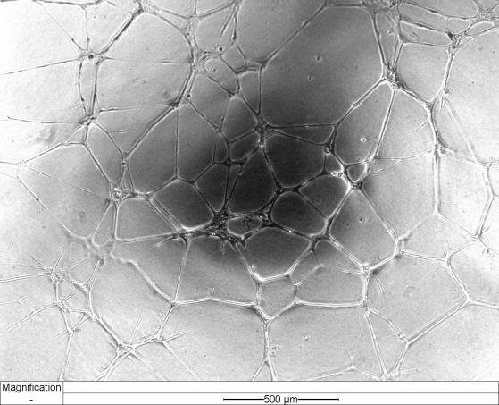
179 Results a) b) PBS c) d) TRAP-6 e) f) TRAP-6+ 15(S)-HETE 162
180 Results Figure 9.2 Images of HMEC-1 tube formation in the presence of releasates from TRAP-6-stimulated platelets. HMEC-1 in the presence of releasates obtained from human PRP stimulated with a-b) PBS, c-d) TRAP-6 (3 µm) or e-f) TRAP-6 and 15(S)- HETE (1 µm). Images were captured using Olympus IX81 inverted microscope (1X) with Hamamatsu Orca-ER digital camera after 16 hour incubation (37 C; 5% CO 2 ) in phase or after the addition of a fluorescent complex, calcein (n=3). Image acquisition was performed using Cell M software (Olympus). TRAP-6 stimulated platelet releasates caused a robust increase in tube formation compared to PBSstimulated controls. 15(S)-HETE did not show any additional effect compared to TRAP-6-stimulated releasates alone. 163

 and in the presence of a-b) vehicle or c-d) 15(S)-HETE (1 µm).")
 in phase or after the addition of a fluorescent complex, calcein (n=3).")

181 Results a) b) TRAP-6+ aspirin c) d) TRAP-6+ aspirin+ 15(S)-HETE Figure 9.3 Images of HMEC-1 in the presence of releasates from TRAP-6-stimulated and aspirin-treated platelets. HMEC-1 in the presence of releasates obtained from human PRP pre-treated with aspirin (1 µm) and stimulated with TRAP-6 (3 µm) and in the presence of a-b) vehicle or c-d) 15(S)-HETE (1 µm). Images were captured using Olympus IX81 inverted microscope (1X) with Hamamatsu Orca-ER digital camera after 16 hour incubation (37 C; 5% CO 2 ) in phase or after the addition of a fluorescent complex, calcein (n=3). Image acquisition was performed using Cell M software (Olympus). Aspirin-treated platelet releasates showed an impairment of the tubule structure compared to untreated platelet releasates. The responses were restored in aspirin-treated platelet releasates when 15(S)-HETE (1 µm) was added. 164
182 Branch points per field Branch points per field Branch points per field Results a) 4 hours b) 8 hours 4 4 * * 3 * * 3 * * PBS TRAP-6 TRAP6+15(S)HETE TRAP-6+aspirin+15(S)HETE PBS TRAP-6 TRAP6+15(S)HETE TRAP-6+aspirin+15(S)HETE c) 16 hours 6 * * * 4 2 PBS TRAP-6 TRAP6+15(S)HETE TRAP-6+aspirin+15(S)HETE Figure 9.4 HMEC-1 tube formation in the presence of releasates from TRAP-6- stimulated platelets. HMEC-1 resuspended with releasates obtained from PRP stimulated with TRAP-6 (3 µm) or TRAP-6 (3 µm) and 15(S)-HETE (1 µm). Tube formation on Matrigel was determined by time-lapse microscopy and the number of branch points was measured after a) 4h incubation (4X magnitude), b) 8h incubation (4X magnitude) and c) 16h incubation (1X magnitude) at 37 C and 5% CO 2. One-way ANOVA (*p.5); n=3. TRAP-6 stimulated platelet releasates significantly increased tube formation compared to those stimulated with PBS. Pretreatment of platelets with aspirin strongly inhibited tubule-like structure formation but the addition of 15(S)-HETE fully restored the cell response. ANOVA with Bonferroni post-test. Differences were taken to be significant at P values of less than.5 (*p.5); n=3. 165


.")

183 Results a) b) collagen c) d) collagen+ 15(S)-HETE Figure 9.5 Images of HMEC-1 tube formation in the presence of releasates from collagen-stimulated platelets. HMEC-1 in the presence of releasates obtained from human PRP stimulated with a-b) collagen (3µg/ml) or c-d) collagen (3µg/ml) and 15(S)-HETE (1µM). Images were captured using Olympus IX81 inverted microscope (1X) with Hamamatsu Orca-ER digital camera after 16 hour incubation (37 C; 5% CO 2 ) in phase or after the addition of a fluorescent complex, calcein (n=3). Image acquisition was performed using Cell M software (Olympus). 166


 and 15(S)EHETE (1 µm).")
 in phase or")
184 Results a) b) collagen+ aspirin c) d) collagen+ aspirin+ 15(S)-HETE Figure 9.6 Images of HMEC-1 in the presence of releasates from collagen-stimulated and aspirin-treated platelets. HMEC-1 in the presence of platelet releasates obtained from human PRP pre-treated with aspirin and stimulated with a-b) collagen (3 µg/ml) or c-d) collagen (3 µg/ml) and 15(S)EHETE (1 µm). Images were captured using Olympus IX81 inverted microscope (1X) with Hamamatsu Orca-ER digital camera after 16 hour incubation (37 C; 5% CO 2 ) in phase or after the addition of a fluorescent complex, calcein (n=3). Image acquisition was performed using Cell M software (Olympus). 167
185 Branch points per field Branch points per field Branch points per field Results a) 4 hours b) 8 hours 4 4 * * 3 * 3 * * * PBS collagen+15(s)hete collagen+aspirin collagen+aspirin+15(s)hete PBS collagen+15(s)hete collagen+aspirin collagen+aspirin+15(s)hete c) 16 hours 6 * * * 4 2 PBS collagen+15(s)hete collagen+aspirin collagen+aspirin+15(s)hete Figure 9.7 HMEC-1 tube formation in the presence of releasates from collagenstimulated platelets. HMEC-1 cells were resuspended with releasates obtained from PRP stimulated with collagen (3 µg/ml) or collagen (3 µg/ml) and 15(S)-HETE (1 µm). Tube formation on Matrigel was determined by time-lapse microscopy and the number of branch points was measured after a) 4h incubation (4X magnitude), b) 8h incubation (4X magnitude) and c) 16h incubation (1X magnitude) at 37 C and 5% CO 2. One-way ANOVA (*p.5); n=3. Collagen-stimulated platelet releasates significantly increased tube formation compared to PBS-stimulated platelet releasates. Pretreatment of platelets with aspirin strongly inhibited tubule-like structure formation, but the addition of 15(S)-HETE fully restored the cell response. ANOVA with Bonferroni post-test. Differences were taken to be significant at P values of less than.5 (*p.5); n=3. 168
186 % field coverage % field coverage Results * * * 8 6 * PBS TRAP-6 TRAP-6+aspirin TRAP-6+aspirin+15(S)HETE PBS collagen collagen+aspirin collagen+aspirin+15(s)hete Figure 9.8 Effect of 15(S)-HETE on HMEC-1 migration. HMEC-1 were used when 1% confluent on a 6-well plate. A scratch was performed with a 2 pipette tip. Platelet releasates were then added and incubated for 2h at 37 C, 5% CO 2. Cell migration in the presence of releasated obtained from a) TRAP-6- or b) collagenstimulated platelets was determined by time-lapse microscopy using Olympus IX81 inverted microscope (1X) with Hamamatsu Orca-ER digital camera after 2 hour incubation (37 C; 5% CO 2 ) in phase. Image acquisition was performed using Cell M software (Olympus) and the percentage (%) of field coverage measured using MCell software. ANOVA with Bonferroni post-test. ANOVA with Bonferroni post-test. Differences were taken to be significant at P values of less than.5 (*p.5); n=4. 169
187 % number of sprouts Results 1 * * * PBS PBS+VEGF PBS+15(S)HETE TRAP-6+aspirin TRAP-6+aspirin+15(S)HETE Figure 9.9 Effect of 15(S)-HETE on formation of angiogenic sprouts from rat aortic rings in the presence of releasates from TRAP-6-stimulated platelets. Platelet releasates were obtained from human (n=3) PRP treated in vitro with or without aspirin (1 µm) and stimulated with PBS or TRAP-6 (3 µm) and finally kept at - 8 C. Releasates were thawed, diluted in OptiMEM+1% FBS (1:3) and supplemented with TRAP-6 and/or aspirin in order to have all samples equally treated. Samples stimulated with PBS were treated with or without VEGF (1 ng/ml) or 15(S)-HETE (1 µm), while those stimulated with TRAP-6 with or without 15(S)-HETE (1 µm). Rat aortic rings (5 per condition) were fed with platelet releasates+optimem+1% FBS and incubated for 5 days at 37 C and 5% CO 2. Images were taken in phase (4X) and the number of sprouts counted using Mcell software. PBS alone did not stimulate formation of sprouts. Conversely, the presence of VEGF significantly increased the number of sprouts while 15(S)-HETE produced a moderate increase. Releasates from TRAP-6 stimulated platelets significantly promoted the formation of sprouts which was strongly inhibited by the presence of aspirin. The addition of 15(S)HETE (1 µm) to aspirin-treated releasates was able to restore the formation of angiogenic sprouts. ANOVA with Bonferroni post-test. Differences were taken to be significant at P values of less than.5 (*p.5). 17

-HETE")


 with")
.")
188 Results PBS PBS+VEGF PBS+15(S)-HETE TRAP-6 TRAP-6+aspirin TRAP-6+aspirin+ 15(S)-HETE Figure 9.1 Images of the effect of 15(S)-HETE on formation of angiogenic sprouts from rat aortic rings in the presence of releasates from TRAP-6-stimulated platelets. Images were taken using Olympus IX81 inverted microscope (1X) with Hamamatsu Orca-ER digital camera after 4 days incubation (37 C; 5% CO 2 ). Image acquisition was performed using Cell M software (Olympus). 171
189 %number of sprouts Results 12 * PBS PBS+VEGF PBS+15(S)HETE collagen collagen+aspirin collagen+aspirin+15(s)hete Figure 9.11 Effect of 15(S)-HETE on formation of angiogenic sprouts from rat aortic rings in the presence of releasates from collagen-stimulated platelets. Platelet releasates were obtained from human (n=3) PRP treated in vitro with or without aspirin (1 µm) and stimulated with PBS or collagen (3 µg/ml) and finally kept at -8 C. Releasates were thawed, diluted in OptiMEM+1% FBS (1:3) and supplemented with collagen and/or aspirin in order to have all samples equally treated. Samples stimulated with PBS were treated with or without VEGF (1 ng/ml) or 15(S)-HETE (1 µm), while those stimulated with TRAP-6 with or without 15(S)- HETE (1 µm). Rat aortic rings (5 per condition) were fed with platelet releasates+optimem+1% FBS and incubated for 5 days at 37 C and 5% CO 2. Images were taken in phase (4X) and the number of sprouts counted using Mcell software. PBS alone did not stimulate formation of sprouts. Conversely, the presence of VEGF significantly increased the number of sprouts while 15(S)-HETE produced a moderate increase. Releasates stimulated with collagen significantly promoted the formation of sprouts which was strongly inhibited by the presence of aspirin. The addition of 15(S)HETE (1 µm) to aspirin-treated releasates was able to restore the formation of angogenic sprouts. ANOVA with Bonferroni post-test. Differences were taken to be significant at P values of less than.5 (*p.5). 172

-HETE")


 with")
.")
190 Results PBS PBS+VEGF PBS+15(S)-HETE collagen collagen+ aspirin collagen+aspirin +15(S)-HETE Figure 9.12 Images of the effect of 15(S)-HETE on formation of angiogenic sprouts from rat aortic rings in the presence of collagen-stimulated platelet releasates. Images were taken using Olympus IX81 inverted microscope (1X) with Hamamatsu Orca-ER digital camera after 4 days incubation (37 C; 5% CO 2 ). Image acquisition was performed using Cell M software (Olympus). 173
EICOSANOID METABOLISM
 1 EICOSANOID METABOLISM EICOSANOIDS C20 polyunsaturated fatty acids e.g. Arachidonic acid Eicosanoids physiologically, pathologically and pharmacologically active compounds PG Prostaglandins TX - Thromboxanes
Enzymatic oxidation of lipids: mechanisms and functions.
 Enzymatic oxidation of lipids: mechanisms and functions. Valerie B. O Donnell, PhD. Cardiff University. Enzymatic lipid oxidation: involves an enzyme catalyst, and gives very specific stereo- and regiospecific
Prostaglandins And Other Biologically Active Lipids
 Prostaglandins And Other Biologically Active Lipids W. M. Grogan, Ph.D. OBJECTIVES After studying the material of this lecture, the student will: 1. Draw the structure of a prostaglandin, name the fatty
Eicosanoid synthesis
 Seminar 12 Eicosanoid synthesis Examples of eicosanoids prostaglandins prostacyclins thromboxanes leukotrienes epoxyeicosatrienoic acids (EET) They have roles in: inflammation fever regulation of blood
PHM142 Lecture 4: Platelets + Endothelial Cells
 PHM142 Lecture 4: Platelets + Endothelial Cells 1 Hematopoiesis 2 Platelets Critical in clotting - activated by subendothelial matrix proteins (e.g. collagen, fibronectin, von Willebrand factor) and thrombin
ANSC/NUTR 618 Lipids & Lipid Metabolism
 I. Nonessential fatty acids ANSC/NUTR 618 Lipids & Lipid Metabolism A. Synthesized completely by the fatty acid synthase reaction (e.g., myristic and palmitic acid). B. Produced by the modification of
Physiology Unit 1 CELL SIGNALING: CHEMICAL MESSENGERS AND SIGNAL TRANSDUCTION PATHWAYS
 Physiology Unit 1 CELL SIGNALING: CHEMICAL MESSENGERS AND SIGNAL TRANSDUCTION PATHWAYS In Physiology Today Cell Communication Homeostatic mechanisms maintain a normal balance of the body s internal environment
Objectives By the end of lecture the student should:
 Objectives By the end of lecture the student should: Illustrate α oxidation of fatty acids. Understand ω oxidation of fatty acids. List sources and fates of active acetate. Discuss eicosanoids. 2- α Oxidation
INFLAMMATION & REPAIR
 INFLAMMATION & REPAIR Lecture 7 Chemical Mediators of Inflammation Winter 2013 Chelsea Martin Special thanks to Drs. Hanna and Forzan Course Outline i. Inflammation: Introduction and generalities (lecture
OBJECTIVE. Lipids are largely hydrocarbon derivatives and thus represent
 Paper 4. Biomolecules and their interactions Module 20: Saturated and unsaturated fatty acids, Nomenclature of fatty acids and Essential and non-essential fatty acids OBJECTIVE The main aim of this module
Cell Signaling. Bruno Sopko
 Cell Signaling Bruno Sopko Content Signal Transduction Pathways Organization Signals Receptors Soluble Receptors Transmembrane Receptors Enzyme Coupled Receptors G-Protein Coupled Receptors Ion-Channel
Lecture Outline. Hormones & Chemical Signaling. Communication Basics: Overview. Communication Basics: Methods. Four methods of cell communication
 Lecture Outline Hormones & Chemical Signaling Communication Basics Communication Overview Communication Methods Signal pathways Regulation (modulation) of signal pathways Homeostasis... again Endocrine
Cell Signaling (part 1)
") 15 Cell Signaling (part 1) Introduction Bacteria and unicellular eukaryotes respond to environmental signals and to signaling molecules secreted by other cells for mating and other communication. In multicellular
15 Cell Signaling (part 1) Introduction Bacteria and unicellular eukaryotes respond to environmental signals and to signaling molecules secreted by other cells for mating and other communication. In multicellular
Cell-Derived Inflammatory Mediators
 Cell-Derived Inflammatory Mediators Introduction about chemical mediators in inflammation Mediators may be Cellular mediators cell-produced or cell-secreted derived from circulating inactive precursors,
Impact of Asthma in the U.S. per Year. Asthma Epidemiology and Pathophysiology. Risk Factors for Asthma. Childhood Asthma Costs of Asthma
 American Association for Respiratory Care Asthma Educator Certification Prep Course Asthma Epidemiology and Pathophysiology Robert C. Cohn, MD, FAARC MetroHealth Medical Center Cleveland, OH Impact of
Detection of Lipid Peroxidation Products From Free Radical and Enzymatic Processes. Jason D. Morrow M.D. Vanderbilt University School of Medicine
 Detection of Lipid Peroxidation Products From Free Radical and Enzymatic Processes Jason D. Morrow M.D. Vanderbilt University School of Medicine Question? Which one of the following is the most accurate
Lipid Mediators: Synthesis, Metabolism, and Function. Overview
 Lipid Mediators: Synthesis, Metabolism, and Function Biochemistry 201 Oct 23, 2007 Electa Park Overview Biological function of lipid mediators Arachidonic acid (AA) synthesis AA metabolites and related
Figure 13-1: Antiplatelet Action of Aspirin (Modified After Taneja et.al 2004) ASPIRIN RESISTANCE
 ASPIRIN RESISTANCE") CHAPTER 13 ASPIRIN Action Aspirin Resistance Aspirin Dose Therapeutic Efficacy - Secondary prevention - Acute coronary syndromes - Primary prevention Limitations and Side Effects Aspirin Aspirin should
CHAPTER 13 ASPIRIN Action Aspirin Resistance Aspirin Dose Therapeutic Efficacy - Secondary prevention - Acute coronary syndromes - Primary prevention Limitations and Side Effects Aspirin Aspirin should
Chapter 5 Control of Cells by Chemical Messengers
 Chapter 5 Control of Cells by Chemical Messengers = How hormones and other signals work Intercellular Communication = Intercellular Signal Transmission Chemical communication Electrical communication Intercellular
Steroid Hormones Synthesis
 *I ll try my best to incorporate the Slides in this Sheet; you don t need to study the slides if you study this sheet. Steroid Hormones Synthesis - The figure to the right is the Steroid nucleus, it has
Biochemistry of the Eicosanoids: Cyclooxygenase and Lipoxygenase Products of Polyunsaturated Fatty Acids
 Lipids in Modern Nutrition, edited by M. Horisberger and U. Bracco. Nestle Nutrition, Vevey/Raven Press, New York 1987. Biochemistry of the Eicosanoids: Cyclooxygenase and Lipoxygenase Products of Polyunsaturated
ulcer healing role 118 Bicarbonate, prostaglandins in duodenal cytoprotection 235, 236
 Subject Index Actin cellular forms 48, 49 epidermal growth factor, cytoskeletal change induction in mucosal repair 22, 23 wound repair 64, 65 polyamine effects on cytoskeleton 49 51 S-Adenosylmethionine
Attribution: University of Michigan Medical School, Department of Microbiology and Immunology
 Attribution: University of Michigan Medical School, Department of Microbiology and Immunology License: Unless otherwise noted, this material is made available under the terms of the Creative Commons Attribution
Eicosapentaenoic Acid and Docosahexaenoic Acid: Are They Different?
 Eicosapentaenoic Acid and Docosahexaenoic Acid: Are They Different? Trevor A Mori, Ph.D., Professor, School of Medicine and Pharmacology, Royal Perth Hospital Unit, University of Western Australia, Perth,
Chapter 26 Biochemistry 5th edition. phospholipids. Sphingolipids. Cholesterol. db=books&itool=toolbar
 http://www.ncbi.nlm.nih.gov/sites/entrez? db=books&itool=toolbar 1 The surface of a soap bubble is a bilayer formed by detergent molecules 2 Chapter 26 Biochemistry 5th edition phospholipids Sphingolipids
G-Protein Signaling. Introduction to intracellular signaling. Dr. SARRAY Sameh, Ph.D
 G-Protein Signaling Introduction to intracellular signaling Dr. SARRAY Sameh, Ph.D Cell signaling Cells communicate via extracellular signaling molecules (Hormones, growth factors and neurotransmitters
Outline Anti-coagulant and anti-thrombotic drugs Haemostasis and Thrombosis Year 3 Dentistry
 Outline Anti-coagulant and anti-thrombotic drugs Year 3 Dentistry Professor Yotis Senis Cellular Haemostasis y.senis@bham.ac.uk I. Haemostasis and II. Coagulation and anti-coagulants III. Platelets and
Vascular reactivity in sepsis and platelet dysfunction in septic shock
 Vascular reactivity in sepsis and platelet dysfunction in septic shock Benjamin Reddi Discipline of Physiology School of Medical Science University of Adelaide Thesis submitted for the degree of Doctor
Inflammation and the Role of Essential Fatty Acids
 Functional Diagnostic Medicine Training Program Module 5 * FMDT 541B Inflammation and the Role of Essential Fatty Acids Limits of Liability & Disclaimer of Warranty We have designed this book to provide
Hashem Al-Dujaily Abdul Aziz ALShamali Abdul Aziz ALShamali Mousa Al-Abbadi
 8 Hashem Al-Dujaily Abdul Aziz ALShamali Abdul Aziz ALShamali Mousa Al-Abbadi Mediators of Inflammation The mediators of inflammation are the substances that initiate and regulate inflammatory reactions.
Propagation of the Signal
 OpenStax-CNX module: m44452 1 Propagation of the Signal OpenStax College This work is produced by OpenStax-CNX and licensed under the Creative Commons Attribution License 3.0 By the end of this section,
Oral Anticoagulant Drugs
 Oral Anticoagulant Drugs Spoiled sweet clover caused hemorrhage in cattle(1930s). Substance identified as bishydroxycoumarin. Initially used as rodenticides, still very effective, more than strychnine.
Cell Signaling part 2
 15 Cell Signaling part 2 Functions of Cell Surface Receptors Other cell surface receptors are directly linked to intracellular enzymes. The largest family of these is the receptor protein tyrosine kinases,
Receptors Families. Assistant Prof. Dr. Najlaa Saadi PhD Pharmacology Faculty of Pharmacy University of Philadelphia
 Receptors Families Assistant Prof. Dr. Najlaa Saadi PhD Pharmacology Faculty of Pharmacy University of Philadelphia Receptor Families 1. Ligand-gated ion channels 2. G protein coupled receptors 3. Enzyme-linked
LIPIDS oxylipins. Marek Vecka
 LIPIDS oxylipins Marek Vecka OXYLIPINS OXYLIPINS = oxygenated fatty acids biologically active as lipid mediators eicosanoids - products of transformation of FA with 20 C AA (20:4n-6), EPA (20:5n-3), DHGLA
Emerging roles of pro-resolving lipid mediators in immunological and adaptive responses to exercise-induced muscle injury
 110 Exercise roles of bioactive lipid mediators Emerging roles of pro-resolving lipid mediators in immunological and adaptive responses to exercise-induced muscle injury James F. Markworth 1, Krishna Rao
AVERTING RISKS ASSOCIATED WITH UTILISING SMALL ANIMAL NSAIDS
 Vet Times The website for the veterinary profession https://www.vettimes.co.uk AVERTING RISKS ASSOCIATED WITH UTILISING SMALL ANIMAL NSAIDS Author : Catherine F Le Bars Categories : Vets Date : April 6,
Understanding How Allergic Responses End: The Allergy Resolvome. Lipid mediators
 Understanding How Allergic Responses End: The Allergy Resolvome Lipid mediators Koichiro Asano Tokai University School of Medicine, Kanagawa, JAPAN ko-asano@tokai-u.jp Resolution of granulocytic inflammation
Principles of Genetics and Molecular Biology
 Cell signaling Dr. Diala Abu-Hassan, DDS, PhD School of Medicine Dr.abuhassand@gmail.com Principles of Genetics and Molecular Biology www.cs.montana.edu Modes of cell signaling Direct interaction of a
A comprehensive analysis of the influence of drug binding kinetics on drug action at molecular and systems levels. Supplementary Material
 A comprehensive analysis of the influence of drug binding kinetics on drug action at molecular and systems levels Ning Yin, Jianfeng Pei and Luhua Lai Supplementary Material The reactions for the arachidonic
: Overview of EFA metabolism
 Figure 1 gives an overview of the metabolic fate of the EFAs when consumed in the diet. The n-6 and n-3 PUFAs when consumed in the form of dietary triglyceride from various food sources undergoes digestion
Chapter 19 Blood Lecture Outline
 Chapter 19 Blood Lecture Outline Cardiovascular system Circulatory system Blood 1. distribution 2. regulation 3. protection Characteristics: ph 7.4 38 C 4-6 L Composition: Plasma Formed elements Erythrocytes
Chapter 15: Signal transduction
 Chapter 15: Signal transduction Know the terminology: Enzyme-linked receptor, G-protein linked receptor, nuclear hormone receptor, G-protein, adaptor protein, scaffolding protein, SH2 domain, MAPK, Ras,
Receptors and Drug Action. Dr. Subasini Pharmacology Department Ishik University, Erbil
 Receptors and Drug Action Dr. Subasini Pharmacology Department Ishik University, Erbil Receptors and Drug Action Receptor Receptor is defined as a macromolecule or binding site located on the surface or
Hemostasis. Learning objectives Dr. Mária Dux. Components: blood vessel wall thrombocytes (platelets) plasma proteins
 plasma proteins") Hemostasis Learning objectives 14-16 Dr. Mária Dux Components: blood vessel wall thrombocytes (platelets) plasma proteins Hemostatic balance! procoagulating activity anticoagulating activity 1 Thrombocytes
Hemostasis Learning objectives 14-16 Dr. Mária Dux Components: blood vessel wall thrombocytes (platelets) plasma proteins Hemostatic balance! procoagulating activity anticoagulating activity 1 Thrombocytes
DIETARY LIPIDS, IMMUNE FUNCTION AND PATHOGENESIS OF DISEASE IN FISH
 DIETARY LIPIDS, IMMUNE FUNCTION AND PATHOGENESIS OF DISEASE IN FISH Santosh P. Lall and Joyce E. Milley National Research Council Canada, Institute for Marine Biosciences, 1411 Oxford Street, Halifax,
number Done by Corrected by Doctor
 number 9 Done by Corrected by Doctor 1 Cellular Infiltration The movement of cells from their original region to a new region is called cellular infiltration. In our case, this applies to the cells that
Anti-Inflammatory Effects of Fish Oils
 Anti-Inflammatory Effects of Fish Oils A. Macrophage Production of Eicosanoids B. w-3 PUFA Supplementation of Macrophages C. w-3 PUFA in IR & diabetes Oswald Quehenberger Departments of Medicine and Pharmacology
Hemostasis and Thrombosis
 Hemostasis Hemostasis and Thrombosis Normal hemostasis is a consequence of tightly regulated processes that maintain blood in a fluid state in normal vessels, yet also permit the rapid formation of a hemostatic
Nafith Abu Tarboush DDS, MSc, PhD
 Nafith Abu Tarboush DDS, MSc, PhD natarboush@ju.edu.jo www.facebook.com/natarboush Lipids (cholesterol, cholesterol esters, phospholipids & triacylglycerols) combined with proteins (apolipoprotein) in
10. Which of the following immune cell is unable to phagocytose (a) neutrophils (b) eosinophils (c) macrophages (d) T-cells (e) monocytes
 neutrophils (b) eosinophils (c) macrophages (d) T-cells (e) monocytes") Chapter 2. Acute and chronic inflammation(6): 1. In acute inflammation, which events occur in the correct chronological order? (Remembered from 2000, 2004 exam.) p50 (a) transient vasoconstriction, stasis
Chapter 2. Acute and chronic inflammation(6): 1. In acute inflammation, which events occur in the correct chronological order? (Remembered from 2000, 2004 exam.) p50 (a) transient vasoconstriction, stasis
Lecture 9: Cell Communication I
 02.05.10 Lecture 9: Cell Communication I Multicellular organisms need to coordinate cellular functions in different tissues Cell-to-cell communication is also used by single celled organisms to signal
Δοκιμασίες λειτουργικότητας αιμοπεταλίων και PCI Εμμανουήλ Βαβουρανάκης
 Δοκιμασίες λειτουργικότητας αιμοπεταλίων και PCI Εμμανουήλ Βαβουρανάκης Αναπλ. Καθηγητής Καρδιολογίας Ιπποκράτειο ΓΝΑ Haematology Research Laboratory!! Platelets Small anucleate discoid cells Involved
Autocrine signalling. Signals are not always diffusible but may be membrane bound or a part of the extracellular matrix. The inflammatory response
 Paracrine signals Proteins, lipids that diffuse short distances Include growth factors and cytokines Secreted or synthesized as membrane-bound precursor (pro-peptide) that is proteolytically processed
Lipids and Membranes
 Lipids and Membranes Presented by Dr. Mohammad Saadeh The requirements for the Pharmaceutical Biochemistry I Philadelphia University Faculty of pharmacy Membrane transport D. Endocytosis and Exocytosis
Phospholipids Metabolism
 Chapter VI: Phospholipids Metabolism Dr. Sameh Sarray Hlaoui Phospholipids Features: Amphipatic: - Hydrophobic head: fatty acids - Hydropholic head: P group+ alcohol Composed of alcohol attached by a phosphodiester
Chapter 20. Cell - Cell Signaling: Hormones and Receptors. Three general types of extracellular signaling. endocrine signaling. paracrine signaling
 Chapter 20 Cell - Cell Signaling: Hormones and Receptors Three general types of extracellular signaling endocrine signaling paracrine signaling autocrine signaling Endocrine Signaling - signaling molecules
Tandem mass spectrometry analysis of prostaglandins and isoprostanes
 Tandem mass spectrometry analysis of prostaglandins and isoprostanes Jeevan Prasain jprasain@uab.edu 6-2612 verview Introduction to PGs and their synthesis Mass spectrometry characterization of PGs and
Name: Class: "Pharmacology NSAIDS (1) Lecture
 Lecture") I Name: Class: "Pharmacology NSAIDS (1) Lecture د. احمد الزهيري Inflammation is triggered by the release of chemical mediators from injured tissues and migrating cells. The specific mediators vary with
I Name: Class: "Pharmacology NSAIDS (1) Lecture د. احمد الزهيري Inflammation is triggered by the release of chemical mediators from injured tissues and migrating cells. The specific mediators vary with
Receptor mediated Signal Transduction
 Receptor mediated Signal Transduction G-protein-linked receptors adenylyl cyclase camp PKA Organization of receptor protein-tyrosine kinases From G.M. Cooper, The Cell. A molecular approach, 2004, third
Part IV Antithrombotics, Anticoagulants and Fibrinolytics
 Part IV Antithrombotics, Anticoagulants and Fibrinolytics "The meaning of good and bad, of better and worse, is simply helping or hurting" Emerson Chapter 16: Blood Coagulation and Fibrinolytic System
Lecture 36: Review of membrane function
 Chem*3560 Lecture 36: Review of membrane function Membrane: Lipid bilayer with embedded or associated proteins. Bilayers: 40-70% neutral phospholipid 10-20% negative phospholipid 10-30% cholesterol 10-30%
Regulation of cell function by intracellular signaling
 Regulation of cell function by intracellular signaling Objectives: Regulation principle Allosteric and covalent mechanisms, Popular second messengers, Protein kinases, Kinase cascade and interaction. regulation
Subject Index. Bcl-2, apoptosis regulation Bone marrow, polymorphonuclear neutrophil release 24, 26
 Subject Index A1, apoptosis regulation 217, 218 Adaptive immunity, polymorphonuclear neutrophil role 31 33 Angiogenesis cancer 178 endometrium remodeling 172 HIV Tat induction mechanism 176 inflammatory
Ch. 45 Blood Plasma proteins, Coagulation and Fibrinolysis Student Learning Outcomes: Describe basic components of plasma
 Chapt. 45 Ch. 45 Blood Plasma proteins, Coagulation and Fibrinolysis Student Learning Outcomes: Describe basic components of plasma Inheritance of X-linked gene for Factor VIII hemophilia A Explain the
Signal Transduction Cascades
 Signal Transduction Cascades Contents of this page: Kinases & phosphatases Protein Kinase A (camp-dependent protein kinase) G-protein signal cascade Structure of G-proteins Small GTP-binding proteins,
Sarah Jaar Marah Al-Darawsheh
 22 Sarah Jaar Marah Al-Darawsheh Faisal Mohammad Receptors can be membrane proteins (for water-soluble hormones/ligands) or intracellular (found in the cytosol or nucleus and bind to DNA, for lipid-soluble
1) Mononuclear phagocytes : 2) Regarding acute inflammation : 3) The epithelioid cells of follicular granulomas are :
 Mononuclear phagocytes : 2) Regarding acute inflammation : 3) The epithelioid cells of follicular granulomas are :") Pathology Second 1) Mononuclear phagocytes : - Are the predominant cells in three day old wounds - Are common in liver, spleen and pancreasd - Produce fibroblast growth factor - Secrete interferon-g -
Pathology Second 1) Mononuclear phagocytes : - Are the predominant cells in three day old wounds - Are common in liver, spleen and pancreasd - Produce fibroblast growth factor - Secrete interferon-g -
Omega-3 Fatty Acids and Athletics. Current Sports Medicine Reports July 2007, 6:
 Omega-3 Fatty Acids and Athletics 1 Current Sports Medicine Reports July 2007, 6:230 236 Artemis P. Simopoulos, MD The Center for Genetics, Nutrition and Health, Washington, DC, USA EIB exercise-induced
Studies of prostaglandin E 2 formation in human monocytes. Sofia Karlsson. Karlstad University Studies
 Faculty of Technology and Science Biomedical Sciences Sofia Karlsson Studies of prostaglandin E 2 formation in human monocytes Karlstad University Studies 2009:43 Sofia Karlsson Studies of prostaglandin
Signal Transduction: G-Protein Coupled Receptors
 Signal Transduction: G-Protein Coupled Receptors Federle, M. (2017). Lectures 4-5: Signal Transduction parts 1&2: nuclear receptors and GPCRs. Lecture presented at PHAR 423 Lecture in UIC College of Pharmacy,
C-Phycocyanin (C-PC) is a n«sjfc&c- waefc-jduble phycobiliprotein. pigment isolated from Spirulina platensis. This water- soluble protein pigment is
 is a n«sjfc&c- waefc-jduble phycobiliprotein. pigment isolated from Spirulina platensis. This water- soluble protein pigment is") ' ^Summary C-Phycocyanin (C-PC) is a n«sjfc&c- waefc-jduble phycobiliprotein pigment isolated from Spirulina platensis. This water- soluble protein pigment is of greater importance because of its various
' ^Summary C-Phycocyanin (C-PC) is a n«sjfc&c- waefc-jduble phycobiliprotein pigment isolated from Spirulina platensis. This water- soluble protein pigment is of greater importance because of its various
BIOLOGY. Cell Communication CAMPBELL. Reece Urry Cain Wasserman Minorsky Jackson. Lecture Presentation by Nicole Tunbridge and Kathleen Fitzpatrick
 CAMPBELL BIOLOGY TENTH EDITION Reece Urry Cain Wasserman Minorsky Jackson 11 Cell Communication Lecture Presentation by Nicole Tunbridge and Kathleen Fitzpatrick Cellular Messaging Cells can signal to
Chapter 11. Cell Communication
 Chapter 11 Cell Communication Overview: The Cellular Internet Cell-to-cell communication Is absolutely essential for multicellular organisms Concept 11.1: External signals are converted into responses
CHAPTER 28 LIPIDS SOLUTIONS TO REVIEW QUESTIONS
 HAPTER 28 LIPIDS SLUTINS T REVIEW QUESTINS 1. The lipids, which are dissimilar substances, are arbitrarily classified as a group on the basis of their solubility in fat solvents and their insolubility
PHOSPHOLIPID MEMBRANE ABNORMALITIES AND REDUCED NIACIN SKIN FLUSH RESPONSE IN SCHIZOPHRENIA
 PHOSPHOLIPID MEMBRANE ABNORMALITIES AND REDUCED NIACIN SKIN FLUSH RESPONSE IN SCHIZOPHRENIA Alena Buretić-Tomljanović 1, Jasminka Giacometti 1, Sergej Nadalin 1, Gordana Rubeša 2, Mirjana Vulin 3, Draško
BCH Graduate Survey of Biochemistry
 BCH 5045 Graduate Survey of Biochemistry Instructor: Charles Guy Producer: Ron Thomas Director: Glen Graham Lecture 58 Slide sets available at: http://hort.ifas.ufl.edu/teach/guyweb/bch5045/index.html
Pharmacogenomics for the anti-platelet drug
 Pharmacogenomics for the anti-platelet drug Su-Jun Lee, PhD Dept. of Pharmacology, Pharmacogenomics Research Center, Inje University College of Medicine, Busan Conflict of interest disclosure None Committee
General Principles of Endocrine Physiology
 General Principles of Endocrine Physiology By Dr. Isabel S.S. Hwang Department of Physiology Faculty of Medicine University of Hong Kong The major human endocrine glands Endocrine glands and hormones
Chapter 19 Cardiovascular System Blood: Functions. Plasma
 Chapter 19 Cardiovascular System Blood: Functions 19-1 Plasma Liquid part of blood. Colloid: liquid containing suspended substances that don t settle out of solution 91% water. Remainder proteins, ions,
Cell Communication. Chapter 11. PowerPoint Lectures for Biology, Seventh Edition. Lectures by Chris Romero. Neil Campbell and Jane Reece
 Chapter 11 Cell Communication PowerPoint Lectures for Biology, Seventh Edition Neil Campbell and Jane Reece Lectures by Chris Romero Overview: The Cellular Internet Cell-to-cell communication Is absolutely
Chapter 9. Cellular Signaling
 Chapter 9 Cellular Signaling Cellular Messaging Page 215 Cells can signal to each other and interpret the signals they receive from other cells and the environment Signals are most often chemicals The
Cell Communication. Local and Long Distance Signaling
 Cell Communication Cell to cell communication is essential for multicellular organisms Some universal mechanisms of cellular regulation providing more evidence for the evolutionary relatedness of all life
The role of prostacyclin synthase and thromboxane synthase signaling in the development and progression of cancer
 Accepted Manuscript The role of prostacyclin synthase and thromboxane synthase signaling in the development and progression of cancer Mary-Clare Cathcart, John V. Reynolds, Kenneth J. O Byrne, Graham P.
Zool 3200: Cell Biology Exam 4 Part I 2/3/15
 Name: Key Trask Zool 3200: Cell Biology Exam 4 Part I 2/3/15 Answer each of the following questions in the space provided, explaining your answers when asked to do so; circle the correct answer or answers
G protein-coupled Signal Transduction
 Theresa Filtz, hd har 735, Winter 2006 G protein-coupled Signal Transduction Main Objectives (the big chunks) Describe in molecular detail the cascades of events in a generalized G protein-coupled signaling
Cell Communication. Chapter 11. Biology Eighth Edition Neil Campbell and Jane Reece. PowerPoint Lecture Presentations for
 Chapter 11 Cell Communication PowerPoint Lecture Presentations for Biology Eighth Edition Neil Campbell and Jane Reece Lectures by Chris Romero, updated by Erin Barley with contributions from Joan Sharp
CHAPTER 28 LIPIDS SOLUTIONS TO REVIEW QUESTIONS
 28 09/16/2013 17:44:40 Page 415 APTER 28 LIPIDS SLUTINS T REVIEW QUESTINS 1. The lipids, which are dissimilar substances, are arbitrarily classified as a group on the basis of their solubility in fat solvents
Signal-Transduction Cascades - 2. The Phosphoinositide Cascade
 Signal-Transduction Cascades - 2 The Phosphoinositide Cascade Calcium ion as a second messenger Tyrosine kinase and receptor dimerization scribd.com Faisal Khatib JU The Phosphoinositide Cascade Used by
Enzyme-coupled Receptors. Cell-surface receptors 1. Ion-channel-coupled receptors 2. G-protein-coupled receptors 3. Enzyme-coupled receptors
 Enzyme-coupled Receptors Cell-surface receptors 1. Ion-channel-coupled receptors 2. G-protein-coupled receptors 3. Enzyme-coupled receptors Cell-surface receptors allow a flow of ions across the plasma
INTERACTION DRUG BODY
 INTERACTION DRUG BODY What the drug does to the body What the body does to the drug Receptors - intracellular receptors - membrane receptors - Channel receptors - G protein-coupled receptors - Tyrosine-kinase
Lecture 15. Signal Transduction Pathways - Introduction
 Lecture 15 Signal Transduction Pathways - Introduction So far.. Regulation of mrna synthesis Regulation of rrna synthesis Regulation of trna & 5S rrna synthesis Regulation of gene expression by signals
Prostaglandins and related substances a practical approach
 O Prostaglandins and related substances a practical approach Edited by c Benedetto Institute of Obstetrics and Gynecology, Universita di Torino, Via Ventimiglia 3, Torino 10126, Italy R C McDonald-Gibson
UNIT VI. Chapter 37: Platelets Hemostasis and Blood Coagulation Presented by Dr. Diksha Yadav. Copyright 2011 by Saunders, an imprint of Elsevier Inc.
 UNIT VI Chapter 37: Platelets Hemostasis and Blood Coagulation Presented by Dr. Diksha Yadav Hemostasis: Prevention of Blood Loss Vascular constriction Formation of a platelet plug Formation of a blood
Chapter 18. Essential Fatty Acids. Arthur A. Spector, MD OUTLINE COMMON ABBREVIATIONS
 Chapter 18 Essential Fatty Acids Arthur A. Spector, MD OUTLINE HISTORICAL PERSPECTIVE STRUCTURE OF POLYUNSATURATED FATTY ACIDS ω6 Polyunsaturated Fatty Acids ω3 Polyunsaturated Fatty Acids Highly Unsaturated
Identification, characterization and evaluation of novel leukotriene biosynthesis inhibitors
 Identification, characterization and evaluation of novel leukotriene biosynthesis inhibitors Identifizierung, Charakterisierung und Bewertung neuartiger Hemmstoffe der Leukotrienbiosynthese Dissertation
Drug Receptor Interactions and Pharmacodynamics
 Drug Receptor Interactions and Pharmacodynamics Dr. Raz Mohammed MSc Pharmacology School of Pharmacy 22.10.2017 Lec 6 Pharmacodynamics definition Pharmacodynamics describes the actions of a drug on the
Chapter 10. Introduction to Nutrition and Metabolism, 3 rd edition David A Bender Taylor & Francis Ltd, London 2002
 Chapter 10 Introduction to Nutrition and Metabolism, 3 rd edition David A Bender Taylor & Francis Ltd, London 2002 Chapter 10: Integration and Control of Metabolism Press the space bar or click the mouse
Platelet function in diabetes
 Platelet function in diabetes Agneta Siegbahn, MD, PhD, FESC Professor Clinical Coagulation Science Department of Medical Sciences and Uppsala Clinical Research Center Uppsala University Disclosures: None