Laminar Thickness: Correlation Against Symptoms of Spinal Stenosis and Changes with Age
|
|
|
- Ellen Davis
- 5 years ago
- Views:
Transcription
1 Laminar Thickness: Correlation Against Symptoms of Spinal Stenosis and Changes with Age Hamed Shalikar, MD 1, Sean L. Borkowski, MS 2, Juan Pablo Villablanca, MD 1, Sophia N. Sangiorgio, PhD 2, Edward Ebramzadeh, PhD 2, Arya N. Shamie, MD 1. 1 UCLA, Los Angeles, CA, USA, 2 Orthopaedic Institute for Children/UCLA, Los Angeles, CA, USA. Disclosures: H. Shalikar: None. S.L. Borkowski: None. J. Villablanca: None. S.N. Sangiorgio: 5; Zimmer, DePuy, TriMed, Extremity Medical, Biomet, NuVasive. E. Ebramzadeh: 5; Zimmer, DePuy, TriMed, Extremity Medical, Biomet, NuVasive. A.N. Shamie: 1; Seaspine. 3B; Biomet. 4; SI Bone, Vertiflex. Introduction: Spinal stenosis is a significant cause of low back pain and a leading reason for orthopaedic visits and subsequent surgery [1]. Despite its large burden on medical care costs, the pathogenesis of spinal stenosis is not fully understood. Spinal lamina may become thicker due to Wolff s law [2], that is, stress over a patient s lifespan, potentially contributing to narrowing of the spinal canal, resulting in spinal stenosis; however, this hypothesis has not been tested previously. The purposes of this study were to measure the laminar thickness in a retrospective cohort of stenosis and non-stenosis patients, to evaluate the effects of laminar thickness on canal size, and to correlate laminar thickness measures to patient gender and age. Methods: Following IRB approval, we retrospectively analyzed lumbar spine computed tomography (CT) scans from a blinded database of patients in a large academic hospital setting, acquired over a one year period (3/1/12-3/1/13), regardless of the indication. Patients with previous lumbar vertebral surgery, vertebral metastasis, and/or severe scoliosis were excluded. Additionally, patients younger than 18 years old or older than 80 years old were excluded. Patients with pain/fatigue and either an aggravating factor or alleviating factor, were included in the spinal stenosis group. Based on these criteria, two groups were defined: stenosis (n=18) and non-stenosis patients (n=133). Patients had a mean age of 53 years old (18-80 years). All CT scans were reviewed to ensure standardized, reliable and repeatable measurements from each patient. The procedure included the helical acquisition of 3mm thick sections in the axial plane, with coronal and sagittal reformations. The field of view was 170mm, with a matrix size of 512 x 512, bone window (40Hs) kernels. Soft tissue kernels (B30, medium smooth) and bone window kernels (B60, sharp) were used. Using Vitrea software in the Vitals Workstation (Vital Images, Inc., Minnetonka, Mn), a 3D reconstruction of a given patient s spine was created using CT source data. Following 3D reconstruction, a custom axial oblique slice of the spine was obtained which passed through the axial plane of the intervertebral disc of interest, and was parallel to the vertebral endplates. This process was repeated separately for both the L4-L5 and the L5-S1 levels of each patient. Then, the laminae were measured in six locations: left and right spinous process-lamina junction, left and right juxta-cortical lamina, and left and right mid-lamina. Statistical analyses were performed using SPSS 19.0 statistical software (IBM, Armonk, NY). The input variables were presence of spinal stenosis symptoms, gender, age, and measurement location. The primary output variable was laminar thickness. For each level, that is L4-L5 and L5-S1, three separate multivariate general linear models were created to evaluate the effects of stenosis symptoms, gender, and age, on laminar thickness at each of the measurement locations: (1) the left+right spinous processlamina junction; (2) the left+right juxta-cortical lamina; and (3) the left+right mid-lamina. Paired samples
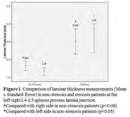
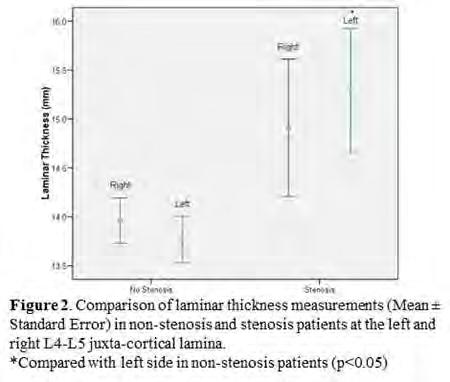
2 t-tests were performed to compare the laminar thicknesses between the left and right lamina at each of the three measurement locations. The Pearson correlation coefficient was calculated to quantify the correlations between laminar thickness and patient age. Finally, separate Pearson correlation analyses were performed to evaluate the repeatability and reliability of the CT laminar thickness measurements. Results: Among the 151 measured patients, the largest laminar thicknesses were found at the juxtacortical lamina locations. In general, laminar thickness at different locations was larger in the patients with stenosis (n=18) compared with the patients without stenosis (n=133), though not all locations were statistically significant. Specifically, mean laminar thickness ranged from mm in stenosis patients, compared to only mm in non-stenosis patients. The results varied by lamina thickness measurement location, with significantly larger thicknesses found in the left juxta-cortical lamina, and the left and right spinous process-lamina junction of L4-L5 (Figure 1 and 2). At the L5-S1 level, significantly larger thicknesses were found at the right and left juxta-cortical lamina locations (Figure 3).There were positive correlations between age and laminar thickness in all measurement locations at both the L4-L5 and L5-S1 levels. With the exception of the left spinous process-lamina junction of L4-L5 (p=0.095), all locations demonstrated significant positive correlations, with Pearson correlation coefficients ranging from to (p<0.01). There were no significant differences between male and female patients. The intraobserver and interobserver measurements were significantly correlated at all measurement locations, with Pearson correlation coefficients ranging from to (p<0.02). Discussion: Laminar thickness was larger in patients with stenosis symptoms compared to those without stenosis symptoms. Furthermore, laminar thickness increased with aging. As such, laminar thickness may be a potential factor in the pathogenesis of lumbar spinal stenosis. By establishing the associations between laminar thickness, a readily available and quantifiable parameter, and clinical factors, including stenosis, age and gender, high risk groups in the population may be identified. Identifying these groups may allow clinicians to improve patient treatment through early conservative interventions, potentially delaying future surgery or preventing it altogether. Significance: Despite the large burdens associated with spinal stenosis, this is the first study to evaluate the potential role of laminar thickness in the pathogenesis of stenosis. The increased thickness reported in the present study suggests its role in this pathogenesis, and will lead to future studies of the factors contributing to spinal stenosis, ultimately improving a clinicians understanding, and in turn, a clinicians ability to treat patients with these conditions.
3
4

5 ORS 2015 Annual Meeting Poster No: 1629
Analyzing Early Onset Scoliosis In 3d: How Does Growing Rod Surgery Affect The Three Planes Of Deformity?
 Analyzing Early Onset Scoliosis In 3d: How Does Growing Rod Surgery Affect The Three Planes Of Deformity? Diana A. Glaser, PhD 1, Burt Yaszay, MD 1, Gregory M. Mundis, MD 2, Behrooz A. Akbarnia, MD 2.
Quantitative Analysis of Vascular Canals in Vertebral Endplate
 Quantitative Analysis of Vascular Canals in Vertebral Endplate Kristine Tan 1, Won C. Bae, PhD 1, Tomonori Yamaguchi, MS 1,2, Kelli Xu, BS 1, Iris Shieh, BS 1, Jade He, BS 1, Robert L. Sah, MD, ScD 1,
Disclosures: T. Yoshii: None. T. Yamada: None. T. Taniyama: None. S. Sotome: None. T. Kato: None. S. Kawabata: None. A. Okawa: None.
 Dynamic Changes in Spinal Cord Compression by Cervical Ossification of the Posterior Longitudinal Ligament Evaluated by Kinematic Computed Tomography Myelogram Toshitaka Yoshii, Tsuyoshi Yamada, Takashi
Facet orientation in patients with lumbar degenerative spondylolisthesis
 35 J. Tokyo Med. Univ., 71 1 35 0 Facet orientation in patients with lumbar degenerative spondylolisthesis Wuqikun ALIMASI, Kenji ENDO, Hidekazu SUZUKI, Yasunobu SAWAJI, Hirosuke NISHIMURA, Hidetoshi TANAKA,
Pediatric CT Protocols (18 years old or less)
") Pediatric CT Protocols (18 years old or less) Ped1: Head CT Ped2: Cervical spine CT Ped3: Sinus CT Ped4: Neck CT Ped5: Chest CT Ped6: Abdomen and pelvis CT Ped7: Thoracic or lumbar spine CT Ped8: Extremity
Pediatric CT Protocols (18 years old or less) Ped1: Head CT Ped2: Cervical spine CT Ped3: Sinus CT Ped4: Neck CT Ped5: Chest CT Ped6: Abdomen and pelvis CT Ped7: Thoracic or lumbar spine CT Ped8: Extremity
X-Ray & CT Physics / Clinical CT
 Computed Tomography-Basic Principles and Good Practice X-Ray & CT Physics / Clinical CT INSTRUCTORS: Dane Franklin, MBA, RT (R) (CT) Office hours will be Tuesdays from 5pm to 6pm CLASSROOM: TIME: REQUIRED
Revised Dec Spine MR Protocols
 Spine MR Protocols Sp 1: Cervical spine MRI without contrast Sp 2: Pre- and post-contrast cervical spine MRI Sp 3: Pre- and post-contrast cervical spine MRI (multiple sclerosis protocol) Sp 4: Thoracic
B. CT protocols for the spine
 B. CT protocols for the spine Poster No.: A-003 Congress: ECR 2010 Type: Invited Speaker Topic: Neuro Authors: B. Tins; Oswestry/UK Keywords: CT, spine, diagnostic imaging protocol DOI: 10.1594/ecr2010/A-003
Systematic Review of Clinical Outcome using Titanium Fiber Mesh Porous Ingrowth Surfaces
 Systematic Review of Clinical Outcome using Titanium Fiber Mesh Porous Ingrowth Surfaces Timothy L. Tan, BS 1, Brenna C. Moeljadi, BS 2, Edward Ebramzadeh, PhD 3, Patricia Campbell, PhD 3, Sophia Nicole
The Occupancy of the Components in the Cervical Spine and Their Changes with Extension and Flexion
 396 Original Article GLOBAL SPINE JOURNAL THIEME The Occupancy of the Components in the Cervical Spine and Their Changes with Extension and Flexion Emrah Sayıt 1 Bayan Aghdasi 2 Michael D. Daubs 3 Jeffrey
CT of the Head, Spine, and Cerebral Vessels
 CT of the Head, Spine, and Cerebral Vessels Objectives Determine specific imaging plane used to acquire or reformat CT scan, i.e. sagittal, coronal, transverse, and offaxis or oblique. Assess and evaluate
Original Article Application of structural autologous cervical laminae as bone graft in anterior cervical discectomy and fusion
 Int J Clin Exp Med 2016;9(7):14012-14017 www.ijcem.com /ISSN:1940-5901/IJCEM0026133 Original Article Application of structural autologous cervical laminae as bone graft in anterior cervical discectomy
The effect of body mass index on lumbar lordosis on the Mizuho OSI Jackson spinal table
 35 35 40 The effect of body mass index on lumbar lordosis on the Mizuho OSI Jackson spinal table Authors Justin Bundy, Tommy Hernandez, Haitao Zhou, Norman Chutkan Institution Orthopaedic Department, Medical
Lumbar Disc Prolapse. Dr. Ahmed Salah Eldin Hassan. Professor of Neurosurgery & Consultant spinal surgeon
 Lumbar Disc Prolapse By Dr. Ahmed Salah Eldin Hassan Professor of Neurosurgery & Consultant spinal surgeon 1-What are the Functions of the Spine Structural support for upright posture Protection of Spinal
Hidayatullah Hamidi. MD Consultant Radiologist. Lumbar Spine MR Imaging Interpretation
 Hidayatullah Hamidi. MD Consultant Radiologist Lumbar Spine MR Imaging Interpretation 13/12/2018 Presenter Hidayatullah Hamidi Consultant Radiologist, Radiology PGME program director, FMIC, Kabul, Afghanistan
Added value of MR myelography using 3D COSMIC sequence in the diagnosis of lumbar canal stenosis: comparison with routine MR imaging
 Added value of MR myelography using 3D COSMIC sequence in the diagnosis of lumbar canal stenosis: comparison with routine MR imaging Poster No.: C-1099 Congress: ECR 2012 Type: Authors: Scientific Exhibit
Artificial intervertebral disc
 The University of Toledo The University of Toledo Digital Repository Master s and Doctoral Projects Artificial intervertebral disc Vikas Ghai Medical University of Ohio Follow this and additional works
Influence of Pedicle Screw Design on the Fatigue Fixation Strength in the Human Lumbar Spine
 Influence of Pedicle Screw Design on the Fatigue Fixation Strength in the Human Lumbar Spine Rebecca Kueny, MSc. 1, Reza Danyali 1, Bartosz Nowak, PhD 2, Klaus Pueschel, Dr med. 3, Michael M. Morlock,
of the lumbar facet joints there
 Skeletal Radiol (1999) 28:215±219 International Skeletal Society 1999 A R T I C L E Dominik Weishaupt Marco Zanetti Norbert Boos Juerg Hodler MR imaging and CT in osteoarthritis of the lumbar facet joints
A Computational Model of Annulus Fiber Deformation in Cervical Discs During In Vivo Dynamic Flexion\Extension, Rotation and Lateral Bending
 A Computational Model of Annulus Fiber Deformation in Cervical Discs During In Vivo Dynamic Flexion\Extension, Rotation and Lateral Bending William Anderst, Mara Palmer, Joon Lee, William Donaldson, James
Lauren M. Burke, Warren D. Yu, Anthony Ho, Timothy Wagner, Joseph R. O Brien. Department of Orthopaedic Surgery George Washington University
 Lauren M. Burke, Warren D. Yu, Anthony Ho, Timothy Wagner, Joseph R. O Brien Department of Orthopaedic Surgery George Washington University O Brien: Consultant for Globus, Relivant, Stryker. Royalties:
3D imaging reformation was obtained. The 3D color imaging reformation was reviewed in a different high resolution setting.
 POST OPERATIVE SPINE WITH CONTRAST CLINICAL INDICATION: Low back pain, Patient is post operative status for L4/5 diskectomy TECHNIQUE: MRI of the lumbosacral spine was performed with multiplanar imaging
MEDICAL IMAGING OF THE VERTEBRAE
 MEDICAL IMAGING OF THE VERTEBRAE Vertebrae are your friends Matthew Harper MS-IV LECTURE OBJECTIVES INTRODUCE THE MOST COMMON MODALITIES OF MEDICAL IMAGING AND BASIC TECHNIQUES FOR READING THESE IMAGES
Magnetic resonance of adolescent idiopathic scoliosis
 Australian Institute of Radiography The Radiographer 2005; 52 (2): 7 14 Magnetic resonance of adolescent idiopathic scoliosis University of Queensland, St Lucia Campus, Brisbane, Queensland, Australia
How to interpret computed tomography of the lumbar spine
 REVIEW Ann R Coll Surg Engl 2014; 96: 502 507 doi 10.1308/003588414X13946184902361 How to interpret computed tomography of the lumbar spine Z Ahmad 1, R Mobasheri 2,TDas 3, S Vaidya 4, S Mallik 5, M El-Hussainy
5/27/2016. Stand-Alone Lumbar Lateral Interbody Fusion (LLIF) vs. Supplemental Fixation. Disclosures. LLIF Approach
 vs. Supplemental Fixation. Disclosures. LLIF Approach") Stand-Alone Lumbar Lateral Interbody Fusion (LLIF) vs. Supplemental Fixation Joseph M. Zavatsky, M.D. Spine & Scoliosis Specialists Tampa, FL Disclosures Consultant - Zimmer / Biomet, DePuy Synthes Spine,
Stand-Alone Lumbar Lateral Interbody Fusion (LLIF) vs. Supplemental Fixation Joseph M. Zavatsky, M.D. Spine & Scoliosis Specialists Tampa, FL Disclosures Consultant - Zimmer / Biomet, DePuy Synthes Spine,
International Journal of Pharma and Bio Sciences
 Original Research Article Anatomy and Allied sciences International Journal of Pharma and Bio Sciences ISSN 0975-6299 SCREENING FOR ANOMALIES IN OCCIPITO-CERVICAL JUNCTION USING CRANIOMETRY IN COMPUTED
Kinematic Analysis of Lumbar Spine Undergoing Extension and Dynamic Neural Foramina Cross Section Measurement
 Copyright c 2008 ICCES ICCES, vol.7, no.2, pp.57-62 Kinematic Analysis of Lumbar Spine Undergoing Extension and Dynamic Neural Foramina Cross Section Measurement Yongjie Zhang 1, Boyle C. Cheng 2, Changho
NECK AND BACK PAIN AN INTRODUCTION TO
 AN INTRODUCTION TO NECK AND BACK PAIN This booklet provides general information on neck and back pain. It is not meant to replace any personal conversations that you might wish to have with your physician
Supplementary Online Content
 Supplementary Online Content Schlaeger R, Papinutto N, Zhu AH, et al. Association between thoracic spinal cord gray matter atrophy and disability in multiple sclerosis. JAMA Neurol. Published online June
DynaWell L-Spine Compression Device
 L-Spine Compression Device The L-Spine was developed specifically to apply gravity to a patient s spine in supine position to simulate the upright position on your patient when scanned in your CT and/or
A new sagittal parameter to estimate pelvic tilt using the iliac cortical density line and iliac tilt: a retrospective X-ray measurement study
 Doi et al. Journal of Orthopaedic Surgery and Research (2015) 10:115 DOI 10.1186/s13018-015-0262-0 RESEARCH ARTICLE Open Access A new sagittal parameter to estimate pelvic tilt using the iliac cortical
"D10-D11 Facet enlargement"
 "D10-D11 Facet enlargement" Poster No.: C-0898 Congress: ECR 2011 Type: Scientific Exhibit Authors: J. Jalal Shokouhi, A. A. Ameri, H. Mohammad pour ; Tehran/IR, 1 2 2 1 2 tehran/ir Keywords: Spine, Neuroradiology
Departement of Neurosurgery A.O.R.N A. Cardarelli- Naples.
 Percutaneous posterior pedicle screw fixation in the treatment of thoracic, lumbar and thoraco-lumbar junction (T12-L1) traumatic and pathological spine fractures. Report of 45 cases. G. Vitale, A. Punzo,
2/5/2019. Facet Joint Pain. Biomechanics
 Facet Arthropathy as a Pain Source Evaluation and Management Shelby Spine Jan 31 st Feb 2 nd, 2019 Kushagra Verma MD, MS Adult and Pediatric Scoliosis And Spine Deformity Beach Orthopaedics Specialty Institute
Am I eligible for the TOPS study? Possibly, if you suffer from one or more of the following conditions:
 Am I eligible for the TOPS study? Possibly, if you suffer from one or more of the following conditions: Radiating leg pain Greater leg / buttock pain than back pain Severe pain sets in when walking as
U.S. MARKET FOR MINIMALLY INVASIVE SPINAL IMPLANTS
 U.S. MARKET FOR MINIMALLY INVASIVE SPINAL IMPLANTS idata_usmis15_rpt Published in December 2014 By idata Research Inc., 2014 idata Research Inc. Suite 308 4211 Kingsway Burnaby, British Columbia, Canada,
Porcine model for early onset scoliosis created with a posterior mini-invasive method
 E-Poster #P72 Porcine model for early onset scoliosis created with a posterior mini-invasive method WANG Bin, ZHENG Xin, QIU Yong*, QIAN Bang-ping, SUN Xu, ZHU Zezhang, YU Yang Spine surgery, the Affiliated
A Morphometric study of lamina of lumbar vertebrae: A dry bone study conducted in RIMS, Imphal
 IOSR Journal of Dental and Medical Sciences (IOSR-JDMS) e-issn: 2279-0853, p-issn: 2279-0861.Volume 17, Issue 11 Ver. 4 (November. 2018), PP 35-39 www.iosrjournals.org A Morphometric study of lamina of
TORNIER BLUEPRINT. 3D Planning + PSI SCAN PROTOCOL
 TORNIER BLUEPRINT 3D Planning + PSI SCAN PROTOCOL Contents 3 Introduction 3 Patient preparation 3 Scanning instructions 4 Image instructions 5 Scanning parameters 6 Technical instructions 2 BLUEPRINT 3D
The spinal vacuum sign or vacuum phenomenon. Is a Persistent Vacuum Phenomenon a Sign of Pseudarthrosis After Posterolateral Spinal Fusion?
 An Original Study Is a Persistent Vacuum Phenomenon a Sign of Pseudarthrosis After Posterolateral Spinal Fusion? Arjun A. Dhawale, MD, Steven Falcone, MD, Barth A. Green, MD, and Nathan H. Lebwohl, MD
Association between Sacral Slanting and Adjacent Structures in Patients with Adolescent Idiopathic Scoliosis
 Original Article Clinics in Orthopedic Surgery 17;9:57-62 https://doi.org/10.4055/cios.17.9.1.57 Association between Sacral Slanting and Adjacent Structures in Patients with Adolescent Idiopathic Scoliosis
Optimal Baseplate Rotational Alignment in Reverse Total Shoulder Arthroplasty: A Three-Dimensional Computer-Aided Design Study.
 Optimal Baseplate Rotational Alignment in Reverse Total Shoulder Arthroplasty: A Three-Dimensional Computer-Aided Design Study. Byron F. Stephens, MD 1, Casey T. Hebert 2, Thomas W. Throckmorton, MD 1,
Module: #15 Lumbar Spine Fusion. Author(s): Jenni Buckley, PhD. Date Created: March 27 th, Last Updated:
: Jenni Buckley, PhD. Date Created: March 27 th, Last Updated:") Module: #15 Lumbar Spine Fusion Author(s): Jenni Buckley, PhD Date Created: March 27 th, 2011 Last Updated: Summary: Students will perform a single level lumbar spine fusion to treat lumbar spinal stenosis.
Module: #15 Lumbar Spine Fusion Author(s): Jenni Buckley, PhD Date Created: March 27 th, 2011 Last Updated: Summary: Students will perform a single level lumbar spine fusion to treat lumbar spinal stenosis.
Degenerative spondylolisthesis is four times more
 Degenerative spondylolisthesis DEVELOPMENTAL OR ACQUIRED? T. W. Love, A. B. Fagan, R. D. Fraser From the Royal Adelaide Hospital, Adelaide, Australia Degenerative spondylolisthesis is four times more common
Hi RES Extremity - (04/18/2011) CTDI: ~13 mgy per acquisition Used for evaluation of: Ankle Elbow Hand Wrist Foot /Calcaneous Toes Fingers
 CTDI: ~13 mgy per acquisition Used for evaluation of: Ankle Elbow Hand Wrist Foot /Calcaneous Toes Fingers") P a g e 1 Hi RES Extremity - (04/18/2011) CTDI: ~13 mgy per acquisition Used for evaluation of: Ankle Elbow Hand Wrist Foot /Calcaneous Toes Fingers Billing: 1. CT Upper/Lower Extremity of concern without
P a g e 1 Hi RES Extremity - (04/18/2011) CTDI: ~13 mgy per acquisition Used for evaluation of: Ankle Elbow Hand Wrist Foot /Calcaneous Toes Fingers Billing: 1. CT Upper/Lower Extremity of concern without
A rare case of osteoblastoma combined with severe scoliosis deformity, coronal and sagittal imbalance
 Wang et al. BMC Musculoskeletal Disorders (2017) 18:538 DOI 10.1186/s12891-017-1902-9 CASE REPORT Open Access A rare case of osteoblastoma combined with severe scoliosis deformity, coronal and sagittal
CT FINDINGS OF THORACOLUMBAR SPINE LESIONS IN DOGS
 CT FINDINGS OF THORACOLUMBAR SPINE LESIONS IN DOGS C. DARABAN 1, V. VULPE 1, FLORENTINA BOCĂNEŢI 1, GIUSEPPINA MENNONNA 2, M. SACCONE 2, G. FATONE 2, L. MEOMARTINO 2 1 University of Agriculture Science
Depiction of Lateral Ligament Complex of the Ankle using 3D MRI in Healthy Subjects and Patients with Chronic Ankle Instability.
 Depiction of Lateral Ligament Complex of the Ankle using 3D MRI in Healthy Subjects and Patients with Chronic Ankle Instability. Satoshi Yamaguchi, M.D. 1, Hiroshi Matsumoto, R.T. 2, Atsuya Watanabe, M.D.
SCALING Radiographic Technique
 SCALING Radiographic Technique SCALING FOR DIGITAL X-RAYS As images become filmless. Current planning practices with acetate sheets become difficult or obsolete. When images are printed to film sometimes
Abdomen and Pelvis CT (1) By the end of the lecture students should be able to:
 By the end of the lecture students should be able to:") RAD 451 Abdomen and Pelvis CT (1) By the end of the lecture students should be able to: State the common indications for Abdomen and pelvis CT exams Identify possible contra indications for Abdomen and
RAD 451 Abdomen and Pelvis CT (1) By the end of the lecture students should be able to: State the common indications for Abdomen and pelvis CT exams Identify possible contra indications for Abdomen and
A minimally invasive surgical approach reduces cranial adjacent segment degeneration in patients undergoing posterior lumbar interbody fusion
 A minimally invasive surgical approach reduces cranial adjacent segment degeneration in patients undergoing posterior lumbar interbody fusion T. Tsutsumimoto, M. Yui, S. Ikegami, M. Uehara, H. Kosaku,
SUBAXIAL CERVICAL SPINE TRAUMA- DIAGNOSIS AND MANAGEMENT
 SUBAXIAL CERVICAL SPINE TRAUMA- DIAGNOSIS AND MANAGEMENT 1 Anatomy 3 columns- Anterior, middle and Posterior Anterior- ALL, Anterior 2/3 rd body & disc. Middle- Posterior 1/3 rd of body & disc, PLL Posterior-
3 脊椎変形の相互関係とリスク分析の今後の方向
 Reprint request: DEGENERATIVE SPINE AND OSTEOPOROSIS IN ELDERLY PEOPLE Toshitaka NAKAMURA Department of Orthopedics, University of Occupational and Environmental Health School of Medicine Vertebral osteophytes,
Spinal Stenosis Surgical
 Spinal Stenosis Surgical Disclaimer This movie is an educational resource only and should not be used to make a decision on. All decisions about surgery must be made in conjunction with your surgeon or
EVALUATE, TREAT AND WHEN TO REFER RED FLAGS Mid Atlantic Occupational Regional Conference and Environmental Medicine October 6, 2018
 EVALUATE, TREAT AND WHEN TO REFER RED FLAGS Mid Atlantic Occupational Regional Conference and Environmental Medicine October 6, 2018 Marc J. Levine, MD Rothman Institute Director Spine Surgery Program
The effect of vertebral rotation of the lumbar spine on dual energy X-ray absorptiometry measurements: observational study
 JCY Cheng HL Sher X Guo VWY Hung AYK Cheung Key words: Absorptiometry, Bone density; Densitometry, X-ray; Lumbar vertebrae; Scoliosis "#$%& "# X HKMJ 2001;7:241-5 The Chinese University of Hong Kong, Prince
RADIATION PROTECTION OF THE PATIENT IN PAEDIATRIC RADIOLOGY. Bahnarel Ion, Dimov Nicolae, Coretchi Liuba, Cujba Natalia
 RADIATION PROTECTION OF THE PATIENT IN PAEDIATRIC RADIOLOGY Bahnarel Ion, Dimov Nicolae, Coretchi Liuba, Cujba Natalia Medical Diagnostic Centre Magnific Chisinau, Republic of Moldova, e-mail: Ndimov@mail.ru
Bilateral spondylolysis of inferior articular processes of the fourth lumbar vertebra: a case report
 Upsala Journal of Medical Sciences. 2012; 117: 72 77 CASE REPORT Bilateral spondylolysis of inferior articular processes of the fourth lumbar vertebra: a case report TOMOAKI KOAKUTSU 1,2, NAOKI MOROZUMI
Bony framework of the vertebral column Structure of the vertebral column
 5.1: Vertebral column & back. Overview. Bones o vertebral column. o typical vertebra. o vertebral canal. o spinal nerves. Joints o Intervertebral disc. o Zygapophyseal (facet) joint. Muscles o 2 compartments:
ORIGINAL PAPER. Department of Orthopedic Surgery, Chubu Rosai Hospital, Nagoya, Japan ABSTRACT
 Nagoya J. Med. Sci. 77. 221 ~ 228, 2015 ORIGINAL PAPER RANGE OF MOTION DETERMINED BY MULTIDETECTOR-ROW COMPUTED TOMOGRAPHY IN PATIENTS WITH CERVICAL OSSIFICATION OF THE POSTERIOR LONGITUDINAL LIGAMENT
Metal Artefact Reduction in CT
 Metal Artefact Reduction in CT DANIEL MARRINER Metal Artefact Reduction in CT Metal Artefact Clinical Indications for MAR SEMAR and How It Works Technical Considerations Case Studies utilising SEMAR Metal
University of Groningen. Thoracolumbar spinal fractures Leferink, Vincentius Johannes Maria
 University of Groningen Thoracolumbar spinal fractures Leferink, Vincentius Johannes Maria IMPORTANT NOTE: You are advised to consult the publisher's version (publisher's PDF) if you wish to cite from
TORNIER. scan protocol V2.1. Tornier Upper Extremities
 TORNIER TM scan protocol V2.1 Tornier Upper Extremities Contents 4 Introduction 4 Patient Preparation 4 Scanning Instructions 5 Image Instructions 6 Scanning Parameters 7 Technical Instructions Scan Protocol
AO CLASSIFICATIONS THORACO-LUMBAR SPINAL INJURIES
 AO CLASSIFICATIONS THORACO-LUMBAR SPINAL INJURIES T H E A O / A S I F ( A R B E I T S G E M E I N S C H A F T F Ü R O S T E O S Y N T H E S E F R A G E N / A S S O C I A T I O N F O R T H E S T U D Y O
Life can cause spinal stenosis. Take yours back with Superion.
 Indirect Decompression System Life can cause spinal stenosis. Take yours back with Superion. Superion can help you take back your life with a simple new procedure. What is spinal stenosis? Your spine is
A Comparison of Cobb Angle: Standing Versus Supine Images of Late-Onset Idiopathic Scoliosis
 Signature: Pol J Radiol, 216; 81: 27-276 DOI: 1.12659/PJR.895949 ORIGINAL ARTICLE Received: 215.9.13 Accepted: 215.11.22 Published: 216.6.1 Authors Contribution: A Study Design B Data Collection C Statistical
Pedicle screw insertion : robotic assistance versus conventional C-arm fluoroscopy
 Acta Orthop. Belg., 2012, 78, 240-245 ORIGINAL STUDY Pedicle screw insertion : robotic assistance versus conventional C-arm fluoroscopy Constantin SCHizAS, Eric THEin, Barbara KwiATKOwSKi, Gerit KULiK
HI-Res Extremity Sensation 16
 Page 1 Routine Extremity - (2/14/2013) CTDI: ~20 mgy per acquisition Used for evaluation of: Humerus Forearm Femur Knee Tib/Fib Billing: 1. CT Upper/Lower Extremity of concern without contrast, with contrast,
Kyung Hee University Spine Centre Kyung Hee University Hospital at Gang Dong Seoul, South Korea
 Report for APSS-Depuy-Synthes Clinical Fellowship 2013 Centre: Kyung Hee University Spine Centre Kyung Hee University Hospital at Gang Dong Seoul, South Korea Supervisor: Prof Kim Ki-Tack Fellow: Dr Tony
Morphological changes of the cervical spinal canal and cord due to aging on MR imaging
 Morphological changes of the cervical spinal canal and cord due to aging on MR imaging Shigeru Kobayashi, MD,PhD 1, Katsuhiko Hayakawa, MD, PhD 2, Takashi Nakane, MD, PhD 2, Riya Kosaka MD,PhD 3. 1 Department
The Fusion of. Strength, Design. Performance. and
 The Fusion of Strength, Design and Performance Interbody Fusion Platform Interbody Fusion Platform The DePuy Spine Interbody Fusion platform is designed to provide an open architecture implant in a load-sharing
Induction and Maintenance of Lordosis in MultiLevel ACDF Using Allograft. Saad Khairi, MD Jennifer Murphy Robert S. Pashman, MD
 Induction and Maintenance of Lordosis in MultiLevel ACDF Using Allograft Saad Khairi, MD Jennifer Murphy Robert S. Pashman, MD Purpose Is lordosis induced by multilevel cortical allograft ACDF placed on
PROPHECY. Preoperative Navigation Guides ANKLE CT SCAN PROTOCOL
 PROPHECY Preoperative Navigation Guides ANKLE CT SCAN PROTOCOL 90 FIGURE 1 Examples FIGURE 1 Examples of neutral ankle positioning. PROPHECY Ankle CT Scan Protocol PROPHECY INBONE and PROPHECY INFINITY
Original article: Multidetector computed tomographic evaluation of cervical spine trauma
 Original article: Multidetector computed tomographic evaluation of cervical spine trauma 1Sajid Ansari *, 2 R.K. Rauniyar, 3 Kaleem Ahmad, 4 Mukesh Kumar Gupta 1Assistant Professor, Department of Radiodiagnosis,
PREOPERATIVE RETROLISTHESIS IS A RISK FACTOR OF LUMBAR DISC HERNIATION AFTER FENESTRATION WITHOUT DISCECTOMY
 PREOPERATIVE RETROLISTHESIS IS A RISK FACTOR OF LUMBAR DISC HERNIATION AFTER FENESTRATION WITHOUT DISCECTOMY Shota Takenaka*, Noboru Hosono, Yoshihiro Mukai, Kosuke Tateishi, Takeshi Fuji Osaka Kosei-nenkin
Magnetic resonance imaging findings in patients with low backache
 Original Article Magnetic resonance imaging findings in patients with low backache Narayan Bikram Thapa 1, Suraj Bajracharya 2 1 Department of Radiology, KIST Medial College Teaching Hospital, Lalitpur,
Assessment of cervical spine morphology by computed tomography and its utility as a presurgical planning tool
 The University of Toledo The University of Toledo Digital Repository Theses and Dissertations 2004 Assessment of cervical spine morphology by computed tomography and its utility as a presurgical planning
Powered by. Dedicated MRI
 Powered by Dedicated MRI Provides the latest software and hardware upgrade configuration powered by exp technology: boosting productivity, increasing image quality, and adding new acquisition techniques.
BACK PAIN. Disclaimer. Integrated web marketing. Multimedia Health Education
 BACK PAIN Disclaimer This movie is an educational resource only and should not be used to make a decision on. All decisions about surgery must be made in conjunction with your surgeon or a licensed healthcare
Facet Joint Arthrosis Disc Degeneration and Lumbago. Dr.Ruchira Sethi Dr. Vishram Singh Department of Anatomy Santosh University, India
 Facet Joint Arthrosis Disc Degeneration and Lumbago Dr.Ruchira Sethi Dr. Vishram Singh Department of Anatomy Santosh University, India INTRODUCTION The initial division of spine into three columns for
ESSENTIALS OF PLAIN FILM INTERPRETATION: SPINE DR ASIF SAIFUDDIN
 ESSENTIALS OF PLAIN FILM INTERPRETATION: SPINE DR ASIF SAIFUDDIN Consultant Musculoskeletal Radiologist Royal National Orthopaedic Hospital Stanmore,UK. INTRODUCTION 2 INTRODUCTION 3 INTRODUCTION Spinal
Skeletal System. Axial Division
 Skeletal System Axial Division The Axial Skeleton You will see that each bone has special features (overviewed in section I below) that provide Sites of Attachment (for muscles, ligaments, tendons, etc.)
LIV selection in selective thoracic fusions
 Russian Research Institute for Traumatology and Orthopedics named after R.R.Vreden, St.Petersburg LIV selection in selective thoracic fusions Ptashnikov D. Professor, The chief of spine surgery & oncology
Adverse events in adult spinal deformity procedures.
 Adverse events in adult spinal deformity procedures. Evalina L BURGER*, Michael S CHANG**, Sean MOLLOY*** Vincent FIERE**** * Department of Orthopedic Surgery, University of Colorado,Aurora, USA ** Department
INDEPENDENT LEARNING: DISC HERNIATION IN THE NATIONAL FOOTBALL LEAGUE: ANATOMICAL FACTORS TO CONSIDER IN REVIEW
 INDEPENDENT LEARNING: DISC HERNIATION IN THE NATIONAL FOOTBALL LEAGUE: ANATOMICAL FACTORS TO CONSIDER IN REVIEW CDC REPORT - CAUSES OF DISABILITY, 2005 REVIEW QUESTIONS ABOUT DISC HERNIATION IN THE NATIONAL
AXIAL SKELETON FORM THE VERTICAL AXIS OF THE BODY CONSISTS OF 80 BONES INCLUDES BONES OF HEAD, VERTEBRAL COLUMN, RIBS,STERNUM
 AXIAL SKELETON FORM THE VERTICAL AXIS OF THE BODY CONSISTS OF 80 BONES INCLUDES BONES OF HEAD, VERTEBRAL COLUMN, RIBS,STERNUM APPENDICULAR SKELETON BONES OF THE FREE APPENDAGES & THEIR POINTS OF ATTACHMENTS
Raymond Wiegand, D.C. Spine Rehabilitation Institute of Missouri
 2D Pattern matching of frontal plane radiograph to 3D model identifies structural and functional deficiencies of the spinal pelvic system in consideration of mechanical spine pain (AKA Spine distortion
Outline. Epidemiology Indications for C-spine imaging Modalities Interpretation Types of fractures
 C-Spine Plain Films Outline Epidemiology Indications for C-spine imaging Modalities Interpretation Types of fractures Epidemiology 7000-10000 c-spine injuries treated each year Additional 5000 die at the
Mineral Density Of Subchondral Bone May Be Quantitatively Evaluated Using A Clinical Cone Beam Computed Tomography Scanner
 Mineral Density Of Subchondral Bone May Be Quantitatively Evaluated Using A Clinical Cone Beam Computed Tomography Scanner Mikael J. Turunen, PhD 1, Juha Töyräs, PhD 1, Harri Kokkonen, PhD 2, Jukka S.
The Leg Length Discrepancy in The Entire Lower Limbs after Total Hip Arthroplasty Influences The Coronal Alignment of Pelvis and Spine.
 The Leg Length Discrepancy in The Entire Lower Limbs after Total Hip Arthroplasty Influences The Coronal Alignment of Pelvis and Spine. Hiroshi Fujimaki, MD 1, Yutaka Inaba, MD, PhD 2, Naomi Kobayashi,
A Study of Measurements of Spinal Canal at the Level of Lower Three Lumbar Vertebra by 16 Slice CT Scanner in Nepalese Population
 Journal of College of Medical Sciences-Nepal, Vol-10, No 4, Oct-Dec 014 ISSN: 2091-0657 (Print); 2091-0673 (Online) Open Access A Study of Measurements of Spinal Canal at the Level of Lower Three Lumbar
Computed Tomography; The how and why of its importance
 Computed Tomography; The how and why of its importance Presented by KENNETH HABLE, MD, BSRT Director of Engineering & Training Technical Prospects, LLC 7/18/2017 1 About me (a little shameless self promotion)
2. The vertebral arch is composed of pedicles (projecting from the body) and laminae (uniting arch posteriorly).
 and laminae (uniting arch posteriorly).") VERTEBRAL COLUMN 2018zillmusom I. VERTEBRAL COLUMN - functions to support weight of body and protect spinal cord while permitting movements of trunk and providing for muscle attachments. A. Typical vertebra
VERTEBRAL COLUMN 2018zillmusom I. VERTEBRAL COLUMN - functions to support weight of body and protect spinal cord while permitting movements of trunk and providing for muscle attachments. A. Typical vertebra
A rare case of spinal injury: bilateral facet dislocation without fracture at the lumbosacral joint
 J Orthop Sci (2012) 17:189 193 DOI 10.1007/s00776-011-0082-y CASE REPORT A rare case of spinal injury: bilateral facet dislocation without fracture at the lumbosacral joint Kei Shinohara Shigeru Soshi
Lumbar Facet Joint Replacement
 Rome Spine 2011 THE SPINE TODAY International Congress Rome 6-7th December 2011 Lumbar Facet Joint Replacement Prof. Dr. Karin Büttner-Janz Past President International Society for the Advancement of Spine
Signal intensity changes of the posterior elements of the lumbar spine in symptomatic adults
 ORIGINAL ARTICLE SPINE SURGERY AND RELATED RESEARCH Signal intensity changes of the posterior elements of the lumbar spine in symptomatic adults Kosuke Sugiura, Toshinori Sakai, Fumitake Tezuka, Kazuta
Edward K. Nomoto, M.D.
 Page 1 of 5 Appointments CURRICULUM VITAE Edward K. Nomoto, M.D. 444 S. San Vicente Blvd, Suite 800 Los Angeles, CA 90048 (310) 423-9780 (work) Cedars-Sinai Medical Center, Spine Center Faculty, Orthopaedic
Evaluation of Wedging of Lower Thoracic and Upper Lumbar Vertebral Bodies in the Pediatric Population
 Pediatric Imaging Original Research Gaca et al. Evaluation of Wedging of Lumbar Vertebral Bodies in Children Pediatric Imaging Original Research Ana Maria Gaca 1 Huiman X. Barnhart 2 George S. Bisset,