INDEPENDENT LEARNING: DISC HERNIATION IN THE NATIONAL FOOTBALL LEAGUE: ANATOMICAL FACTORS TO CONSIDER IN REVIEW
|
|
|
- Laurence Rodgers
- 5 years ago
- Views:
Transcription
1 INDEPENDENT LEARNING: DISC HERNIATION IN THE NATIONAL FOOTBALL LEAGUE: ANATOMICAL FACTORS TO CONSIDER IN REVIEW
2 CDC REPORT - CAUSES OF DISABILITY, 2005
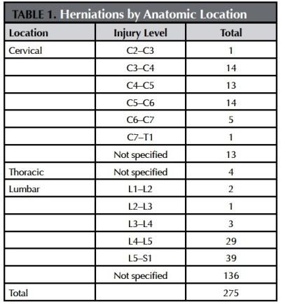
3 REVIEW QUESTIONS ABOUT DISC HERNIATION IN THE NATIONAL FOOTBALL LEAGUE QUESTION 1- The most common site of disc herniation was at level L5-S1 which was much greater than the frequency of occurrence at L1-L2, L2-L3 or L3-L4. Based upon your knowledge of vertebral anatomy, what are two factors that could contribute to the high frequency of occurrence at L5-S1?

4 DISC HERNIATIONS IN DIFFERENT REGIONS
5
6 NUMBER OF HERNIATIONS PER YEAR
7 REVIEW ANATOMY: WEIGHT SUPPORTED BY VERTEBRAE Cervical (neck) - support ONLY head Thoracic (chest) - support head, neck + upper extremities Lumbar (lower back) - support head + neck + upper extremities + thorax Sacral (pelvis) - fused vertebrae; transmit forces to pelvis WEIGHT SUPPORTED BY VERTEBRAL COLUMN - increases from cervical to thoracic to lumbar regions


8 2) LOWER SPINE EXTENSION : MOVEMENTS PERMITTED IN LUMBAR REGION OF SPINE spinous process lamina pedicle Bodies - hefty Pedicles - stout Lamina - thick Spinous Processes - broad body posterior view Articular processes in sagittal plane - permit flexionextension movements of spine
9 E. MOVEMENTS OF VERTEBRAL COLUMN a. Extension b. Flexion a. Extension b. Flexion - Bend anterior - Bend posterior Flexion-Extension of back occurs in Lumbar region of spine
10 REVIEW QUESTIONS ABOUT DISC HERNIATION IN THE NATIONAL FOOTBALL LEAGUE QUESTION 1- The most common site of disc herniation was at level L5-S1 which was much greater than the frequency of occurrence at L1-L2, L2-L3 or L3-L4. Based upon your knowledge of vertebral anatomy, what are two factors that could contribute to the high frequency of occurrence at L5-S1? Article page 'In the lumbar spine, high loading from the weight of the linemen, rigorous weight training, and forcible lower spine extension during blocking likely play a role in lumbar herniations'

11 ARTICLE ALSO DISCUSSES INCREASE IN WEIGHT OF FOOTBALL PLAYERS IN NFL F = ma force = mass X acceleration Source of data: Associate press study reported in See also: Weigh in Results for Lineman 2014:


12 FORCIBLE LOWER SPINE EXTENSION DURING BLOCKING: PRACTICE DRILL LUMBAR EXTENSION START POSITION INITIAL CONTACT PUSH PLAYER BACK from: Techniques & Drills for Creating Championship Lineman

13
14 QUESTION 2- The second most common region for disc herniation was in the Cervical spine. What aspect of the anatomy of cervical vertebrae could contribute to the relatively high frequency of occurrence of herniation in this region?
15 B. REGIONS OF VERTEBRAL COLUMN Cervical (neck) - 7 vertebrae (C1-C7) Thoracic (chest) - 12 vertebrae (T1-T12) Lumbar (lower back) - 5 vertebrae (L1-L5) Sacral (pelvis) - 5 fused vertebrae (S1-S5) Coccygeal (tail) vertebrae (Co1-Co3) Note: Bodies increase in size from rostral to caudal = superior to inferior

16 NOSE BODY CERVICAL VERTEBRA ant. - body is small TRANSVERSE PROCESS post. Helmet to Helmet contact in NFL - force transmitted through cervical vertebrae
17 QUESTION 2- The second most common region for disc herniation was in the Cervical spine. What aspect of the anatomy of cervical vertebrae could contribute to the relatively high frequency of occurrence of herniation in this region? Review Anatomy: 1) vertebral bodies increase in size from superior to inferior 2) bodies of cervical vertebrae (and, therefore intervertebral discs) are small
18 QUESTION 3- Disc herniation was rare in the thoracic region. Name one specialization of thoracic vertebrae that could contribute to the infrequency of occurrence of herniations in this region?
 bone and metal appear white, air is black; soft tissues")
19 CT OF THORACIC VERTEBRA body of Vertebra T10 rib rib transverse process spine Note: In radiographic images (CT= Computed Tomography and X rays) bone and metal appear white, air is black; soft tissues appear grey


20 THORACIC VERTEBRAE ARE STABILIZE BY RIBS WHICH ATTACH ANTERIORLY TO STERNUM Dorsal view of skeleton Ventral view of thorax Vertebrae Sternum Ribs Ribs Vertebrae

21 LATERAL VIEW OF THORACIC VERTEBRA Superior Articular process Inferior Articular process post ant 3. Costal Facets for Ribs - Body - Facets for Heads of rib; Transverse Process - Facets for Tubercle 5. Articular Processes in coronal plane THORAX IS NOT BENT WHEN SPINE IS FLEXED permit some rotation - little or no flex-extend (also limited by ribs); useful - no flex down on heart, lungs
22 QUESTION 3- Disc herniation was rare in the thoracic region. Name one specialization of thoracic vertebrae that could contribute to the infrequency of occurrence of herniations in this region? REVIEW ANATOMY - 1) Thoracic vertebrae are stable due to posterior attachment of ribs. Anteriorly ribs are attached anteriorly to form thoracic cage. 2) Articular process - In thorax, articular processes are in coronal plane, preventing flexion-extension movements.
23 QUESTION 4- The highest number of injuries to spinal nerves occurred in the Cervical region. Based upon your knowledge of vertebral anatomy, what could contribute to the high rate of occurrence of nerve injuries in this region?
24 TYPE OF INJURIES: NERVE INJURIES IN CERVICAL REGION
25 NOSE BODY TRANSVERSE PROCESS CERVICAL VERTEBRA ant. - body is small ARTICULAR FACETS - angled superiorly & medially - considerable freedom of movement permit considerable flexion-extension, lateral flexion, rotation - useful - move head


26 MOVEMENTS OF HEAD AND NECK: body language YES FLEXION - anterior MAYBE EXTENSION - posterior NO LATERAL FLEXION - head on shoulder, face forward ROTATION - face turned, look over shoulder
 has no body only ring of bone 2) Anterior and Poste")
 has Foramina Transversaria 5) superior articular facets to occipital bone of skull; permits Flex-Ext yes movement of head SECOND")
27 FIRST CERVICAL VERTEBRA = C1 (ATLAS) LATERAL MASS ANTERIOR ARCH POSTERIOR ARCH Ant. tubercle Post. tubercle 1) has no body only ring of bone 2) Anterior and Posterior Arches and Lateral mass 3) bumps on arches - Ant. and Post. Tubercles 4) has Foramina Transversaria 5) superior articular facets to occipital bone of skull; permits Flex-Ext yes movement of head SECOND CERVICAL VERTEBRA = C2 (AXIS) DENS 1) has peg-like Odontoid process = Dens (may be fused body of C1) 2) joint between C1-C2 is pivot joint allowing rotation; Rotation = no movement of head; joint is important in hanging

28 NOSE BODY TRANSVERSE PROCESS CERVICAL VERTEBRA ant. - body is small ARTICULAR FACETS - angled superiorly & medially - considerable freedom of movement permit considerable flexion-extension, lateral flexion, rotation - useful - move head


29 SPINAL CORD GENERAL TERMINOLOGY: ORGANIZATION OF SPINAL CORD WHITE MATTER Grey and white matter - some nervous tissue appears white because of myelin. Lateral Spinothalamic tract section of cord removed GREY MATTER Dorsal Columns Lat. Corticospinal - descending 1. 'White matter' of nervous system is composed of myelinated axons. Ascending and descending tracts (axons from and to other places). ex. Ascending - Dorsal Columns Spinothalamic tract ex. Descending - Corticospinal tract 2. 'Grey matter' is composed of everything else in nervous system (mainly cell bodies, unmyelinated axons, etc.). Neurons form synapses mostly in Grey matter. histological section (thin section) of spinal cord

30 NERVE DAMAGE SPINAL CORD SPINAL NERVE DORSAL PRIMARY RAMUS INTER- VERTEBRAL DISC SYMPATHETIC CHAIN VENTRAL PRIMARY RAMUS SPINAL NERVE Excessive bending of neck can damage Spinal Nerves


31 EXCESSIVE BENDING OF NECK CAN INJURE SPINAL NERVES Excessive bending of neck can damage Spinal Nerves Tackling in NFL - photo of Ahmed Brooks tackling Drew Brees (quarterback of New Orleans Saints) Brachial Plexus innervates upper extremity - most ventral primary rami from Cervical spinal nerves (C5-T1)
VERTEBRAL COLUMN VERTEBRAL COLUMN
 VERTEBRAL COLUMN FUNCTIONS: 1) Support weight - transmits weight to pelvis and lower limbs 2) Houses and protects spinal cord - spinal nerves leave cord between vertebrae 3) Permits movements - *clinical
2. The vertebral arch is composed of pedicles (projecting from the body) and laminae (uniting arch posteriorly).
 and laminae (uniting arch posteriorly).") VERTEBRAL COLUMN 2018zillmusom I. VERTEBRAL COLUMN - functions to support weight of body and protect spinal cord while permitting movements of trunk and providing for muscle attachments. A. Typical vertebra
VERTEBRAL COLUMN 2018zillmusom I. VERTEBRAL COLUMN - functions to support weight of body and protect spinal cord while permitting movements of trunk and providing for muscle attachments. A. Typical vertebra
Axial Skeleton: Vertebrae and Thorax
 Axial Skeleton: Vertebrae and Thorax Function of the vertebral column (spine or backbone): 1) 2) 3) Composition of Vertebral column The vertebral column is formed by 33 individual vertebrae (some of which
Vertebral Column. Backbone consists of 26 vertebrae. Five vertebral regions. Cervical
 Vertebral Column Backbone consists of 26 vertebrae. Five vertebral regions Cervical vertebrae (7) in the neck. Thoracic vertebrae (12) in the thorax. Lumbar vertebrae (5) in the lower back. Sacrum (5,
Copyright 2010 Pearson Education, Inc.
 E. VERTEBRAL COLUMN 1. The vertebral column extends from the skull to the pelvis and forms the vertical axis of the skeleton. 2. The vertebral column is composed of vertebrae that are separated by intervertebral
Human Anatomy and Physiology - Problem Drill 07: The Skeletal System Axial Skeleton
 Human Anatomy and Physiology - Problem Drill 07: The Skeletal System Axial Skeleton Question No. 1 of 10 Which of the following statements about the axial skeleton is correct? Question #01 A. The axial
Copyright 2010 Pearson Education, Inc. Copyright 2010 Pearson Education, Inc. Figure Sectioned spinous process. Interspinous.
 PowerPoint Lecture Slides prepared by Janice Meeking, Mount Royal College C H A P T E R 7 The Skeleton: Part B Vertebral Column Transmits weight of trunk to lower limbs Surrounds and protects spinal cord
THE VERTEBRAL COLUMN. Average adult length: In male: about 70 cms. In female: about 65 cms.
 THE VERTEBRAL COLUMN Average adult length: In male: about 70 cms. In female: about 65 cms. 1 Vertebral Column (Regions and Curvatures) Curvatures of the vertebral column: A. Primary curvature: C-shaped;
Overview of the Skeleton: Bone Markings
 Name Overview of the Skeleton: Bone Markings Match the terms in column B with the appropriate description in column A. Column A 1. sharp, slender process* 2. small rounded projection* 3. narrow ridge of
AXIAL SKELETON FORM THE VERTICAL AXIS OF THE BODY CONSISTS OF 80 BONES INCLUDES BONES OF HEAD, VERTEBRAL COLUMN, RIBS,STERNUM
 AXIAL SKELETON FORM THE VERTICAL AXIS OF THE BODY CONSISTS OF 80 BONES INCLUDES BONES OF HEAD, VERTEBRAL COLUMN, RIBS,STERNUM APPENDICULAR SKELETON BONES OF THE FREE APPENDAGES & THEIR POINTS OF ATTACHMENTS
Clarification of Terms
 Clarification of Terms The Spine, Spinal Column, and Vertebral Column are synonymous terms referring to the bony components housing the spinal cord Spinal Cord = made of nervous tissue Facet = a small,
THE THORACIC WALL. Boundaries Posteriorly by the thoracic part of the vertebral column. Anteriorly by the sternum and costal cartilages
 THE THORACIC WALL Boundaries Posteriorly by the thoracic part of the vertebral column Anteriorly by the sternum and costal cartilages Laterally by the ribs and intercostal spaces Superiorly by the suprapleural
Clarification of Terms
 Clarification of Terms The Spine, Spinal Column, and Vertebral Column are synonymous terms referring to the bony components housing the spinal cord Spinal Cord = made of nervous tissue Facet = a small,
Clarification of Terms
 Clarification of Terms The Spine, Spinal Column, and Vertebral Column are synonymous terms referring to the bony components housing the spinal cord Spinal Cord = made of nervous tissue Facet = a small,
Chapter 7. Skeletal System
 Chapter 7 Skeletal System 1 Skull A. The skull is made up of 22 bones: 8 cranial bones, 13 facial bones, and the mandible. B. The Cranium encloses and protects the brain, provides attachments for muscles,
The Thoracic Cage ANATOMY 2: THORACIC CAGE AND VERTEBRAL COLUMN
 ANATOMY 2: THORACIC CAGE AND VERTEBRAL COLUMN PSK 4U Mr. S. Kelly North Grenville DHS The Thoracic Cage 7 true ribs 3 false ribs 2 floating ribs Clavicle = collarbone Manubrium Sternum Xiphoid Process
Chapter 7 Part B The Skeleton
 Chapter 7 Part B The Skeleton 7.2 The Vertebral Column General Characteristics Extends from skull to pelvis Also called spine or spinal column Functions to transmit weight of trunk to lower limbs, surround
Yara saddam & Dana Qatawneh. Razi kittaneh. Maher hadidi
 1 Yara saddam & Dana Qatawneh Razi kittaneh Maher hadidi LECTURE 10 THORAX The thorax extends from the root of the neck to the abdomen. The thorax has a Thoracic wall Thoracic cavity and it is divided
Human Anatomy - Problem Drill 06: The Skeletal System Axial Skeleton & Articualtions
 Human Anatomy - Problem Drill 06: The Skeletal System Axial Skeleton & Articualtions Question No. 1 of 10 Instructions: (1) Read the problem and answer choices carefully, (2) Work the problems on paper
ANATOMY & PHYSIOLOGY I Laboratory Version B Name Section. REVIEW SHEET Exercise 10 Axial Skeleton
 ANATOMY & PHYSIOLOGY I Laboratory Version B Name Section REVIEW SHEET Exercise 10 Axial Skeleton 1 POINT EACH. THE SKULL MULTIPLE CHOICE 1. The major components of the axial skeleton include the 7. The
Any of the vertebra in the cervical (neck) region of the spinal column. The cervical vertebra are the smallest vertebra in the spine, reflective of th
 region of the spinal column. The cervical vertebra are the smallest vertebra in the spine, reflective of th") Any of the vertebra in the cervical (neck) region of the spinal column. The cervical vertebra are the smallest vertebra in the spine, reflective of the fact that they support the least load. In humans,
Any of the vertebra in the cervical (neck) region of the spinal column. The cervical vertebra are the smallest vertebra in the spine, reflective of the fact that they support the least load. In humans,
You have 24 vertebrae in your spinal column. Two are special enough to be individually named.
 You have 24 vertebrae in your spinal column. Two are special enough to be individually named. Your atlas (C01) and axis (C02) are very important vertebrae. Without them, head and neck movement would be
The Thoracic Cage. Role of the Thoracic Cage 2/13/2019. Anatomy 2: Thoracic Cage and Vertebral Column
 PSK 4U Mr. S. Kelly North Grenville DHS Anatomy 2: Thoracic Cage and Column The Thoracic Cage 7 true ribs 3 false ribs 2 floating ribs Clavicle = collarbone Manubrium Sternum Xiphoid Process 12 thoracic
Anatomy and Physiology II. Spine
 Anatomy and Physiology II Spine Bones and Other Structures Vertibrae Contains Cervical, Thoracic, Lumbar, Sacral and Coccygeal regions We use Capital letters to refer to these (C, T, L, S, and Co) and
Chest cavity, vertebral column and back muscles. Respiratory muscles. Sándor Katz M.D., Ph.D.
 Chest cavity, vertebral column and back muscles. Respiratory muscles. Sándor Katz M.D., Ph.D. Chest cavity - bony structures Chest cavity- bony structures Sternum Ribs True ribs: first seven pairs connect
THEME 2. VERTEBRAE (GENERAL DATA). CERVICAL, THORACIC AND LUMBAR VERTEBRAE. SACRUM. COCCYX. THE VERTEBRAL COLUMN AS A WHOLE
. CERVICAL, THORACIC AND LUMBAR VERTEBRAE. SACRUM. COCCYX. THE VERTEBRAL COLUMN AS A WHOLE") THEME 2. VERTEBRAE (GENERAL DATA). CERVICAL, THORACIC AND LUMBAR VERTEBRAE. SACRUM. COCCYX. THE VERTEBRAL COLUMN AS A WHOLE Osteology of the Vertebral Column Bone Description vertebra Notes a vertebra
THEME 2. VERTEBRAE (GENERAL DATA). CERVICAL, THORACIC AND LUMBAR VERTEBRAE. SACRUM. COCCYX. THE VERTEBRAL COLUMN AS A WHOLE Osteology of the Vertebral Column Bone Description vertebra Notes a vertebra
MEDICAL IMAGING OF THE VERTEBRAE
 MEDICAL IMAGING OF THE VERTEBRAE Vertebrae are your friends Matthew Harper MS-IV LECTURE OBJECTIVES INTRODUCE THE MOST COMMON MODALITIES OF MEDICAL IMAGING AND BASIC TECHNIQUES FOR READING THESE IMAGES
APPENDICULAR SKELETON 126 AXIAL SKELETON SKELETAL SYSTEM. Cranium. Skull. Face. Skull and associated bones. Auditory ossicles. Associated bones.
 SKELETAL SYSTEM 206 AXIAL SKELETON 80 APPENDICULAR SKELETON 26 Skull Skull and associated s 29 Cranium Face Auditory ossicles 8 4 6 Associated s Hyoid Thoracic cage 25 Sternum Ribs 24 Vertebrae 24 column
Sports Medicine Part I : ANATOMY OF THE SPINE, ABDOMEN AND SHOULDER COMPLEX
 Sports Medicine 25 1.1 Part I : ANATOMY OF THE SPINE, ABDOMEN AND SHOULDER COMPLEX c.w.p. Wagner High School, Sports Medicine, A. Morgan, T. Morgan 2008 Anatomy of the Upper Body In this section of the
Cranium Facial bones. Sternum Rib
 Figure 7.1 The human skeleton. Skull Thoracic cage (ribs and sternum) Cranium Facial bones Sternum Rib Bones of pectoral girdle Vertebral column Sacrum Vertebra Bones of pelvic girdle (a) Anterior view
The Trunk and Spinal Column Kinesiology Cuneyt Mirzanli Istanbul Gelisim University
 The Trunk and Spinal Column Kinesiology Cuneyt Mirzanli Istanbul Gelisim University The Trunk and Spinal Column Vertebral column 24 articulating vertebrae 31 pairs of spinal nerves Abdominal muscles some
Human Anatomy Biology 351
 nnnnn 1 Human Anatomy Biology 351 Exam #2 Please place your name on the back of the last page of this exam. You must answer all questions on this exam. Because statistics demonstrate that, on average,
Chapter 7: Skeletal System: Gross Anatomy
 Chapter 7: Skeletal System: Gross Anatomy I. General Considerations A. How many bones in an average adult skeleton? B. Anatomic features of bones are based on II. Axial Skeleton A. Skull 1. Functionally
A. Incorrect! The axial skeleton includes bones of the skull, inner ear, chest and spinal column.
 Anatomy and Physiology - Problem Drill 07: The Skeletal System II No. 1 of 10 1. Which of the following statements about the axial skeleton is correct? A. The axial skeleton includes bones of the skull,
human anatomy 2015 lecture four Dr meethak ali ahmed neurosurgeon
 The Vertebral Column the vertebral columnis central pillar of the body.it serve to protect the spinal cord and support the weight of the head trunk, which it transmits to the hip bones & the lower limbs.
Bony framework of the vertebral column Structure of the vertebral column
 5.1: Vertebral column & back. Overview. Bones o vertebral column. o typical vertebra. o vertebral canal. o spinal nerves. Joints o Intervertebral disc. o Zygapophyseal (facet) joint. Muscles o 2 compartments:
Cervical Spine Anatomy and Biomechanics. Typical Cervical Vertebra C3 6. Typical Cervical Vertebra Anterior 10/5/2017
 Cervical Spine Anatomy and Biomechanics Typical Cervical Vertebra C3 6 Small, relatively broad body Bifid SpinousProcess Long and narrow laminae Spinal Canal: large, triangular; remarkably consistent dimensions
Ligaments of the vertebral column:
 In the last lecture we started talking about the joints in the vertebral column, and we said that there are two types of joints between adjacent vertebrae: 1. Between the bodies of the vertebrae; which
o Diaphysis o Area where red marrow is found o Area where yellow marrow is found o Epiphyseal plate AXIAL SKELETON Skull
 64 Anatomy & Physiology Coloring Workbook 7. Figure 5-2A is a midlevel, cross-sectional view of the diaphysis of the femur. Label the membrane that lines the cavity and the membrane that covers the outside
Structure and Function of the Vertebral Column
 Structure and Function of the Vertebral Column Posture Vertebral Alignment Does it really matter? Yes it does! Postural Curves The vertebral column has a series of counterbalancing curves posterior anterior
Exercise 10. The Axial Skeleton
 Exercise 10 The Axial Skeleton The Axial Skeleton Consists of the skeletal structures found along the midline of the body. Includes the skull, hyoid, vertebrae, ribs, sternum, and sacrum. The cartilages
Crafton Hills College Human Anatomy & Physiology Axial Skeleton
 A. Major Divisions Crafton Hills College Human Anatomy & Physiology Axial keleton 1. Axial: Part of skeleton lies along long axis of body 2. Appendicular: Bones & features of the appendages B. AXIAL KELETON
Dr Ajit Singh Moderator Dr P S Chandra Dr Rajender Kumar
 BIOMECHANICS OF SPINE Dr Ajit Singh Moderator Dr P S Chandra Dr Rajender Kumar What is biomechanics? Biomechanics is the study of the consequences of application of external force on the spine Primary
Anatomy notes-thorax.
 Anatomy notes-thorax. Thorax: the part extending from the root of the neck to the abdomen. Parts of the thorax: - Thoracic cage (bones). - Thoracic wall. - Thoracic cavity. ** The thoracic cavity is covered
The vault bones Frontal Parietals Occiput Temporals Sphenoid Ethmoid
 The Vertebral Column Head, Neck and Spine Bones of the head Some consider the bones of the head in terms of the vault bones and the facial bones hanging off the front of them The vault bones Frontal Parietals
Spinal nerves and cervical plexus Prof. Abdulameer Al Nuaimi. E mail: a.al E. mail:
 Spinal nerves and cervical plexus Prof. Abdulameer Al Nuaimi E mail: a.al nuaimi@sheffield.ac.uk E. mail: abdulameerh@yahoo.com Branches of ophthalmic artery Muscles of face A spinal nerve Spinal
Thoracic and Lumbar Spine Anatomy.
 Thoracic and Lumbar Spine Anatomy www.fisiokinesiterapia.biz Thoracic Vertebrae Bodies Pedicles Laminae Spinous Processes Transverse Processes Inferior & Superior Facets Distinguishing Feature Costal Fovea
Anatomy Lecture #19 AN INTRODUCTION TO THE THORAX April 3, 2012
 Page 1 بسم الله الرحمن الرحيم The Thoracic Wall Firstly, when we talk about thorax, we should begin with the thorax wall which means not only bones that construct the thorax but also the muscles which
1TRUNK: BODY WALL AND SPINE
 TRUNK: BODY WALL AND SPINE SURFACE ANATOMY SKELETON JOINTS & LIGAMENTS MUSCLES VASCULATURE NERVES SPINAL CORD & VERTEBRAL CANAL ANTERIOR BODY WALL & MAMMARY GLAND LATERAL BODY WALL INGUINAL REGION SUPERFICIAL
Skeletal System. Axial Division
 Skeletal System Axial Division The Axial Skeleton You will see that each bone has special features (overviewed in section I below) that provide Sites of Attachment (for muscles, ligaments, tendons, etc.)
Thoracolumbar Anatomy Eric Shamus Catherine Patla Objectives
 1 2 Thoracolumbar Anatomy Eric Shamus Catherine Patla Objectives List the muscular and ligamentous attachments of the thoracic and lumbar spine Describe how the muscles affect the spine and upper extremity
NERVOUS SYSTEM ANATOMY
 INTRODUCTION to NERVOUS SYSTEM ANATOMY M1 - Gross and Developmental Anatomy Dr. Milton M. Sholley Professor of Anatomy and Neurobiology and Dr. Michael H. Peters Professor of Chemical and Life Science
The Back. Anatomy RHS 241 Lecture 9 Dr. Einas Al-Eisa
 The Back Anatomy RHS 241 Lecture 9 Dr. Einas Al-Eisa The spine has to meet 2 functions Strength Mobility Stability of the vertebral column is provided by: Deep intrinsic muscles of the back Ligaments
ANATOMY OF SPINAL CORD. Khaleel Alyahya, PhD, MEd King Saud University School of
 ANATOMY OF SPINAL CORD Khaleel Alyahya, PhD, MEd King Saud University School of Medicine @khaleelya OBJECTIVES At the end of the lecture, students should be able to: Describe the external anatomy of the
The Skeletal System. Parts of the skeletal system. Bones (Skeleton) Joints Cartilages Ligaments
 Joints Cartilages Ligaments") The Skeletal System Parts of the skeletal system Bones (Skeleton) Joints Cartilages Ligaments Functions of the Bones Support Internal framework of the body Protection Skull and vertebrae protect brain
The Skeletal System Parts of the skeletal system Bones (Skeleton) Joints Cartilages Ligaments Functions of the Bones Support Internal framework of the body Protection Skull and vertebrae protect brain
LEARN * DREAM * AWAKEN* DISCOVER * ENLIGHTEN * INVESTIGATE * QUESTION * EXPLORE
 S C I E N C E H U M A N Spine & Vertebrate The Spine & Vertebrate in Focus This Enrichment4You e-guide provides a brief overview of the spine and vertebrate. In this e-guide you will: Human Body Spine/Vertebrate
Human Anatomy - Problem Drill 11: The Spinal Cord and Spinal Nerves
 Human Anatomy - Problem Drill 11: The Spinal Cord and Spinal Nerves Question No. 1 of 10 Instructions: (1) Read the problem statement and answer choices carefully, (2) Work the problems on paper as needed,
11/25/2012. Chapter 7 Part 2: Bones! Skeletal Organization. The Skull. Skull Bones to Know Cranium
 Chapter 7 Part 2: Bones! 5) Distinguish between the axial and appendicular skeletons and name the major parts of each 6) Locate and identify the bones and the major features of the bones that compose the
Anatomy of the Spine. Figure 1. (left) The spine has three natural curves that form an S-shape; strong muscles keep our spine in alignment.
 The spine has three natural curves that form an S-shape; strong muscles keep our spine in alignment.") 1 2 Anatomy of the Spine Overview The spine is made of 33 individual bony vertebrae stacked one on top of the other. This spinal column provides the main support for your body, allowing you to stand upright,
1 2 Anatomy of the Spine Overview The spine is made of 33 individual bony vertebrae stacked one on top of the other. This spinal column provides the main support for your body, allowing you to stand upright,
Anatomy and Physiology II. Review Spine and Neck
 Anatomy and Physiology II Review Spine and Neck Spine regions How many cervical vertibrae are there? 7 The curvature is the cervical region posterior? Concave posterior How many thoracic? And curvature?
Yoga Anatomy & Physiology
 Yoga Anatomy & Physiology Anatomy & Physiology Anatomy- One of the basic essential sciences of medicine that studies the structure of an organism. Physiology- The biological study of the functions of living
Note: Please refer to handout Spinal Plexuses and Representative Spinal Nerves for
 Chapter 13 Outline Note: Please refer to handout Spinal Plexuses and Representative Spinal Nerves for what you need to know from Exhibits 13.1 13.4 I. INTRODUCTION A. The spinal cord and spinal nerves
Biology 218 Human Anatomy. Adapted from Martini Human Anatomy 7th ed. Chapter 6 The Skeletal System: Axial Division
 Adapted from Martini Human Anatomy 7th ed. Chapter 6 The Skeletal System: Axial Division Introduction The axial skeleton: Composed of bones along the central axis of the body Divided into three regions:
Anatomy of the Thorax
 Anatomy of the Thorax A) THE THORACIC WALL Boundaries Posteriorly by the thoracic part of the vertebral column Anteriorly by the sternum and costal cartilages Laterally by the ribs and intercostal spaces
NERVOUS SYSTEM ANATOMY
 NTRODUCTON to NERVOUS SYSTEM ANATOMY M1 - Gross and Developmental Anatomy Dr. Milton M. Sholley Professor of Anatomy and Neurobiology and Dr. Michael H. Peters Professor of Chemical and Life Science Engineering
Gross Morphology of Spinal Cord
 Gross Morphology of Spinal Cord Done By : Rahmeh Alsukkar ** I did my best and sorry for any mistake ** the sheet does not contain pictures, tables and some slides so please be careful and go back to slides
Cervical Spine: Pearls and Pitfalls
 Cervical Spine: Pearls and Pitfalls Presenters Dr. Rob Donkin Functional Anatomy Current research Cervical Radiculopathy Dr. Gert Ferreira Red flags Case Study Kinesio Taping Chris Neethling Gonstead adjusting
Main Menu. Trunk and Spinal Column click here. The Power is in Your Hands
 1 The Trunk and Spinal Column click here Main Menu K.9 http://www.handsonlineeducation.com/classes/k9/k9entry.htm[3/27/18, 2:00:55 PM] The Trunk and Spinal Column Vertebral column complex 24 intricate
2 skull, vertebral column, thoracic cage
 CHAPTER 7-SKELTON FILL-IN NOTES 2 skull, vertebral column, thoracic cage 3 Fig. 7.1 pg. 199 4 I. Skull: A. : Encloses and the brain - 8 bones B. : 14 bones Cranium A. Forehead (brain) Anterior part of
Anatomy and Physiology 1 Chapter 7 self quiz Pro, Dima Darwish,MD.
 Anatomy and Physiology 1 Chapter 7 self quiz Pro, Dima Darwish,MD. 1) How many bones make up the axial skeleton? A) 50 B) 60 C) 70 D) 80 E) 90 2) Which of the following is a function of the axial skeleton?
2 Back and Spinal Cord
 2 Back and Spinal Cord Cards 2-1 to 2-24 Bones and Joints 2-1 Vertebral Column 2-2 Cervical Vertebrae 2-3 Thoracic Vertebrae 2-4 Lumbar Vertebra 2-5 Lumbar Vertebrae 2-6 Vertebral Ligaments: Lumbar Region
Anatomy of the Shoulder Girdle. Prof Oluwadiya Kehinde FMCS (Orthop)
") Anatomy of the Shoulder Girdle Prof Oluwadiya Kehinde FMCS (Orthop) www.oluwadiya.com Bony Anatomy Shoulder Complex: Sternum(manubrium) Clavicle Scapula Proximal humerus Manubrium Sterni Upper part of
Anatomy of the Shoulder Girdle Prof Oluwadiya Kehinde FMCS (Orthop) www.oluwadiya.com Bony Anatomy Shoulder Complex: Sternum(manubrium) Clavicle Scapula Proximal humerus Manubrium Sterni Upper part of
Muscle Action Origin Insertion Nerve Innervation Chapter Page. Deltoid. Trapezius. Latissimus Dorsi
 Muscle Action Origin Insertion Nerve Innervation Chapter Page All Fibers Abduct the shoulder (glenohumeral joint) Deltoid Anterior Fibers Flex the shoulder (G/H joint) Horizontally adduct the shoulder
VERTEBRAL COLUMN ANATOMY IN CNS COURSE
 VERTEBRAL COLUMN ANATOMY IN CNS COURSE Vertebral body Sections of the spine Atlas (C1) Axis (C2) What type of joint is formed between atlas and axis? Pivot joint What name is given to a fracture of both
Anatomical Terminology
 Anatomical Terminology Dr. A. Ebneshahidi Anatomy Anatomy : is the study of structures or body parts and their relationships to on another. Anatomy : Gross anatomy - macroscopic. Histology - microscopic.
Classification of the nervous system. Prof. Dr. Nikolai Lazarov 2
 1 1. Formation and general organization 2. Spinal ganglia 3. Zonal and segmental innervation 4. Dorsal rami of the spinal nerves 5. Ventral rami of the spinal nerves 6. Cervical plexus Classification of
Bones of the wrist and ankle Bones that form within tendons (e.g., patella)
") Skeletal System Review Surface Anatomy Dr. Gary Mumaugh Function of Bones Support form the framework that supports the body and cradles soft organs Protection provide a protective case for the brain, spinal
Skeletal System Review Surface Anatomy Dr. Gary Mumaugh Function of Bones Support form the framework that supports the body and cradles soft organs Protection provide a protective case for the brain, spinal
Chapter 7 The Skeletal System:The Axial Skeleton
 Chapter 7 The Skeletal System:The Axial Skeleton Axial Skeleton 80 bones lie along longitudinal axis skull, hyoid, vertebrae, ribs, sternum, ear ossicles Appendicular Skeleton 126 bones upper & lower limbs
Spinal Cord Injuries: The Basics. Kadre Sneddon POS Rounds October 1, 2003
 Spinal Cord Injuries: The Basics Kadre Sneddon POS Rounds October 1, 2003 Anatomy Dorsal columntouch, vibration Corticospinal tract- UMN Anterior horn-lmn Spinothalamic tractpain, temperature (contralateral)
SD School Anatomy Program 1: Bones QuikNotes. Student Notes
 QuikNotes The transverse plane runs from right to left and divides the body into superior (upper) and inferior (lower) sections. Student Notes The frontal plane lies vertically along the body from head
Organisation of the nervous system
 Chapter1 Organisation of the nervous system 1. Subdivisions of the nervous system The nervous system is divided: i) Structurally The central nervous system (CNS) composed of the brain and spinal cord.
Ex. 1 :Language of Anatomy
 Collin College BIOL 2401 : Human Anatomy & Physiology Ex. 1 :Language of Anatomy The Anatomical Position Used as a reference point when referring to specific areas of the human body Body erect Head and
1 Normal Anatomy and Variants
 1 Normal Anatomy and Variants 1.1 Normal Anatomy MR Technique. e standard MR protocol for a routine evaluation of the spine always comprises imaging in sagittal and axial planes, while coronal images are
River North Pain Management Consultants, S.C., Axel Vargas, M.D., Regional Anesthesiology and Interventional Pain Management.
 River North Pain Management Consultants, S.C., Axel Vargas, M.D., Regional Anesthesiology and Interventional Pain Management. Chicago, Illinois, 60611 Phone: (888) 951-6471 Fax: (888) 961-6471 Clinical
THE SKELETAL SYSTEM. Focus on the Skull
 THE SKELETAL SYSTEM Focus on the Skull Review Anatomical Terms Anterior/Posterior Dorsal/Ventral Medial/Lateral Superior/Inferior Bone Markings - Review Projections for attachment of muscles, ligaments
Skeletal system. Prof. Abdulameer Al-Nuaimi. E. mail:
 Skeletal system Prof. Abdulameer Al-Nuaimi E-mail: a.al-nuaimi@sheffield.ac.uk E. mail: abdulameerh@yahoo.com Functions of Bone and The Skeletal System Support: The skeleton serves as the structural framework
_CH01redo.qxd 9/24/07 3:07 PM Page 1. [Half-Title to come]
![_CH01redo.qxd 9/24/07 3:07 PM Page 1. [Half-Title to come]](/thumbs/81/84146690.jpg "_CH01redo.qxd 9/24/07 3:07 PM Page 1. [Half-Title to come]") 10752-01_CH01redo.qxd 9/24/07 3:07 PM Page 1 [Half-Title to come] 10752-01_CH01redo.qxd 9/24/07 3:07 PM Page 2 THE BACK Lippincott Williams & Wilkins atlas of ANATOMY CHAPTER 1 Plate 1-01 Palpable Structures
10752-01_CH01redo.qxd 9/24/07 3:07 PM Page 1 [Half-Title to come] 10752-01_CH01redo.qxd 9/24/07 3:07 PM Page 2 THE BACK Lippincott Williams & Wilkins atlas of ANATOMY CHAPTER 1 Plate 1-01 Palpable Structures
 P R E S E N T S Dr. Mufa T. Ghadiali is skilled in all aspects of General Surgery. His General Surgery Services include: General Surgery Advanced Laparoscopic Surgery Surgical Oncology Gastrointestinal
LESSON ASSIGNMENT. Positioning for Exams of the Spine. After completing this lesson, you should be able to identify:
 LESSON ASSIGNMENT LESSON 4 Positioning for Exams of the Spine. LESSON ASSIGNMENT Paragraphs 4-1 through 4-15. LESSON OBJECTIVES After completing this lesson, you should be able to identify: 4-1. Identify
Human Anatomy. Spinal Cord and Spinal Nerves
 Human Anatomy Spinal Cord and Spinal Nerves 1 The Spinal Cord Link between the brain and the body. Exhibits some functional independence from the brain. The spinal cord and spinal nerves serve two functions:
Anatomy. Anatomy deals with the structure of the human body, and includes a precise language on body positions and relationships between body parts.
 Anatomy deals with the structure of the human body, and includes a precise language on body positions and relationships between body parts. Proper instruction on safe and efficient exercise technique requires
BOGOMOLETS NATIONAL MEDICAL UNIVERSITY. Department of human anatomy
 BOGOMOLETS NATIONAL MEDICAL UNIVERSITY Department of human anatomy GUIDELINES Academic discipline дисципліна HUMAN ANATOMY Module 1 Content module 2 The theme of the lesson Sacrum. Coccyx. Ribs. Chest.
Synapse Homework. Back page last question not counted. 4 pts total, each question worth 0.18pts. 26/34 students answered correctly!
 Synapse Homework Back page last question not counted 26/34 students answered correctly! 4 pts total, each question worth 0.18pts Business TASS hours extended! MWF 1-2pm, Willamette 204 T and Th 9:30-10:30am,
Human Anatomy & Physiology I Dr. Sullivan Unit VIIIa The Axial Skeleton Chapter 8 (Sections )
") Human Anatomy & Physiology I Dr. Sullivan Unit VIIIa The Axial Skeleton Chapter 8 (Sections 8.1-8.3) I. Divisions of the skeletal system a) An adult human skeleton has 206 named bones b) Most are paired
Human Anatomy & Physiology I Dr. Sullivan Unit VIIIa The Axial Skeleton Chapter 8 (Sections 8.1-8.3) I. Divisions of the skeletal system a) An adult human skeleton has 206 named bones b) Most are paired
Sympathetic Nervous System
 Sympathetic Nervous System Lecture Objectives Review the subdivisions of the nervous system. Review the general arrangement and compare the sympathetic and parasympathetic parts. Describe the following
The Thoracic wall including the diaphragm. Prof Oluwadiya KS
 The Thoracic wall including the diaphragm Prof Oluwadiya KS www.oluwadiya.com Components of the thoracic wall Skin Superficial fascia Chest wall muscles (see upper limb slides) Skeletal framework Intercostal
Nervous & Skeletal Systems. Virtual Science University
 Nervous & Skeletal Systems Virtual Science University 1 Nervous & Skeletal Systems Texas TEK B.10(A) The student will interpret the function of systems in organisms (humans) including the nervous and skeletal
Spinal Column. Anatomy Of The Spine
 Anatomy Of The Spine The spine is a flexible column, composed of a stack of individual bones. Each bone is called a vertebra. There are seven vertebrae in the neck (cervical vertebrae) twelve in the thoracic
STERNUM. Lies in the midline of the anterior chest wall It is a flat bone Divides into three parts:
 STERNUM Lies in the midline of the anterior chest wall It is a flat bone Divides into three parts: 1-Manubrium sterni 2-Body of the sternum 3- Xiphoid process The body of the sternum articulates above
Group of students. - Rawan almujabili د. محمد المحتسب - 1 P a g e
 - 14 - Group of students - Rawan almujabili د. محمد المحتسب - 1 P a g e Nerves of the posterior abdominal wall The spinal cord gives off spinal nerves between the vertebrae. In the abdomen, through the