CT of the Head, Spine, and Cerebral Vessels
|
|
|
- Wilfrid Dorsey
- 5 years ago
- Views:
Transcription
1 CT of the Head, Spine, and Cerebral Vessels
2 Objectives Determine specific imaging plane used to acquire or reformat CT scan, i.e. sagittal, coronal, transverse, and offaxis or oblique. Assess and evaluate CT scans for use of contrast media. Differentiate between normal anatomy and pathological processes. Critique CT scan for proper positioning, acquisition methods, parameter selections, and any protocol modifications. Investigate methods to reduce radiation during CT procedures. 2
3 3
4 Patient Preparation Metallic objects Fasting Empty bladder Patient anxiety Contrast injection Medications Comfort Eyes (laser positioning light) 4
5 Challenging Patients Combative or uncooperative patients Body fluid protection 5
 Contrast enhancement http://www.slideshare.")
6 Scanning Protocols - Head IOML line Transverse plane Reformats - thin slices Direct coronals Sinuses (air-fluid levels) Temporal petrous bones/iacs/sella turcica Algorithm (kernel) Contrast enhancement 6
7 Head Positioning Center the patients head in the gantry Identify patients Select protocol Scout Scan range: Base of skull/c1-vertex 7
8 Head Scan Axial vs. helical SFOV: 25 cm, 50cm (sometimes) 120 kvp mas Slice thickness: 5mm 8
9 Head Scan Pitch single: Pitch: multi-detector: Gray/white matter: WL: 35; WW: Bone: WL: 400; WL 3000 Hemorrhage/Hematoma: WL: 75; WW 150 9
10 Head Scan Annotate Non contrast vs. contrast Contrast: ml Infused with power injector Delay 10
11 Head and Face CT is valuable for assessing intracranial pathological conditions, especially in the acute stage: Cerebral infarction Hemorrhage Aneurysm Hematoma Trauma Infection Tumors Congenital defects 11

12 Infarction Figure 17-1 A, Early subtle infarction. B, Late well-defined cerebral infarction. Subtle loss of the distinction between the gray and the white matter is present on the early CT, in the right middle cerebral artery territory, with good delineation of the affected area on the later CT. 12

13 Hematoma Figure 17-2 A, Large left parenchymal hematoma causing a marked midline shift to the right. B, Severe subarachnoid hemorrhage outlining all the cisterns of the skull base. 13
 GBM; it is surrounded by edema, causing mass effect on the")
14 Metastasis Figure 17-4 A, Multiple ring enhancing metastases in both frontal lobes. B, Enhancing bifrontal (butterfly) GBM; it is surrounded by edema, causing mass effect on the frontal horns. 14
15 Other Conditions in the Head and Face Sinus disease Mandible Orbits Trauma Pituitary IACs 15
16 Orbits Axial Plane Thin 1-3 mm parallel to IOML Direct coronals perpendicular to axial plane Soft tissue: WL 40; WW 400 Bone: WL 400; WW 3000 IV contrast 16
17 Sinuses/Facial Bones Axial images parallel to hard palate or direct coronal perpendicular to the axial plane Include all sinuses; facial bones region of interest TMJ 1-3mm (open and closed mouth) Coronal plane best visualizes the osteomeatal complex Soft tissue: WL 40; WL 400 Bone: WL 400; WW 3000 Contrast 17

18 Sinuses Figure 17-6 A, Paranasal sinuses obtained prone to better see the drainage pathways of the maxillary sinuses (known as the ostia). B, Reformatted three-dimensional image of the mandible. The image can be rotated on the computer to evaluate all the margins of this curved bone. 18

19 Trauma Figure 17-3 A, Orbital roof fractures; the transverse image on orbit settings shows the bone fragment spearing the right medial rectus muscle and the sagittal image on bone windows shows the rotation of this fragment. B, Comminuted depressed skull fracture with subcutaneous emphysema and fluid in the right frontal sinus. C, Wooden stick piercing the right temporal bone and lobe, displacing a fragment of bone into the brain, better appreciated on wide windows (it mimics air on routine brain windows, not shown). 19
 coronal images of the internal auditory canals (IAC), on soft tissue (C) and bone (D) algorithms.")
20 Pituitary and IACs Figure 17-7 A and B, Very large pituitary adenoma, filling the sphenoid sinus but not abutting on the optic chiasm, imaged in transverse plane and reformatted in coronal plane. C and D, Direct (i.e., with neck extension) coronal images of the internal auditory canals (IAC), on soft tissue (C) and bone (D) algorithms. E, Left petrous bone, acquired in direct coronal plane, showing the ossicles in the middle ear, surrounded by air. 20
21 Spine Trauma Masses/tumors Infections Arthritis Scoliosis 21
22 Spine Compliment to radiographs Reformats excellent view of alignment and vertebrae Trauma vs. routine Arms extended down Remove metal Hold still, no swallowing or breathing. 22
23 Spine Place patient in the center of table Scan Range: C-spine: EAM through T1 T-spine: area of interest L-spine: L2-S1 Lateral laser light mid coronal plane of spine 23
24 Spine kvp mas Slice thickness: 2 mm Slice interval: 2 mm Pitch:
25 Spine Slice thickness: Reconstruction: 3-5 mm SFOV: cm DFOV: cm Pitch: Contrast: ml; 2-3 ml/sec 25
26 Cervical spine Matrix 512 x 512 Standard algorithm: WW 400 WL 50 Bone algorithm WW 2000 WL

27 Fractures Figure A, Reformatted image of the cervical spine showing fractures of C2 and C5. Multiple spinal fractures are not uncommon. B, Metal causes streak artifacts but still demonstrates the C1 fracture. C, Alignment is a critical component of spinal assessment. A facet lock is present. 27
28 Myelogram Intrathecal administration-subarachnoid space Spinal cord, nerves, soft tissue structures 28
29 Cerebral Blood Vessels CT Angiography Stenosis Thrombosis Cerebral infarction CT perfusion 29
30 CT Angiography (CTA) Technical requirements Patient preparation Acquisition parameters Contrast medium administration Image postprocessing techniques Mulitplanar reconstruction Maximum intensity projection Volume rendering Interactive cine 30
31 Brain Angiography Cranial blood vessels (Circle of Willis) Contrast: ml; 3-4 ml/sec. Delay: seconds Thin sections: mm; helical Thin sections reconstructed with minimum of 50% overlap Reconstructions: MIP MPR 3-D 31
32 Radiographic Technique ma, kv, helical pitch, rotation time Slice thickness Matrix size Reconstruction algorithms 32
33 Brain Perfusion Level of blood flow within the brain, evaluates the perfusion of contrast through vessels of the brain Cerebral blood volume (CVB) Cerebral blood flow (CBF) Mean transit time (MTT) Central Volume Principle Contrast: Precontrast scan CTA brain and carotids 50 ml at 4-7 ml/sec. 33
34 Carotid CTA Used to evaluate carotid arteries Slice Thickness: mm; helical Aortic arch-base of skull ma minimal: kvp: Contrast: ml; 3-4 ml/sec; delay seconds Thin sections: 50% overlap 34

35 CTA of Carotid Bifurcation Figure 17-8 A, Normal CTA of the common carotid bifurcation, showing the Y-shaped arteries and the large jugular vein posterior to it. B, Normal CTA of the circle of Willis, showing the anterior communicating artery and both middle cerebral arteries. 35
36 Angiography Replacing conventional angiography Reconstruction in multiplanar sections Imaging of hepatic, renal, messentric, iliac, and femoral arties Contrast and timing Positioning 36
37 Angiography Mid chest through symphysis Spiral:.625 mm; 50% overlap SFOV DFOV 37
38 Angiography kvp: Auto ma;.5-.8 seconds MPR, MIP, and VR 38
39 Angiography Post processing Reformations 39
40 Run-off Angiography Diaphragm-below ankles kvp: 100 Auto ma;.5-.8 seconds MPR, MIP, and VR 40
41 Angiography CTA timing Trigger: HU Injection rate: 4-5 ml/second second delay Amount: ml 41
 demonstrating a femoral graft on the right (arrow) replacing the occluded superficial femoral artery.")
42 CT Runoff Angiogram Figure CT runoff angiogram demonstrating the entire vascular tree from the aorta to the lower calves. 3D reconstruction of the aorta and iliac arteries (A). Maximum intensity projection image of the thighs (B) demonstrating a femoral graft on the right (arrow) replacing the occluded superficial femoral artery. Maximum intensity projection image of the calves (C) demonstrating that the graft (arrow) has been anastomosed to one of the runoff branches. Most of the bony structures have been segmented and removed. (Images courtesy Dr. Mike Martin, Vancouver Hospital.) 42
43 Musculoskeletal System Protocols tailored to each of the different anatomical regions of the musculoskeletal system Tailor to the problem CT used to visualize bony abnormalities in correlation with plain radiographs Precise positioning is important When possible both unaffected and affected sides structures assessed for comparison Indications: masses/tumors, trauma, osteomyelitis 43
44 Musculoskeletal System Supine/prone/oblique Feet or head first Extremity centered 44
45 Examination Preparation Spiral SFOV 25 cm DFOV 12 cm or smaller Departmental protocols 45
46 Examination Matrix 512 x 512 kvp mas Slice thickness Reconstruction 1.25 mm and interval of 0.6 Departmental protocols arttext&pid=s &lng=es&tlng=en 46
47 Examination Pitch Bone algorithm WL 300; WW 2000 Soft Tissue WL 50; WW 400 Reconstruction arttext&pid=s &lng=es&tlng=en 47
48 Examination Direct axial Oblique axial Sagittal Coronal arttext&pid=s &lng=es&tlng=en 48
49 Arthrography Evaluation of joint after an injection of contrast into the intra-articular joint space Detail of soft tissue structures Contrast: administration into joint space ml Double contrast 49
50 References Seeram, Euclid, Computed Tomography Physical Principles, Clinical Applications, and Quality Control. Elsevier, 4 th Ed. Demaio, D. MEd. RT ( R), CT Mosby s Exam Review for Computed Tomography. Elsevier. 3 rd ed. 50
HEAD AND NECK IMAGING. James Chen (MS IV)
") HEAD AND NECK IMAGING James Chen (MS IV) Anatomy Course Johns Hopkins School of Medicine Sept. 27, 2011 OBJECTIVES Introduce cross sectional imaging of head and neck Computed tomography (CT) Review head
HEAD AND NECK IMAGING James Chen (MS IV) Anatomy Course Johns Hopkins School of Medicine Sept. 27, 2011 OBJECTIVES Introduce cross sectional imaging of head and neck Computed tomography (CT) Review head
Vascular CT Protocols
 Vascular CT Protocols V 1D: Chest and abdominal CT angiogram (aortic dissection protocol) V 1T: Chest CT angiogram (aortic trauma protocol) V 2: Abdominal and pelvis CT angiogram (aortic aneurysm protocol)
NEURO PROTOCOLS MRI NEURO PROTOCOLS (SIEMENS SCANNERS)
") Page 1 NEURO PROTOCOLS Brain Stroke Brain Brain with contrast Brain for seizures Brain for MS Brain for Pineal gland Sella FAST Scan for hydrocephalus MRA/MRV Brain MRA carotids 8 th nerve Cranial nerves
Page 1 NEURO PROTOCOLS Brain Stroke Brain Brain with contrast Brain for seizures Brain for MS Brain for Pineal gland Sella FAST Scan for hydrocephalus MRA/MRV Brain MRA carotids 8 th nerve Cranial nerves
General Imaging. Imaging modalities. Incremental CT. Multislice CT Multislice CT [ MDCT ]
![General Imaging. Imaging modalities. Incremental CT. Multislice CT Multislice CT [ MDCT ]](/thumbs/76/74079340.jpg "General Imaging. Imaging modalities. Incremental CT. Multislice CT Multislice CT [ MDCT ]") General Imaging Imaging modalities Conventional X-rays Ultrasonography [ US ] Computed tomography [ CT ] Radionuclide imaging Magnetic resonance imaging [ MRI ] Angiography conventional, CT,MRI Interventional
General Imaging Imaging modalities Conventional X-rays Ultrasonography [ US ] Computed tomography [ CT ] Radionuclide imaging Magnetic resonance imaging [ MRI ] Angiography conventional, CT,MRI Interventional
CT angiography techniques. Boot camp
 CT angiography techniques Boot camp Overview Basic concepts Contrast administration arterial opacification Time scan acquisition during the arterial phase Protocol examples Helical non-gated CTA Pulmonary
Pediatric CT Protocols (18 years old or less)
") Pediatric CT Protocols (18 years old or less) Ped1: Head CT Ped2: Cervical spine CT Ped3: Sinus CT Ped4: Neck CT Ped5: Chest CT Ped6: Abdomen and pelvis CT Ped7: Thoracic or lumbar spine CT Ped8: Extremity
Pediatric CT Protocols (18 years old or less) Ped1: Head CT Ped2: Cervical spine CT Ped3: Sinus CT Ped4: Neck CT Ped5: Chest CT Ped6: Abdomen and pelvis CT Ped7: Thoracic or lumbar spine CT Ped8: Extremity
Case Report 1. CTA head. (c) Tele3D Advantage, LLC
 Tele3D Advantage, LLC") Case Report 1 CTA head 1 History 82 YEAR OLD woman with signs and symptoms of increased intra cranial pressure in setting of SAH. CT Brain was performed followed by CT Angiography of head. 2 CT brain Extensive
Case Report 1 CTA head 1 History 82 YEAR OLD woman with signs and symptoms of increased intra cranial pressure in setting of SAH. CT Brain was performed followed by CT Angiography of head. 2 CT brain Extensive
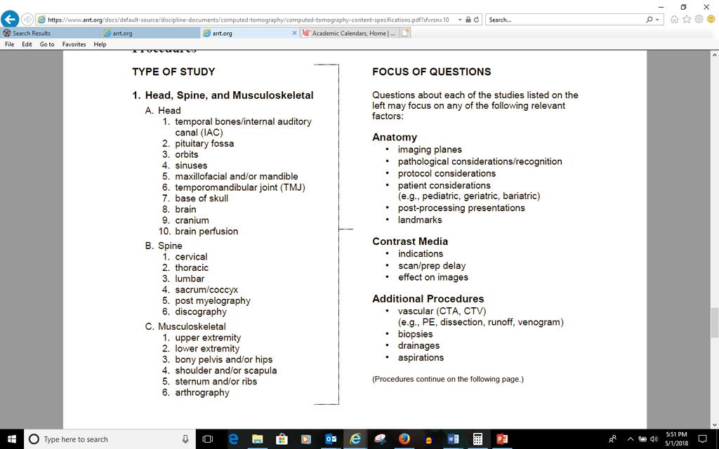
EXAMINATION CONTENT SPECIFICATIONS ARRT BOARD APPROVED: JANUARY 2017 IMPLEMENTATION DATE: JULY 1, 2017
 EXAMINATION CONTENT SPECIFICATIONS Computed Tomography The purpose of the computed tomography examination is to assess the knowledge and cognitive skills underlying the intelligent performance of the tasks
Diagnostic Imaging
 www.fisiokinesiterapia.biz Diagnostic Imaging Diagnostic Imaging is no longer limited to radiography. Major technological advancements have lead to the use of new and improved imaging technologies. The
The Computed Tomography Examination
 CONTENT SPECIFICATIONS The Computed Tomography Examination The purpose of The American Registry of Radiologic Technologists (ARRT ) Computed Tomography Examination is to assess the knowledge and cognitive
STRUCTURED EDUCATION REQUIREMENTS IMPLEMENTATION DATE: JULY 1, 2017
 STRUCTURED EDUCATION REQUIREMENTS IMPLEMENTATION DATE: JULY 1, 2017 Computed Tomography The purpose of structured education is to provide the opportunity for individuals to develop mastery of discipline-specific
STRUCTURED EDUCATION REQUIREMENTS EFFECTIVE: JANUARY 1, 2016
 Computed Tomography The purpose of structured education is to provide the opportunity for individuals to develop mastery of discipline-specific knowledge that, when coupled with selected clinical experiences,
Class Time: 9/12,10-10,11-21 Days: Listed Saturdays 1p-4p Room:HS208. Office Phone: Home Phone:
 Semester/Year: FALL 2015 CASPER COLLEGE COURSE SYLLABUS RDTK 1920 H1 Computed Tomography Procedures I Lecture Hours: 3 Lab Hours: 0 Credit Hours: 3 Class Time: 9/12,10-10,11-21 Days: Listed Saturdays 1p-4p
Customizing Contrast Injection for Body MDCT: Algorithmic Approach
 Customizing Contrast Injection for Body MDCT: Algorithmic Approach Lincoln L. Berland, M.D., F.A.C.R. University of Alabama at Birmingham Before Contrast Prep and Hydration Hydration single most important
CT - Brain Examination
 CT - Brain Examination Submitted by: Felemban 1 CT - Brain Examination The clinical indication of CT brain are: a) Chronic cases (e.g. headache - tumor - abscess) b) ER cases (e.g. trauma - RTA - child
2
 1 2 3 4 5 6 7 8 9 10 11 12 13 Cine loop of tomosynthesis slice images through the chest. 14 Standard PA chest radiograph (left) and single slice from the tomosynthesis image dataset (right) of a patient
Hi RES Extremity - (04/18/2011) CTDI: ~13 mgy per acquisition Used for evaluation of: Ankle Elbow Hand Wrist Foot /Calcaneous Toes Fingers
 CTDI: ~13 mgy per acquisition Used for evaluation of: Ankle Elbow Hand Wrist Foot /Calcaneous Toes Fingers") P a g e 1 Hi RES Extremity - (04/18/2011) CTDI: ~13 mgy per acquisition Used for evaluation of: Ankle Elbow Hand Wrist Foot /Calcaneous Toes Fingers Billing: 1. CT Upper/Lower Extremity of concern without
P a g e 1 Hi RES Extremity - (04/18/2011) CTDI: ~13 mgy per acquisition Used for evaluation of: Ankle Elbow Hand Wrist Foot /Calcaneous Toes Fingers Billing: 1. CT Upper/Lower Extremity of concern without
Neuroradiology MR Protocols
 Neuroradiology MR Protocols Brain protocols N 1: Brain MRI without contrast N 2: Pre- and post-contrast brain MRI N 3 is deleted N 4: Brain MRI without or pre-/post-contrast (seizure protocol) N 5: Pre-
Fundamentals, Techniques, Pitfalls, and Limitations of MDCT Interpretation and Measurement
 Fundamentals, Techniques, Pitfalls, and Limitations of MDCT Interpretation and Measurement 3 rd Annual Imaging & Physiology Summit November 20-21, 21, 2009 Seoul, Korea Wm. Guy Weigold, MD, FACC Cardiovascular
HONG KONG COLLEGE OF RADIOLOGISTS. Higher Training (Radiology) Subspecialty Training in Computed Tomography
 Subspecialty Training in Computed Tomography") HONG KONG COLLEGE OF RADIOLOGISTS Higher Training (Radiology) Subspecialty Training in Computed Tomography [The following guidelines should be read in conjunction with the General Guidelines on Higher
HONG KONG COLLEGE OF RADIOLOGISTS Higher Training (Radiology) Subspecialty Training in Computed Tomography [The following guidelines should be read in conjunction with the General Guidelines on Higher
X-Ray & CT Physics / Clinical CT
 Computed Tomography-Basic Principles and Good Practice X-Ray & CT Physics / Clinical CT INSTRUCTORS: Dane Franklin, MBA, RT (R) (CT) Office hours will be Tuesdays from 5pm to 6pm CLASSROOM: TIME: REQUIRED
CNS Imaging. Dr Amir Monir, MD. Lecturer of radiodiagnosis.
 CNS Imaging Dr Amir Monir, MD Lecturer of radiodiagnosis www.dramir.net Types of radiological examinations you know Plain X ray X ray with contrast GIT : barium (swallow, meal, follow through, enema) ERCP
Head CT Scan Interpretation: A Five-Step Approach to Seeing Inside the Head Lawrence B. Stack, MD
 Head CT Scan Interpretation: A Five-Step Approach to Seeing Inside the Head Lawrence B. Stack, MD Five Step Approach 1. Adequate study 2. Bone windows 3. Ventricles 4. Quadrigeminal cistern 5. Parenchyma
HI-Res Extremity Sensation 16
 Page 1 Routine Extremity - (2/14/2013) CTDI: ~20 mgy per acquisition Used for evaluation of: Humerus Forearm Femur Knee Tib/Fib Billing: 1. CT Upper/Lower Extremity of concern without contrast, with contrast,
AXIAL SKELETON SKULL
 AXIAL SKELETON SKULL CRANIAL BONES (8 total flat bones w/ 2 paired) 1. Frontal forms forehead & upper portion of eyesocket (orbital) 2. Parietal paired bones; form superior & lateral walls of cranium 3.
Reduce Exposure Eyes. CT Imaging Procedures. Head Positioning. Head Pathology 5/16/2008. CT Head & Posterior Fossa
 CT Head & Posterior Fossa CT Imaging Procedures Scoutview = Lateral OML ( 10 to 20 degrees) 15 degrees ( above OML) Time (fastest obtain info) Slide # 1 Slide # 2 2 CT Head & Posterior Fossa Reduce Exposure
The central nervous system
 Sectc.qxd 29/06/99 09:42 Page 81 Section C The central nervous system CNS haemorrhage Subarachnoid haemorrhage Cerebral infarction Brain atrophy Ring enhancing lesions MRI of the pituitary Multiple sclerosis
Head & Neck Clinical Sub Group. Network Agreed Imaging Guidelines for UAT and Thyroid Cancer. Measure Nos: 11-1C-105i & 11-1C-106i
 Greater Manchester, Lancashire & South Cumbria Strategic Clinical Network & Senate Head & Neck Clinical Sub Group Network Agreed Imaging Guidelines for UAT and Thyroid Cancer Measure Nos: 11-1C-105i &
Biology 218 Human Anatomy. Adapted from Martini Human Anatomy 7th ed. Chapter 6 The Skeletal System: Axial Division
 Adapted from Martini Human Anatomy 7th ed. Chapter 6 The Skeletal System: Axial Division Introduction The axial skeleton: Composed of bones along the central axis of the body Divided into three regions:
screening; including image post processing CT, heart; without contrast material; with Requires authorization
 0042T Cerebral perfusion analysis using CT; with ; including of parametric maps with determination of cerebral blood flow, cerebral blood volume, and mean transit time 74263 Computed tomographic (CT) colonography,
CT Chest. Verification of an opacity seen on the straight chest X ray
 CT Chest Indications: To assess equivocal plain x-ray findings Staging of lung neoplasm Merastatic workup of extra thoraces malignancies Diagnosis of diffuse lung diseases with HRCT Assessment of bronchietasis
Principles Arteries & Veins of the CNS LO14
 Principles Arteries & Veins of the CNS LO14 14. Identify (on cadaver specimens, models and diagrams) and name the principal arteries and veins of the CNS: Why is it important to understand blood supply
TRANSVERSE SECTION PLANE Scalp 2. Cranium. 13. Superior sagittal sinus
 TRANSVERSE SECTION PLANE 1 1. Scalp 2. Cranium 3. Superior sagittal sinus 4. Dura mater 5. Falx cerebri 6. Frontal lobes of the cerebrum 7. Middle meningeal artery 8. Cortex, grey matter 9. Cerebral vessels
LESSON ASSIGNMENT. Positioning for Exams of the Cranium, Sinuses, and Mandible. After completing this lesson, you should be able to:
 LESSON ASSIGNMENT LESSON 5 Positioning for Exams of the Cranium, Sinuses, and Mandible. LESSON ASSIGNMENT Paragraphs 5-1 through 5-9. LESSON OBJECTIVES After completing this lesson, you should be able
Major Anatomic Components of the Orbit
 Major Anatomic Components of the Orbit 1. Osseous Framework 2. Globe 3. Optic nerve and sheath 4. Extraocular muscles Bony Orbit Seven Bones Frontal bone Zygomatic bone Maxillary bone Ethmoid bone Sphenoid
CT: Common Protocols. Michael Steigner, M.D. Director, Vascular CT/MR Assistant Professor of Radiology
 CT: Common Protocols BRIGHAM AND WOMEN S HOSPITAL Heart & Vascular Center Harvard Medical School Teaching Hospital Michael Steigner, M.D. Director, Vascular CT/MR Assistant Professor of Radiology msteigner@bwh.harvard.edu
The diagnostic value of Computed Tomography in evaluation of maxillofacial Trauma
 The diagnostic value of Computed Tomography in evaluation of maxillofacial Trauma Qais H. Muassa FICMS College of Dentistry, Babylon University Ibrahim S. Gataa, BDS, FICMS College of Dentistry, Sulaimania
Anthem Blue Cross and Blue Shield Virginia Advanced Imaging Procedures Requiring Precertification Revised 02/13/2013
 Anthem Blue Cross and Blue Shield Virginia Advanced Imaging Procedures Requiring Precertification Revised 02/13/2013 Modality and CT Head CTA Head: Cerebrovascular MRI Head MRA Head: Cerebrovascular Functional
Clinician s Guide To Ordering NeuroImaging Studies
 Clinician s Guide To Ordering NeuroImaging Studies MRI CT South Jersey Radiology Associates The purpose of this general guide is to assist you in choosing the appropriate imaging test to best help your
Anatomy. Contents Brain (Questions)
") Anatomy 12 Contents 12.1 Brain (Questions).................................................... 683 12.2 Head and Neck (Questions)............................................. 685 12.3 Thorax (Questions)...................................................
Anatomy 12 Contents 12.1 Brain (Questions).................................................... 683 12.2 Head and Neck (Questions)............................................. 685 12.3 Thorax (Questions)...................................................
SPECIFIC PRINCIPLES FOR DOSE REDUCTION IN HEAD CT IMAGING. Rajiv Gupta, MD, PhD Neuroradiology, Massachusetts General Hospital Harvard Medical School
 SPECIFIC PRINCIPLES FOR DOSE REDUCTION IN HEAD CT IMAGING Rajiv Gupta, MD, PhD Neuroradiology, Massachusetts General Hospital Harvard Medical School OUTLINE 1 st Presentation: Dose optimization strategies
Multidetector CTA for Diagnosing Blunt Cerebrovascular Injuries
 Multidetector CTA for Diagnosing Blunt Cerebrovascular Injuries 4 th Nordic Trauma Course 2006 Stuart E. Mirvis, M.D., FACR Department of Diagnostic Radiology and Nuclear Medicine, University of Maryland
Pearls and Pitfalls in Neuroradiology of Cerebrovascular Disease The Essentials with MR and CT
 Pearls and Pitfalls in Neuroradiology of Cerebrovascular Disease The Essentials with MR and CT Val M. Runge, MD Wendy R. K. Smoker, MD Anton Valavanis, MD Control # 823 Purpose The focus of this educational
ADI Procedure Codes. August 2016 Revised April 2017 Page 1 of 7 ADI Procedure Codes
 Code Description 70450 CT Head without contrast 70460 CT Head with contrast 70470 CT Head with & without contrast 70480 CT Orbit, et al without contrast 70481 CT Orbit, et al with contrast 70482 CT Orbit,
Assignable revenue codes: Explanation of services:
 computed tomography Chest/Cardiac Assignable revenue codes: Explanation of services: 0350 CT Scan General Classification 0351 CT Scan Head Scan 0352 CT Scan Body Scan 0359 CT Scan Other CT Scans Known
Skull-2. Norma Basalis Interna Norma Basalis Externa. Dr. Heba Kalbouneh Associate Professor of Anatomy and Histology
 Skull-2 Norma Basalis Interna Norma Basalis Externa Dr. Heba Kalbouneh Associate Professor of Anatomy and Histology Norma basalis interna Base of the skull- superior view The interior of the base of the
Attenuation value in HU From -500 To HU From -10 To HU From 60 To 90 HU. From 200 HU and above
 Brain Imaging Common CT attenuation values Structure Air Fat Water Brain tissue Recent hematoma Calcifications Bone Brain edema and infarction Normal liver parenchyma Attenuation value in HU From -500
Medical Review Guidelines Magnetic Resonance Angiography
 Medical Review Guidelines Magnetic Resonance Angiography Medical Guideline Number: MRG2001-05 Effective Date: 2/13/01 Revised Date: 2/14/2006 OHCA Reference OAC 317:30-5-24. Radiology. (f) Magnetic Resonance
Applicable Neuroradiology
 For the Clinical Neurology Clerkship LSU Medical School New Orleans Amy W Voigt, MD Clerkship Director Introduction The field of Radiology first developed following the discovery of X-Rays by Wilhelm Roentgen
AIM 2014 CPT Radiology & Cardiac Codes Requiring Review
 AIM 2014 CPT Radiology & Cardiac Codes Requiring Review Modality Body Part CT Head 1 70480 CT orbit, sella or posterior fossa; w/o contrast 1 CT Head 1 70481 CT orbit, sella or posterior fossa; with CT
Human Anatomy and Physiology - Problem Drill 07: The Skeletal System Axial Skeleton
 Human Anatomy and Physiology - Problem Drill 07: The Skeletal System Axial Skeleton Question No. 1 of 10 Which of the following statements about the axial skeleton is correct? Question #01 A. The axial
Appendix Sample CT Protocols
 Appendix Sample CT Protocols The protocols in the following Appendix are designed to serve as a general guide to setting up comprehensive CT protocols on your scanner. The protocols are independent of
Anatomy and Physiology. Bones, Sutures, Teeth, Processes and Foramina of the Human Skull
 Anatomy and Physiology Chapter 6 DRO Bones, Sutures, Teeth, Processes and Foramina of the Human Skull Name: Period: Bones of the Human Skull Bones of the Cranium: Frontal bone: forms the forehead and the
Index. radiologic.theclinics.com. Note: Page numbers of article titles are in boldface type.
 Index Note: Page numbers of article titles are in boldface type. A ALCAPA. See Anomalous left coronary artery from the pulmonary artery. Angiosarcoma computed tomographic assessment of, 809 811 Anomalous
Combined Anatomical and Functional Imaging with Revolution * CT
 GE Healthcare Case studies Combined Anatomical and Functional Imaging with Revolution * CT Jean-Louis Sablayrolles, M.D. Centre Cardiologique du Nord, Saint-Denis, France Case 1 Whole Brain Perfusion and
B. CT protocols for the spine
 B. CT protocols for the spine Poster No.: A-003 Congress: ECR 2010 Type: Invited Speaker Topic: Neuro Authors: B. Tins; Oswestry/UK Keywords: CT, spine, diagnostic imaging protocol DOI: 10.1594/ecr2010/A-003
Contrast Guidelines for Common CT/CTA & MRI/MRA
 Contrast Guidelines for Common /A & /MRA Body Imaging Gastrointestinal CLINICAL GUIDELINES EXAM DESCRIPTION /A CPT CODES EXAM DESCRIPTION /MRA CPT CODES Abdominal mass Abdomen & Pelvis w 74177 Abdomen
APPENDICULAR SKELETON 126 AXIAL SKELETON SKELETAL SYSTEM. Cranium. Skull. Face. Skull and associated bones. Auditory ossicles. Associated bones.
 SKELETAL SYSTEM 206 AXIAL SKELETON 80 APPENDICULAR SKELETON 26 Skull Skull and associated s 29 Cranium Face Auditory ossicles 8 4 6 Associated s Hyoid Thoracic cage 25 Sternum Ribs 24 Vertebrae 24 column
Chapter 7 Part A The Skeleton
 Chapter 7 Part A The Skeleton Why This Matters Understanding the anatomy of the skeleton enables you to anticipate problems such as pelvic dimensions that may affect labor and delivery The Skeleton The
(Non-EKG Gated) CTA Thoracic Aorta = CTA Chest
 CTA Thoracic Aorta = CTA Chest") (Non-EKG Gated) CTA Thoracic Aorta = CTA Chest Reviewed By: Dan Verdini, MD, Rachael Edwards, MD Last Reviewed: January 2019 Contact: (866) 761-4200, Option 1 In accordance with the ALARA principle, TRA
(Non-EKG Gated) CTA Thoracic Aorta = CTA Chest Reviewed By: Dan Verdini, MD, Rachael Edwards, MD Last Reviewed: January 2019 Contact: (866) 761-4200, Option 1 In accordance with the ALARA principle, TRA
 www.oralradiologists.com CONE BEAM CT REPORT CASE ---- Case Information Referring Doctor: - Patient Name: - Scan Date: December 1, 2015 Patient DOB: - Reason for Exam: - Study Details: icat Flex, 160x160x112
Advances in Emergency Imaging
 Hampton Symposium,, October 16 th, 2010 Advances in Emergency Imaging Robert A. Novelline, MD Professor of Radiology, Harvard Medical School Director of Emergency Radiology, Massachusetts General Hospital
Anatomy 2 nd Paper. Past Papers
 Anatomy 2 nd Paper Past Papers September 2010 April 2010 September 2009 April 2009 September 2008 April 2008 September 2007 April 2007 September 2006 May 2006 September 2005 April 2005 September 2004 April
MR Advance Techniques. Vascular Imaging. Class II
 MR Advance Techniques Vascular Imaging Class II 1 Vascular Imaging There are several methods that can be used to evaluate the cardiovascular systems with the use of MRI. MRI will aloud to evaluate morphology
Clinical Applications
 C H A P T E R 16 Clinical Applications In selecting pulse sequences and measurement parameters for a specific application, MRI allows the user tremendous flexibility to produce variations in contrast between
CT PROCEDURE REFERENCE GUIDE 2017
 Head CT PROCEDURE REFERENCE GUIDE 2017 Procedure Contrast Scan Field Preparatio n Base of Skull to Vertex Sinuses Orbits Mastoids/IAC/ Temporal Bones Facial Bones ST Neck Low Dose Lung Screening Routine
MOLINA HEALTHCARE OF MICHIGAN PRIOR AUTHORIZATION / PRE-SERVICE REVIEW GUIDE IMAGING CODES REQUIRING PRIOR AUTHORIZATION EFFECTIVE 1/1/2014
 70336 MRI MRI, temporomandibular joint(s) 70450 CT/CTA CT, head or brain; without contrast material 70460 CT/CTA CT, head or brain; with contrast material(s) 70470 CT/CTA CT, head or brain; without contrast
ANATOMY & PHYSIOLOGY I Laboratory Version B Name Section. REVIEW SHEET Exercise 10 Axial Skeleton
 ANATOMY & PHYSIOLOGY I Laboratory Version B Name Section REVIEW SHEET Exercise 10 Axial Skeleton 1 POINT EACH. THE SKULL MULTIPLE CHOICE 1. The major components of the axial skeleton include the 7. The
Neuro CT What s a Good Head Exam?
 Neuro CT What s a Good Head Exam? Rajiv Gupta, PhD, MD Neuroradiology Massachusetts General Hospital Harvard Medical School Outline What we need to see? Routine Head CT protocols Dose optimization strategies
W/ (2) (3) (4) (5) (5) (6) (6) CTA
 (3) (4) (5) (5) (6) (6) CTA") Index Abdomen W/ and W/Out (2) Abdomen Pelvis W/Out (3) Abdomen Pelvis W/ (4) Pelvis W/ (5) Chest W/Out (5) Chest/Abdomen/Pelvis W/ (6) Chest W/ (6) CTA ( 7-8) Neuro (8-9) Musculoskeletal (10) Trauma (11)
Index Abdomen W/ and W/Out (2) Abdomen Pelvis W/Out (3) Abdomen Pelvis W/ (4) Pelvis W/ (5) Chest W/Out (5) Chest/Abdomen/Pelvis W/ (6) Chest W/ (6) CTA ( 7-8) Neuro (8-9) Musculoskeletal (10) Trauma (11)
Abdomen and Pelvis CT (1) By the end of the lecture students should be able to:
 By the end of the lecture students should be able to:") RAD 451 Abdomen and Pelvis CT (1) By the end of the lecture students should be able to: State the common indications for Abdomen and pelvis CT exams Identify possible contra indications for Abdomen and
RAD 451 Abdomen and Pelvis CT (1) By the end of the lecture students should be able to: State the common indications for Abdomen and pelvis CT exams Identify possible contra indications for Abdomen and
Magnetic Resonance Angiography
 Magnetic Resonance Angiography 1 Magnetic Resonance Angiography exploits flow enhancement of GR sequences saturation of venous flow allows arterial visualization saturation of arterial flow allows venous
Emerging Applications in Musculoskeletal CT Imaging
 Emerging pplications in Musculoskeletal CT Imaging y K Murali MD(RD), PDCC, Director of Interventional Radiology, G. Francis DMRD, DN (RD), Consultant Radiologist, and R. Madan, MS, MD, Consultant Radiologist,
University of Michigan continues fine-tuning neuro ExamCards
 I s s u e 3 1 - M a rc h 2 0 0 7 F i e l d Strength Publication for the Philips MRI Community University of Michigan continues fine-tuning neuro ExamCards Drs. Mukherji, Parmar create Achieva 3.0T ExamCards
05/02/ CPT Preauthorization Groupings Effective May 2, Computerized Tomography (CT) Abdomen 6. CPT Description SEGR CT01
 Abdomen 6. CPT Description SEGR CT01") Computerized Tomography (CT) 6 & 101 5 Upper Extremity 11 Lower Extremity 12 Head 3 Orbit 1 Sinus 2 Neck 4 7 Cervical Spine 8 Thoracic Spine 9 Lumbar Spine 10 Colon 13 CPT Preauthorization Groupings CPT
Computerized Tomography (CT) 6 & 101 5 Upper Extremity 11 Lower Extremity 12 Head 3 Orbit 1 Sinus 2 Neck 4 7 Cervical Spine 8 Thoracic Spine 9 Lumbar Spine 10 Colon 13 CPT Preauthorization Groupings CPT
PTA 106 Unit 1 Lecture 3
 PTA 106 Unit 1 Lecture 3 The Basics Arteries: Carry blood away from the heart toward tissues. They typically have thicker vessels walls to handle increased pressure. Contain internal and external elastic
2012 CPT Radiology Codes Requiring Review Blue Cross and Blue Shield of Louisiana
 2012 CPT Radiology Codes Requiring Review Blue Cross and Blue Shield of Louisiana CT Head 70480 CT orbit, sella or posterior fossa; w/o CT Head 70481 CT orbit, sella or posterior fossa; with CT Head 70482
Human Anatomy - Problem Drill 06: The Skeletal System Axial Skeleton & Articualtions
 Human Anatomy - Problem Drill 06: The Skeletal System Axial Skeleton & Articualtions Question No. 1 of 10 Instructions: (1) Read the problem and answer choices carefully, (2) Work the problems on paper
The dura is sensitive to stretching, which produces the sensation of headache.
 Dural Nerve Supply Branches of the trigeminal, vagus, and first three cervical nerves and branches from the sympathetic system pass to the dura. Numerous sensory endings are in the dura. The dura is sensitive
Imaging the musculoskeletal system. An Introduction
 Imaging the musculoskeletal system An Introduction Objectives Discuss: commonly used imaging modalities in the musculoskeletal system normal imaging anatomy in the extremities fracture description Imaging
Improvement of Image Quality with ß-Blocker Premedication on ECG-Gated 16-MDCT Coronary Angiography
 16-MDCT Coronary Angiography Shim et al. 16-MDCT Coronary Angiography Sung Shine Shim 1 Yookyung Kim Soo Mee Lim Received December 1, 2003; accepted after revision June 1, 2004. 1 All authors: Department
TORNIER BLUEPRINT. 3D Planning + PSI SCAN PROTOCOL
 TORNIER BLUEPRINT 3D Planning + PSI SCAN PROTOCOL Contents 3 Introduction 3 Patient preparation 3 Scanning instructions 4 Image instructions 5 Scanning parameters 6 Technical instructions 2 BLUEPRINT 3D
Structure Location Function
 Frontal Bone Cranium forms the forehead and roof of the orbits Occipital Bone Cranium forms posterior and inferior portions of the cranium Temporal Bone Cranium inferior to the parietal bone forms the
The Human Body: An Orientation
 The Human Body: An Orientation Body standing upright Anatomical Position feet slightly apart palms facing forward thumbs point away from body Directional Terms Superior and inferior toward and away from
, David Stultz, MD. Cardiac CT. David Stultz, MD Cardiology Fellow, PGY 6 March 28, 2006
 Cardiac CT David Stultz, MD Cardiology Fellow, PGY 6 March 28, 2006 Courtesy Tom Kracus Courtesy Kettering Tom Medical Kracus Cente Kettering Medical Center 2003-2006, David Stultz, MD Courtesy Tom Kracus
Meninges and Ventricles
 Meninges and Ventricles Irene Yu, class of 2019 LEARNING OBJECTIVES Describe the meningeal layers, the dural infolds, and the spaces they create. Name the contents of the subarachnoid space. Describe the
PTERYGOPALATINE FOSSA
 PTERYGOPALATINE FOSSA Outline Anatomical Structure and Boundaries Foramina and Communications with other spaces and cavities Contents Pterygopalatine Ganglion Especial emphasis on certain arteries and
Regional Human Anatomy (HBA 461/561/540): Course Objectives
: Course Objectives") Regional Human Anatomy (HBA 461/561/540): Course Objectives This is a 5-credit course that consists of 1-hour lectures followed by 3-hour labs. It is organized into three modules (see syllabus): Module
Regional Human Anatomy (HBA 461/561/540): Course Objectives This is a 5-credit course that consists of 1-hour lectures followed by 3-hour labs. It is organized into three modules (see syllabus): Module
Inferior view of the skull showing foramina (Atlas of Human Anatomy, 5th edition, Plate 12)
") Section 1 Head and Neck Skull, Basal View Incisive foramen Choanae Foramen ovale Foramen lacerum Foramen spinosum Carotid canal Jugular fossa Mastoid process Inferior view of the skull showing foramina
Section 1 Head and Neck Skull, Basal View Incisive foramen Choanae Foramen ovale Foramen lacerum Foramen spinosum Carotid canal Jugular fossa Mastoid process Inferior view of the skull showing foramina
Skeletal system. Prof. Abdulameer Al-Nuaimi. E. mail:
 Skeletal system Prof. Abdulameer Al-Nuaimi E-mail: a.al-nuaimi@sheffield.ac.uk E. mail: abdulameerh@yahoo.com Functions of Bone and The Skeletal System Support: The skeleton serves as the structural framework
Bones of the skull & face
 Bones of the skull & face Cranium= brain case or helmet Copyright The McGraw-Hill Companies, Inc. Permission required for reproduction or display. The cranium is composed of eight bones : frontal Occipital
Cardiac Computed Tomography
 Cardiac Computed Tomography Authored and approved by Koen Nieman Stephan Achenbach Francesca Pugliese Bernard Cosyns Patrizio Lancellotti Anastasia Kitsiou Contents CARDIAC COMPUTED TOMOGRAPHY Page 1.
OBJECTIVES. At the end of the lecture, students should be able to: List the cerebral arteries.
 DR JAMILA EL MEDANY OBJECTIVES At the end of the lecture, students should be able to: List the cerebral arteries. Describe the cerebral arterial supply regarding the origin, distribution and branches.
Temporal fossa Infratemporal fossa Pterygopalatine fossa Terminal branches of external carotid artery Pterygoid venous plexus
 Outline of content Temporal fossa Infratemporal fossa Pterygopalatine fossa Terminal branches of external carotid artery Pterygoid venous plexus Boundary Content Communication Mandibular division of trigeminal
JlntSocPlastination, Vol4:16-22,
 JlntSocPlastination, Vol4:16-22, 1990 16 SECTIONAL ANATOMY: STANDARDIZED METHODOLOGY Alexander Lane, Coordinator of Anatomy and Physiology, Triton College, Visiting Associate Professor, University of Illinois
TORNIER. scan protocol V2.1. Tornier Upper Extremities
 TORNIER TM scan protocol V2.1 Tornier Upper Extremities Contents 4 Introduction 4 Patient Preparation 4 Scanning Instructions 5 Image Instructions 6 Scanning Parameters 7 Technical Instructions Scan Protocol
CTA Pulmonary Embolism CTA Chest W (arterial)
") CTA Pulmonary Embolism CTA Chest W (arterial) Reviewed By: Rachael Edwards, MD; Anna Ellermeier, MD; Brett Mollard, MD Last Reviewed: January 2019 Contact: (866) 761-4200, Option 1 In accordance with the
CTA Pulmonary Embolism CTA Chest W (arterial) Reviewed By: Rachael Edwards, MD; Anna Ellermeier, MD; Brett Mollard, MD Last Reviewed: January 2019 Contact: (866) 761-4200, Option 1 In accordance with the
Computed Tomography - CSB 056
 Computed Tomography - CSB 056 Week 1 - Some intra-abdominal fat is ideal to differentiate organs better - Dual tube/dual energy imaging (2 tubes and 2 detector banks) - CT used: if lesion is large enough