Special Senses Sight Smell Taste Hearing and balance. Touch, not special, and not here (Ch 13)
|
|
|
- Jeffrey Welch
- 5 years ago
- Views:
Transcription
1 Special Senses Sight Smell Taste Hearing and balance Touch, not special, and not here (Ch 13)

2 Eye, matey.
3 Eye and Associated Structures 70% of all body sensory receptors are in the eye About half of the cerebral cortex is involved in visual processing 5/6 ths of the eye is protected by a cushion of fat and the bony orbit
4 Eye and Associated Structures Accessory structures include eyebrows, eyelids, conjunctiva, lacrimal apparatus, and extrinsic eye muscles
5 Eyebrows Coarse hairs that overlie the supraorbital margins Functions include: Shading the eye Preventing perspiration from reaching the eye Associated muscles Orbicularis oculi muscle depresses the eyebrows Corrugator muscles move the eyebrows medially


6 Eyebrows Orbicularis oculi muscle depresses the eyebrows Corrugator muscles move the eyebrows medially
7 Palpebrae (Eyelids) Protect the eye anteriorly Palpebral fissure separates eyelids Canthi medial and lateral angles (commissures) Lacrimal caruncle contains glands that secrete a whitish, oily secretion: (Sandman s eye sand)
8 Conjunctiva Transparent membrane that: Lines the eyelids as the palpebral (or tarsal) conjunctiva Covers the whites of the eyes as the bulbar (or ocular) conjunctiva Lubricates and protects the eye

9 Palpebrae (Eyelids) Palpebral conjunctiva Bulbar conjunctiva
10 Lacrimal Apparatus Consists of the lacrimal gland and associated ducts Lacrimal glands secrete tears Tears Contain mucus, antibodies, and lysozyme Enter the eye via superolateral excretory ducts Exit the eye medially via the lacrimal punctum Drain into the nasolacrimal duct

11 Lacrimal Apparatus Lacrimal sac Lacrimal gland Excretory ducts of lacrimal glands Lacrimal punctum Lacrimal canaliculus Nasolacrimal duct
12 Extrinsic Eye Muscles Six straplike extrinsic eye muscles Enable the eye to follow moving objects Maintain the shape of the eyeball Four rectus muscles originate from the annular ring Two oblique muscles move the eye in the vertical plane
 Lateral view of the")
13 Extrinsic Eye Muscles Superior oblique muscle Superior oblique tendon Superior rectus muscle Lateral rectus muscle Inferior rectus Inferior oblique muscle muscle (a) Lateral view of the right eye
 Superior view of the right")
14 Extrinsic Eye Muscles Trochlea Superior oblique muscle Superior oblique tendon Superior rectus muscle Axis at center of eye Inferior rectus muscle Medial rectus muscle Lateral rectus muscle Common tendinous ring (b) Superior view of the right eye

15 Let s play, Pin the Lacrimal Gland on the Eye. Superior Or Inferior Lateral view of R eye
16 Let s play, Pin the Lacrimal Gland on the Eye. Medial Or Lateral Superior view of R eye

17 Let s play, Pin the Lacrimal Gland on the Eye. Ok, Superior and Lateral, right? So, now name this muscle. And, this one. Superior view of R eye

18 Summary of Cranial Nerves and Muscle Actions Names, actions, and cranial nerve innervation of the extrinsic eye muscles Muscle Lateral rectus Medial rectus Superior rectus Inferior rectus Inferior oblique Superior oblique Action Moves eye laterally Moves eye medially Elevates eye and turns it medially Depresses eye and turns it medially Elevates eye and turns it laterally Depresses eye and turns it laterally Controlling cranial nerve VI (abducens) III (oculomotor) III (oculomotor) III (oculomotor) III (oculomotor) IV (trochlear) (c) Summary of muscle actions and innervating cranial nerves
19 Structure of the Eyeball A slightly irregular hollow sphere with anterior and posterior poles The wall is composed of three tunics fibrous, vascular, and sensory The internal cavity is filled with fluids called humors The lens separates the internal cavity into anterior and posterior segments
20 Structure of the Eyeball Sclera Choroid Retina Fibrous Vascular Sensory Anterior segment (contains aqueous humor) Posterior segment (contains vitreous humor) (a) Diagrammatic view. The vitreous humor is illustrated only in the bottom part of the eyeball. Hyaloid canal
21 Fibrous Tunic Forms the outermost coat of the eye and is composed of: Opaque sclera (posteriorly) Clear cornea (anteriorly) The sclera protects the eye and anchors extrinsic muscles The cornea lets light enter the eye
22 Structure of the Eyeball: Fibrous Tunic Cornea Sclera Choroid Retina Vascular Sensory Anterior pole Anterior segment (contains aqueous humor) Posterior segment (contains vitreous humor)
23 Vascular Tunic (Uvea): Has three regions: choroid, ciliary body, and iris Choroid region A dark brown membrane that forms the posterior portion of the uvea Supplies blood to all eye tunics
24 Vascular Tunic (Uvea): Choroid Region Cornea Sclera Choroid Retina Sensory Anterior segment (contains aqueous humor) Posterior segment (contains vitreous humor)
25 Vascular Tunic (Uvea): Ciliary Body A thickened ring of tissue surrounding the lens Composed of smooth muscle bundles (ciliary muscles) Anchors the suspensory ligament that holds the lens in place
26 Vascular Tunic (Uvea): Ciliary Body Ciliary body Ciliary zonule (suspensory ligament) Cornea Sclera Choroid Retina Sensory Anterior segment (contains aqueous humor) Posterior segment (contains vitreous humor)
27 Vascular Tunic (Uvea): Iris The colored part of the eye Pupil central opening of the iris Regulates the amount of light entering the eye during: Close vision and bright light pupils constrict Distant vision and dim light pupils dilate Changes in emotional state pupils dilate when the subject matter is appealing or requires problem-solving skills
28 Vascular Tunic (Uvea): Iris Ciliary body Ciliary zonule (suspensory ligament) Iris Sclera Choroid Retina Sensory Anterior segment (contains aqueous humor) Posterior segment (contains vitreous humor)
 Sphincter pupillae Dilator pupillae Dilator pupillae muscle contraction increases pupil size.")
29 Pupil Dilation and Constriction Parasympathetic + Sympathetic + Sphincter pupillae muscle contraction decreases pupil size. Iris (two muscles) Sphincter pupillae Dilator pupillae Dilator pupillae muscle contraction increases pupil size.
30 Sensory Tunic: Retina A delicate two-layered membrane Pigmented layer the outer layer that absorbs light and prevents its scattering Neural layer, which contains: Photoreceptors that transduce light energy Bipolar cells and ganglion cells Amacrine and horizontal cells
31 Structure of the Eyeball: Sensory Tunic Ciliary body Ciliary zonule (suspensory ligament) Sclera Choroid Retina Iris Anterior segment (contains aqueous humor) Posterior segment (contains vitreous humor)
32 Sensory Tunic: Retina Pathway of light Neural layer of retina Pigmented layer of retina Choroid Sclera (a) Posterior aspect of the eyeball
33 Ganglion cells Bipolar cells Photoreceptors Rod Cone Pathway of light Amacrine cell Horizontal cell Pathway of signal output Pigmented Pathway of light layer of retina (b) Cells of the neural layer of the retina
34 Nuclei of ganglion cells Outer segments of rods and cones Choroid Axons of Nuclei Nuclei of ganglion of bipolar rods and cells cells cones (c) Photomicrograph of retina Pigmented layer of retina
35 The Retina: Ganglion Cells and the Optic Disc Ganglion cell axons: Run along the inner surface of the retina Leave the eye as the optic nerve The optic disc: Is the site where the optic nerve leaves the eye Lacks photoreceptors (the blind spot)
36 Ganglion cells Bipolar cells Photoreceptors Rod Cone Pathway of light Amacrine cell Horizontal cell Pathway of signal output Pigmented Pathway of light layer of retina (b) Cells of the neural layer of the retina
 Posterior aspect of the")
37 Sensory Tunic: Retina Pathway of light Optic disc Neural layer of retina Pigmented layer of retina Choroid Sclera Optic nerve (a) Posterior aspect of the eyeball
38 The Retina: Optic Disc Optic disc Blind Spot Retina
39 The Retina: Photoreceptors Rods: Respond to dim light Are used for peripheral vision Cones: Respond to bright light Have high-acuity color vision Are found in the macula lutea Are concentrated in the fovea centralis
40 Ganglion cells Bipolar cells Photoreceptors Rod Cone Pathway of light Amacrine cell Horizontal cell Pathway of signal output Pigmented Pathway of light layer of retina (b) Cells of the neural layer of the retina
41 The Retina: Optic Disc Macula lutea Optic disc Blind Spot Retina
42 Sensory Tunic: Retina Bipolar cells and ganglion cells Carry the action potential to the optic nerve Amacrine and horizontal cells Assist with visual processing Modify output of bipolar cells
 Cells of the neural layer of the retina")
43 Sensory Tunic: Retina Ganglion cells Bipolar cells Photoreceptors Rod Cone Pathway of light Amacrine cell Horizontal cell Pathway of signal output Pigmented Pathway of light layer of retina (b) Cells of the neural layer of the retina
44 Blood Supply to the Retina The neural retina receives its blood supply from two sources The outer third receives its blood from the choroid The inner two-thirds is served by the central artery and vein Small vessels radiate out from the optic disc and can be seen with an ophthalmoscope

45 The Retina: Central artery and vein Central artery and vein emerging from the optic disc Optic disc Retina
46 Inner Chambers and Fluids The lens separates the internal eye into anterior and posterior segments The posterior segment is filled with a clear gel called vitreous humor that: Transmits light Supports the posterior surface of the lens Holds the neural retina firmly against the pigmented layer Contributes to intraocular pressure
47 Inner Chambers and Fluids Posterior segment (contains vitreous humor)
48 Anterior Segment Composed of two chambers Anterior Ch. between the cornea and the iris Posterior Ch. between the iris and the lens Aqueous humor A plasmalike fluid that fills the anterior segment Produced by the ciliary processes Drains via the scleral venous sinus (Canal of Schlemm) Supports, nourishes, and removes wastes
49 Anterior Segment Cornea Lens Anterior segment (contains aqueous humor) Anterior chamber Posterior chamber Scleral venous sinus 3 1 Ciliary processes Ciliary muscle Ciliary body
50 Lens A biconvex, transparent, flexible, avascular structure that: Allows precise focusing of light onto the retina Is composed of epithelium and lens fibers

51 Anterior Segment Cornea Lens Lens epithelium Lens 2 Ciliary processes Ciliary muscle Ciliary body
52 Lens Is composed of epithelium and lens fibers Lens epithelium anterior, cuboidal cells that differentiate into lens fibers Lens fibers cells filled with the transparent protein crystallin

53 Anterior Segment Figure 15.8

54 Lens With age, the lens becomes more compact and dense and loses its elasticity, and you need
55 Focusing Light on the Retina Pathway of light entering the eye: cornea, aqueous humor, lens, vitreous humor, neural layer of the retina to the photoreceptors Light is refracted: At the cornea Entering the lens Leaving the lens The lens curvature and shape allow for fine focusing of an image

56 Focusing for Distant Vision Light from a distance needs little adjustment for proper focusing Far Point of Vision the distance beyond which the lens does not need to change shape to focus (20 ft.)

57 Focusing for Distant Vision Nearly parallel rays from distant object Lens Sympathetic activation Ciliary zonule Ciliary muscle (a) Lens is flattened for distant vision. Sympathetic input relaxes the ciliary muscle, tightening the ciliary zonule, and flattening the lens. Inverted image
58 Focusing for Close Vision Close vision requires: Accommodation changing the lens shape by ciliary muscle contraction and lens ligament relaxation to increase refractory power Constriction the pupillary reflex constricts the pupils to prevent divergent light rays from entering the eye Convergence medial rotation of the eyeballs toward the object being viewed

59 Focusing for Close Vision Divergent rays from close object Parasympathetic activation Ciliary muscle Inverted image Lens ligaments (b) Lens bulges for close vision. Parasympathetic input contracts the ciliary muscle, loosening the ciliary zonule, allowing the lens to bulge.
60 Visual Pathways Axons of retinal ganglion cells form the optic nerve Medial fibers of the optic nerve decussate at the optic chiasm Optic chiasm
61 Visual Pathways
62 Visual Pathways Most fibers of the optic tracts continue to the lateral geniculate body of the thalamus (gateway to the cortex, conscious senses all pass through the thalamus)
63 Visual Pathways
64 Visual Pathways Other optic tract fibers end in superior colliculi (initiating visual reflexes) and pretectal nuclei (involved with pupillary reflexes) Optic radiations travel from the thalamus to the visual cortex

65 Visual Pathways
66 End, matey.
67 New senses. Taste and Smell.
68 Chemical Senses Chemical senses = olfaction (smell) and gustation (taste) Their chemoreceptors respond to chemicals in aqueous solution Smell to substances dissolved in fluids of the nasal membranes Taste to substances dissolved in saliva
69 Sense of Smell (this one s easy) The organ of smell is the olfactory epithelium, which covers the superior nasal concha

")
70 Olfactory Receptors Olfactory epithelium Olfactory tract Olfactory bulb (a)
71 Sense of Smell (this one s easy) Olfactory receptor cells are bipolar neurons with radiating olfactory cilia Olfactory receptors are surrounded and cushioned by supporting cells Basal cells lie at the base of the epithelium
72 Olfactory Receptors Olfactory epithelium Olfactory tract Olfactory tract Olfactory bulb Cribriform plate of ethmoid bone Filaments of olfactory nerve (a) Route of inhaled air Olfactory gland Olfactory epithelium Mucus Axon Basal cell Olfactory receptor cell Supporting cell Dendrite Olfactory cilia (b) Route of inhaled air containing odor molecules
73 Olfactory Pathway Olfactory receptor cells synapse with mitral cells at glomeruli Mitral cells process odor signals Mitral cells send impulses to: The olfactory cortex (via the thalamus for conscious perception of the sense of smell) The hypothalamus, amygdala, and limbic system (for unconscious perceptions and emotional response)
 Route of inhaled air Olfactory epithelium Mucus (b) Route of inhaled air containing odor")
74 Olfactory Receptors Olfactory epithelium Olfactory tract Mitral cell (output cell) Glomeruli Olfactory tract Olfactory bulb Nasal conchae Olfactory gland (a) Route of inhaled air Olfactory epithelium Mucus (b) Route of inhaled air containing odor molecules
75 Olfactory Receptors, end. (told U it was easy)
76 Taste
77 Taste Buds Most of the 10,000 or so taste buds are found on the tongue Some are other places in the mouth. Taste buds are found in papillae of the tongue mucosa Papillae come in three types: filiform (foliate), fungiform, and circumvallate (vallate)
 Taste buds are associated with fungiform, foliate, and circumvallate (vallate)")
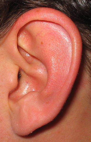
78 Taste Buds Epiglottis Palatine tonsil Lingual tonsil Foliate papillae Circumvallate (vallate) Fungiform papillae (a) Taste buds are associated with fungiform, foliate, and circumvallate (vallate) papillae.
 Enlarged section of a circumvallate")
79 Taste Buds Circumvallate papilla Taste bud (b) Enlarged section of a circumvallate papilla.
80 Structure of a Taste Bud Gourd-shaped
 cells Taste pore Stratified squamous epithelium of tongue (c) Enlarged view of a taste")
81 Taste Buds Connective tissue Taste fibers of cranial nerve Gustatory hair Basal cells Gustatory (taste) cells Taste pore Stratified squamous epithelium of tongue (c) Enlarged view of a taste bud.
82 Structure of a Taste Bud Each gourd-shaped taste bud consists of three major cell types Supporting cells insulate the receptor Basal cells dynamic stem cells, replace gustatory cells every 7-10 days. Gustatory cells taste cells, transmit signals to sensory dendrites of Cranial Nerves VII and IX (facial and?, to thalamus )
 cells Taste pore Stratified squamous epithelium of")
83 Taste Buds Connective tissue Taste fibers of cranial nerve Gustatory hair Basal cells Gustatory (taste) cells Taste pore Stratified squamous epithelium of tongue
84 Taste Sensations There are five basic taste sensations Sweet sugars, saccharin, alcohol, and some amino acids Salt metal ions (NaCl = Na+ and Cl-) Sour hydrogen ions, acids Bitter alkaloids such as quinine and nicotine Umami elicited by the amino acid glutamate (MSG) And, maybe now a 6 th, calcium. Most tastes are combinations
85 Physiology of Taste In order to be tasted, a chemical: Must be dissolved in saliva Must contact gustatory hairs Binding of the food chemical: Depolarizes the taste cell membrane, releasing neurotransmitter to the sensory dendrite
86 Gustatory Pathway Cranial Nerves VII (Facial) and IX (? *Hint, well named for tongue stuff) carry impulses from taste buds to the thalamus and then to the gustatory cortex (Conscious perception of taste)
 Glossopharyngeal nerve")
87 Gustatory cortex (in insula) Thalamus Pons Solitary nucleus in medulla oblongata Facial nerve (VII) Glossopharyngeal nerve (IX)
88 Influence of Other Sensations on Taste Taste is 80% smell, food tastes bland when you have a cold. Thermoreceptors, mechanoreceptors, nociceptors also influence tastes Temperature and texture enhance or detract from taste

89 End of Taste and Smell.
90 The Ear: Hearing and Balance

91 The Ear: Hearing and Balance Wake up, sit up, pay attention, this one is complicated. Sitzender Junge ("Sitting youngster") by Werner Stötzer, 1956
92 The Ear: Hearing and Balance The three parts of the ear are the inner, outer, and middle ear The outer and middle ear are involved with hearing The inner ear functions in both hearing and equilibrium
93 Three regions of the ear External ear Middle ear Internal ear (labyrinth)
94 Outer Ear The auricle (pinna) contains: The helix (rim) The lobule (earlobe) External auditory canal Short, curved tube filled with ceruminous glands
95 The Ear: Hearing and Balance External ear Middle ear Internal ear (labyrinth) Auricle (pinna) Helix Lobule External acoustic meatus
96 Outer Ear Tympanic membrane (eardrum) Thin connective tissue membrane that vibrates in response to sound Transfers sound energy to the middle ear ossicles Boundary between outer and middle ears
 Tympanic")
97 The Ear: Hearing and Balance External ear Middle ear Internal ear (labyrinth) Tympanic membrane
98 Middle Ear (Tympanic Cavity) A small, air-filled, mucosa-lined cavity Flanked laterally by the eardrum (tympanic membrane) Flanked medially by the oval and round windows
99 Middle Ear Oval window (deep to stapes) Tympanic membrane Round window
100 Middle Ear: Ear Ossicles The tympanic cavity contains three small bones: malleus, incus, and stapes Transmit vibratory motion of the eardrum to the oval window Dampened by the tensor tympani (tube to malleus) and stapedius muscles (cavity to the stapes)
 Incu (anvil) Stapes")
101 Middle Ear: Ear Ossicles Auditory ossicles Malleus (hammer) Incu (anvil) Stapes (stirrup)
102 Middle Ear (Tympanic Cavity) Pharyngotympanic tube connects the middle ear to the nasopharynx Equalizes pressure in the middle ear cavity with the external air pressure

103 Middle Ear Pharyngotympanic (auditory) tube
104 The Inner Ear: Hearing and Balance Receptors for hearing and balance: Respond to separate stimuli Are activated independently
105 Inner Ear: Hearing and Balance
106 Inner Ear: Hearing and Balance Bony labyrinth Tortuous channels worming their way through the temporal bone Contains the vestibule, the cochlea, and the semicircular canals Filled with perilymph, similar to CSF
107 Inner Ear: Hearing and Balance Semicircular ducts in semicircular canals Anterior Posterior Lateral Utricle in vestibule Saccule in vestibule Cochlear duct in cochlea
108 Inner Ear: Hearing and Balance Bony labyrinth Tortuous channels worming their way through the temporal bone Contains the vestibule, the cochlea, and the semicircular canals Filled with perilymph, similar to CSF, supports the Membranous labyrinth Series of membranous sacs within the bony labyrinth Filled with endolymph, similar to intracellular fluid
109 Inner Ear: Hearing and Balance Membranous labyrinth inside bony labyrinth inside which bone? Semicircular ducts in semicircular canals Temporal bone Anterior Posterior Lateral Balance Cristae ampullares in the membranous ampullae Utricle in vestibule Saccule in vestibule Stapes in oval window Hearing Maculae Spiral organ (of Corti) Cochlear duct in cochlea Round window

110 Mechanisms of Equilibrium and Orientation
111 Mechanisms of Equilibrium and Orientation Vestibular apparatus equilibrium receptors in the semicircular canals and vestibule Maintains our orientation and balance in space Vestibular receptors monitor static equilibrium Semicircular canal receptors monitor dynamic equilibrium

112 Inner Ear: Balance: The Vestibule
113 Inner Ear: Balance: The Vestibule The Vestibule The central cavity of the bony labyrinth Suspended in its perilymph are two sacs: the utricle and saccule The utricle extends into the semicircular canals (balance) (U is a semi circle) The saccule extends into the cochlea (cochlea is for hearing)
114 Inner Ear: Balance: The Vestibule Utricle in vestibule Saccule in vestibule
115 Inner Ear: Balance: The Vestibule The utricle and saccule Have equilibrium receptors called maculae Respond to gravity and changes in the position of the head
116 Inner Ear: Balance: The Vestibule Maculae Utricle in vestibule Saccule in vestibule

117 Inner Ear: Balance: The Vestibule Utricle Macula Saccule
118 Anatomy of Maculae Maculae are the sensory receptors for static equilibrium Contain supporting cells and hair cells Each hair cell has stereocilia and kinocilium embedded in an otolithic membrane Otolithic membrane jellylike mass studded with tiny CaCO 3 stones called otoliths Utricular hairs respond to horizontal movement Saccular hairs respond to vertical movement

119 Inner Ear: Balance: The Macula Stereocilia Kinocilium Otoliths Otolithic membrane Hair bundle Macula of utricle Macula of saccule Vestibular nerve fibers Hair cells Supporting cells

120 Inner Ear: Balance: The Macula Otolithic membrane Kinocilium Stereocilia Receptor potential Nerve impulses generated in vestibular fiber Depolarization When hairs bend toward the kinocilium, the hair cell depolarizes, exciting the nerve fiber, which generates more frequent action potentials. Hyperpolarization When hairs bend away from the kinocilium, the hair cell hyperpolarizes, inhibiting the nerve fiber, and decreasing the action potential frequency.

121 Inner Ear: Balance: The Semicircular Canals
122 Inner Ear: Balance: The Semicircular Canals Three canals that each define two-thirds of a circle and lie in the three planes of space Membranous semicircular ducts line each canal and communicate with the utricle

123 Inner Ear: Balance: The Semicircular Canals Semicircular ducts in semicircular canals Anterior Posterior Lateral
124 Inner Ear: Balance: The Ampulla The ampulla is the swollen end of each canal and it houses equilibrium receptors in a region called the crista ampullaris These receptors respond to angular movements of the head

125 Inner Ear: Balance: The Ampulla Semicircular ducts in semicircular canals Anterior Posterior Lateral Cristae ampullares in the membranous ampullae
126 Crista Ampullaris and Dynamic Equilibrium The crista ampullaris (or crista): Is the receptor for dynamic equilibrium Is located in the ampulla of each semicircular canal Responds to angular movements

127 Inner Ear: Balance: The Ampulla Crystea ampullares in the ampullea
128 Crista Ampullaris and Dynamic Equilibrium Each crista has support cells and hair cells that extend into a gel-like mass called the cupula Dendrites of vestibular nerve fibers encircle the base of the hair cells
 Hair cell Supporting")
129 Inner Ear: Balance: The Ampulla Cupula Endolymph Membranous Crista labyrinth ampullaris Fibers of vestibular nerve Hair bundle (kinocilium plus stereocilia) Hair cell Supporting cell

130 Inner Ear: Balance: The Ampulla Cupula (b) Scanning electron micrograph of a crista ampullaris (200x)
 Movement of the cupula during rotational acceleration and deceleration During rotational acceleration, endolymph moves inside the semicircular canals in the direction opposite the rotation (it")
131 Inner Ear: Balance: The Ampulla Section of ampulla, filled with endolymph Cupula Fibers of vestibular nerve Flow of endolymph At rest, the cupula stands upright. (c) Movement of the cupula during rotational acceleration and deceleration During rotational acceleration, endolymph moves inside the semicircular canals in the direction opposite the rotation (it lags behind due to inertia). Endolymph flow bends the cupula and excites the hair cells. As rotational movement slows, endolymph keeps moving in the direction of the rotation, bending the cupula in the opposite direction from acceleration and inhibiting the hair cells.
 Cerebellum Central nervous system processing Vestibular nuclei (in brain stem) Oculomotor control (cranial nerve")
132 Inner Ear: Balance Input: Information about the body s position in space comes from three main sources and is fed into two major processing areas in the central nervous system. Vestibular receptors Visual receptors Somatic receptors (from skin, muscle and joints) Cerebellum Central nervous system processing Vestibular nuclei (in brain stem) Oculomotor control (cranial nerve nuclei III, IV, VI) (eye movements) Spinal motor control (cranial nerve XI nuclei and vestibulospinal tracts) (neck movements) Output: Fast reflexive control of the muscles serving the eye and neck, limb, and trunk are provided by the outputs of the central nervous system.
133 Inner Ear: Hearing: The Cochlea
134 Inner Ear: Hearing: The Cochlea The Cochlea A spiral, conical, bony chamber that: Extends from the anterior vestibule (saccule area) Coils around a bony pillar called the modiolus Contains the cochlear duct, which ends at the cochlear apex (helicotrema) Contains the organ of Corti (hearing receptor)

135 Modiolus The Cochlea Helicotrema
136 Inner Ear: Hearing: The Cochlea The cochlea is divided into three chambers: Scala vestibuli Scala media Scala tympani
 Spiral organ (of Corti) Scala tympani (contains")
137 Inner Ear: Hearing: The Cochlea Cochlear duct (scala media; contains endolymph) Scala vestibuli (contains perilymph) Spiral organ (of Corti) Scala tympani (contains perilymph)
138 Inner Ear: Hearing: The Cochlea The cochlea is divided into three chambers: Scala vestibuli Continuous with vestibule, abuts the oval window Scala media The cochlear duct, the hearing part Scala tympani Abuts round window, meets the scala vestibuli at the helicotrema.

139 The Cochlea and Hearing
140 The Cochlea The floor of the scala media (aka cochlear duct) is composed of: The osseous/bony spiral lamina The basilar membrane, which supports the organ of Corti The cochlear branch of nerve VIII runs from the organ of Corti to the brain (CN VIII is.?) Vestibulocochlear
141 The Cochlea Osseous spiral lamina Cochlear duct (scala media; contains endolymph) Spiral organ (of Corti) Basilar membrane Scala vestibuli (contains perilymph) Scala tympani (contains perilymph) Spiral ganglion Cochlear nerve, division of the vestibulocochlear nerve (VIII)

142 Organ of Corti Tectorial membrane Hairs (stereocilia) Outer hair cells Inner hair cell Afferent nerve fibers Supporting cells Fibers of cochlear nerve Basilar membrane
143 Air pressure A little physio: Sound and Mechanisms of Hearing Wavelength Area of high pressure (compressed molecules) Area of low pressure (rarefaction) Crest Trough Distance Amplitude (a) A struck tuning fork alternately compresses and rarefies the air molecules around it, creating alternate zones of high and low pressure. (b) Sound waves radiate outward in all directions.
144 A little physio: Sound and Mechanisms of Hearing Sound vibrations beat against the eardrum The eardrum pushes against the ossicles, which press fluid in the inner ear against the oval and round windows This movement sets up shearing forces that pull on hair cells Moving hair cells stimulates the cochlear nerve that sends impulses to the brain
 Route of sound waves through the ear 1 Sound waves vibrate the tympanic membrane. 2 Auditory ossicles vibrate. Pressure is amplified.")
145 Transmission of Sound to the Inner Ear Auditory ossicles Malleus Incus Stapes Cochlear nerve Oval window Scala vestibuli Helicotrema Scala tympani 2 3 Cochlear duct Basilar membrane 1 Tympanic Round membrane window (a) Route of sound waves through the ear 1 Sound waves vibrate the tympanic membrane. 2 Auditory ossicles vibrate. Pressure is amplified. 3 Pressure waves created by the stapes pushing on the oval window move through fluid in the scala vestibuli. Sounds with frequencies below hearing travel through the helicotrema and do not excite hair cells. Sounds in the hearing range go through the cochlear duct, vibrating the basilar membrane and deflecting hairs on inner hair cells.
 Fibers of basilar membrane Apex (long, floppy fibers) Low-frequency sounds displace the basilar membrane near the apex.")
146 Transmission of Sound to the Inner Ear Basilar membrane High-frequency sounds displace the basilar membrane near the base. Medium-frequency sounds displace the basilar membrane near the middle. Base (short, stiff fibers) Fibers of basilar membrane Apex (long, floppy fibers) Low-frequency sounds displace the basilar membrane near the apex. (b) Different sound frequencies cross the basilar membrane at different locations. Frequency (Hz)
147 Transmission of Sound to the Inner Ear The route of sound to the inner ear follows this pathway: Outer ear pinna, auditory canal, eardrum Middle ear malleus, incus, and stapes to the oval window Inner ear scalas vestibuli and tympani to the cochlear duct/scala media Stimulation of the organ of Corti Generation of impulses in the cochlear nerve
 Midbrain Cochlear nuclei Vibrations Vibrations Medulla Vestibulocochlear nerve Spiral ganglion of cochlear nerve Bipolar cell Spiral organ (of")
148 Inner Ear: Hearing: The Cochlea Medial geniculate nucleus of thalamus Hearing Primary auditory cortex in temporal lobe Inferior colliculus Lateral lemniscus Superior olivary nucleus (pons-medulla junction) Midbrain Cochlear nuclei Vibrations Vibrations Medulla Vestibulocochlear nerve Spiral ganglion of cochlear nerve Bipolar cell Spiral organ (of Corti)
149 Deafness Conduction deafness something hampers sound conduction to the fluids of the inner ear (e.g., impacted earwax, perforated eardrum, osteosclerosis of the ossicles) Sensorineural deafness results from damage to the neural structures at any point from the cochlear hair cells to the auditory cortical cells

150 End
Taste buds Gustatory cells extend taste hairs through a narrow taste pore
 The Special Senses Objectives Describe the sensory organs of smell, and olfaction. Identify the accessory and internal structures of the eye, and explain their function. Explain how light stimulates the
SPECIAL SENSES PART I: OLFACTION & GUSTATION
 SPECIAL SENSES PART I: OLFACTION & GUSTATION 5 Special Senses Olfaction Gustation Vision Equilibrium Hearing Olfactory Nerves Extend through cribriform plate into nasal cavity on both sides of nasal septum
o A cushion of fat surrounds most of the eye
 Name Period SPECIAL SENSES The Senses of touch o Temperature o Pressure o Pain o Smell o Taste o Sight o Hearing o Equilibrium The Eye and Vision are in the eyes has over a o Most of the eye is enclosed
The Senses. Chapter 10 7/8/11. Introduction
 Chapter 10 The Senses Introduction A. Sensory receptors detect changes in the environment and stimulate neurons to send nerve impulses to the brain. B. A sensation is formed based on the sensory input.
The Nervous System: General and Special Senses Pearson Education, Inc.
 18 The Nervous System: General and Special Senses Introduction Sensory information arrives at the CNS Information is picked up by sensory receptors Sensory receptors are the interface between the nervous
o A cushion of fat surrounds most of the eye
 Name Period SPECIAL SENSES The Senses General senses of touch o Temperature o Pressure o Pain Special senses o Smell o Taste o Sight o Hearing o Equilibrium The Eye and Vision 70 percent of all sensory
Special Senses. Accessory Structures of the Eye. The Eye and Vision. Accessory Structures of the Eye. Accessory Structures of the Eye
 8 PART A Special Senses PowerPoint Lecture Slide Presentation by Jerry L. Cook, Sam Houston University ESSENTIALS OF HUMAN ANATOMY & PHYSIOLOGY EIGHTH EDITION ELAINE N. MARIEB The Senses General senses
Sensory system. Dr. Carmen E. Rexach Anatomy 35 Mt San Antonio College
 Sensory system Dr. Carmen E. Rexach Anatomy 35 Mt San Antonio College Sensory receptors Detect stimuli Classified by structure Origin Distribution Modality Structural Classification naked nerve endings
THE SPECIAL SENSES. Introduction Vision
 THE SPECIAL SENSES Introduction Vision RECEPTORS Structures designed to respond to stimuli Variable complexity RECEPTORS: GENERAL PROPERTIES Transducers Receptor Potential Generator Potential RECEPTORS
Introduction. Senses our perception of what is out there 2 groups. General senses Special senses
 Introduction Senses our perception of what is out there 2 groups General senses Special senses Central Processing and Adaptation Adaptation the loss of sensitivity after continuous stimulation Tonic receptors
The Special Senses: Part A
 PowerPoint Lecture Slides prepared by Janice Meeking, Mount Royal College CHAPTER 15 The Special Senses: Part A Warm Up What is the function of the eyeball? List any structures of the eyeball that you
Chapter 10. The Senses
 Chapter 10 The Senses 1 Introduction A. Sensory receptors detect changes in the environment and stimulate neurons to send nerve impulses to the brain. B. A sensation is formed based on the sensory input.
Unit 8: The Special Senses
 Unit 8: The Special Senses I. The Senses A. General senses of touch 1. Temperature 2. Pressure 3. Pain B. Special senses 1. Smell 2. Taste 3. Sight 4. Hearing 5. Equilibrium II. The Eye and Vision A. 70%
Copyright 2009 Pearson Education, Inc.
 Outline Nervous System Sensory Systems I. II. III. IV. V. VI. Biol 105 Lecture 11 Chapter 9 Senses Sensory receptors Touch Vision Hearing and balance Smell Senses Sensory receptor cells Sensory receptors
The white of the eye and the part that maintains its shape is know n as the:
 Scrub In The white of the eye and the part that maintains its shape is know n as the: a. Cornea b. Pupil c. Retina d. Sclera The structure that is found in the ear and contains the organ of hearing is
Special Senses. The Senses. General senses. Special senses. Yong Jeong, MD, PhD Department of Bio and Brain Engineering
 8 Special Senses Yong Jeong, MD, PhD Department of Bio and Brain Engineering The Senses General senses Touch Pressure Pain Temperature Proprioception Special senses Smell Taste Sight Hearing Equilibrium
Essentials of Human Anatomy & Physiology. Chapter 8. Special Senses. Slides Lecture Slides in PowerPoint by Jerry L.
 Essentials of Human Anatomy & Physiology Elaine N. Marieb Seventh Edition Chapter 8 Special Senses Slides 8.1 8.19 Lecture Slides in PowerPoint by Jerry L. Cook Special Senses Title Somatosensation Essential
Chapter 18 Senses SENSORY RECEPTION 10/21/2011. Sensory Receptors and Sensations. Sensory Receptors and Sensations. Sensory Receptors and Sensations
 SENSORY RECEPTION Chapter 18 Senses s convert stimulus energy to action potentials s 1. Are specialized cells, or 2. Specialized endings that detect stimuli All stimuli are forms of energy s in eyes detect
The Special Senses. Smell, taste, vision, hearing and equilibrium Housed in complex sensory organs
 The Special Senses Smell, taste, vision, hearing and equilibrium Housed in complex sensory organs Chemical Senses Interaction of molecules with receptor cells Olfaction (smell) and gustation (taste) Both
20-20,000 Hertz range of human hearing
 20-20,000 Hertz range of human hearing accommodation automatic adjustment in focal length of the lens of the eye; changing the shape of the lens aqueous humor Watery fluid in the anterior chambers of the
Unit 8 - The Special Senses 1
 Unit 8 - The Special Senses 1 I. Unit 8: The Special Senses A. The Senses 1. General senses a) Light touch (1) Meissner's corpuscles b) Temperature c) Pressure (1) Pacinian corpuscles; also called lamellar
Special Senses. Mechanoreception Electroreception Chemoreception Others
 Special Senses Mechanoreception Electroreception Chemoreception Others Recall our receptor types Chemically regulated: Respond to particular chemicals Voltage regulated: respond to changing membrane potential
Chapter 15 Lecture Outline
 Chapter 15 Lecture Outline See separate PowerPoint slides for all figures and tables preinserted into PowerPoint without notes. Copyright 2016 McGraw-Hill Education. Permission required for reproduction
Head: Special Senses. Taste Smell Vision Hearing/Balance
 Head: Special Senses Taste Smell Vision Hearing/Balance TASTE: how does it work? Taste buds on tongue on fungiform papillae ( mushroom-like projections) Each bud contains several cell types in microvilli
a. The neural layer possesses an optic disc (blind spot), where the optic nerve exits the eye, and lacks photoreceptors. b. Lateral to the blind spot
, where the optic nerve exits the eye, and lacks photoreceptors. b. Lateral to the blind spot") The Special Senses Outline PART 1 THE EYE AND VISION (pp. 545 565; Figs. 15.1 15.19) 15.1 The eye has three layers, a lens, and humors and is surrounded by accessory structures (pp. 549 557; Figs. 15.1
The Special Senses Outline PART 1 THE EYE AND VISION (pp. 545 565; Figs. 15.1 15.19) 15.1 The eye has three layers, a lens, and humors and is surrounded by accessory structures (pp. 549 557; Figs. 15.1
Chapter 17, Part 2! The Special Senses! Hearing and Equilibrium!
 Chapter 17, Part 2! The Special Senses! Hearing and Equilibrium! SECTION 17-5! Equilibrium sensations originate within the inner ear, while hearing involves the detection and interpretation of sound waves!
Chapter 17, Part 2! Chapter 17 Part 2 Special Senses! The Special Senses! Hearing and Equilibrium!
 Chapter 17, Part 2! The Special Senses! Hearing and Equilibrium! SECTION 17-5! Equilibrium sensations originate within the inner ear, while hearing involves the detection and interpretation of sound waves!
Essentials of Human Anatomy and Physiology, 11e (Marieb) Chapter 8 Special Senses. 8.1 Multiple Choice Part I Questions
 Chapter 8 Special Senses. 8.1 Multiple Choice Part I Questions") Essentials of Human Anatomy and Physiology, 11e (Marieb) Chapter 8 Special Senses 8.1 Multiple Choice Part I Questions Using Figure 8.1, identify the following: 1) The auricle (pinna) is indicated by.
Essentials of Human Anatomy and Physiology, 11e (Marieb) Chapter 8 Special Senses 8.1 Multiple Choice Part I Questions Using Figure 8.1, identify the following: 1) The auricle (pinna) is indicated by.
Senses and Sense Organs
 Senses and Sense Organs SENSORY SYSTEMS Human experience is effected by both internal and external stimuli. Humans are able to distinguish among many different types of stimuli by means of a highly developed
Special Senses: The Eye
 Unit 4 Special Senses: The Eye ESSENTIALS OF HUMAN ANATOMY & PHYSIOLOGY The Senses General senses of touch Temperature Pressure Pain Special senses Smell Taste Sight Hearing Equilibrium The Eye and Vision
Ear. Utricle & saccule in the vestibule Connected to each other and to the endolymphatic sac by a utriculosaccular duct
 Rahaf Jreisat *You don t have to go back to the slides. Ear Inner Ear Membranous Labyrinth It is a reflection of bony labyrinth but inside. Membranous labyrinth = set of membranous tubes containing sensory
Anatomy of the Ear Region. External ear Middle ear Internal ear
 Ear Lecture Objectives Make a list of structures making the external, middle, and internal ear. Discuss the features of the external auditory meatus and tympanic membrane. Describe the shape, position,
Principles of Anatomy and Physiology
 Principles of Anatomy and Physiology 14 th Edition CHAPTER 17 The Special Senses Olfaction: Sense of Smell Smell and taste are chemical senses. The human nose contains 10 million to 100 million receptors
Chapter 18. The Nervous System. General and Special Senses. Lecture Presentation by Steven Bassett Southeast Community College
 Chapter 18 The Nervous System General and Special Senses Lecture Presentation by Steven Bassett Southeast Community College Introduction Every plasmalemma functions as a receptor for the cell Plasmalemma
AUDITORY APPARATUS. Mr. P Mazengenya. Tel 72204
 AUDITORY APPARATUS Mr. P Mazengenya Tel 72204 Describe the anatomical features of the external ear Describe the tympanic membrane (ear drum) Describe the walls of the middle ear Outline the structures
Human Biology 175 Lecture Notes: Special Senses Section 1 Eye
 Human Biology 175 Lecture Notes: Special Senses Section 1 Eye A) Accessory Eye Structures 1) Protects 2) a) mucous membrane covers anterior sclera and inner eyelid b) lubricate/rinse the surface c) Conjunctivitis:
4. Which letter in figure 9.1 points to the fovea centralis? Ans: b
 Chapter 9: The Sensory System 1. Proprioceptors are involved in the sense of A) pain. B) temperature. C) pressure. D) movement of limbs. 2. Which are chemoreceptors? A) taste B) olfactory C) proprioceptors
The Senses Help to maintain homeostasis General senses receptors located throughout the body
 The Senses Help to maintain homeostasis General senses receptors located throughout the body Within the skin, organs & joints Sense of touch Special senses receptors in the head Sight Smell Taste Hearing
4/17/2019. Special Senses. Special sensory receptors. Vision - 70% of body's sensory receptors in eye Taste Smell Hearing Equilibrium
 Special Senses Special sensory receptors Distinct, localized receptor cells in head Vision - 70% of body's sensory receptors in eye Taste Smell Hearing Equilibrium The Eye and Accessory Structures The
4/22/16. Eye. External Anatomy of Eye. Accessory Structures. Bio 40B Dr. Kandula
 Eye Bio 40B Dr. Kandula External Anatomy of Eye Accessory Structures l Eyebrows l Levator Palpebrae Superioris - opens eye l Eyelashes l Ciliary glands modified sweat glands l Small sebaceous glands l
THE SPECIAL SENSES (1) THE CHEMICAL SENSES: TASTE (GUSTATION) AND SMELL (OLFACTION)
 THE CHEMICAL SENSES: TASTE (GUSTATION) AND SMELL (OLFACTION)") THE SPECIAL SENSES Senses allow the body to maintain homeostasis by constantly receiving information regarding internal and external environmental changes. There are many ways we sense things, but there
THE SPECIAL SENSES Senses allow the body to maintain homeostasis by constantly receiving information regarding internal and external environmental changes. There are many ways we sense things, but there
Biology 218 Human Anatomy
 Chapter 22 Adapted form Tortora 10 th ed. LECTURE OUTLINE A. Special Senses 1. Olfaction: Sense of Smell (p. 672) i. The olfactory epithelium is located in the superior portion of the nasal cavity and
Activity 1: Anatomy of the Eye and Ear Lab
 Activity 1: Anatomy of the Eye and Ear Lab 1. Launch the view! Launch Human Anatomy Atlas. Navigate to Quizzes/Lab Activities, find the Eye and Ear Lab section. Launch Augmented Reality mode and scan the
Chapter 15 Hearing & Equilibrium
 Chapter 15 Hearing & Equilibrium ANATOMY OF THE OUTER EAR EAR PINNA is the outer ear it is thin skin covering elastic cartilage. It directs incoming sound waves to the EXTERNAL AUDITORY CANAL, which is
Olfaction. The Special Senses. The Special Senses. Olfaction. The Ethmoid. Olfactory Receptors. The five special senses are
 The Special Senses The Special Senses Chapter 14 in Open Stax Chapter 17 in Martini The five special senses are Olfaction Gustation Equilibrium Hearing Vision Olfaction Olfaction The sense of smell, or
Lab Activities 16, 17, & 18
 Lab Activities 16, 17, & 18 Olfaction & Taste Vision Hearing & Equilibrium Portland Community College BI 232 Lingual Papilla Papilla are epithelial projections on the superior surface of the tongue Circumvallate
ACTIVITIES. Complete Diagrams PNS 18 and 19 Complete PNS 23 Worksheet 3 #1 only Complete PNS 24 Practice Quiz
 ACTIVITIES Complete Diagrams PNS 18 and 19 Complete PNS 23 Worksheet 3 #1 only Complete PNS 24 Practice Quiz THE SPECIAL SENSES Introduction Vision RECEPTORS Structures designed to respond to stimuli Variable
7/24/2018. Special Senses. Special sensory receptors. Vision - 70% of body's sensory receptors in eye Taste Smell Hearing Equilibrium.
 Special Senses Special sensory receptors Distinct, localized receptor cells in head Vision - 70% of body's sensory receptors in eye Taste Smell Hearing Equilibrium Sense of Vision Ora serrata Ciliary body
For this lab you will use parts of Exercise #18 in your Wise lab manual. Please be sure to read those sections before coming to lab
 Bio 322 Human Anatomy Objectives for the laboratory exercise The Eye and Ear Required reading before beginning this lab: Saladin, KS: Human Anatomy 5 th ed (2017) Chapter 17 For this lab you will use parts
is the clear, transparent part at the front of the eye. It allows light to enter the eye and it also refracts (focuses) the light onto the retina.
 the light onto the retina.") Senses- Vision Light is a small part (1/70th) of the total electromagnetic (EM) spectrum. The EM band extends from radio waves at one extreme to x-rays at the other. The eye detects light and converts
Senses- Vision Light is a small part (1/70th) of the total electromagnetic (EM) spectrum. The EM band extends from radio waves at one extreme to x-rays at the other. The eye detects light and converts
Special Senses PART A
 8 Special Senses PART A PowerPoint Lecture Slide Presentation by Jerry L. Cook, Sam Houston University ESSENTIALS OF HUMAN ANATOMY & PHYSIOLOGY EIGHTH EDITION ELAINE N. MARIEB The Senses General senses
Essential questions. What are the structures of the sensory system? 3.03 Remember the structures of the sensory system 2
 Essential questions What are the structures of the sensory system? 3.03 Remember the structures of the sensory system 2 The Senses Eyes Sight Ears Hearing Nose Smell Tongue Taste Skin Touch 3.03 Remember
Senses- Ch. 12. Pain receptors- respond to tissue damage in all tissues except in the brain
 Senses- Ch. 12 5 general types of sensory neurons or receptors are known. These specialized neurons detect stimuli from the eyes, ears, nose, mouth, and skin. The stimuli are changed into electrical signals
Sense of Vision. Chapter 8. The Eye and Vision. The Eye Orbit. Eyebrows, Eyelids, Eyelashes. Accessory Organs 5/3/2016.
 Sense of Vision Chapter 8 Special Senses The Eye and Vision 70 percent of all sensory receptors are in the eyes Each eye has over 1 million nerve fibers Protection for the eye Most of the eye is enclosed
TASTE: Taste buds are the sense organs that respond to gustatory stimuli. Chemoreceptors that respond to chemicals broken down from food in the saliva
 UNIT 5: Nervous System- Senses Somatic Senses Somatic senses are associated with receptors in the skin, muscles, joints, and viscera (organs of the body) Include senses of touch, pressure, temperature,
Chapter 18. The Senses SENSORY RECEPTION. Introduction: Superhuman Senses. Introduction: Superhuman Senses
 Introduction: Superhuman Senses Chapter 18 The Senses! Three senses found in some animals but not humans Echolocation locating objects by detecting echoes of emitted sound waves Electroreception ability
Sensory Systems. BIOLOGY OF HUMANS Concepts, Applications, and Issues. Judith Goodenough Betty McGuire
 BIOLOGY OF HUMANS Concepts, Applications, and Issues Fifth Edition Judith Goodenough Betty McGuire 9 Sensory Systems Lecture Presentation Anne Gasc Hawaii Pacific University and University of Hawaii Honolulu
Overview of Sensory Receptors
 Sensory Systems Chapter 45 Overview of Sensory Receptors Sensory receptors provide information from our internal and external environments that is crucial for survival and success -Exteroceptors sense
High graded potential at receptor ending causes rapid firing of its afferent neuron. Afferent neuron. Fig. 6-1, p. 142
 What are receptor neurons? Specialized neurons that respond to physical or chemical stimuli Respond by changing ion channels, altering graded potentials Afferent neuron High graded potential at receptor
Where sensations get received
 What are receptor neurons? Specialized neurons that respond to physical or chemical stimuli Respond by changing ion channels, altering graded potentials Afferent neuron High graded potential at receptor
GENERAL REFLEX ARC. Sense Organs. Lecture Overview. Senses. Melissa Gonzales McNeal 1
 Nervous System Central Nervous System Peripheral Nervous System Sense Organs One definition of man is an intelligence served by organs Ralph Waldo Emerson Brain Spinal Cord Cranial Nerves Anatomical Classification
Chapter 16B. The Special Senses. The Special Senses. Olfactory Epithelium. Chemical Senses
 The Special Senses Chapter 16B Smell, taste, vision, hearing and equilibrium Housed in complex sensory organs The Special Senses 1 2 Chemical Senses Interaction of molecules with chemoreceptor cells Olfaction
Equilibrium (Balance) *
 *") OpenStax-CNX module: m63740 1 Equilibrium (Balance) * Steven Telleen Based on Sensory Perception by OpenStax This work is produced by OpenStax-CNX and licensed under the Creative Commons Attribution License
OpenStax-CNX module: m63740 1 Equilibrium (Balance) * Steven Telleen Based on Sensory Perception by OpenStax This work is produced by OpenStax-CNX and licensed under the Creative Commons Attribution License
The Special Senses. Chapter 17
 The Special Senses Chapter 17 Objective Describe the structure of vertebrate sensory organs and relate structure to function in vertebrate sensory systems. The 5 Special Senses 1. Olfaction 2. Gustation
Chapter 29 The Senses
 Chapter 29 The Senses PowerPoint Lectures for Biology: Concepts & Connections, Sixth Edition Campbell, Reece, Taylor, Simon, and Dickey Copyright 2009 Pearson Education, Inc. Lecture by Edward J. Zalisko
Unit VIII Problem 8 Anatomy: Orbit and Eyeball
 Unit VIII Problem 8 Anatomy: Orbit and Eyeball - The bony orbit: it is protecting our eyeball and resembling a pyramid: With a base directed: anterolaterally. And an apex directed: posteromedially. Notes:
Presentation On SENSATION. Prof- Mrs.Kuldeep Kaur
 Presentation On SENSATION Prof- Mrs.Kuldeep Kaur INTRODUCTION:- Sensation is a specialty area within Psychology that works at understanding how are senses work and how we perceive stimuli in the environment.
Otoconia: Calcium carbonate crystals Gelatinous mass. Cilia. Hair cells. Vestibular nerve. Vestibular ganglion
 VESTIBULAR SYSTEM (Balance/Equilibrium) The vestibular stimulus is provided by Earth s, and. Located in the of the inner ear, in two components: 1. Vestibular sacs - gravity & head direction 2. Semicircular
Biology. Slide 1 of 49. End Show. Copyright Pearson Prentice Hall
 Biology 1 of 49 2 of 49 Sensory Receptors Neurons that react directly to stimuli from the environment are called sensory receptors. Sensory receptors react to stimuli by sending impulses to other neurons
Sensory Physiology. Sensory Range Varies. Introduction to the Special Senses. How do we sense the world around us?
 Sensory Physiology How do we sense the world around us? We do not see things as they are; we see things as we are. --Anais Nin Anais Nin, French author 1903-1977 Sensory Range Varies Introduction to the
SPECIAL SENSES. Anatomy & Physiology
 SPECIAL SENSES Anatomy & Physiology BELL WORK: DEFINE LACRIMAL ACHROMATIC OTOSCOPE TENNITIS VERTIGO STANDARD 25) Define key terms associated with vision disorders, ear disorders, nose disorders, and mouth
-Detect heat or cold and help maintain body temperature
 Sensory Receptors -Transduce stimulus energy and transmit signals to the central nervous system -Reception occurs when a receptor detectd a stimulus -Perception occurs in the brain as this information
Gathering information the sensory systems; Vision
 Visual System Gathering information the sensory systems; Vision The retina is the light-sensitive receptor layer at the back of the eye. - Light passes through the cornea, the aqueous chamber, the lens,
Unit VIII Problem 9 Anatomy of The Ear
 Unit VIII Problem 9 Anatomy of The Ear - The ear is an organ with 2 functions: Hearing. Maintenance of equilibrium/balance. - The ear is divided into 3 parts: External ear. Middle ear (which is also known
Chapter 15 Taste, Smell and Vision
 Chapter 15 Taste, Smell and Vision The special senses are so named because they are associated with specific areas of the cortex. Touch is a general sense, so it s not included with the special senses.
Hearing. By: Jimmy, Dana, and Karissa
 Hearing By: Jimmy, Dana, and Karissa Anatomy - The ear is divided up into three parts - Sound enters in through the outer ear and passes into the middle where the vibrations are received and sent to the
The Special Senses. PowerPoint Lecture Presentations prepared by Jason LaPres. Lone Star College North Harris Pearson Education, Inc.
 17 The Special Senses PowerPoint Lecture Presentations prepared by Jason LaPres Lone Star College North Harris An Introduction to the Special Senses Five Special Senses 1. Olfaction 2. Gustation 3. Vision
Chap Senses. 1. Give an example of something a general sensory receptor would detect.
 Carl Christensen, PhD Chap. 17 - Senses Bio. 2304 Human Anatomy 1. Give an example of something a general sensory receptor would detect. 2. Classification of Sensory Receptors a. mechanoreceptors b. thermoreceptors
2. WINDOWS OF KNOWLEDGE
 CONTENT 2. WINDOWS OF KNOWLEDGE Vision - The protective measures of eyes. - Structure of human eye, Working of eye lens, - Photo receptors in the retina, Sense of vision. - Disorders & diseases of eyes,
The Sense Organs 10/13/2016. The Human Eye. 1. Sclera 2. Choroid 3. Retina. The eye is made up of three layers:
 The human body gathers information from the outside world by using the five senses of: The Sense Organs 12.3 Sight Hearing Taste Smell Touch This information is essential in helping the body maintain homeostasis.
The olfactory epithelium is located at the roof of the nasal cavity. Nasal conchae cause turbulance of incoming air
 Special Senses I. Olfaction II. Gustation A. Anatomy and general info The olfactory epithelium is located at the roof of the nasal cavity Nasal conchae cause turbulance of incoming air Olfactory glands
Special Senses. Unit 6.7 (6 th Edition) Chapter 7.7 (7 th Edition)
 Chapter 7.7 (7 th Edition)") Special Senses Unit 6.7 (6 th Edition) Chapter 7.7 (7 th Edition) 1 Learning Objectives Identify the five special senses. Identify the four general senses. Trace the pathway of light rays as they pass
Special Senses Unit 6.7 (6 th Edition) Chapter 7.7 (7 th Edition) 1 Learning Objectives Identify the five special senses. Identify the four general senses. Trace the pathway of light rays as they pass
CHAPTER 17 LECTURE OUTLINE
 CHAPTER 17 LECTURE OUTLINE I. INTRODUCTION A. Receptors for the special senses - smell, taste, vision, hearing, and equilibrium - are housed in complex sensory organs. B. Ophthalmology is the science that
Bi 121 Lab OLFACTION. olfactory bulb, olfactory nerve (=cranial nerve I), olfactory foramina, olfactory epithelium
, olfactory foramina, olfactory epithelium") Bi 121 Lab Week 9: THE SPECIAL SENSES The special senses include smell, taste, vision, hearing, and balance. In this laboratory exercise, we will look at many of the structures that provide for these senses.
Bi 121 Lab Week 9: THE SPECIAL SENSES The special senses include smell, taste, vision, hearing, and balance. In this laboratory exercise, we will look at many of the structures that provide for these senses.
INTRODUCTION: ****************************************************************************************************
 BIOLOGY 211: HUMAN ANATOMY & PHYSIOLOGY **************************************************************************************************** EYES AND VISION ****************************************************************************************************
FIGURES C. The interneurons form the olfactory tracts, which extend to the olfactory cortex in the temporal lobes. FIGURES 15.4 and 15.
 OLFACTION 1. Olfaction is the sense of smell. 2. Neural components. FIGURES 15.1-15.3 A. The olfactory neurons are bipolar neurons located in the olfactory epithelium in the superior part of the nasal
Chapter 7. Audition, the Body Senses, and the Chemical Senses. Copyright Allyn & Bacon 2004
 Chapter 7 Audition, the Body Senses, and the Chemical Senses This multimedia product and its contents are protected under copyright law. The following are prohibited by law: any public performance or display,
Chapter 17 The Special Senses Lecture Outline
 Chapter 17 The Special Senses Lecture Outline Five special senses Olfaction = smell Gustation = taste Vision = sight Hearing Equilibrium Special sensory receptors: 1. Distinct cells 2. Complex organ /
Special Senses. Chapter 17
 Special Senses Chapter 17 Overview of Special Senses Special senses: Sense of smell.olfaction. Sense of taste.gustation. Sense of sight.vision. Sense of hearing and balance.auditory and equilibrium. Visual
The Sensory Systems. Lesson 7.1: The Eye Lesson 7.2: The Ear Lesson 7.3: Smell and Taste
 7 The Sensory Systems Lesson 7.1: The Eye Lesson 7.2: The Ear Lesson 7.3: Smell and Taste Chapter 7: The Sensory Systems Lesson 7.1 The Eye The Eye anatomy of the eye external internal vision injuries,
LESSON ASSIGNMENT. After completing this lesson, you should be able to:
 LESSON ASSIGNMENT LESSON 13 The Special Senses. LESSON ASSIGNMENT Paragraphs 13-1 through 13-24. LESSON OBJECTIVES After completing this lesson, you should be able to: 13-1. Identify functions of structures
SOCM EAP The General and Special Senses PFN: SOMAPL19. Terminal Learning Objective. References. Hours: 2.0
 SOCM EAP The General and Special Senses PFN: SOMAPL19 Hours: 2.0 Slide 1 Terminal Learning Objective Action: Communicate knowledge of The General and Special Senses Condition: Given a lecture in a classroom
The Senses 7/4/12. Lecture Sensory receptors General senses Vision Chemical senses Lab: Hearing and equilibrium Anatomy of the ear.
 The Senses Lecture Sensory receptors General senses Vision Chemical senses Lab: Hearing and equilibrium Anatomy of the ear Viper Moth following trail of pheromones Eye Infrared receptor http://highered.mcgraw-hill.com/sites/0070271348/student_view0/chapter32/
to vibrate the fluid. The ossicles amplify the pressure. The surface area of the oval window is
 Page 1 of 6 Question 1: How is the conduction of sound to the cochlea facilitated by the ossicles of the middle ear? Answer: Sound waves traveling through air move the tympanic membrane, which, in turn,
Classifying receptors
 Sense organs Specialized nerves that detect changes in external environment Translate via nerve impulses to CNS Classifying receptors Chemoreceptors Electroreceptors Mechanoreceptors Photo (radiation)
Chapter 14: Nervous System Guided Notes (A-day)
") Chapter 14: Nervous System Guided Notes (A-day) Nervous System Overview Major Function: Control the body's and. Divided into the Nervous System (CNS=Brain and Spinal Cord) and the Nervous System (PNS=Cranial
Chapter 14: Nervous System Guided Notes (A-day) Nervous System Overview Major Function: Control the body's and. Divided into the Nervous System (CNS=Brain and Spinal Cord) and the Nervous System (PNS=Cranial
13031_ch 10 8/15/08 10:01 AM Page 152. Overview
 13031_ch 10 8/15/08 10:01 AM Page 152 Overview The sensory system enables us to detect changes taking place both internally and externally. These changes are detected by specialized structures called receptors.
Ganglion Cells Blind Spot Cornea Pupil Visual Area of the Bipolar Cells Thalamus Rods and Cones Lens Visual cortex of the occipital lobe
 How We See How We See Cornea Ganglion Cells whose axons form the optic nerve Blind Spot the exit point at the back of the retina Pupil which is controlled by the iris Bipolar Cells Visual Area of the Thalamus
The Organs of Special Senses
 8 The Organs of Special Senses Special senses are those other than touch, pain, temperature, and proprioception. Vision, hearing, and equilibrium are the special senses discussed in this chapter. The Eye