PATHOLOGY OF THE MANDIBLE
|
|
|
- Asher Warner
- 5 years ago
- Views:
Transcription

1 PATHOLOGY OF THE MANDIBLE BOTOND TIMÁR The material of the lecture is exclusively for university teaching!


 Rarely maxillary")

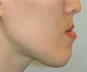
2 MICROGNATHIA / RETROGNATHISM Retrognathism: is a type of malocclusion which refers to an abnormal posterior positioning of the maxilla or mandible, particularly the mandible, relative to the facial skeleton and soft tissues. Micrognathia (undersized jaw) Rarely maxillary anteroposition or hyperplasia Micrognathia Potentially life-threatening condition in neonates (airway obstruction!) Frequent spontaneous correction in puberty
 Patau-syndrome (trisomy 13)")
3 MICROGNATHIA Can be part of different syndromes Cri-du-chat syndrome (deletion in Chr 5) Marfan-syndrome Pierre-Robin syndrome - (micrognathia, cleft palate, glossoptosis) - can be part of other syndromes - eating difficulty, difficulty of speach - crowded teeth Progeria Edwards-syndrome (trisomy 18) Patau-syndrome (trisomy 13) Turner-syndrome
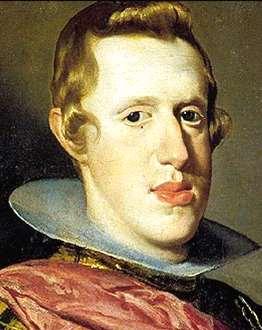

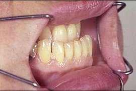
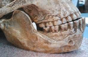
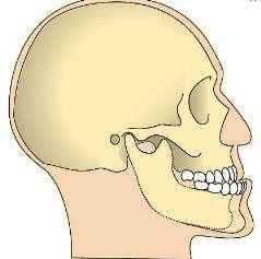
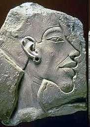
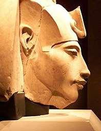
4 PROGNATHISM Angle s classification:malocclusio(iii.cl.) May also be normal variant
 Ferdinand")


5 Spanish Habsburgs V. (Aragon) Ferdinand Castilian Isabel Burgundy Maria I. (Ausztrian) Miksa Mad Johanna I. (Nice) Philip Charles V. Philip II. Philip III. autosomalis domináns prognathia Philip IV. Charles II.
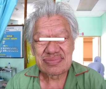
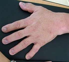
6 ACROMEGALY Teeth malpositioning Malocclusion


7 BENIGN FIBRO-OSSEAL LESIONS Cementoossifying fibroma Fibrous dysplasia Cementoosseous dysplasia
 Osteoprogenitor cells: expansion decreased marrow fibrosis Osteoblasts: abnormal bone matrix production, irregular")

8 FIBROSUS DYSPLASIA Non-hereditary, hamartomatous tissue proliferation Activating mutation in GNAS gene (20q13.3) Osteoprogenitor cells: expansion decreased marrow fibrosis Osteoblasts: abnormal bone matrix production, irregular trabecules Monostotic polyostotic Mainly in childhood or in puberty Slowly growing, painless, spindle-shaped Arrested eruption, movement of teeth Facial asymmetry Maxilla > mandible Mandible: mainly molar/praemolar Maxilla: may spread into the sinuses

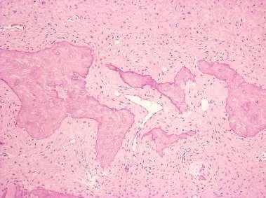
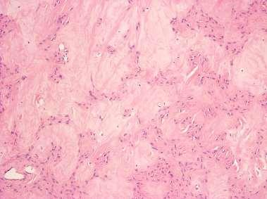
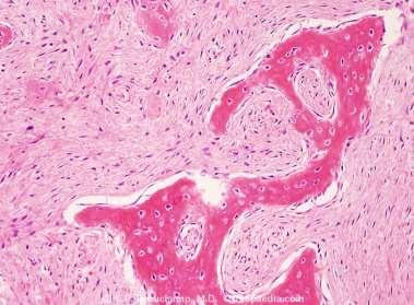
9 FIBROSUS DYSPLASIA


10 FIBROSUS DYSPLASIA Normal bone structure Lamellar bone Fibrous dysplasia: Woven bone


11 X-Ray: variable appearance Ill defined border ( ossifying fibroma) FIBROSUS DYSPLASIA Facultative praeblastomatosis (1 4 %) Radioresistent! (irradiation may cause malignant transformation [fibrosarcoma])
12 OSTEONECROSIS AND OSTEOMYELITIS Osteonecrosis Many underlying etiological factors Medication (drug) related osteonecrosis Irradiation-related necrosis Inflammatory (eg. VZV) Osteomyelitis Unlike in other bones, infection and inflammation of the jaw (osteitis) is a common occurence due to dental infections Dental infections (periodontitis, pulpitis, periapical granuloma)


13 Osteoradionecrosis MANDIBLE NECROSIS 20% following local irradiation endarteritis obliterans end arteries coagulative necrosis sterile, symptomless prone to fracture, infection infected: rapid spread osteomyelitis Bisphosphonate-necrosis (BRONJ Bisphosphonate related ON of the Jaw) trauma necrosis in the devitalized bone frequent after extraction 6-10 % pathological fracture, fistule formation
14 BISPHOSPHONATES Applications: Diseases with bone resorption - malignant tumors with lytic bone met. - Plasma cell myeloma (multiple myeloma ) - osteoporosis P C P basic structure
15 BISPHOSPHONATE MECHANISM decreased production, decreased angiogenesis immune modulation arrested epithel and fibroblast proliferation, delayed wound healing direct toxic effect Absorption with oral administration: ~ 3 % Half-life: can be many decades! (low rate metabolism)
16 BISPHOSPHONATES Osteoclasts: Early cell death, decrease of VEGF-production Osteoblasts: Deregulation of apoptosis, increase of precursor cells Tumor cells: Increased apoptotic rate, inhibition of cell division
17 BRONJ Bisphosphonat-related osteonecrosis of the jaw More frequent in malignant diseases than in osteoporosis Mandible: 70%, maxilla: 30 % Most frequently the molar region is involved Predisposing local factors: extraction implant periapical focus continuous pressure from protesis 75% painful (visible signs of inflammation) Histology: necrosis, inflammatory cells, Actinomyces exclude metastasis! PATIENT Hx!!!


18 BRONJ Bisphosphonat-related osteonecrosis of the jaw 0. st.: aspecific symptoms (toothache, dull bone pain, unexpected tooth loosening) 1. st.: necrotic bone surface, but symptomless 2. st.: necrotic bone surface, + infection, pain 3. st.: as above + fistules, pathologic fracture
19 OSTEOMYELITIS Several classification schemes! 1. Acute osteomyelitis (< 1 month) 2. Chronic osteomyelitis - chronic suppurative (secondary chronic osteomyelitis) (spread from dental focus) - chronic non-suppurative osteomyelitis (no pus, no fistule) 3. Diffuse sclerosing osteomyelitis (unknown etiology) 4. Chronic recurrent multifocal osteomyelitis 5. Garré-osteomyelitis (ossifying periostitis; immature bone tissue outside the cortical) Zurich-classification: Acute osteomyelitis secondary, chronic osteomyelitis Primary chronic osteomyelitis
20 MANDIBULAR CYST Mandibular (jaw) cysts Nasolabial (nasoalveolar) Odontogenic 90% Non odontogenic 10% Aneurysmal bone cyst Inflammatory 60-70% Developmental Nasopalatinal cyst (foramen incisivum)
21 ODONTOGENIC CYSTS Inflammatory cysts Paradental cysts Radicular cysts (60-70%) Developmental cysts Orthokeratinised odontogenic keratocyst Follicular (dentigerous) cyst Primordial cyst Gingival cyst Lateral periodontal cyst Glandularis odontogenic cyst Cystic neoplasms Unicystic ameloblastoma Calcifying cystic odontogenic tumor Odontogenickeratocyst (Keratocystic odontogenic tumor)

 Around necrotised, non-vital tooth")


22 RADICULAR CYST Csont Ciszta Caries 1 apical 2 lateral 3 residual 4 paradental (next to partially erupted wisdom tooth, associated with pericoronitis) Around necrotised, non-vital tooth Periapical granuloma cytokines, growth factors proliferation of rests of Malassez-f. cyst formation > Degeneration, death of the central cells, microcysts, confluent microcysts > Infected root canal, bacteria, lytic effect


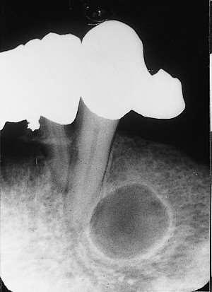
23 RADICULAR CYST Radicular cyst Residual cyst

24 RADICULAR CYST


25 RADICULAR CYST gr Malassezrests Malassezrests 1. Periapical granuloma, bacterial endotoxins (mitogenic effect on the epithel cells + cytokines) - IL-1, IL-6, TNF, PDGF, TGF - 2. Center: hypoxic necrosis, high protein content 3. Osmotic fluid influx more necrosis more elevatad protein content larger cavityüreg 4. Central cavitation becomes independent of the inflammation 5. May result in a bone resorption


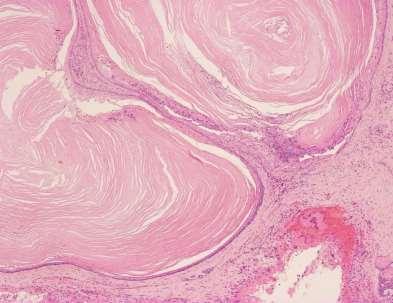
26 RADICULAR CYST Hyperplastic, non-keratinizing squamous epithelium Rushton-bodies (10 %) Cholesterin clefts Chronic inflammatory stroma


 Inflammation is usually")
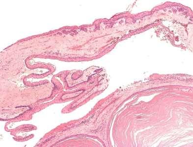
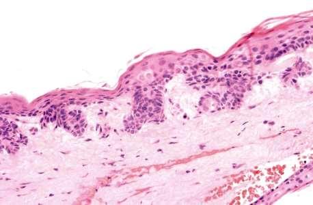
27 FOLLICULAR (DENTIGEROUS) CYST Encloses part of all of the crown of an unerupted tooth Mechanism is largely unknown Wide age distribution Painless Most frequent at the area of molar 3 Unilocular 4-5 layers, non-keratinizing squamous epithelium ( mucinous metaplasia) Inflammation is usually absent
 Teenagers, young adults frequently")

 (ddg.")
 May be secondarily inflamed No association with")
28 KERATOCYSTS Odontogenic keratocyst Orthokeratinized odontogenic keratocyst (WHO: keratocystic odontogenic tumor) Teenagers, young adults frequently more agressive than other cystsl Male predominance (60-70%) more frequent recurrance rate Mainly in the posterior part of mandible may be associated with nevoid basal cell cc syndr.l 70% associated with impacted tooth 65-75% mandible (mainly molar) (ddg.: follicular cyst) Young adults Usually unilocular Unilateral, sclerotic rim Rare recurrence rate ( 3 %) May be secondarily inflamed No association with Gorlin-syndrome Pk Gr Ø K Gr B Pap Ø ConT B Pap Ø ConT
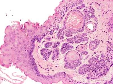
29 KERATOCYSTIC ODONTOGENIC TUMOR


30 TORUS (EXOSTOSIS) Palate or mandible Following chronic, local traumas or surgical interventions Mandible: lingual surface, praemolar region frequently bilateral may be multiple slowly growing bony mass

 hemangioma chondroma 2.")

31 MANDIBULA DAGANATAI Rare 1. ossifying fibroma - circumscribed, usually encapsulated (ddg.: fibrous dysplasia) hemangioma chondroma 2. osteosarcoma, chrondrosarcoma, plasmocytoma, Langerhans-cell histiocytosis 3. Metastatic tumors (1% of malignant tumors) breast, lung, kidney osteolytic, osteoblastic, mixed Birbeck granules
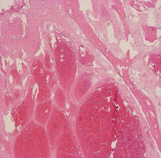
32 HAEMANGIOMA CAVERNOSUM


33 Maxillary osteosarcoma Intraosseal squamous cell cc.
34 THANK YOU!
Origin of Odontogenic Cysts & Tumors
 Origin of Odontogenic Cysts & Tumors Odontogenic Apparatus Origin of Odontogenic Cysts & Tumors Odontogenic Apparatus Remnants of dental lamina Reduced enamel epithelium Odontogenic rests Basal cell layer
RADIOGRAPHIC INTERPRETATION Differential Diagnosis
 RADIOGRAPHIC INTERPRETATION Differential Diagnosis MODULE 1: The Introduction. Chief complaint Demographics Age Sex Race Historical findings Physical findings Clinical Radiographic Location Maxilla/mandible
The clinical appearance and diagnosis of odontogenic cysts. SE Arc-Állcsont-Szájsebészeti és Fogászati Klinika BUDAPEST
 The clinical appearance and diagnosis of odontogenic cysts SE Arc-Állcsont-Szájsebészeti és Fogászati Klinika BUDAPEST DEFINITION A cyst is a sac with walls of connective tissue, lined by epithelium, containing
Disclosure. Educational Objectives. Terminology. Odontogenic Cysts. Terminology
 Disclosure Lisa J. Koenig BChD, DDS, MS Professor & Program Director, Oral Medicine and Oral Radiology Marquette University School of Dentistry Consultant to Soredex for the Scanora 3D and 3Dx Author/Editor
Problem diagnoses. Current issues in Anatomic pathology. Problem Diagnoses in Tumors of the Oral Cavity 5/29/2009
 Current issues in Anatomic pathology Problem Diagnoses in Tumors of the Oral Cavity Richard Jordan DDS PhD FRCPath Professor of Oral Pathology & Pathology Director, UCSF Oral Pathology Diagnostic Laboratory
IMAGING OF CYSTS OF THE JAWS July 2002 N. Serman
 IMAGING OF CYSTS OF THE JAWS July 2002 N. Serman This is an area where radiology plays an important role in assisting with the diagnosis, determining the size of the lesion and the relationship to adjacent
Inter-radicular Radiolucencies
 Inter-radicular Radiolucencies Differential Diagnosis Laterally Displaced Radicular Cyst Accessory canals Root fracture Lateral Periodontal Cyst Botryoid variant Odontogenic Keratocyst Incisive Canal Cyst
DISEASES OF THE JAWS I
 DISEASES OF THE JAWS I ODONTOGENIC AND PERIODONTAL INFECTIONS ODONTOGENIC INFECTIONS PERIAPICAL GRANULOMA PERIAPICAL ABSCESS APICAL PERIODONTAL CYST PHOENIX ABSCESS FISTULA, DRAINING SINUS SPACE INFECTIONS
Index. oralmaxsurgery.theclinics.com. Note: Page numbers of article titles are in boldface type.
 Index Note: Page numbers of article titles are in boldface type. A Adenomatoid odontogenic tumor, pediatric, 50 51 Ameloblastic carcinoma, pediatric, 17, 49 Ameloblastic fibro-odontoma, pediatric, 54 Ameloblastic
Study of maxillary and mandibular cystic lesions
 Study of maxillary and mandibular cystic lesions Poster No.: C-1428 Congress: ECR 2013 Type: Educational Exhibit Authors: M. L. Rozas Rodríguez, M. E. Banegas Illescas, M. Y. Torres Sousa, R. M. Fernández
The resident will be assigned to be on call with the Oral and Maxillofacial service. Call will be set according to PARO guidelines.
 Goals and Objectives for the Otolaryngology-Head & Neck Resident on the Oral and Maxillofacial Surgery (OMFS) Rotation St. Catharines General Hospital (1 four-week rotational block) During the second year
Large Dentigerous Cyst
 Volume 16.2.1 Feb 2016 This Lecture Series qualifies for 0.5 Informal CPD Learning Hours Large Dentigerous Cyst By Dr Hassem Geha A 55 year-old male presented with a painless swelling in the right mandible.
Vascular. Extravasated blood. Melanocytic. Tattoo. Epidermolysis bullosa. Lichen planus. Pemphigoid Pemphigus Lupus. Candidosis. Surface Epithelial
 Oral Soft Tissue Pathology Epithelial Thickening (white) Combination Erythema migrans Epithelial atrophy (red) Surface Lesions Clinical Impression Enlargements Surface Debris Pigmented Vesicular Ulcerated
Pre-reading - radiolucencies
 Pre-reading - radiolucencies Multiple radiolucencies o Suggests a systemic cause o Most likely: cherubism or KCOT s of nevoid basal cell carcinoma syndrome o Sometimes: florid osseous dysplasia (if limited
Inherited & developmental disorders:
 Inherited & developmental disorders: Osteogenesis Imperfecta: Excessive fragility of bone Defect in synthesis of type I collagen Inadequate formation of bone generalized osteoporosis Slender and fracture
Metabolic & Endocrine disorders of bone:
 Metabolic & Endocrine disorders of bone: Osteoporosis: Bone apposition < bone resorption Risk factors: Postmenopausal women Hyperthyroidism Hyperparathyroidism Cushing s syndrome bone quantity: thin cortex
Common/Important Radiolucencies. B. Most Common Location Apex of permanent first molar, rare in primary teeth.
 Cincinnati Dental Association Breakfast at Tiffany s: The Jewels and Gems of Oral Pathology November 17, 2010 John A. Svirsky, DDS, MEd Virginia Commonwealth University 804-828-0547 FAX: 804-828-6234 EMAIL:
Differential Diagnosis of Radiolucent Lesions of the Jaws
 Differential Diagnosis of Radiolucent Lesions of the Jaws Multilocular Multilocular Radiolucencies Odontogenic Keratocyst Botryoid Odontogenic Cyst Glandular odontogenic Cyst Invasive Ameloblastoma Central
Differential Diagnosis of Oral Masses. Gingival Lesions
 Differential Diagnosis of Oral Masses Gingival Lesions Gingival/Alveolar Ridge Masses Parulis Periodontal Abscess Tori and Exostoses Reactive Proliferations Peripheral Odontogenic Cysts Peripheral Odontogenic
Course Description 343 DDS- Clinical Oral and Maxillofacial Radiology II ( )
") King Saud University College of Dentistry Dept. of Oral Medicine & Diagnostic Sciences Division of Oral & Maxillofacial Radiology Course Description 343 DDS- Clinical Oral and Maxillofacial Radiology II
King Saud University College of Dentistry Dept. of Oral Medicine & Diagnostic Sciences Division of Oral & Maxillofacial Radiology Course Description 343 DDS- Clinical Oral and Maxillofacial Radiology II
IN THE NAME OF GOD. Dr.kheirandish DDS,MSC Oral and maxillofacial pathology
 IN THE NAME OF GOD Dr.kheirandish DDS,MSC Oral and maxillofacial pathology ODONTOGENIC CYSTS AND TUMORS Chapter 15 I. DENTIGEROUS CYST II. III. IV. ERUPTION CYST ODONTOGENIC KERATOCYST Orthokeratinized
4/2/17. Panoramic Radiography: Normal Variants and Pathology. Composite of in-focused and blurred images. It s a type of Tomogram.
 Fundamentals Panoramic Radiography: Normal Variants and Pathology It s a type of Tomogram Jimmie L. Harper D.D.S., M.S. Cincinnati Oral and Maxillofacial Surgery, Inc. Volunteer Assistant Professor, Division
Course Description 343 DDS- Clinical Oral and Maxillofacial Radiology II ( )
") King Saud University College of Dentistry Dept. of Oral Medicine & Diagnostic Sciences Division of Oral & Maxillofacial Radiology Course Description 343 DDS- Clinical Oral and Maxillofacial Radiology II
King Saud University College of Dentistry Dept. of Oral Medicine & Diagnostic Sciences Division of Oral & Maxillofacial Radiology Course Description 343 DDS- Clinical Oral and Maxillofacial Radiology II
University of Palestine. Final Exam 2 ed Semester 2014/2015 Total Grade: 60
 Question One: MCQ: 1- Dens in dent occurs most commonly in the : A- Maxillary canines B- Mandibular premolars C- Mandibular second molars D- Maxillary lateral incisors 2- A cyst occurring under the tongue
10/23/2014. features to image interpretation what to look for and what it means. interpretation vs. diagnosis. science or art? image investigation
 features to image interpretation what to look for and what it means interpretation vs. diagnosis ERNEST LAM, DMD, MSc, PhD, FRCD(C) PROFESSOR AND THE DR. LLOYD & MRS. KAY CHAPMAN CHAIR IN CLINICAL SCIENCES
INFLAMMATORY DENTIGEROUS CYST OR INFLAMMATORY CYSTIC LESIONS OF MIXED DENTITION?: A REPORT OF THREE CASES
 Case Report International Journal of Dental and Health Sciences Volume 03, Issue 03 INFLAMMATORY DENTIGEROUS CYST OR INFLAMMATORY CYSTIC LESIONS OF MIXED DENTITION?: A REPORT OF THREE CASES Pritam K Mankapure
Radiographic features of cysts and benign tumors of the jaws. Cyst. Effects on adjacent structures. Types. Odontogenic Cysts. Non-Odontogenic cysts
 Radiographic features of cysts and benign tumors of the jaws Cyst A Cyst is a benign pathologic cavity filled with fluid, lined by epithelium, and surrounded by a connective tissue wall Steven R. Singer,
SYLLABUS FOR EXAMINATION OF PRECLINICS IN ORAL AND MAXILLOFACIAL SURGERY
 SYLLABUS FOR EXAMINATION OF PRECLINICS IN ORAL AND MAXILLOFACIAL SURGERY 1. Asepsis and antisepsis in the oral and maxillofacial surgery. Preparation of the hands of the surgeon and the operative field
Odontomes and Odontogenic tumours
 Odontomes and Odontogenic tumours Odontomes Developmental hamartoma Hamartoma: normal tissue in abnormal location Any cells to be neoplastic it must be able to replicate, which is not seen in hamartoma
Benign Fibro-osseous Lesions
 Benign Fibro-osseous Lesions Plus Vision is the art of seeing things invisible. Jonathan Swift 1667-1745 Steven R. Singer, DDS srs2@columbia.edu 212.305.5674 Benign Fibro-osseous Lesions A group of lesions
Simple diagnostic approach for mandible and maxilla lesions.
 Simple diagnostic approach for mandible and maxilla lesions. Poster No.: C-1285 Congress: ECR 2015 Type: Educational Exhibit Authors: A. Gargallo Vaamonde, A. Burguete, N. Baraibar Argota, M. M. 1 2 1
Role of MDCT and VR reconstructions in the diagnosis and characterization of maxillary cystic lesions.
 Role of MDCT and VR reconstructions in the diagnosis and characterization of maxillary cystic lesions. Poster No.: C-0704 Congress: ECR 2011 Type: Scientific Exhibit Authors: M. Coronella 1, P. V. Foti
POST-TEST FOR UNIT 10: Jawbone Osteonecrosis
 POST-TEST FOR UNIT 10: Jawbone Osteonecrosis This is a printable version of the Unit 10 Test for IAOMT Accreditation. It is for your records only. To achieve credit, you MUST TAKE THIS TEST ONLINE AT https://www.cvent.com/d/tvq54j
高雄醫學大學 口腔醫學院 口腔病理影像科 牙科 X 光影像判讀 教學範例
 高雄醫學大學 口腔醫學院 口腔病理影像科 牙科 X 光影像判讀 教學範例 Content Image No. 001 Dentigerous cyst over left upper embedded canine--------------------- 頁 1 Image No. 002---------------------------------------------------------------
CYSTS OF THE JAW AND SOFT TISSUES
 CYSTS OF THE JAW AND SOFT TISSUES Definition: it is a pathological cavity lined by epithelium containing fluid or semifluid (true cyst). If the cyst not lined by epithelium it is pseudo- cyst. Classification
Surgical management of Ossifying Fibroma of the mandible with inferior alveolar nerve involvement
 Yadegari A,et al J Res Dentomaxillofac Sci e(issn): 2383-2754 Journal of Research in Dental and Maxillofacial Sciences Surgical management of Ossifying Fibroma of the mandible with inferior alveolar nerve
Maxilla and mandible benign lesions: Radiologic Findings and Differential Diagnosis in CT
 Maxilla and mandible benign lesions: Radiologic Findings and Differential Diagnosis in CT Poster No.: C-0964 Congress: ECR 2012 Type: Scientific Exhibit Authors: N. Lopez 1, E. Marcos Naranjo 2, M. D.
Jaws: Cysts and Odontogenic Neoplasms
 Topic 10: Jaw Cysts General Features of Jaw Cysts Sources of Epithelium in Cysts Radiographic Features of Jaw Cysts Microscopic Features of Jaw Cysts Treatment and Prognosis of Jaw Cysts Classification
MALIGNANT TUMOURS OF THE JAWS
 MALIGNANT TUMOURS OF THE JAWS MALIGNANT TUMOURS OF THE JAWS Squamous cell carcinoma Osteogenic sarcoma Chondrosarcoma Fibrosarcoma Malignant lymphomas (incl. Burkitt s) Multiple myeloma Ameloblastoma Secondary
Chapter 5. Developmental Disorders. Copyright 2014, 2009, 2004, 2000, 1996, 1992 by Saunders, an imprint of Elsevier Inc 1
 Chapter 5 Developmental Disorders Copyright 2014, 2009, 2004, 2000, 1996, 1992 by Saunders, an imprint of Elsevier Inc 1 Outline Ø Embryonic Development of the Face, Oral Cavity, and Teeth Ø Developmental
Index. Note: Page numbers of article titles are in boldface type.
 Note: Page numbers of article titles are in boldface type. A Actinomycosis, 200 201 Adenoid cystic carcinoma, 148 150 Adenomatoid odontogenic tumors, 134, 135 Ameloblastic fibro-odontoma, 134 Ameloblastoma,
Epithelial Sources. Rests of Serres Rests of Malassez Reduced Enamel Epithelium Surface Mucosa
 ODONTOGENIC CYSTS Epithelial Sources Rests of Serres Rests of Malassez Reduced Enamel Epithelium Surface Mucosa Epithelial Sources Surface Epithelium Rests of Serres Reduced Enamel Epithelium Rests of
Incidental finding of dentigerous cyst - a case report
 Case Report Incidental finding of dentigerous cyst - a case report Pulivarthi Sushma 1, Sowbhagya M.B 2, Balaji P 3, Mahesh Kumar T.S 4 1 Postgraduate, 2 Reader, 3 Professor and Head of department, 4 Senior
Differential Diagnosis of Oral Lesions. An Interactive Lecture Using Audience Response Polling. John L. Alonge, MS, DDS
 Differential Diagnosis of Oral Lesions An Interactive Lecture Using Audience Response Polling John L. Alonge, MS, DDS Goals 1. Review the diagnostic process needed to formulate a differential diagnosis
Educational Cases EQA November T.J. Palmer Raigmore Hospital Inverness
 Educational Cases EQA November 2013 T.J. Palmer Raigmore Hospital Inverness Case 2 Clinical Details Dob 11 February 1951 PMH: 1964 Extraction of 45 aet 13 yr 1966 Cyst between 44 and 46 enucleated 1973
Malignant Lesions Steven R. Singer, DDS
 Definitions Malignant Lesions Steven R. Singer, DDS srs2@columbia.edu 212.305.5674 Malignancies are uncontrolled growths of tissue Primary tumors represent de novo tumors in their initial site Metastatic
PACIFIC JOURNAL OF MEDICAL SCIENCES ISSN:
 PACIFIC JOURNAL OF MEDICAL SCIENCES {Formerly: Medical Sciences Bulletin} ISSN: 2072 1625 Pac. J. Med. Sci. (PJMS) www.pacjmedsci.com. Email: pacjmedsci@gmail.com. ADENOMATOID ODONTOGENIC TUMOR WITH RARE
An Expansile Large Odontogenic Keratocyst Maxilla: A Case Report.
 RESEARCH AND REVIEWS: JOURNAL OF DENTAL SCIENCES An Expansile Large Odontogenic Keratocyst Maxilla: A Case Report. Nasib Chand Khabra 1, Ish Pandhi 1 *, Kiran DN 2, Sunil Alipuria 1, Bhawna Gulati 1, and
Case Report An Unusual Case of Tooth in the Floor of the Orbit: The Libyan Experience
 Case Reports in Dentistry Volume 2012, Article ID 954789, 5 pages doi:10.1155/2012/954789 Case Report An Unusual Case of Tooth in the Floor of the Orbit: The Libyan Experience Y. Naresh Shetty, Irfan Adil
Management of a Dentigerous Cyst Associated with Inverted and Fused Mesiodens: A Rare Case Report
 Management of a Dentigerous Cyst Associated with Inverted and Fused Mesiodens: A Rare Case Report Kiran Patel 1, Nishtha Patel 2, Karthik Venkataraghavan 3 1 Sr. Lecturer, Department of Oral & Maxillofacial
[ 06-10] Dr. B. Siva Reddy, Dr. B. Ajay Reginald, Dr. D. Sireesha, Dr. Meda Samatha India Abstract: Keywords ARTICLE 20/07/ /09/2018
![[ 06-10] Dr. B. Siva Reddy, Dr. B. Ajay Reginald, Dr. D. Sireesha, Dr. Meda Samatha India Abstract: Keywords ARTICLE 20/07/ /09/2018](/thumbs/87/95905171.jpg "[ 06-10] Dr. B. Siva Reddy, Dr. B. Ajay Reginald, Dr. D. Sireesha, Dr. Meda Samatha India Abstract: Keywords ARTICLE 20/07/ /09/2018") Dentigerous Cyst Associated with an Impacted Mesiodens: A Rare Case Report with Review of Literature [PP: 06-10] Dr. B. Siva Reddy, Dr. B. Ajay Reginald, Dr. D. Sireesha, Dr. Meda Samatha, Department of
Dentigerous Cyst Associated with an Impacted Mesiodens: A Rare Case Report with Review of Literature [PP: 06-10] Dr. B. Siva Reddy, Dr. B. Ajay Reginald, Dr. D. Sireesha, Dr. Meda Samatha, Department of
IMAGING NONODONTOGENIC TUMORS OF THE JAWBONES
 IMAGING NONODONTOGENIC TUMORS OF THE JAWBONES N. Serman. Sept, 2002 W. & P Ch. 20 + 21. Stafne & Gibilisco Ch 15 Oral and Maxillofacial diagnostic Imaging Ch. 8 Non agenesis or non eruption of teeth often
MANSOURA UNIVERSITY FACULTY OF DENTISTRY ORAL PATHOLOGY DEPT
 MANSOURA UNIVERSITY FACULTY OF DENTISTRY ORAL PATHOLOGY DEPT THIRD YEAR Course Director: Dr. Nadia M. Lotfy Professor of Oral Pathology Dr. Manal Mohamed Zyada Associate Professor of Oral Pathology Oral
Pictorial Essay. CT of Calcifying Jaw Bone Diseases
 Pictorial Essay CT of Calcifying Jaw one Diseases Koichi Yonetsu 1 and Takashi Nakamura Downloaded from www.ajronline.org by 46.3.204.207 on 01/08/18 from IP address 46.3.204.207. Copyright RRS. For personal
Periapical central giant cell granuloma misdiagnosed as odontogenic cyst
 doi: 10.1111/j.1365-2591.2006.01107.x CLINICAL ARTICLE Periapical central giant cell granuloma misdiagnosed as odontogenic cyst T. Lombardi 1, M. Bischof 1,2, R. Nedir 1,2, D. Vergain 1, C. Galgano 3,
Mousa Al-Abadi. Abd. Kharabsheh. Rand Abu Anzeh
 7 Mousa Al-Abadi Abd. Kharabsheh Rand Abu Anzeh 1 Recap The histological appearance of Giant cell tumor of bone shows only multi-nucleated giant cells. The histological appearance of Aneurysmal bone cyst
Case Report Intraosseous Follicular Adenomatoid Odontogenic Tumour A Case Report
 International Dentistry Volume 2009, Article ID 597483, 4 pages doi:10.1155/2009/597483 Case eport Intraosseous Follicular Adenomatoid Odontogenic Tumour A Case eport Farhan Durrani 1 and oyana Singh 2
AMELOBLASTIC FIBROMA: A RARE CASE REPORT
 Case Report International Journal of Dental and Health Sciences Volume 04, Issue 03 AMELOBLASTIC FIBROMA: A RARE CASE REPORT Namratha Patil 1 1.Sr lecturer, dept of oral medicine and radiology, KAHES VK
Pathology of the Alimentary System. Lecture 3 Teeth, tonsils, salivary glands & tongue
 Systemic Pathology I - VPM 221 Pathology of the Alimentary System Lecture 3 Teeth, tonsils, salivary glands & tongue Enrique Aburto Fall 2014 II. Diseases of teeth & dental tissues Structure & function
SQUAMOUS ODONTOGENIC TUMOUR: REPORT OF FIVE CASES FROM NIGERIA AND REVIEW OF LITERATURE
 African Journal of Oral Health Volume 3 Numbers 1&2, 2006:1-5 REFEREED ARTICLE SQUAMOUS ODONTOGENIC TUMOUR: REPORT OF FIVE CASES FROM NIGERIA AND REVIEW OF LITERATURE Adebiyi K.E., Odukoya O., Taiwo, E.O.
Glandular Odontogenic Cyst Coexisting with a Dentigerous Cyst: Case Report
 SmyrnaMed Case 2018;2(1): 1-5 ISSN (Online): 2564-6869 www.smyrnamed.com Glandular Odontogenic Cyst Coexisting with a Dentigerous Cyst: Case Report Assist.Prof.Dr. Serap Keskin Tunç 1, Dt. Erkan Feslihan
Proceedings of the 36th World Small Animal Veterinary Congress WSAVA
 www.ivis.org Proceedings of the 36th World Small Animal Veterinary Congress WSAVA Oct. 14-17, 2011 Jeju, Korea Next Congress: http://www.ivis.org October 14(Fri) ~ 17(Mon) 2011 ICC Jeju, Korea 2011 WSAVA
Odontogenic Cysts - An Overview
 Namita V Nayyer Michaelina Macluskey and William Keys Odontogenic Cysts - An Overview Abstract: This article aims to discuss the clinical features, radiological assessment, histopathology and management
Six cases report of differential diagnosis of periapical diseases
 Int J Oral Sci (2011) 3: 153-159. www.ijos.org.cn CLINICAL ARTICLE Six cases report of differential diagnosis of periapical diseases Wen-wei Xia, Ya-qin Zhu, Xiao-yi Wang* Department of Endodontics and
INFECTED DENTIGEROUS CYST IN IMPACTED CANINE- A case report
 Case Report INFECTED DENTIGEROUS CYST IN IMPACTED CANINE- A case report Gazala Fatima Parveen, M.D. Akheel 1 Department of oral & maxillofacial surgery, MCDRC Lucknow, U.P.,India 1-Department of Oral &
Tapping the teeth with a tongue depressor will cause tingling sensation because of involvement of the root of the teeth.
 Odontogenic Cysts of upper jaw an analysis March 26, 2013 Rhinology 1 Professor Balasubramanian Thiagarajan Balasubramanian Thiagarajan 1 Stanley Medical College Thiagarajan B. Odontogenic Cysts of upper
Only 40% of the Story
 X-RAY, X-RAY, READ ALL ABOUT IT! The Use and Utility of Dental Radiographs in Practice Lisa Fink, DVM, DAVDC Dentistry & Oral Surgery Service October 4, 2015 Only 40% of the Story Radiographs of teeth
Diagnosing the most common odontogenic cystic and osseous lesions of the jaws for the practicing pathologist
 S96 2017 USCAP, Inc All rights reserved 0893-3952/17 $32.00 Diagnosing the most common odontogenic cystic and osseous lesions of the jaws for the practicing pathologist Robert A Robinson Department of
International Journal of Health Sciences and Research ISSN:
 International Journal of Health Sciences and Research www.ijhsr.org ISSN: 2249-9571 Case Report Orthokeratinized Odontogenic Cyst: A Case Report- A Milder Variant of OKC or an Independent Entity Mariyam
For the Patient: Bisphosphonates and Oral Health in Multiple Myeloma
 For the Patient: Bisphosphonates and Oral Health in Multiple Myeloma Regular dental care is very important for all cancer patients. As soon as possible after your cancer diagnosis, your dentist should
Bone Tumors Clues and Cues
 William Herring, M.D. 2002 Bone Tumors Clues and Cues In Slide Show mode, advance the slides by pressing the spacebar All Photos Retain the Copyright of their Authors Clues by Appearance of Lesion Patterns
Pericoronal radiolucency associated with incomplete crown
 Imaging Science in entistry 2013; 43: 295-301 http://dx.doi.org/10.5624/isd.2013.43.4.295 Pericoronal radiolucency associated with incomplete crown Kyung-Soo Nah 1, * 1 epartment of Oral and Maxillofacial
Primary bone tumors > metastases from other sites Primary bone tumors widely range -from benign to malignant. Classified according to the normal cell
 Primary bone tumors > metastases from other sites Primary bone tumors widely range -from benign to malignant. Classified according to the normal cell counterpart and line of differentiation. Among the
Impacted third molars can be. Multiple Multilocular Dentigerous Cysts with Intra-osseous and Extra-osseous Third Molar Displacement: A Case Report
 Multiple Multilocular Dentigerous Cysts with Intra-osseous and Extra-osseous Third Molar Displacement: A Case Report Kevin E Lung, BSc, DDS, MSc, FRCD(C); Seema Ganatra, BSc, DDS, MSD, FRCD(C); Christopher
Chronic Incisor Periodontal Disease with Cemental Hyperplasia and Hypoplasia in Horses
 Published in IVIS with the permission of the AAEP Close this window to return to IVIS Chronic Incisor Periodontal Disease with Cemental Hyperplasia and Hypoplasia in Horses Robert C. Gregory, DVM; Joanne
The Radiology Assistant : Bone tumor - well-defined osteolytic tumors and tumor-like lesions
 Bone tumor - well-defined osteolytic tumors and tumor-like lesions Henk Jan van der Woude and Robin Smithuis Radiology department of the Onze Lieve Vrouwe Gasthuis, Amsterdam and the Rijnland hospital,
Orthokeratinized Odontogenic Cyst: A Rarity
 aijoc AIJOC Case Report 1 Heena Sonawane, 2 Freny R Karjodkar, 3 Kaustubh Sansare, 4 Nimish Prakash ABSTRACT Orthokeratinized odontogenic cyst (OOC) was first identified as the rare variant of keratocystic
Aggressive Juvenile Ossifying Fibroma of the Anterior Mandible
 Case Report Aggressive Juvenile Ossifying Fibroma of the Anterior Mandible Dr.Ravikumar.R 1, Dr.Raghavendra.K 2, Dr.Santhosh Kumar 3 1 Senior Lecturer, 2 Reader Department of Oral Surgery, Sri Siddhartha
AMERICAN JOURNAL OF BIOLOGICAL AND PHARMACEUTICAL RESEARCH
 AMERICAN JOURNAL OF BIOLOGICAL AND PHARMACEUTICAL RESEARCH e-issn - 2348-2184 Print ISSN - 2348-2176 Journal homepage: www.mcmed.us/journal/ajbpr SOLID AND MULTICYSTIC FOLLICULAR AMELOBLASTOMA - A CASE
Relation between the complex diagnostics and the complications of the wisdom-tooth surgery
 Relation between the complex diagnostics and the complications of the wisdom-tooth surgery Changes of the "surgical profile" at the Dental Surgery Department of the Oral & Maxillofacial Surgery Clinic,
An unusual site of Adenomatoid Odontogenic Tumor: A rare case report
 J. Int Oral Health 2010 Case Report All right reserved An unusual site of Adenomatoid Odontogenic Tumor: A rare case report Sapna Panjwani*, Anjana Bagewadi**, Vaishali Keluskar*** *Post Graduate Student
Keratocystic Odontogenic Tumor : What radiologist needs to know?
 Keratocystic Odontogenic Tumor : What radiologist needs to know? Poster No.: C-0444 Congress: ECR 2014 Type: Authors: Keywords: DOI: Educational Exhibit K. El Karzazi, J. M. Villanueva Rincón, R. Corrales,
COMBINED PERIODONTAL-ENDODONTIC LESION. By Dr. P.K. Agrawal Sr. Prof and Head Dept. Of Periodontia Govt. Dental College, Jaipur
 COMBINED PERIODONTAL-ENDODONTIC LESION By Dr. P.K. Agrawal Sr. Prof and Head Dept. Of Periodontia Govt. Dental College, Jaipur Differential diagnosis For differential diagnostic purposed the endo-perio
A SURVEY OF ORAL AND MAXILLOFACIAL BIOPSIES IN CHILDREN. A SINGLE-CENTER RETROSPECTIVE STUDY OF 20 YEARS IN PELOTAS-BRAZIL
 www.fob.usp.br/jaos or www.scielo.br/jaos J Appl Oral Sci. 2008;6(6):97-402 A SURVEY OF ORAL AND MAXILLOFACIAL BIOPSIES IN CHILDREN. A SINGLE-CENTER RETROSPECTIVE STUDY Giana da Silveira LIMA, Silvia Terra
TRICOLLEGIATE DIPLOMA OF MEMBERSHIP IN ORAL SURGERY (M. Oral Surgery) APPENDIX A LEARNING OUTCOMES & BLUEPRINT
 APPENDIX A LEARNING OUTCOMES & BLUEPRINT") TRICOLLEGIATE DIPLOMA OF MEMBERSHIP IN ORAL SURGERY (M. Oral Surgery) APPENDIX A LEARNING OUTCOMES & BLUEPRINT A - LEARNING OUTCOMES In brackets below the headings are cross references to corresponding
TRICOLLEGIATE DIPLOMA OF MEMBERSHIP IN ORAL SURGERY (M. Oral Surgery) APPENDIX A LEARNING OUTCOMES & BLUEPRINT A - LEARNING OUTCOMES In brackets below the headings are cross references to corresponding
Oral Tumors in Dogs Gingival Enlargement
 Oral Tumors in Dogs Is that lump you re seeing in your dog s mouth normal? Or is it something to be concerned about? The easiest way to know for sure is to have it evaluated by a veterinarian. When you
 www.oralradiologists.com CONE BEAM CT REPORT CASE XXXX Patient information Patient Name: - Referring Doctor: - Patient DOB: - Scan Date: [Start date] Reason for Exam: Maxillary facial pain Doctor Notes:
TRAUMATIC BONE CYST OF IDIOPATHIC ORIGIN? A REPORT OF TWO CASES
 Traumatic Bone Cyst Kumar S. et al 183 CASE REPORT TRAUMATIC BONE CYST OF IDIOPATHIC ORIGIN? A REPORT OF TWO CASES Kumar Satish 1, S. Padmashree 1, Jayalekshmy Rema 1 ABSTRACT BACKGROUND: Traumatic bone
DENTAL RADIOGRAPH INTERPRETATION
 DENTAL RADIOGRAPH INTERPRETATION Brook A. Niemiec, DVM Diplomate, American Veterinary Dental College Fellow, Academy of Veterinary Dentistry www.vetdentaltraning.com www.vetdentalrad.com Interpreting dental
EXTENSIVE PERIAPICAL CYST IN THE MAXILLARY SINUS A CASE REPORT
 May, 2012 INTERNATIONAL DENTAL JOURNAL OF STUDENT S RESEARCH: CASE REPORT Article Code: IDJSR 0002 EXTENSIVE PERIAPICAL CYST IN THE MAXILLARY SINUS A CASE REPORT Authors Bouguezzi Adel, DDS, Resident 1
Case Report An Extrafollicular Adenomatoid Odontogenic Tumor Mimicking a Periapical Cyst
 Hindawi Case Reports in Radiology Volume 2018, Article ID 6987050, 5 pages https://doi.org/10.1155/2018/6987050 Case Report An Extrafollicular Adenomatoid Odontogenic Tumor Mimicking a Periapical Cyst
The future of health is digital
 Dated: XX/XX/XXXX Name: XXXXXXXX XXXXXXXXXXX Birth Date: XX/XX/XXXX Date of scan: XX/XX/XXXX Examination of the anatomical volume: The following structures are reviewed and evaluated for bilateral symmetry,
Panoramic Radiology. Seminars on Maxillofacial Imaging and Interpretation. Bearbeitet von Allan G Farman
 Panoramic Radiology Seminars on Maxillofacial Imaging and Interpretation Bearbeitet von Allan G Farman 1. Auflage 2007. Buch. xiv, 232 S. Hardcover ISBN 978 3 540 46229 3 Format (B x L): 19,3 x 27 cm Gewicht:
Calcifying Cystic Odontogenic Tumor: Review with Discussion
 DOI 10.7603/s40782-014-0006-9 GST International Journal of Advances in edical Research (JAR) Vol.1.1, ay 2014 Calcifying Cystic Odontogenic Tumor: Review with Discussion Dr. Ritika Jindal BDS, 1 Dr. Ravikiran
Radiology. & supporting structures. Lec. 14 Common diseases of teeth Dr. Areej
 Radiology Lec. 14 Common diseases of teeth Dr. Areej & supporting structures A radiograph is only one part of the diagnostic process. Usually one does NOT make a diagnosis solely from a radiograph. A diagnosis
Arun V Subramaniam et al. / International Journal of Biopharmaceutics. 2014; 5(3): International Journal of Biopharmaceutics
: International Journal of Biopharmaceutics") 225 e- ISSN 0976-1047 Print ISSN 2229-7499 International Journal of Biopharmaceutics Journal homepage: www.ijbonline.com IJB ODONTOGENIC KERATOCYSTS: VARIOUS RADIOGRAPHIC APPEARANCES Arun Subramaniam*
225 e- ISSN 0976-1047 Print ISSN 2229-7499 International Journal of Biopharmaceutics Journal homepage: www.ijbonline.com IJB ODONTOGENIC KERATOCYSTS: VARIOUS RADIOGRAPHIC APPEARANCES Arun Subramaniam*
WHO Histological typing of odontogenic tumors, A. Epithelial Odontogenic Tumors
 Cheng-Chung Lin, Prof. in Oral Pathology College of Dental Medicine, KMU 2007 Classification: The following classification is based upon the inductive effect of one dental tissue upon another. In normal
Periodontal Disease. Radiology of Periodontal Disease. Periodontal Disease. The Role of Radiology in Assessment of Periodontal Disease
 Radiology of Periodontal Disease Steven R. Singer, DDS srs2@columbia.edu 212.305.5674 Periodontal Disease! Includes several disorders of the periodontium! Gingivitis! Marginal Periodontitis! Localized