Malignant Lesions Steven R. Singer, DDS
|
|
|
- Sydney Rogers
- 6 years ago
- Views:
Transcription

 2. Sarcomas (mesenchymal origin) 3. Hematopoietic origin 4.")
 Dysphagia (difficulty swallowing) Dysphonia (difficulty speaking)")


1 Definitions Malignant Lesions Steven R. Singer, DDS Malignancies are uncontrolled growths of tissue Primary tumors represent de novo tumors in their initial site Metastatic tumors originate from distant primary growths Malignancies are generally classified by tissue of origin Four Categories 1. Carcinomas (epithelial origin) 2. Sarcomas (mesenchymal origin) 3. Hematopoietic origin 4. Metastases Definitions Clinical Presentation of Malignant Lesions Displaced or mobile teeth Ulceration Foul odor Swelling Paresthesia Dysesthesia Pain Dysgeusia (decreased taste) Dysphagia (difficulty swallowing) Dysphonia (difficulty speaking) Dysphasia (impaired speech) Exposed bone Poorly healing or nonhealing surgical or trauma sites Sensory or neural deficits Weight loss Hemorrhage Clinical Presentation of Malignant Lesions Onset of symptoms of malignancies is often rapid Prevalence of oral malignancies is low. Due to lack of experience, detection is often delayed. This results in larger tumors, metastases, and poorer prognosis Survival rates for oral cancers have not improved over the years. This is thought to be due to late diagnosis The Role of Radiology Initial Diagnosis Spread of the lesion Size and location of the lesion for surgical planning 1


2 Radiographic features of oral malignancies Location Varies depending on the type. Carcinomas: soft tissue locations Sarcomas: mandible and posterior region of jaws Metastatic lesions : common in the posterior mandible and maxilla and within the follicles of developing teeth Radiographic features of malignancies Periphery and shape Ill defined border with lack of cortication and absence of encapsulation. Associated nonhealing soft tissue ulceration and or swelling is highly suggestive Shape is generally irregular Radiographic features of malignancies Internal Architecture As most malignancies do not produce bone or stimulate the formation of reactive bone, internal aspect is typically radiolucent Lesions such as osteosarcomas produce frank sclerosis, whereas some tumors such as prostate and breast metastatic lesions can induce bone formation at distant sites Radiographic features of malignancies Effects on adjacent structures Ill-defined, invasive borders Bone destruction Adapted from White & Pharoah, fifth edition Radiographic features of malignancies Effects on adjacent structures Radiographic features of malignancies Effects on adjacent structures Destruction of cortical borders Soft tissue mass Adapted from White & Pharoah, fifth edition Invasion of PDL Widened PDL space Adapted from White & Pharoah, fifth edition 2


3 Radiographic features of malignancies Effects on adjacent structures Radiographic features of malignancies Effects on adjacent structures Destruction of bone at apices Displacement of developing tooth Adapted from White & Pharoah, fifth edition Teeth appear to float due to bone destruction Adapted from White & Pharoah, fifth edition Radiographic features of malignancies: Effects on cortical bone Cortical bone destruction without periosteal reaction Laminated periosteal reaction + cortical bone destruction Codman s triangle Sunray or sunburst periosteal reaction Adapted from White & Pharoah, fifth edition Codman's triangle Codman's triangle is the radiographic appearance of the rim of new subperiosteal bone which forms when a lesion such as a tumor lifts the periosteum away from the bone. The small triangle of bone is seen at the advancing margin of the lesion. The three main causes for a Codman's triangle are: Ewing's sarcoma Subperiosteal abscess Layering of the new bone may result in an "onion skin" appearance. Source: Carcinomas Lesions of Epithelial Origin 3


4 Malignant tumor from surface epithelium Invades Deeper soft tissue Connective tissue Underlying bone Local and regional nodes Metastases to liver, lung, and skeleton Clinical Appearance Red, white, or mixed lesion Ulcerated Indurated or rolled borders Can be painful or painless Rubbery or hard lymph nodes that are fixed to underlying structures. Usually occurs in patients >50 years More common in males Location Radiographic features Location Often on lateral border of the tongue Therefore, it is seen radiographically in the posterior mandible Lesions in lip and floor of the mouth may invade anterior mandible Ginigival lesions may initially mimic periodontal disease Radiographic features Shape and Borders Commonly irregular and ill-defined borders Finger-like projections demonstrating invasion Occasionally, the lesion may have smooth borders, indicating erosion Pathologic fractures may occur. Sharp, thin edges may be evident Radiographic features Internal architecture Squamous cell carcinoma tends to be completely radiolucent. There may be trapped pieces of residual bone within the lesion 4


5 Radiographic features Effects on adjacent structures Periodontal ligament space will initially appear to widen. Eventually, teeth will appear to float in the lesion, and may be displaced as lesion expands Tumor may spread along the mandibular canal, giving a widened appearance Adjacent cortical borders may be effaced (destroyed) Images courtesy of Ashai University School of Dentistry 5


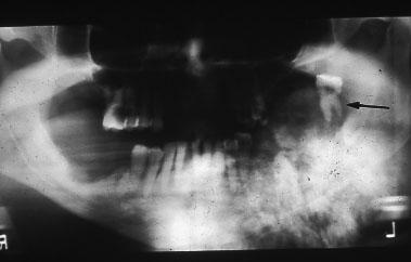
6 Soft tissue origin Invasive from the floor of the mouth Case courtesy of Dr. Maano Milles, Dept of Oral and Maxillofacial Surgery UMDNJ Soft tissue origin Soft tissue origin Presurgical panoramic radiograph Cropped panoramic radiograph Originating in a cyst Uncommon lesion May arise from Periapical inflammatory cysts Residual cysts Dentigerous cysts Odontogenic keratocysts (OKC) Originating in a cyst Clinical Features Pain Dull Several months duration Swelling Pathological fracture Regional lymphadenopathy Maxillary lesions may invade sinus 6


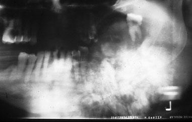
7 Originating in a cyst Location Tooth-bearing areas Most occur in the mandible Shape and Borders Initially indistinguishable from a cyst. Smooth, corticated and hydraulic Advanced lesions are ill-defined, diffuse, and lack cortication Originating in a cyst Internal Architecture Entirely radiolucent Effect on adjacent structures Destroys cortices and adjacent lamina dura of teeth. Capable of destroying alveolar processes Residual cyst with Squamous Cell Carcinoma Residual cyst with Squamous Cell Carcinoma Cropped panoramic radiograph Originating in bone Originating in base of the tongue Adapted from White & Pharoah, fifth edition 7

8 Central Mucoepidermoid Carcinoma Central Mucoepidermoid Carcinoma Epithelial tumor arising in bone Possibly originates from pluripotential odontogenic epithelium or from the lining of a cyst Leaves cortical plates intact Central Mucoepidermoid Carcinoma Clinical Features Mimics benign lesions such as a cyst or tumor Painless swelling May displace teeth or cause asymmetry May cause tenderness or paresthesia More common in females Central Mucoepidermoid Carcinoma Location Twice as common in the mandible than the maxilla Usually in the premolar or molar region Occurs superior to the mandibular canal. This might indicate odontogenic origin Central Mucoepidermoid Carcinoma Borders and shape Unilocular or multilocular mass Thick, corticated borders Internal architecture Multilocular soap bubble appearance similar to ameloblastoma or odontogenic myxoma Septae are from remodeled residual bone Central Mucoepidermoid Carcinoma Effects on adjacent structures Expands buccal and lingual cortices Expands inferior border of mandible May thin or scallop cortices Similar effects to benign tumors 8


 Three major types 1.")
9 Central Mucoepidermoid Carcinoma Central Mucoepidermoid Carcinoma Case courtesy of the Korean Academy of Oral & Maxillofacial Radiology Cropped panoramic radiograph Case courtesy of White & Pharoah, fifth edition The boy is right. We must take a break. Sarcomas Lesions of Mesenchymal Origin Malignant neoplasm of bone New bone is produced by the lesion (not by reactive bone formation of surrounding osteoclasts) Three major types 1. Chondroblastic 2. Osteoblastic 3. Fibroblastic 9

10 Clinical Features Rare. Jaws account for only 7% of all osteosarcomas 2:1 Male: Female ratio Peak in 4 th decade Initially reported due to swelling or bleeding Location More common in the mandible Usually arises in the posterior mandible. The molar areas and ramus are most commonly affected In maxilla, usually arises in the posterior. The ridge, sinus, and palate are most commonly affected Borders and shape Ill-defined Radiolucent without capsule or surrounding osteosclerosis If the periosteum is involved, sunray spicules (aka: hair-on-end trabeculae, or orthoradial striations) may be present Internal architecture May be radiolucent, mixed density, or completely opaque May have varied osseous appearances, such as granular, cotton wool, wisps, etc. In all cases, normal trabeculation is lost Effects on adjacent structures Widening of the PDL Destruction of cortices May destroy or widen the cortex of the inferior alveolar canal Codman s triangles are seen PA View Images courtesy of Nagasaki University School of Dentistry 10


11 Axial CT Bone window Images courtesy of Nagasaki University School of Dentistry Axial CT Soft tissue window Images courtesy of Nagasaki University School of Dentistry Images courtesy of Ashai University School of Dentistry Panoramic View Cropped Panoramic View 11

12 Chondrosarcoma Clinical Features Malignancy of cartilaginous origin Firm to hard bony mass of long duration Four subtypes 1. Clear cell 2. Dedifferentiated 3. Myxoid 4. Mesenchymal Occurs within the bone, peripheral to the bone, or, less commonly, in soft tissue Mean age: 47 yrs Affects males and females equally Cropped Occlusal View Chondrosarcoma Location Unusual in the facial bones. Accounts for only 10% of all cases Occurs equally in maxilla and mandible near cartilage Maxillary lesions tend toward the anterior, while mandibular lesions occur in the coronoid process, head of the condyle and neck, and sometimes in the mandibular symphysis Chondrosarcoma Borders and Shape Round, ovoid, or lobulated Borders can range from smooth and well corticated to indistinct If the periosteum is involved, sunray spicules (aka: hair-on-end trabeculae, or orthoradial striations) Chondrosarcoma Internal architecture May appear as multilocular lucencies to highly calcified lesions. Usual appearance is mixed density Radiographic appearance may be flocculent (snow-like) Moth eaten appearance may be seen, amid islands of unaffected bone Chondrosarcoma Effects on adjacent structures Expand cortical boundaries due to slow growth Can remodel condyle and glenoid fossa Widened PDL and lack of lamina dura of associated teeth 12


13 Chondrosarcoma In general, chondrosarcomas share the general radiographic features of malignant neoplasms Chondrosarcoma Differential Diagnosis Benign fibro osseous lesions Odontogenic myxoma Fibroma Osteoma Ameloblastoma Central bone malignancies Chondrosarcoma Chondrosarcoma Images courtesy Marquette University School of Dentistry Ewing s Sarcoma Ewing s Sarcoma Clinical Features Rare in Jaws Generally found in long bones Origin is uncertain Most common in second decade of life 2:1 M:F ratio Distribution of cases 13


14 Ewing s Sarcoma Location 2:1 mandibular to maxillary cases Found in posterior Lesions start in marrow spaces and expand to involve cortices Ewing s Sarcoma Shape and Borders Poorly demarcated Non-corticated borders There is no typical shape to lesions of Ewing s sarcoma Internal Architecture Radiolucent Ewing s Sarcoma Ewing s Sarcoma Effects on adjacent structures May stimulate the periosteum to lay down new bone in sunray pattern or Codman s triangles Will destroy cortices of normal anatomy such as lamina dura of teeth Ewing s sarcoma of the ramus Ewing s Sarcoma Ewing s Sarcoma Ewing s sarcoma of the fibula Ewing s sarcoma of the fibula 14


15 Ewing s Sarcoma Fibrosarcoma Composed of malignant fibroblasts that produce collagen and elastin Unknown etiology May arise in tissues that have been irradiated Ewing s sarcoma Fibrosarcoma Clinical Features M=F Generally occurs in 4 th decade of life Slowly to rapidly enlarging mass If entirely within bone, the lesion is often painful May exit bone and invade soft tissue, or may begin peripherally Fibrosarcoma Location Mandible Premolar and molar region Borders and Shape Ill-defined, ragged borders Poorly demarcated and non-corticated Non-encapsulated Fibrosarcoma Fibrosarcoma Internal Architecture Usually radiolucent May include reactive bone formation Effects on adjacent structure Destruction Alveolar cortices Inferior cortex of the mandible Follicular cortices Floor of the maxillary sinus Displacement of teeth (rather than resorption) 15


16 Fibrosarcoma Hematopoietic Origin Multiple Myeloma Malignant neoplasm of plasma cells Most common malignancy of bone in adults Multiple Myeloma Clinical features M+F ratio Average age 60 yrs Multiple Myeloma Radiographic features Location Uncommon in the jaws More frequent in the mandible than the maxilla In the mandible, usually found in the posterior body and ramus In the maxilla, it is usually found in posterior areas Multiple Myeloma Radiographic features Shape and Borders Well-defined, punched out lesions Non-corticated borders No bone reaction seen Some lesions have ragged borders, although most are round or ovoid 16


17 Multiple Myeloma Multiple Myeloma Radiographic features Internal architecture No apparent internal architecture Uniformly radiolucent appearance Effects on adjacent structures Teeth may appear excessively opaque, due to the radiolucent appearance of demineralized bone Lamina dura may be lost, along with other cortices Cortical borders may be effaced Multiple Myeloma Multiple Myeloma Case from AAOMR CRC Session 11/04 Non-Hodgkin s Lymphoma Non-Hodgkin s Lymphoma Refers to a family of tumors Composed of malignant cells of the lymphatic system 17


18 Non-Hodgkin s Lymphoma Clinical features Occurs in all age groups, but is uncommon in first decade Teeth may become mobile as bone is lost Patients may feel unwell and lose weight Night sweats are a common pathonuemonic feature of lymphoma Non-Hodgkin s Lymphoma Radiographic features Location Lesions of the head and neck occur in the lymph nodes Extranodal lesions are found in the maxillary sinus, posterior mandible, and maxilla Non-Hodgkin s Lymphoma Radiographic features Shape and Borders Initial lesions are shaped like the host bone Long standing lesions can destroy the cortices of the bone Borders are poorly-defined and demonstrate invasive processes Lesions in a spaces such as the maxillary sinus may have a smooth border Non-Hodgkin s Lymphoma Radiographic features Internal architecture Uniformly radiolucent Effects on adjacent structures May efface the walls of the maxillary sinus Lymphomas grow in the PDL space of teeth May efface the cortices of the follicles of the developing teeth and displace them superiorly Lymphoma Lymphoma v. Leukemia Lymphoma involving the maxillary sinus Case courtesy of the KAOMFR 18


 Common primary sites include: Breast Kidney Lung Colon")
19 Lymphoma v. Leukemia Lymphoma v. Leukemia Case courtesy of the KAOMFR Case courtesy of the KAOMFR Metastases Metastatic Lesions Pet/CT of non-small cell tumor of the lung with metastases Metastatic Lesions Metastatic tumors are foci of malignant disease that originated in a distant primary tumor Usual pathway is through the bloodstream Metastases located in the jaws generally arise from primary tumors located below the clavicles Usually, the primary has been discovered prior to the discovery of jaw metastases Metastatic Lesions Common type of primary tumor is carcinoma (epithelial origin) Common primary sites include: Breast Kidney Lung Colon Rectum Prostate Thyroid stomach Melanoma Testes Bladder Ovary Cervix 19
20 Metastatic Lesions Clinical Features Most common in 5 th to 7 th decade of life Complaints may include: Pain Numbness Paresthesia Bleeding Pathologic fracture of the mandible Metastatic Lesions Location Posterior regions of the jaws More common in: mandible > maxilla >maxillary sinus > anterior hard palate> mandibular condyle Metastases may be bilateral Lesions may be located in the periodontal ligament space. They may be confused with periodontal or apical inflammatory lesions Metastatic Lesions Borders and Shape Moderately well-demarcated Non-corticated borders May also have ill-defined, invasive borders Polymorphous in shape (i.e.: irregular) Metastatic Lesions Internal architecture Lesions are generally lucent Normal trabeculation may be seen, interspersed with radiolucent areas, representing osteolysis. (bone destruction) May be multiple lesions, which may later coalesce Metastatic Lesions Metastatic bone lesions Effects on adjacent structures Effacement of the lamina dura Widening of the PDL space Periosteal reaction. May perforate cortices and form a soft tissue mass extraorally or intraorally Teeth may float in a soft tissue mass and may be displaced 20

 chordoma From:")

21 Metastatic bone lesions Metastatic bone lesions A B C D A= from breast ca, B= from renal cell cal C and D= lesions from gastric ca Metastatic Lesions Age group & above Age Distribution of Common Primary Tumors of Bone Most common benign lesions simple bone cyst eosinophilic granuloma non-ossifying fibroma fibrous dysplasia simple bone cyst aneurysmal bone cyst osteochondroma (exostosis) osteoid osteoma osteoblastoma chondroblastoma chondromyxoid fibroma enchondroma giant cell tumor osteoma Most common malignant tumors Ewing's sarcoma leukemic involvement metastatic neuroblastoma osteosarcoma, Ewing's sarcoma, adamantinoma chondrosarcoma metastatic tumors myeloma leukemic involvement chondrosarcoma osteosarcoma (Paget's associated) chordoma From: Part Four: Time and Eternity XXVII BECAUSE I could not stop for Death, He kindly stopped for me; The carriage held but just ourselves And Immortality. We slowly drove, he knew no haste, And I had put away My labor, and my leisure too, For his civility. We passed the school where children played At wrestling in a ring; We passed the fields of gazing grain, We passed the setting sun. We paused before a house that seemed A swelling of the ground; The roof was scarcely visible, The cornice but a mound. Emily Dickinson Since then t is centuries; but each Feels shorter than the day I first surmised the horses heads Were toward eternity. 21
Bone Tumors Clues and Cues
 William Herring, M.D. 2002 Bone Tumors Clues and Cues In Slide Show mode, advance the slides by pressing the spacebar All Photos Retain the Copyright of their Authors Clues by Appearance of Lesion Patterns
Periodontal Disease. Radiology of Periodontal Disease. Periodontal Disease. The Role of Radiology in Assessment of Periodontal Disease
 Radiology of Periodontal Disease Steven R. Singer, DDS srs2@columbia.edu 212.305.5674 Periodontal Disease! Includes several disorders of the periodontium! Gingivitis! Marginal Periodontitis! Localized
Benign Fibro-osseous Lesions
 Benign Fibro-osseous Lesions Plus Vision is the art of seeing things invisible. Jonathan Swift 1667-1745 Steven R. Singer, DDS srs2@columbia.edu 212.305.5674 Benign Fibro-osseous Lesions A group of lesions
高雄醫學大學 口腔醫學院 口腔病理影像科 牙科 X 光影像判讀 教學範例
 高雄醫學大學 口腔醫學院 口腔病理影像科 牙科 X 光影像判讀 教學範例 Content Image No. 001 Dentigerous cyst over left upper embedded canine--------------------- 頁 1 Image No. 002---------------------------------------------------------------
Disclosure. Educational Objectives. Terminology. Odontogenic Cysts. Terminology
 Disclosure Lisa J. Koenig BChD, DDS, MS Professor & Program Director, Oral Medicine and Oral Radiology Marquette University School of Dentistry Consultant to Soredex for the Scanora 3D and 3Dx Author/Editor
Course Description 343 DDS- Clinical Oral and Maxillofacial Radiology II ( )
") King Saud University College of Dentistry Dept. of Oral Medicine & Diagnostic Sciences Division of Oral & Maxillofacial Radiology Course Description 343 DDS- Clinical Oral and Maxillofacial Radiology II
King Saud University College of Dentistry Dept. of Oral Medicine & Diagnostic Sciences Division of Oral & Maxillofacial Radiology Course Description 343 DDS- Clinical Oral and Maxillofacial Radiology II
Large Dentigerous Cyst
 Volume 16.2.1 Feb 2016 This Lecture Series qualifies for 0.5 Informal CPD Learning Hours Large Dentigerous Cyst By Dr Hassem Geha A 55 year-old male presented with a painless swelling in the right mandible.
AMELOBLASTIC FIBROMA: A RARE CASE REPORT
 Case Report International Journal of Dental and Health Sciences Volume 04, Issue 03 AMELOBLASTIC FIBROMA: A RARE CASE REPORT Namratha Patil 1 1.Sr lecturer, dept of oral medicine and radiology, KAHES VK
IMAGING NONODONTOGENIC TUMORS OF THE JAWBONES
 IMAGING NONODONTOGENIC TUMORS OF THE JAWBONES N. Serman. Sept, 2002 W. & P Ch. 20 + 21. Stafne & Gibilisco Ch 15 Oral and Maxillofacial diagnostic Imaging Ch. 8 Non agenesis or non eruption of teeth often
RADIOGRAPHIC INTERPRETATION Differential Diagnosis
 RADIOGRAPHIC INTERPRETATION Differential Diagnosis MODULE 1: The Introduction. Chief complaint Demographics Age Sex Race Historical findings Physical findings Clinical Radiographic Location Maxilla/mandible
Primary bone tumors > metastases from other sites Primary bone tumors widely range -from benign to malignant. Classified according to the normal cell
 Primary bone tumors > metastases from other sites Primary bone tumors widely range -from benign to malignant. Classified according to the normal cell counterpart and line of differentiation. Among the
Course Description 343 DDS- Clinical Oral and Maxillofacial Radiology II ( )
") King Saud University College of Dentistry Dept. of Oral Medicine & Diagnostic Sciences Division of Oral & Maxillofacial Radiology Course Description 343 DDS- Clinical Oral and Maxillofacial Radiology II
King Saud University College of Dentistry Dept. of Oral Medicine & Diagnostic Sciences Division of Oral & Maxillofacial Radiology Course Description 343 DDS- Clinical Oral and Maxillofacial Radiology II
Differential Diagnosis of Radiolucent Lesions of the Jaws
 Differential Diagnosis of Radiolucent Lesions of the Jaws Multilocular Multilocular Radiolucencies Odontogenic Keratocyst Botryoid Odontogenic Cyst Glandular odontogenic Cyst Invasive Ameloblastoma Central
Differential Diagnosis of Oral Masses. Gingival Lesions
 Differential Diagnosis of Oral Masses Gingival Lesions Gingival/Alveolar Ridge Masses Parulis Periodontal Abscess Tori and Exostoses Reactive Proliferations Peripheral Odontogenic Cysts Peripheral Odontogenic
MALIGNANT TUMOURS OF THE JAWS
 MALIGNANT TUMOURS OF THE JAWS MALIGNANT TUMOURS OF THE JAWS Squamous cell carcinoma Osteogenic sarcoma Chondrosarcoma Fibrosarcoma Malignant lymphomas (incl. Burkitt s) Multiple myeloma Ameloblastoma Secondary
Radiographic features of cysts and benign tumors of the jaws. Cyst. Effects on adjacent structures. Types. Odontogenic Cysts. Non-Odontogenic cysts
 Radiographic features of cysts and benign tumors of the jaws Cyst A Cyst is a benign pathologic cavity filled with fluid, lined by epithelium, and surrounded by a connective tissue wall Steven R. Singer,
Dr.Sepideh Falah-kooshki
 Dr.Sepideh Falah-kooshki MAXILLA Premaxillary/median palatal suture (radiolucent). Incisive fossa and foramen (radiolucent). Nasal passages (radiolucent). Nasal septum (radiopaque). Anterior nasal spine
TRAUMATIC BONE CYST OF IDIOPATHIC ORIGIN? A REPORT OF TWO CASES
 Traumatic Bone Cyst Kumar S. et al 183 CASE REPORT TRAUMATIC BONE CYST OF IDIOPATHIC ORIGIN? A REPORT OF TWO CASES Kumar Satish 1, S. Padmashree 1, Jayalekshmy Rema 1 ABSTRACT BACKGROUND: Traumatic bone
A Case Report of Odontogenic Keratocyst in Anterior Mandibule Position
 A Case Report of Odontogenic Keratocyst in Anterior Mandibule Position Malihe Moeini 1, Seyed Ehsan Anvar 2, Rasool Barzegari Bafghi 3* 1.Resident of Oral and Maxillofacial Radiology, Faculty of Dentistry,
4/2/17. Panoramic Radiography: Normal Variants and Pathology. Composite of in-focused and blurred images. It s a type of Tomogram.
 Fundamentals Panoramic Radiography: Normal Variants and Pathology It s a type of Tomogram Jimmie L. Harper D.D.S., M.S. Cincinnati Oral and Maxillofacial Surgery, Inc. Volunteer Assistant Professor, Division
The Radiology Assistant : Bone tumor - ill defined osteolytic tumors and tumor-like lesions
 Bone tumor - ill defined osteolytic tumors and tumor-like lesions Henk Jan van der Woude and Robin Smithuis Radiology department of the Onze Lieve Vrouwe Gasthuis, Amsterdam and the Rijnland hospital,
Vascular. Extravasated blood. Melanocytic. Tattoo. Epidermolysis bullosa. Lichen planus. Pemphigoid Pemphigus Lupus. Candidosis. Surface Epithelial
 Oral Soft Tissue Pathology Epithelial Thickening (white) Combination Erythema migrans Epithelial atrophy (red) Surface Lesions Clinical Impression Enlargements Surface Debris Pigmented Vesicular Ulcerated
Maxilla and mandible benign lesions: Radiologic Findings and Differential Diagnosis in CT
 Maxilla and mandible benign lesions: Radiologic Findings and Differential Diagnosis in CT Poster No.: C-0964 Congress: ECR 2012 Type: Scientific Exhibit Authors: N. Lopez 1, E. Marcos Naranjo 2, M. D.
Squamous cell carcinoma of the maxillary sinus mimicking periodontitis
 Squamous cell carcinoma of the maxillary sinus mimicking periodontitis 저자저널명발행기관 NDSL URL Na, Ji Yeon ; Kang, Joo Hyun ; Choi, Seong-Ho ; Jeong, Ho-Gul ; Han, Sang-Sun 大韓齒科醫師協會誌 = The journal of the Korean
Inter-radicular Radiolucencies
 Inter-radicular Radiolucencies Differential Diagnosis Laterally Displaced Radicular Cyst Accessory canals Root fracture Lateral Periodontal Cyst Botryoid variant Odontogenic Keratocyst Incisive Canal Cyst
Introduction to Musculoskeletal Tumors. James C. Wittig, MD Orthopedic Oncologist Sarcoma Surgeon
 Introduction to Musculoskeletal Tumors James C. Wittig, MD Orthopedic Oncologist Sarcoma Surgeon www.tumorsurgery.org Definitions Primary Bone / Soft tissue tumors Mesenchymally derived tumors (Mesodermal)
Bone tumors. RMG: jan
 Bone tumors RMG: jan 217. @Kijohs KIZZA JOHN KIJOHS Diseases arising in bone Lipoma Fibrous cortical defects Non-ossifying fibroma Bone island Benign simple cysts Enchondroma Osteochondroma Osteoid osteoma
Bone Tumours - a synopsis. Dr Zena Slim SpR in Histopathology QAH 2009
 Bone Tumours - a synopsis Dr Zena Slim SpR in Histopathology QAH 2009 Aims General approach to diagnosis Common entities.and not so common ones. Mini quiz Challenge of bone tumour diagnosis Bone tumours
Oral Tumors in Dogs Gingival Enlargement
 Oral Tumors in Dogs Is that lump you re seeing in your dog s mouth normal? Or is it something to be concerned about? The easiest way to know for sure is to have it evaluated by a veterinarian. When you
The Radiology Assistant : Bone tumor - well-defined osteolytic tumors and tumor-like lesions
 Bone tumor - well-defined osteolytic tumors and tumor-like lesions Henk Jan van der Woude and Robin Smithuis Radiology department of the Onze Lieve Vrouwe Gasthuis, Amsterdam and the Rijnland hospital,
Pre-reading - radiolucencies
 Pre-reading - radiolucencies Multiple radiolucencies o Suggests a systemic cause o Most likely: cherubism or KCOT s of nevoid basal cell carcinoma syndrome o Sometimes: florid osseous dysplasia (if limited
Metabolic & Endocrine disorders of bone:
 Metabolic & Endocrine disorders of bone: Osteoporosis: Bone apposition < bone resorption Risk factors: Postmenopausal women Hyperthyroidism Hyperparathyroidism Cushing s syndrome bone quantity: thin cortex
The resident will be assigned to be on call with the Oral and Maxillofacial service. Call will be set according to PARO guidelines.
 Goals and Objectives for the Otolaryngology-Head & Neck Resident on the Oral and Maxillofacial Surgery (OMFS) Rotation St. Catharines General Hospital (1 four-week rotational block) During the second year
Common/Important Radiolucencies. B. Most Common Location Apex of permanent first molar, rare in primary teeth.
 Cincinnati Dental Association Breakfast at Tiffany s: The Jewels and Gems of Oral Pathology November 17, 2010 John A. Svirsky, DDS, MEd Virginia Commonwealth University 804-828-0547 FAX: 804-828-6234 EMAIL:
Problem diagnoses. Current issues in Anatomic pathology. Problem Diagnoses in Tumors of the Oral Cavity 5/29/2009
 Current issues in Anatomic pathology Problem Diagnoses in Tumors of the Oral Cavity Richard Jordan DDS PhD FRCPath Professor of Oral Pathology & Pathology Director, UCSF Oral Pathology Diagnostic Laboratory
Index. oralmaxsurgery.theclinics.com. Note: Page numbers of article titles are in boldface type.
 Index Note: Page numbers of article titles are in boldface type. A Adenomatoid odontogenic tumor, pediatric, 50 51 Ameloblastic carcinoma, pediatric, 17, 49 Ameloblastic fibro-odontoma, pediatric, 54 Ameloblastic
IMAGING OF CYSTS OF THE JAWS July 2002 N. Serman
 IMAGING OF CYSTS OF THE JAWS July 2002 N. Serman This is an area where radiology plays an important role in assisting with the diagnosis, determining the size of the lesion and the relationship to adjacent
Skeletal metastases are the most common variety of bone tumors and should always be considered in the differential diagnosis, particularly in older
 Dr Brajesh Nandan Skeletal metastases are the most common variety of bone tumors and should always be considered in the differential diagnosis, particularly in older patients. Cancers of the breast, prostate,
PACIFIC JOURNAL OF MEDICAL SCIENCES ISSN:
 PACIFIC JOURNAL OF MEDICAL SCIENCES {Formerly: Medical Sciences Bulletin} ISSN: 2072 1625 Pac. J. Med. Sci. (PJMS) www.pacjmedsci.com. Email: pacjmedsci@gmail.com. ADENOMATOID ODONTOGENIC TUMOR WITH RARE
Grading of Bone Tumors
 Grading of Bone Tumors Joon Hyuk Choi, M.D. Department of Pathology College of Medicine, Yeungnam University Introduction to grading system of bone tumor used at Mayo Clinic WHO Histologic Classification
The future of health is digital
 Dated: XX/XX/XXXX Name: XXXXXXXX XXXXXXXXXXX Birth Date: XX/XX/XXXX Date of scan: XX/XX/XXXX Examination of the anatomical volume: The following structures are reviewed and evaluated for bilateral symmetry,
Case Report Basal Cell Ameloblastoma of Mandible: A Rare Case Report with Review
 Case Reports in Dentistry Volume 2013, Article ID 187820, 4 pages http://dx.doi.org/10.1155/2013/187820 Case Report Basal Cell Ameloblastoma of Mandible: A Rare Case Report with Review Hemant Shakya, 1
Primary Intra-Osseous Squamous Cell Carcinoma of the Mandible - A Case Report
 IOSR Journal of Dental and Medical Sciences (IOSR-JDMS) e-issn: 2279-0853, p-issn: 2279-0861.Volume 15, Issue 9 Ver. VI (September). 2016), PP 66-70 www.iosrjournals.org Primary Intra-Osseous Squamous
Clinical details: Details of scan: CONE BEAM CT REPORT: Name: H. B. Gender: Reason for referral: Referred by:
 Name: H. B. Gender: Male DOB: 11/12/1950 Age: 64 Date taken: 16/11/2015 Date reported: 19/11/2015 Clinical details: Reason for referral: Referred by: Investigate symptoms related to left TMJ. Reconstructed
Department of Radiology, University of Szeged. Imaging of the skeleton
 Imaging of the skeleton Methods of examination: plain x-ray (radiography, densitometry) x-ray with contrast material (fistulography, angiography) ultrasound (b-mode, Doppler, color, duplex) computed tomography
CASE REPORT. MANDIBULAR OSTEOSARCOMA Ashutosh Chitnis 1, Bhagirath Kandhare 2, Bhavin Patel 3, Divya Bansal 4
 MANDIBULAR OSTEOSARCOMA Ashutosh Chitnis 1, Bhagirath Kandhare 2, Bhavin Patel 3, Divya Bansal 4 HOW TO CITE THIS ARTICLE: Ashutosh Chitnis, Bhagirath Kandhare, Bhavin Patel, Divya Bansal. Mandibular osteosarcoma.
Typical skeletal location and differential diagnosis of bone tumors.
 Typical skeletal location and differential diagnosis of bone tumors. Poster No.: C-2418 Congress: ECR 2015 Type: Educational Exhibit Authors: M. Barros, L. A. Ferreira, Y. Costa, P. J. V. Coelho, F. Caseiro
Radiology. & supporting structures. Lec. 14 Common diseases of teeth Dr. Areej
 Radiology Lec. 14 Common diseases of teeth Dr. Areej & supporting structures A radiograph is only one part of the diagnostic process. Usually one does NOT make a diagnosis solely from a radiograph. A diagnosis
SURGICAL'MANAGEMENT'OF' PLEXIFORM'AMELOBLASTOMA:'A' CASE'REPORT'
 HARSH,Ashutosh* PUROHIT,Pragya* PUROHIT,NC** ADLAKHA,Twisha*** SURGICALMANAGEMENTOF PLEXIFORMAMELOBLASTOMA:A CASEREPORT ABSTRACT Ameloblastoma is a benign but locally invasive neoplasm of odontogenicepithelium.patientsusuallypresentlateafterthetumor
DENTAL RADIOGRAPH INTERPRETATION
 DENTAL RADIOGRAPH INTERPRETATION Brook A. Niemiec, DVM Diplomate, American Veterinary Dental College Fellow, Academy of Veterinary Dentistry www.vetdentaltraning.com www.vetdentalrad.com Interpreting dental
CENTRAL GIANT CELL GRANULOMA PRESENTING AS UNILOCULAR RADIOLUCENCY IN POSTERIOR MANDIBLE A CASE REPORT
 IJCRR Section: Healthcare Sci. Journal Impact Factor 4.016 Case Report CENTRAL GIANT CELL GRANULOMA PRESENTING AS UNILOCULAR RADIOLUCENCY IN POSTERIOR MANDIBLE A CASE REPORT S. Aruleena Shaminey 1, G.
Bubbly Lesions of Bone
 Residents Section Pattern of the Month w79 08.18.09 Eisenberg Residents Section Pattern of the Month Residents inradiology Ronald L. Eisenberg 1 Eisenberg RL Keywords: bubbly lesions, fegnomashic, skeletal
The clinical appearance and diagnosis of odontogenic cysts. SE Arc-Állcsont-Szájsebészeti és Fogászati Klinika BUDAPEST
 The clinical appearance and diagnosis of odontogenic cysts SE Arc-Állcsont-Szájsebészeti és Fogászati Klinika BUDAPEST DEFINITION A cyst is a sac with walls of connective tissue, lined by epithelium, containing
Pericoronal radiolucency associated with incomplete crown
 Imaging Science in entistry 2013; 43: 295-301 http://dx.doi.org/10.5624/isd.2013.43.4.295 Pericoronal radiolucency associated with incomplete crown Kyung-Soo Nah 1, * 1 epartment of Oral and Maxillofacial
10/23/2014. features to image interpretation what to look for and what it means. interpretation vs. diagnosis. science or art? image investigation
 features to image interpretation what to look for and what it means interpretation vs. diagnosis ERNEST LAM, DMD, MSc, PhD, FRCD(C) PROFESSOR AND THE DR. LLOYD & MRS. KAY CHAPMAN CHAIR IN CLINICAL SCIENCES
Differential Diagnosis of Oral Masses. Palatal Lesions
 Differential Diagnosis of Oral Masses Palatal Lesions Palatal Masses Periapical Abscess Torus Palatinus Mucocele Lymphoid Hyperplasia Adenomatous Hyperplasia Benign Salivary Neoplasms Malignant Salivary
Reconstruction of large mandibular defects
 Immediate Reconstruction of a Large Mandibular Defect of Locally Invasive Benign Lesions (A New Method) Gholamreza Shirani, OMFS, DDS, MS,* Mahnaz Arshad, DDS, 1 Farnoush Mohammadi, OMFS, DDS, MS* Tehran,
Ameloblastic Carcinoma of the Mandible: a Rare Case Report
 IBIMA Publishing Journal of Research and Practice in Dentistry http://www.ibimapublishing.com/journals/dent/dent.html Vol. 2015 (2015), Article ID 672596, 6 pages DOI: 10.5171/2015.672596 Case Report Ameloblastic
Aggressive Juvenile Ossifying Fibroma of the Anterior Mandible
 Case Report Aggressive Juvenile Ossifying Fibroma of the Anterior Mandible Dr.Ravikumar.R 1, Dr.Raghavendra.K 2, Dr.Santhosh Kumar 3 1 Senior Lecturer, 2 Reader Department of Oral Surgery, Sri Siddhartha
Inherited & developmental disorders:
 Inherited & developmental disorders: Osteogenesis Imperfecta: Excessive fragility of bone Defect in synthesis of type I collagen Inadequate formation of bone generalized osteoporosis Slender and fracture
Case Report Concurrent Cemento-Osseous Dysplasia and Osteogenic Sarcoma: Report of Two Cases
 Case Reports in Medicine Volume 2012, Article ID 180561, 4 pages doi:10.1155/2012/180561 Case Report Concurrent Cemento-Osseous Dysplasia and Osteogenic Sarcoma: Report of Two Cases A. A. Olusanya, 1 B.
Surgical management of Ossifying Fibroma of the mandible with inferior alveolar nerve involvement
 Yadegari A,et al J Res Dentomaxillofac Sci e(issn): 2383-2754 Journal of Research in Dental and Maxillofacial Sciences Surgical management of Ossifying Fibroma of the mandible with inferior alveolar nerve
IN THE NAME OF GOD. Dr.kheirandish DDS,MSC Oral and maxillofacial pathology
 IN THE NAME OF GOD Dr.kheirandish DDS,MSC Oral and maxillofacial pathology ODONTOGENIC CYSTS AND TUMORS Chapter 15 I. DENTIGEROUS CYST II. III. IV. ERUPTION CYST ODONTOGENIC KERATOCYST Orthokeratinized
Imaging Findings Of Bone Tumors: A Pictorial Review
 Imaging Findings Of Bone Tumors: A Pictorial Review Poster No.: C-2511 Congress: ECR 2015 Type: Educational Exhibit Authors: M. Limeme, N. Benzina, A. BelKhiria, H. Zaghouani, S. Majdoub, N. Mallat, H.
Chapter 3. Neoplasms. Copyright 2015 Cengage Learning.
 Chapter 3 Neoplasms Terminology Related to Neoplasms and Tumors Neoplasm New growth Tumor Swelling or neoplasm Leukemia Malignant disease of bone marrow Hematoma Bruise or contusion Classification of Neoplasms
NEOPLASIA-I CANCER. Nam Deuk Kim, Ph.D.
 NEOPLASIA-I CANCER Nam Deuk Kim, Ph.D. 1 2 Tumor in the hieroglyphics of the Edwin Smith papyrus (1,600 B.C., Breasted s translation 1930) 3 War on Cancer (National Cancer Act, 1971) 4 Cancer Acts in Korea
Neoplasia part I. Dr. Mohsen Dashti. Clinical Medicine & Pathology nd Lecture
 Neoplasia part I By Dr. Mohsen Dashti Clinical Medicine & Pathology 316 2 nd Lecture Lecture outline Review of structure & function. Basic definitions. Classification of neoplasms. Morphologic features.
MARK D. MURPHEY MD, FACR. Physician-in-Chief, AIRP. Chief, Musculoskeletal Imaging
 ALPHABET SOUP AND CYSTIC LESIONS OF THE BONE MARK D. MURPHEY MD, FACR Physician-in-Chief, AIRP Chief, Musculoskeletal Imaging ALPHABET SOUP AND CYSTIC LESIONS OF THE BONE Giant cell tumor (GCT) Unicameral
SESSION 1: GENERAL (BASIC) PATHOLOGY CONCEPTS Thursday, October 16, :30am - 11:30am FACULTY COPY
 PATHOLOGY CONCEPTS Thursday, October 16, :30am - 11:30am FACULTY COPY") SESSION 1: GENERAL (BASIC) PATHOLOGY CONCEPTS Thursday, October 16, 2008 9:30am - 11:30am FACULTY COPY GOAL: Describe the basic morphologic (structural) changes which occur in various pathologic conditions.
SESSION 1: GENERAL (BASIC) PATHOLOGY CONCEPTS Thursday, October 16, 2008 9:30am - 11:30am FACULTY COPY GOAL: Describe the basic morphologic (structural) changes which occur in various pathologic conditions.
MULTILOCULAR AMELOBLASTOMA OF MANDIBLE-A CASE REPORT
 International Journal of Advancements in Research & Technology, Volume 2, Issue2, February-2013 1 MULTILOCULAR AMELOBLASTOMA OF MANDIBLE-A CASE REPORT DR SANTOSH KUMAR SUBUDHI*, DR SUMIT DASH**, DR.K..PREMANANDA***,
Arun V Subramaniam et al. / International Journal of Biopharmaceutics. 2014; 5(3): International Journal of Biopharmaceutics
: International Journal of Biopharmaceutics") 225 e- ISSN 0976-1047 Print ISSN 2229-7499 International Journal of Biopharmaceutics Journal homepage: www.ijbonline.com IJB ODONTOGENIC KERATOCYSTS: VARIOUS RADIOGRAPHIC APPEARANCES Arun Subramaniam*
225 e- ISSN 0976-1047 Print ISSN 2229-7499 International Journal of Biopharmaceutics Journal homepage: www.ijbonline.com IJB ODONTOGENIC KERATOCYSTS: VARIOUS RADIOGRAPHIC APPEARANCES Arun Subramaniam*
Gorham Disease an Enigma
 Article ID: WMC002898 ISSN 2046-1690 Gorham Disease an Enigma Corresponding Author: Dr. Prashant Kothari, Asso Professor, ODMR MCDRC, 442301 - India Submitting Author: Dr. Prashant Kothari, Asso Professor,
Case Studies in the Skull Base
 Case Studies in the Skull Base Amy C Tsai, MD Neuroradiology Fellow Department of Radiology and Imaging Sciences University of Utah Health Sciences Center Salt Lake City, Utah, USA No disclosures related
Periapical central giant cell granuloma misdiagnosed as odontogenic cyst
 doi: 10.1111/j.1365-2591.2006.01107.x CLINICAL ARTICLE Periapical central giant cell granuloma misdiagnosed as odontogenic cyst T. Lombardi 1, M. Bischof 1,2, R. Nedir 1,2, D. Vergain 1, C. Galgano 3,
APMA 2018 Radiology Track Bone Tumors When to say Gulp!
 APMA 2018 Radiology Track Bone Tumors When to say Gulp! DANIEL P. EVANS, DPM, FACFAOM Professor, Department of Podiatric Medicine and Radiology Dr. Wm. Scholl College of Podiatric Medicine Conflict of
Odontomes and Odontogenic tumours
 Odontomes and Odontogenic tumours Odontomes Developmental hamartoma Hamartoma: normal tissue in abnormal location Any cells to be neoplastic it must be able to replicate, which is not seen in hamartoma
Peripheral Odontogenic Fibroma: A rare case report
 Annals of Dental Research (2013) Vol 3 (1): 10-14 HATAM Publishers: All Rights Reserved Annals of Dental Research www.hgpub.com www.adres.yolasite.com Case Report Peripheral Odontogenic Fibroma: A rare
Malignant bone tumors. Incidence Myeloma 45% Osteosarcoma 24% Chondrosarcoma 12% Lyphoma 8% Ewing s Sarcoma 7%
 Malignant bone tumors Incidence Myeloma 45% Osteosarcoma 24% Chondrosarcoma 12% Lyphoma 8% Ewing s Sarcoma 7% Commonest primary bone sarcoma is osteosarcoma X ray Questions to ask 1. Solitary or Multiple
COPYRIGHT 2004 BY THE JOURNAL OF BONE AND JOINT SURGERY, INCORPORATED
 84 COPYRIGHT 2004 BY THE JOURNAL BONE AND JOINT SURGERY, INCORPORATED Radiographic Evaluation of Pathological Bone Lesions: Current Spectrum of Disease and Approach to Diagnosis BY BENJAMIN G. DOMB, MD,
J of Evolution of Med and Dent Sci/ eissn , pissn / Vol. 3/ Issue 18/May 05, 2014 Page 4859
 CYSTIC DEGENERATION IN FOLLICULAR AMELOBLASTOMA: A CASE REPORT Neeraj Kumar 1, Niharika Rathore 2, Hemant Shakya 3, Anshuman Jamdade 4, Puneet Chitlangia 5 HOW TO CITE THIS ARTICLE: Neeraj Kumar, Niharika
Keratocystic Odontogenic Tumor : What radiologist needs to know?
 Keratocystic Odontogenic Tumor : What radiologist needs to know? Poster No.: C-0444 Congress: ECR 2014 Type: Authors: Keywords: DOI: Educational Exhibit K. El Karzazi, J. M. Villanueva Rincón, R. Corrales,
Role of MDCT and VR reconstructions in the diagnosis and characterization of maxillary cystic lesions.
 Role of MDCT and VR reconstructions in the diagnosis and characterization of maxillary cystic lesions. Poster No.: C-0704 Congress: ECR 2011 Type: Scientific Exhibit Authors: M. Coronella 1, P. V. Foti
Immunohistochemistry in Bone and Soft Tissue Tumors. Sahar Rassi Zankoul, MD
 Immunohistochemistry in Bone and Soft Tissue Tumors Sahar Rassi Zankoul, MD Introduction Bone tumors represent a wide variety of tumors of various origins and malignant potentials. These different tumor
Pleomorphic adenoma of palate: differential diagnosis and case report
 Case report Pleomorphic adenoma of palate: differential diagnosis and case report 1Dr. Jigna S. Shah, 2 Dr. MonaliPrajapati, 3 Dr. Utsav Bhatt 1Professor & Head, Dept of Oral Medicine & Radiology, Govt.
Squamous Cell Carcinoma Arising in a Residual Cyst: A Case Report
 Squamous Cell Carcinoma Arising in a Residual Cyst: A Case Report Abstract Aim: The purpose of this report is to present a case of squamous cell carcinoma (SCC) arising from a mandibular residual cyst.
A CASE OF A Huge Submandibular Pleomorphic Adenoma
 ISPUB.COM The Internet Journal of Head and Neck Surgery Volume 4 Number 2 S VERMA Citation S VERMA.. The Internet Journal of Head and Neck Surgery. 2009 Volume 4 Number 2. Abstract Pleomorphic adenoma
AMERICAN JOURNAL OF BIOLOGICAL AND PHARMACEUTICAL RESEARCH
 AMERICAN JOURNAL OF BIOLOGICAL AND PHARMACEUTICAL RESEARCH e-issn - 2348-2184 Print ISSN - 2348-2176 Journal homepage: www.mcmed.us/journal/ajbpr SOLID AND MULTICYSTIC FOLLICULAR AMELOBLASTOMA - A CASE
AJCC Staging of Head & Neck Cancer (7 th edition, 2010) -LIP & ORAL CAVITY-
 -LIP & ORAL CAVITY-") TX: primary tumor cannot be assessed T0: no evidence of primary tumor Tis: carcinoma in situ. T1: tumor is 2 cm or smaller AJCC Staging of Head & Neck Cancer (7 th edition, 2010) -LIP & ORAL CAVITY- T2:
TX: primary tumor cannot be assessed T0: no evidence of primary tumor Tis: carcinoma in situ. T1: tumor is 2 cm or smaller AJCC Staging of Head & Neck Cancer (7 th edition, 2010) -LIP & ORAL CAVITY- T2:
Aneurysmal Bone Cyst of Mandible: A Case Report and Review of Literature
 CASE REPORT Aneurysmal Bone Cyst of Mandible: A Case Report and Review of Literature ABSTRACT An aneurysmal bone cyst is a rare benign expanding osteolytic lesion of bone characterized by replacement with
Study of maxillary and mandibular cystic lesions
 Study of maxillary and mandibular cystic lesions Poster No.: C-1428 Congress: ECR 2013 Type: Educational Exhibit Authors: M. L. Rozas Rodríguez, M. E. Banegas Illescas, M. Y. Torres Sousa, R. M. Fernández
Glandular Odontogenic Cyst Coexisting with a Dentigerous Cyst: Case Report
 SmyrnaMed Case 2018;2(1): 1-5 ISSN (Online): 2564-6869 www.smyrnamed.com Glandular Odontogenic Cyst Coexisting with a Dentigerous Cyst: Case Report Assist.Prof.Dr. Serap Keskin Tunç 1, Dt. Erkan Feslihan
Tumors of the Paranasal Sinuses:
 Tumors of the Paranasal Sinuses: Approaches to Diagnostic Imaging Nir J. Harish September 2007 Head and Neck Cancers Oral cavity Pharynx Larynx Nasal cavity Paranasal sinuses Salivary glands Incidence
Neoplasia literally means "new growth.
 NEOPLASIA Neoplasia literally means "new growth. A neoplasm, defined as "an abnormal mass of tissue the growth of which exceeds and is uncoordinated with that of the normal tissues and persists in the
Primary bone tumors according to the WHO classification: a review of 13 years with illustrative examples
 Primary bone tumors according to the WHO classification: a review of 13 years with illustrative examples Poster No.: C-1741 Congress: ECR 2015 Type: Educational Exhibit Authors: J. Silva, M. A. Ramírez
My Journey into the World of Salivary Gland Sebaceous Neoplasms
 My Journey into the World of Salivary Gland Sebaceous Neoplasms Douglas R. Gnepp Warren Alpert Medical School at Brown University Rhode Island Hospital Pathology Department Providence RI Asked to present
Unusual transmigration of canines report of two cases in a family
 ISSN: Electronic version: 1984-5685 RSBO. 2014 Jan-Mar;11(1):88-92 Case Report Article Unusual transmigration of canines report of two cases in a family Sulabha A. Narsapur 1 Sameer Choudhari 2 Shrishal
Case Report Primary Squamous Cell Carcinoma of Lung Leading to Metastatic Jaw Tumor
 Case Reports in Pulmonology, Article ID 392616, 5 pages http://dx.doi.org/10.1155/2014/392616 Case Report Primary Squamous Cell Carcinoma of Lung Leading to Metastatic Jaw Tumor Chintamaneni Raja Lakshmi,
This lecture is sponsored by a grant from the Delta Dental of Iowa Foundation IDA Annual Conference Guest Lecture Series.
 This lecture is sponsored by a grant from the Delta Dental of Iowa Foundation IDA Annual Conference Guest Lecture Series. Radiology: Back to the Classroom! Juan F. Yepes DDS, MD, MPH, MS, DrPH Associate