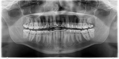
4/2/17. Panoramic Radiography: Normal Variants and Pathology. Composite of in-focused and blurred images. It s a type of Tomogram.
|
|
|
- Scot Singleton
- 5 years ago
- Views:
Transcription


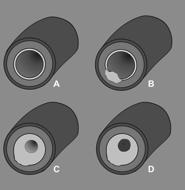
1 Fundamentals Panoramic Radiography: Normal Variants and Pathology It s a type of Tomogram Jimmie L. Harper D.D.S., M.S. Cincinnati Oral and Maxillofacial Surgery, Inc. Volunteer Assistant Professor, Division of Oral & Maxillofacial Surgery, University of Cincinnati path of sliding rotation center anterior rotation center Sliding centre of rotation Three-dimensional curved zone or image layer in which structures are reasonably well defined lateral rotation center Focal trough Formed when the object is in between the center of rotation and image receptor. Composite of in-focused and blurred images 1


2 Cervical vertebrae Body, condyle and ramus of the contralateral side of the mandible Ghost Images Lead Apron Artifact Patient Positioning Errors Cassette Positioning Errors Palate Chin rest (R)or(L) markers of the machine Neck chains Napkin chains Earrings, tongue rings Shoulder straps of protective apron Hyoid bone Double image of vertebral column Cervical spine Inferior border of mandible Earrings Tongue ring Neck chain Chin rest Lead apron (R)or(L)markers of machine Tongue ring Double Images Ghost image of earrings Real images may be double images Double images are formed in zone in central region Common double images include hard palate Positioning/setup errors Know your machine! soft palate hyoid bone 2


3 Common errors Lead apron shadow Shadow of vertebral column, usually from patient not standing straight E.POSITIONING OF THE SPINE If the patient is not sitting or standing with a straight spine, the cervical spine appears as a pyramid shaped radiopacity in the center of the film. POSITIONING OF THE LIPS AND TEETH If the lips are not closed on the bite block, a dark radiolucent shadow obscure the anterior teeth. If the tongue is not in contact with palate, a dark radiolucent shadow obscures the apices of the maxillary teeth. Palatoglossal Air Space Structures smaller on the side to which head is turned; larger on opposite side. 3


4 I.DISTORTION DUE TO PATIENT MOVEMENT Prolonged exposure results in increased horizontal dimension of the image. HEAD TIPPED DOWN Mandibular incisors shortened, V-shaped mandible HEAD TIPPED DOWN HEAD TIPPED UP Squared-off mandible, palate superimposed over maxillary teeth Teeth too posterior Anterior teeth wider and blurred 4


5 Forgot to remove denture Crooked implants? Panoramic Radiography, what are we looking at? Systematic evaluation Is it a good film? Try to evaluate radiographs the same way every time Gestalt Clarity Contrast Symmetry Distortion Look at the films the same way every time Avoid tunnel vision Start on periphery and work your way to center Look at the condyles, spine, orbits, sinuses, nasal cavity, bony borders Use opposite side for comparison Count teeth Evaluate alveolar and basilar bone Identify and describe abnormalities Assess radiograph 5


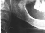
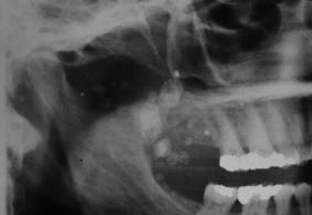
6 Assess radiograph Description of lesions Assessment of Lesion Location Density Size Shape Border Contents Effects on neighboring structures Calcified Stylohyoid Ligament Tonsillolith Lesions in structures adjacent to the jaws AKA is that in the bone? Tonsillolith Tonsillolith Tonsillolith 6

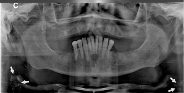
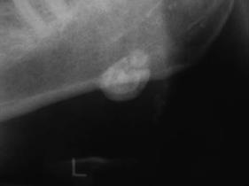
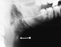
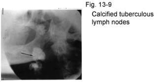
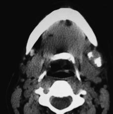
7 Carotid Artery Calcifications Carotid Artery Calcifications Carotid Artery Calcifications Sialolith in Submandibular Gland Calcified Lymph Node Calcified Lymph Nodes Calcified Lymph Node: Scofula Phlebolith in a hemangioma Antral Pseudocyst 7


8 Radicular cyst Trauma Patterns of fracture Description of lesions Location Lesions within the Jawbones Density Size Shape Border Lions and tigers and bears, oh my! Contents Effects on neighboring structures When you hear hoof beats With pathology, think differential diagnosis common things are common 8


")


9 TYPES OF CEMENTO-OSSEOUS DYSPLASIAS: - Based on their clinical and radiological features, grouped into 1. Periapical cemento-osseous dysplasia 2. Focal cemento-osseous dysplasia 3. Florid cemento-osseous dysplasia Radiography: well defined radioopaque up to 3 cm below the apex with intact lamina dura Focal sclerosing osteomyelitis Histopathology Cemento-osseous dysplasia Periapical Cemento-osseous dysplasia. 1. Mandibular incisor 2. More than one teeth is affected. 3. Tooth vital 4. More common in black female. 5. Middle age ( around 40) and rare before Asymptomatic, typical discovered on routine radiographic examination A mixture of benign fibrous tissue, trabeculae of bone and cemnentum like material 9


10 Periapical Cemento-osseous dysplasia Cemento-osseous dysplasia Radiography: 1.Early lesion appears as rounded radiolucent area related to the apex and continue with PDL. 2.Later produce solid radio-opaque mass Early Late Florid cemento-osseous dysplasia: 1. Aka Gigantiform cementoma 2. Less common 3. Represent an exuberant and severe form of periapical type 4. Middle age black women 5. Typically symmetrical and bilateral Radio-opaque masses with radiolucent porder at the root When not associated with a tooth apex called as focal cementoosseous, dysplasia 6. Some times all four quadrants involved CEMENTOBLASTOMA Treatment is enucleation benign ectomesenchymal neoplasm of cementum and forms a mass of cementumlike tissue as an irregular or rounded mass attached to the root of a tooth, usually a mandibular first molar. mainly affect young adults, particularly males. They are slow-growing and the jaw is not usually expanded. And may rarely causes gross bony swelling and pain Radiographically, there is typically a radiopaque mass with thin radiolucent margin, attached to the roots of a tooth. Resorption of related roots is common, but the tooth remains vital. 10


11 Complex odontoma Disordered mass of dental hard tissue Tooth tissues but not teeth! Most common site are posterior mandible or maxilla Compound odontoma Tiny teeth May be malformed Most common sites are anterior maxilla Well defined radiolucent Lesions Is that a cyst? Radicular cyst 11


12 Nasopalatine Duct Cyst Adenomatoid Odontogenic Tumor adenoameloblastoma,glandular ameloblastoma,adenomatoid ameloblastoma Some consider it as a hamartoma uncommon, accounting for 3 to 7% of all odontogenic tumors. limited to young, extremely uncommon in patients > 30 years. Maxilla > mandible Anterior region > posterior region Female > male rarely exceeding 3 cm in diameter lesion appears as a well-circumscribed unilocular radiolucency that involves the crown of an unerupted tooth, frequently a canine.radiolucency extend beyond the cemento-enamel junction Cysts of the maxillary sinus OKC Fibrous dysplasia A self limiting disease characterized by fibrous replacement of medullary bone by metaplastic woven bone that eventually replaced by dense lamellar bone 1. Monostatic 2. Polystatic 12


13 Fibrous dysplasia Monostatic : 1. More common 70%. 2. Any bone affected 3. In the Jaw bone maxilla is more affected 4. Start in childhood. 5. Slow growing painless, smooth, rounded bony swelling with facial asymmetry. 6. Enlargement can cause malocclusion and displacemnet of teeth and sometimes prevent tooth eruption Monostatic fibrous dysplasia Fibrous dysplasia 7. No pain on palpation 8. Maxillary lesion may cause exophthalmos, proptosis and nasal obstruction. 9. Mandibular lesion occur in molar and premolar region 10. Protuberance and increase the depth of jaw Fibrous dysplasia Radiography:. Radiolucent area with fine orange peal textures. Borders are difficult to define because of gradual transition to normal.. Initially resemble cystlike radiolucencies containing faint bony trabeculae Ground glass appearance Paget s disease of Bone 1. The disease is self limiting 2. Large lesion surgical ecountring Cotton wool appearance Dental radiographs also show the classical cotton wool appearance. Extensive hypercementosis can be noted. 13

 is a well")


14 Multiple Myeloma BRONJ OSSIFYING FIBROMA (Cementifying fibroma / Cemento-ossifying fibroma) Ossifying fibroma (OF) is a well circumscribed, sometimes encapsulated neoplasm composed of fibrous tissue containing varying amounts of calcified material. This calcified material may be bone, cementum like spheruls or a mixture of both. It has been suggested that the origin of the tumor is odontogenic or from periodontal ligaments. But identical tumors have been reported in orbital, frontal, ethmoid, sphenoid and temporal bone, leaving these prior theories of origin open to question. 14


 Malignancy of mesenchymal cells that have the ability to produce osteoid or")
15 Ossifying fibroma JUVENILE OSSIFYING FIBROMA Uncommon lesion of bone. Differentiated from ossifying fibroma on the basis of age incidence, site predilection and clinical behavior. However, histologically the distinction from OF is not so clear. Two patterns recognized trabecular and psammomatoid. Signs & symptoms: CLINICAL FEATURES: - Age incidence: Patients younger than 15 years of age. Sex incidence: Equal. Site predilection: Most commonly involves orbital and frontal bones. Maxilla is involved more commonly. Most tumors show rapid growth. In such cases, pain and paresthesia may be noted. Psammomatoid variant frequently appears outside the jaws, mostly arising in the orbital and frontal bone and paranasal sinuses. Cortical expansion and facial asymmetry is seen with jaw lesions. Orbital and sinus involvement may cause exophthalmus, proptosis and nasal obstruction. OSTEOSARCOMA (Osteogenic sarcoma) Malignancy of mesenchymal cells that have the ability to produce osteoid or immature bone. Commonest malignancy arising within the bone along with hematopoietic neoplasms. Majority arise from within the bone (intramedullary), some may be peripheral (juxtacortical) CLINICAL FEATURES: - Age incidence: 3 rd and 4 th decades. Sex incidence: Commoner in males. Site predilection: Long bones and U / L jaws. 15
 may also be noted.")

 To completely radiopaque Periphery of lesions usually indistinct and ill")


16 Signs & symptoms: Swelling and pain - commonest symptoms. Loosening of teeth, paresthesia and nasal obstruction (in case of maxillary tumors) may also be noted. Treatment For smaller lesions, complete local excision. Rapidly growing lesion, wider resection may be required. Recurence rate is about 30% to 58%. RADIOGRAPHIC FEATURES: - Radiographic features vary from densely sclerotic Mixed radiopacity radiolucency (mottled) To completely radiopaque Periphery of lesions usually indistinct and ill defined. The characteristic sunburst appearance can be noted in about 25% of jaw tumors. Produced by osteophytic bone production. CT Reconstruction of Osteogenic Sarcoma In summary: Start with good radiographs Good radiographs start with good patient positioning Systematically evaluate the radiographs Evaluate pathology based on the seven descriptors Correlate with the clinical exam and history Can the diagnosis be made based on current information For many lesions, biopsy is required to make the final diagnosis 16
Common/Important Radiolucencies. B. Most Common Location Apex of permanent first molar, rare in primary teeth.
 Cincinnati Dental Association Breakfast at Tiffany s: The Jewels and Gems of Oral Pathology November 17, 2010 John A. Svirsky, DDS, MEd Virginia Commonwealth University 804-828-0547 FAX: 804-828-6234 EMAIL:
RADIOGRAPHIC INTERPRETATION Differential Diagnosis
 RADIOGRAPHIC INTERPRETATION Differential Diagnosis MODULE 1: The Introduction. Chief complaint Demographics Age Sex Race Historical findings Physical findings Clinical Radiographic Location Maxilla/mandible
高雄醫學大學 口腔醫學院 口腔病理影像科 牙科 X 光影像判讀 教學範例
 高雄醫學大學 口腔醫學院 口腔病理影像科 牙科 X 光影像判讀 教學範例 Content Image No. 001 Dentigerous cyst over left upper embedded canine--------------------- 頁 1 Image No. 002---------------------------------------------------------------
Disclosure. Educational Objectives. Terminology. Odontogenic Cysts. Terminology
 Disclosure Lisa J. Koenig BChD, DDS, MS Professor & Program Director, Oral Medicine and Oral Radiology Marquette University School of Dentistry Consultant to Soredex for the Scanora 3D and 3Dx Author/Editor
The future of health is digital
 Dated: XX/XX/XXXX Name: XXXXXXXX XXXXXXXXXXX Birth Date: XX/XX/XXXX Date of scan: XX/XX/XXXX Examination of the anatomical volume: The following structures are reviewed and evaluated for bilateral symmetry,
Extraoral Imaging. Chapter 42. Copyright 2018, Elsevier Inc. All Rights Reserved. 1
 Extraoral Imaging Chapter 42 Copyright 2018, Elsevier Inc. All Rights Reserved. 1 Learning Objectives Lesson 42.1: Panoramic Imaging 1. Pronounce, define, and spell the key terms. 2. Discuss panoramic
Large Dentigerous Cyst
 Volume 16.2.1 Feb 2016 This Lecture Series qualifies for 0.5 Informal CPD Learning Hours Large Dentigerous Cyst By Dr Hassem Geha A 55 year-old male presented with a painless swelling in the right mandible.
Course Description 343 DDS- Clinical Oral and Maxillofacial Radiology II ( )
") King Saud University College of Dentistry Dept. of Oral Medicine & Diagnostic Sciences Division of Oral & Maxillofacial Radiology Course Description 343 DDS- Clinical Oral and Maxillofacial Radiology II
King Saud University College of Dentistry Dept. of Oral Medicine & Diagnostic Sciences Division of Oral & Maxillofacial Radiology Course Description 343 DDS- Clinical Oral and Maxillofacial Radiology II
Ossifying fibromas of the jaw bone: 20 cases
 (2010) 39, 57 63 2010 The British Institute of Radiology http://dmfr.birjournals.org CASE REPORT Ossifying fibromas of the jaw bone: 20 cases Y Liu 1, M You 1, H Wang*,1, Z Yang 1, J Miao 1, K Shimizutani
Aggressive Juvenile Ossifying Fibroma of the Anterior Mandible
 Case Report Aggressive Juvenile Ossifying Fibroma of the Anterior Mandible Dr.Ravikumar.R 1, Dr.Raghavendra.K 2, Dr.Santhosh Kumar 3 1 Senior Lecturer, 2 Reader Department of Oral Surgery, Sri Siddhartha
Dr.Sepideh Falah-kooshki
 Dr.Sepideh Falah-kooshki MAXILLA Premaxillary/median palatal suture (radiolucent). Incisive fossa and foramen (radiolucent). Nasal passages (radiolucent). Nasal septum (radiopaque). Anterior nasal spine
Course Description 343 DDS- Clinical Oral and Maxillofacial Radiology II ( )
") King Saud University College of Dentistry Dept. of Oral Medicine & Diagnostic Sciences Division of Oral & Maxillofacial Radiology Course Description 343 DDS- Clinical Oral and Maxillofacial Radiology II
King Saud University College of Dentistry Dept. of Oral Medicine & Diagnostic Sciences Division of Oral & Maxillofacial Radiology Course Description 343 DDS- Clinical Oral and Maxillofacial Radiology II
Surgical management of Ossifying Fibroma of the mandible with inferior alveolar nerve involvement
 Yadegari A,et al J Res Dentomaxillofac Sci e(issn): 2383-2754 Journal of Research in Dental and Maxillofacial Sciences Surgical management of Ossifying Fibroma of the mandible with inferior alveolar nerve
Pre-reading - radiolucencies
 Pre-reading - radiolucencies Multiple radiolucencies o Suggests a systemic cause o Most likely: cherubism or KCOT s of nevoid basal cell carcinoma syndrome o Sometimes: florid osseous dysplasia (if limited
Pictorial Essay. CT of Calcifying Jaw Bone Diseases
 Pictorial Essay CT of Calcifying Jaw one Diseases Koichi Yonetsu 1 and Takashi Nakamura Downloaded from www.ajronline.org by 46.3.204.207 on 01/08/18 from IP address 46.3.204.207. Copyright RRS. For personal
Benign Fibro-osseous Lesions
 Benign Fibro-osseous Lesions Plus Vision is the art of seeing things invisible. Jonathan Swift 1667-1745 Steven R. Singer, DDS srs2@columbia.edu 212.305.5674 Benign Fibro-osseous Lesions A group of lesions
Maxilla and mandible benign lesions: Radiologic Findings and Differential Diagnosis in CT
 Maxilla and mandible benign lesions: Radiologic Findings and Differential Diagnosis in CT Poster No.: C-0964 Congress: ECR 2012 Type: Scientific Exhibit Authors: N. Lopez 1, E. Marcos Naranjo 2, M. D.
PACIFIC JOURNAL OF MEDICAL SCIENCES ISSN:
 PACIFIC JOURNAL OF MEDICAL SCIENCES {Formerly: Medical Sciences Bulletin} ISSN: 2072 1625 Pac. J. Med. Sci. (PJMS) www.pacjmedsci.com. Email: pacjmedsci@gmail.com. ADENOMATOID ODONTOGENIC TUMOR WITH RARE
Differential Diagnosis of Radiolucent Lesions of the Jaws
 Differential Diagnosis of Radiolucent Lesions of the Jaws Multilocular Multilocular Radiolucencies Odontogenic Keratocyst Botryoid Odontogenic Cyst Glandular odontogenic Cyst Invasive Ameloblastoma Central
[ 06-10] Dr. B. Siva Reddy, Dr. B. Ajay Reginald, Dr. D. Sireesha, Dr. Meda Samatha India Abstract: Keywords ARTICLE 20/07/ /09/2018
![[ 06-10] Dr. B. Siva Reddy, Dr. B. Ajay Reginald, Dr. D. Sireesha, Dr. Meda Samatha India Abstract: Keywords ARTICLE 20/07/ /09/2018](/thumbs/87/95905171.jpg "[ 06-10] Dr. B. Siva Reddy, Dr. B. Ajay Reginald, Dr. D. Sireesha, Dr. Meda Samatha India Abstract: Keywords ARTICLE 20/07/ /09/2018") Dentigerous Cyst Associated with an Impacted Mesiodens: A Rare Case Report with Review of Literature [PP: 06-10] Dr. B. Siva Reddy, Dr. B. Ajay Reginald, Dr. D. Sireesha, Dr. Meda Samatha, Department of
Dentigerous Cyst Associated with an Impacted Mesiodens: A Rare Case Report with Review of Literature [PP: 06-10] Dr. B. Siva Reddy, Dr. B. Ajay Reginald, Dr. D. Sireesha, Dr. Meda Samatha, Department of
 www.oralradiologists.com CONE BEAM CT REPORT CASE XXXX Patient information Patient Name: - Referring Doctor: - Patient DOB: - Scan Date: [Start date] Reason for Exam: Maxillary facial pain Doctor Notes:
INTERPRETATION RADIOGRAPHIC INTERPRETATION. Law of Symmetry. We will be reviewing: 7/30/16
 RADIOGRAPHIC INTERPRETATION Pam Wood, CDA, RDH, M.Ed, CAGS Community College of Rhode Island pwood@ccri.edu INTERPRETATION the ability to read what is revealed on a dental radiograph any dental professional
10/23/2014. features to image interpretation what to look for and what it means. interpretation vs. diagnosis. science or art? image investigation
 features to image interpretation what to look for and what it means interpretation vs. diagnosis ERNEST LAM, DMD, MSc, PhD, FRCD(C) PROFESSOR AND THE DR. LLOYD & MRS. KAY CHAPMAN CHAIR IN CLINICAL SCIENCES
IMAGING OF CYSTS OF THE JAWS July 2002 N. Serman
 IMAGING OF CYSTS OF THE JAWS July 2002 N. Serman This is an area where radiology plays an important role in assisting with the diagnosis, determining the size of the lesion and the relationship to adjacent
Inter-radicular Radiolucencies
 Inter-radicular Radiolucencies Differential Diagnosis Laterally Displaced Radicular Cyst Accessory canals Root fracture Lateral Periodontal Cyst Botryoid variant Odontogenic Keratocyst Incisive Canal Cyst
Cemento-Ossifying Fibroma A Radiographic Diagnostic Dilemma
 Case Report Cemento-Ossifying Fibroma A Radiographic Diagnostic Dilemma Zahra Dalili 1, Somayeh Nemati 2, Fatemeh Shahsavari 3 1 Associate Professor, Department of Maxillofacial Radiology, Guilan University
Problem diagnoses. Current issues in Anatomic pathology. Problem Diagnoses in Tumors of the Oral Cavity 5/29/2009
 Current issues in Anatomic pathology Problem Diagnoses in Tumors of the Oral Cavity Richard Jordan DDS PhD FRCPath Professor of Oral Pathology & Pathology Director, UCSF Oral Pathology Diagnostic Laboratory
Inherited & developmental disorders:
 Inherited & developmental disorders: Osteogenesis Imperfecta: Excessive fragility of bone Defect in synthesis of type I collagen Inadequate formation of bone generalized osteoporosis Slender and fracture
Vascular. Extravasated blood. Melanocytic. Tattoo. Epidermolysis bullosa. Lichen planus. Pemphigoid Pemphigus Lupus. Candidosis. Surface Epithelial
 Oral Soft Tissue Pathology Epithelial Thickening (white) Combination Erythema migrans Epithelial atrophy (red) Surface Lesions Clinical Impression Enlargements Surface Debris Pigmented Vesicular Ulcerated
Malignant Lesions Steven R. Singer, DDS
 Definitions Malignant Lesions Steven R. Singer, DDS srs2@columbia.edu 212.305.5674 Malignancies are uncontrolled growths of tissue Primary tumors represent de novo tumors in their initial site Metastatic
A case report of cemento-ossifying fibroma presenting as a mass of. the ethmoid sinus
 Received: 27.3.2010 Accepted: 1.8.2010 Case Report A case report of cemento-ossifying fibroma presenting as a mass of the ethmoid sinus Ali Hekmatnia a, Amirhossein Ghazavi* b, Masih Saboori c, Parvin
A Case Report of Odontogenic Keratocyst in Anterior Mandibule Position
 A Case Report of Odontogenic Keratocyst in Anterior Mandibule Position Malihe Moeini 1, Seyed Ehsan Anvar 2, Rasool Barzegari Bafghi 3* 1.Resident of Oral and Maxillofacial Radiology, Faculty of Dentistry,
Radiology. & supporting structures. Lec. 14 Common diseases of teeth Dr. Areej
 Radiology Lec. 14 Common diseases of teeth Dr. Areej & supporting structures A radiograph is only one part of the diagnostic process. Usually one does NOT make a diagnosis solely from a radiograph. A diagnosis
MALIGNANT TUMOURS OF THE JAWS
 MALIGNANT TUMOURS OF THE JAWS MALIGNANT TUMOURS OF THE JAWS Squamous cell carcinoma Osteogenic sarcoma Chondrosarcoma Fibrosarcoma Malignant lymphomas (incl. Burkitt s) Multiple myeloma Ameloblastoma Secondary
The clinical appearance and diagnosis of odontogenic cysts. SE Arc-Állcsont-Szájsebészeti és Fogászati Klinika BUDAPEST
 The clinical appearance and diagnosis of odontogenic cysts SE Arc-Állcsont-Szájsebészeti és Fogászati Klinika BUDAPEST DEFINITION A cyst is a sac with walls of connective tissue, lined by epithelium, containing
Odontomes and Odontogenic tumours
 Odontomes and Odontogenic tumours Odontomes Developmental hamartoma Hamartoma: normal tissue in abnormal location Any cells to be neoplastic it must be able to replicate, which is not seen in hamartoma
Radiographic features of cysts and benign tumors of the jaws. Cyst. Effects on adjacent structures. Types. Odontogenic Cysts. Non-Odontogenic cysts
 Radiographic features of cysts and benign tumors of the jaws Cyst A Cyst is a benign pathologic cavity filled with fluid, lined by epithelium, and surrounded by a connective tissue wall Steven R. Singer,
Case Report Concurrent Cemento-Osseous Dysplasia and Osteogenic Sarcoma: Report of Two Cases
 Case Reports in Medicine Volume 2012, Article ID 180561, 4 pages doi:10.1155/2012/180561 Case Report Concurrent Cemento-Osseous Dysplasia and Osteogenic Sarcoma: Report of Two Cases A. A. Olusanya, 1 B.
WHO Histological typing of odontogenic tumors, A. Epithelial Odontogenic Tumors
 Cheng-Chung Lin, Prof. in Oral Pathology College of Dental Medicine, KMU 2007 Classification: The following classification is based upon the inductive effect of one dental tissue upon another. In normal
AMELOBLASTIC FIBROMA: A RARE CASE REPORT
 Case Report International Journal of Dental and Health Sciences Volume 04, Issue 03 AMELOBLASTIC FIBROMA: A RARE CASE REPORT Namratha Patil 1 1.Sr lecturer, dept of oral medicine and radiology, KAHES VK
Index. oralmaxsurgery.theclinics.com. Note: Page numbers of article titles are in boldface type.
 Index Note: Page numbers of article titles are in boldface type. A Adenomatoid odontogenic tumor, pediatric, 50 51 Ameloblastic carcinoma, pediatric, 17, 49 Ameloblastic fibro-odontoma, pediatric, 54 Ameloblastic
DENTAL RADIOGRAPH INTERPRETATION
 DENTAL RADIOGRAPH INTERPRETATION Brook A. Niemiec, DVM Diplomate, American Veterinary Dental College Fellow, Academy of Veterinary Dentistry www.vetdentaltraning.com www.vetdentalrad.com Interpreting dental
FOCAL CEMENTO-OSSEOUS DYSPLASIA OF LOWER JAW IN AN ELDERLY WOMAN: CLINICOPATHOLOGICAL FEATURES AND MANAGEMENT IN A CASE REPORT
 www.djas.co.in ISSN No-31-148 DJAS 4(III), 08-1, 016 All rights are reserved CASE REPORT Dental JOURNAL of Advance Studies FOCAL CEMENTO-OSSEOUS DYSPLASIA OF LOWER JAW IN AN ELDERLY WOMAN: CLINICOPATHOLOGICAL
 2016 course three self-study course The Ohio State University College of Dentistry is a recognized provider for ADA CERP credit. ADA CERP is a service of the American Dental Association to assist dental
Periapical Radiography
 Periapical Radiography BARBARA E. DIXON B.D.S., M.Sc., D.P.D.S. Main Indications Detection of Apical infection/inflammation Assessment of the periodontal status After trauma Assessment of Unerupted teeth
Clinical details: Details of scan: CONE BEAM CT REPORT: Name: H. B. Gender: Reason for referral: Referred by:
 Name: H. B. Gender: Male DOB: 11/12/1950 Age: 64 Date taken: 16/11/2015 Date reported: 19/11/2015 Clinical details: Reason for referral: Referred by: Investigate symptoms related to left TMJ. Reconstructed
Squamous cell carcinoma of the maxillary sinus mimicking periodontitis
 Squamous cell carcinoma of the maxillary sinus mimicking periodontitis 저자저널명발행기관 NDSL URL Na, Ji Yeon ; Kang, Joo Hyun ; Choi, Seong-Ho ; Jeong, Ho-Gul ; Han, Sang-Sun 大韓齒科醫師協會誌 = The journal of the Korean
Case Report An Extrafollicular Adenomatoid Odontogenic Tumor Mimicking a Periapical Cyst
 Hindawi Case Reports in Radiology Volume 2018, Article ID 6987050, 5 pages https://doi.org/10.1155/2018/6987050 Case Report An Extrafollicular Adenomatoid Odontogenic Tumor Mimicking a Periapical Cyst
Dental panoramic tomography: An approach for the general radiologist
 Pictorial Essay Australasian Radiology (2006) 50, 526 533 Dental panoramic tomography: An approach for the general radiologist R Boeddinghaus and A Whyte Perth Radiological Clinic, Perth, Western Australia,
Jaws: Cysts and Odontogenic Neoplasms
 Topic 10: Jaw Cysts General Features of Jaw Cysts Sources of Epithelium in Cysts Radiographic Features of Jaw Cysts Microscopic Features of Jaw Cysts Treatment and Prognosis of Jaw Cysts Classification
Normal Radiographic Anatomy Maxillary Central Area
 VARIA Normal Radiographic Anatomy Maxillary Central Area Carmen Elena Georgescu 1, Gabriela Tãnase 2, Augustin Mihai 3 Bucharest, Romania Summary The radiopgraphic recognition of disease requires knowledge
Unusual transmigration of canines report of two cases in a family
 ISSN: Electronic version: 1984-5685 RSBO. 2014 Jan-Mar;11(1):88-92 Case Report Article Unusual transmigration of canines report of two cases in a family Sulabha A. Narsapur 1 Sameer Choudhari 2 Shrishal
This lecture is sponsored by a grant from the Delta Dental of Iowa Foundation IDA Annual Conference Guest Lecture Series.
 This lecture is sponsored by a grant from the Delta Dental of Iowa Foundation IDA Annual Conference Guest Lecture Series. Radiology: Back to the Classroom! Juan F. Yepes DDS, MD, MPH, MS, DrPH Associate
The resident will be assigned to be on call with the Oral and Maxillofacial service. Call will be set according to PARO guidelines.
 Goals and Objectives for the Otolaryngology-Head & Neck Resident on the Oral and Maxillofacial Surgery (OMFS) Rotation St. Catharines General Hospital (1 four-week rotational block) During the second year
 www.oralradiologists.com CONE BEAM CT REPORT CASE ---- Case Information Referring Doctor: - Patient Name: - Scan Date: December 1, 2015 Patient DOB: - Reason for Exam: - Study Details: icat Flex, 160x160x112
Anatomy and Physiology. Bones, Sutures, Teeth, Processes and Foramina of the Human Skull
 Anatomy and Physiology Chapter 6 DRO Bones, Sutures, Teeth, Processes and Foramina of the Human Skull Name: Period: Bones of the Human Skull Bones of the Cranium: Frontal bone: forms the forehead and the
Periodontal Disease. Radiology of Periodontal Disease. Periodontal Disease. The Role of Radiology in Assessment of Periodontal Disease
 Radiology of Periodontal Disease Steven R. Singer, DDS srs2@columbia.edu 212.305.5674 Periodontal Disease! Includes several disorders of the periodontium! Gingivitis! Marginal Periodontitis! Localized
Upper arch. 1Prosthodontics. Dr.Bassam Ali Al-Turaihi. Basic anatomy & & landmark of denture & mouth
 1Prosthodontics Lecture 2 Dr.Bassam Ali Al-Turaihi Basic anatomy & & landmark of denture & mouth Upper arch Palatine process of maxilla: it form the anterior three quarter of the hard palate. Horizontal
IMAGING NONODONTOGENIC TUMORS OF THE JAWBONES
 IMAGING NONODONTOGENIC TUMORS OF THE JAWBONES N. Serman. Sept, 2002 W. & P Ch. 20 + 21. Stafne & Gibilisco Ch 15 Oral and Maxillofacial diagnostic Imaging Ch. 8 Non agenesis or non eruption of teeth often
A Radiographic technique for differentiating enamel and dentin in odontogenic tumors
 ISPUB.COM The Internet Journal of Radiology Volume 12 Number 1 A Radiographic technique for differentiating enamel and dentin in odontogenic tumors D Shetty, A Urs, R Kaur Citation D Shetty, A Urs, R Kaur.
International Journal of Health Sciences and Research ISSN:
 International Journal of Health Sciences and Research www.ijhsr.org ISSN: 2249-9571 Case Report Ossifying Fibroma of Jaw: A Clinico-Radiographic Case Series Dr Satyapal Johaley 1, Dr Freny R Karjodkar
Only 40% of the Story
 X-RAY, X-RAY, READ ALL ABOUT IT! The Use and Utility of Dental Radiographs in Practice Lisa Fink, DVM, DAVDC Dentistry & Oral Surgery Service October 4, 2015 Only 40% of the Story Radiographs of teeth
Role of MDCT and VR reconstructions in the diagnosis and characterization of maxillary cystic lesions.
 Role of MDCT and VR reconstructions in the diagnosis and characterization of maxillary cystic lesions. Poster No.: C-0704 Congress: ECR 2011 Type: Scientific Exhibit Authors: M. Coronella 1, P. V. Foti
TRAUMATIC BONE CYST OF IDIOPATHIC ORIGIN? A REPORT OF TWO CASES
 Traumatic Bone Cyst Kumar S. et al 183 CASE REPORT TRAUMATIC BONE CYST OF IDIOPATHIC ORIGIN? A REPORT OF TWO CASES Kumar Satish 1, S. Padmashree 1, Jayalekshmy Rema 1 ABSTRACT BACKGROUND: Traumatic bone
Case Report An Unusual Case of Tooth in the Floor of the Orbit: The Libyan Experience
 Case Reports in Dentistry Volume 2012, Article ID 954789, 5 pages doi:10.1155/2012/954789 Case Report An Unusual Case of Tooth in the Floor of the Orbit: The Libyan Experience Y. Naresh Shetty, Irfan Adil
Structure Location Function
 Frontal Bone Cranium forms the forehead and roof of the orbits Occipital Bone Cranium forms posterior and inferior portions of the cranium Temporal Bone Cranium inferior to the parietal bone forms the
Unusually large erupted complex odontoma: A rare case report
 Imaging Science in Dentistry 2015; 45: 49-54 http://dx.doi.org/10.5624/isd.2015.45.1.49 Unusually large erupted complex odontoma: A rare case report Shivanand B. Bagewadi 1, *, Rahul Kukreja 1, Gundareddy
Erupting Compound Odontome - A case report
 IOSR Journal of Dental and Medical Sciences (IOSR-JDMS) e-issn: 2279-0853, p-issn: 2279-0861.Volume 13, Issue 3 Ver. V. (Mar. 2014), PP 26-30 Erupting Compound Odontome - A case report Dr. Sahana Srinath
An Expansile Large Odontogenic Keratocyst Maxilla: A Case Report.
 RESEARCH AND REVIEWS: JOURNAL OF DENTAL SCIENCES An Expansile Large Odontogenic Keratocyst Maxilla: A Case Report. Nasib Chand Khabra 1, Ish Pandhi 1 *, Kiran DN 2, Sunil Alipuria 1, Bhawna Gulati 1, and
Study of maxillary and mandibular cystic lesions
 Study of maxillary and mandibular cystic lesions Poster No.: C-1428 Congress: ECR 2013 Type: Educational Exhibit Authors: M. L. Rozas Rodríguez, M. E. Banegas Illescas, M. Y. Torres Sousa, R. M. Fernández
INFECTED DENTIGEROUS CYST IN IMPACTED CANINE- A case report
 Case Report INFECTED DENTIGEROUS CYST IN IMPACTED CANINE- A case report Gazala Fatima Parveen, M.D. Akheel 1 Department of oral & maxillofacial surgery, MCDRC Lucknow, U.P.,India 1-Department of Oral &
Peripheral Odontogenic Fibroma: A rare case report
 Annals of Dental Research (2013) Vol 3 (1): 10-14 HATAM Publishers: All Rights Reserved Annals of Dental Research www.hgpub.com www.adres.yolasite.com Case Report Peripheral Odontogenic Fibroma: A rare
IN THE NAME OF GOD. Dr.kheirandish DDS,MSC Oral and maxillofacial pathology
 IN THE NAME OF GOD Dr.kheirandish DDS,MSC Oral and maxillofacial pathology ODONTOGENIC CYSTS AND TUMORS Chapter 15 I. DENTIGEROUS CYST II. III. IV. ERUPTION CYST ODONTOGENIC KERATOCYST Orthokeratinized
Response Type axium Adult Comprehensive Oral Examination (COE)
") Page 1 1. RADIOGRAPHIC EVALUATION 1001 Panoramic image (PAN) Maxillary sinuses Nasal cavity TMJ complex Mandibular canal visualization?bone anomalies (eg. radiopacity/radiolucency) Soft tissue abnormalities
Page 1 1. RADIOGRAPHIC EVALUATION 1001 Panoramic image (PAN) Maxillary sinuses Nasal cavity TMJ complex Mandibular canal visualization?bone anomalies (eg. radiopacity/radiolucency) Soft tissue abnormalities
You know you would like to stop swearing at the computer after each shot. Troubleshooting oral radiography
 You know you would like to stop swearing at the computer after each shot Troubleshooting oral radiography Goals of oral radiology Achieve diagnostic images of the teeth and surrounding bone. Images should
INFLAMMATORY DENTIGEROUS CYST OR INFLAMMATORY CYSTIC LESIONS OF MIXED DENTITION?: A REPORT OF THREE CASES
 Case Report International Journal of Dental and Health Sciences Volume 03, Issue 03 INFLAMMATORY DENTIGEROUS CYST OR INFLAMMATORY CYSTIC LESIONS OF MIXED DENTITION?: A REPORT OF THREE CASES Pritam K Mankapure
Management of a Dentigerous Cyst Associated with Inverted and Fused Mesiodens: A Rare Case Report
 Management of a Dentigerous Cyst Associated with Inverted and Fused Mesiodens: A Rare Case Report Kiran Patel 1, Nishtha Patel 2, Karthik Venkataraghavan 3 1 Sr. Lecturer, Department of Oral & Maxillofacial
Charles Kim, Brianne Henderson, Calvin Biddle. Introduction
 Introduction What is conventional imaging? List its pros/cons o Panoramic radiographs, intra-oral radiographs, lateral cephalometrics o Advantages: superior spatial resolution, low cost, easy access (readily
Six cases report of differential diagnosis of periapical diseases
 Int J Oral Sci (2011) 3: 153-159. www.ijos.org.cn CLINICAL ARTICLE Six cases report of differential diagnosis of periapical diseases Wen-wei Xia, Ya-qin Zhu, Xiao-yi Wang* Department of Endodontics and
Proceedings of the 36th World Small Animal Veterinary Congress WSAVA
 www.ivis.org Proceedings of the 36th World Small Animal Veterinary Congress WSAVA Oct. 14-17, 2011 Jeju, Korea Next Congress: http://www.ivis.org October 14(Fri) ~ 17(Mon) 2011 ICC Jeju, Korea 2011 WSAVA
IMPACTED CANINES. Unfortunately, this important tooth is the second most common tooth to be impacted after third molars
 IMPACTED CANINES After we talked about impacted third molars, today we ll discuss about maxillary impacted canines in upper dental arch, how to manage these cases as a dental surgeon. You will study about
Panoramic Radiology. Seminars on Maxillofacial Imaging and Interpretation. Bearbeitet von Allan G Farman
 Panoramic Radiology Seminars on Maxillofacial Imaging and Interpretation Bearbeitet von Allan G Farman 1. Auflage 2007. Buch. xiv, 232 S. Hardcover ISBN 978 3 540 46229 3 Format (B x L): 19,3 x 27 cm Gewicht:
Fundamental & Preventive Curvatures of Teeth and Tooth Development. Lecture Three Chapter 15 Continued; Chapter 6 (parts) Dr. Margaret L.
 Dr. Margaret L.") Fundamental & Preventive Curvatures of Teeth and Tooth Development Lecture Three Chapter 15 Continued; Chapter 6 (parts) Dr. Margaret L. Dennis Proximal contact areas Contact areas are on the mesial and
Fundamental & Preventive Curvatures of Teeth and Tooth Development Lecture Three Chapter 15 Continued; Chapter 6 (parts) Dr. Margaret L. Dennis Proximal contact areas Contact areas are on the mesial and
Medical NBDE-II. Dental Board Exams Part I.
 Medical NBDE-II Dental Board Exams Part I http://killexams.com/exam-detail/nbde-ii Question: 149 Anatomically, the term "clinical root" can be defined as which of the following: A. The space in the tooth
JOURNAL TAOMFR Mandiblular Ameloblastic fibro-odontoma
 Ameloblastic Fibro-Odontoma of the Mandible with Emphasis on Evaluation Using Cone Beam Computed Tomography Jing-Yi Chen 1, Frank Lei 2, Yu-Fong Chen 3,4 * Divisions of 1 Oral Pathology, 2 Periodotology,
ANATOMY & PHYSIOLOGY I Laboratory Version B Name Section. REVIEW SHEET Exercise 10 Axial Skeleton
 ANATOMY & PHYSIOLOGY I Laboratory Version B Name Section REVIEW SHEET Exercise 10 Axial Skeleton 1 POINT EACH. THE SKULL MULTIPLE CHOICE 1. The major components of the axial skeleton include the 7. The
Case Report Intraosseous Follicular Adenomatoid Odontogenic Tumour A Case Report
 International Dentistry Volume 2009, Article ID 597483, 4 pages doi:10.1155/2009/597483 Case eport Intraosseous Follicular Adenomatoid Odontogenic Tumour A Case eport Farhan Durrani 1 and oyana Singh 2
ACADEMY FOR DENTAL ASSISTANTS
 ACADEMY FOR DENTAL ASSISTANTS 12 Week Dental Assisting Program Section 2 Quiz Chapter 38 1. Exposure to radiation: a. no matter how small, has the potential to cause harmful biologic changes. b. has a
Complex Odontoma in Both the Jaws: A Rare Case Report
 10.5005/jp-journals-10026-1013 Adit Srivastava et al CASE REPORT Complex Odontoma in Both the Jaws: A Rare Case Report Adit Srivastava, AG Annaji, Sanjay B Nyamati, Govind Singh, GC Shivakumar, S Sahana
Erupted odontomas: a report of two unusual cases
 ISSN: Printed version: 1806-7727 Electronic version: 1984-5685 RSBO. 2012 Apr-Jun;9(2):199-203 Case Report Article Erupted odontomas: a report of two unusual cases Santosh Patil¹ Farzan Rahman² Shoaib
CHAPTER 8 SECTION 1.4 ORAL SURGERY TRICARE/CHAMPUS POLICY MANUAL M DEC 1998 SPECIAL BENEFIT INFORMATION
 TRICARE/CHAMPUS POLICY MANUAL 6010.47-M DEC 1998 SPECIAL BENEFIT INFORMATION CHAPTER 8 SECTION 1.4 Issue Date: October 8, 1986 Authority: 32 CFR 199.4(e)(10) I. DESCRIPTION There are certain oral surgical
Juvenile Trabecular And Psammomatoid Variant Of Ossifying Fibroma Of The Maxilla With Secondary Aneurysmal Bone Cyst.
 ISPUB.COM The Internet Journal of Otorhinolaryngology Volume 14 Number 1 Juvenile Trabecular And Psammomatoid Variant Of Ossifying Fibroma Of The Maxilla With Secondary L Emerson., M Alexander, A Job Citation
Annals and Essences of Dentistry
 aedj.2014.6.3.2.4 Central Calcifying Cystic Odontogenic Tumor Of Mandible A Case Report 1 Rajasekhar Gali 2 Madan Mohan Reddy 3 Vandana Raghunath 4 Sajan Anand 1 Associate Professor 2 Reader 3 Professor
Cone Beam Computed Tomography Findings in Calcifying Cystic Odontogenic Tumor Associated with Odontome: A Case Report
 Phulambrikar T., et al. Dent Shiraz Univ Med Sci., December 2015; 16(4): 374-379. Case Report Cone Beam Computed Tomography Findings in Calcifying Cystic Odontogenic Tumor Associated with Odontome: A Case
Simpo PDF Merge and Split Unregistered Version -
 CHAPTER 21 BENIGN TUMORS OF THE JAWS as ameloblastic fioro-odontomas, when they are really developing odontomas. Clinical Features. The clinical features are similar to odontomas, often associated with
Case Report Fibrous Dysplasia versus Juvenile Ossifying Fibroma: A Dilemma
 Case Reports in Dentistry Volume 2016, Article ID 6439026, 4 pages http://dx.doi.org/10.1155/2016/6439026 Case Report Fibrous Dysplasia versus Juvenile Ossifying Fibroma: A Dilemma Sreelakshmi N. Nair,
Coincidence of Compound Odontoma and Cemento Ossifying Fibroma; A Rare Case Report
 Journal of Dental School 2016; 34(2): 123-8 Original Article Coincidence of Compound Odontoma and Cemento Ossifying Fibroma; A Rare Case Report Sedigheh Bakhtiari 1 Fatemeh Mashhadi Abbas 2 Seyyed Hasan
Research Article Incidental Findings on Cone Beam Computed Tomography Images
 International Dentistry Volume 2012, Article ID 871532, 9 pages doi:10.1155/2012/871532 Research Article Incidental Findings on Cone Beam Computed Tomography Images Veeratrishul Allareddy, 1 Steven D.