In Vivo Evaluation of BioSphere Bioactive Bone Graft Putty: Improved Bone Formation
|
|
|
- Patrick Spencer
- 5 years ago
- Views:
Transcription
1 In Vivo Evaluation of BioSphere Bioactive Bone Graft : Improved Bone Formation ABSTRACT BioSphere is a novel bone graft product that was developed using spherical particles of bioactive glass with a narrow, bimodal size range. The shape and the size of the bioactive glass particles were modified in order to optimize the ion release profile and improve the bone forming abilities of the glass. In this study, BioSphere was implanted in a critically sized femoral defect in New Zealand white rabbits. The product was compared against a conventional bioactive glass putty composed of irregular particles (Novabone ) and a putty composed of silicated hydroxyapatite (Actifuse ). Femurs were assessed at 6 and 12 weeks using radiographic, histologic, and histomorphometric analysis (HMA). The results showed more bone in the BioSphere group compared to the Novabone and Actifuse Putties. At 6 weeks, bone was seen growing throughout the entire defect in the BioSphere group while the Novabone and Actifuse groups showed bone formation only at the periphery of the defect. By 12 weeks, The BioSphere defects were completely healed and were undergoing remodeling. In the Novabone and Actifuse groups, additional bone growth was seen at 12 weeks. However, some specimens still showed partial filling of the defect with bone. HMA analysis confirmed the qualitative observations and showed that the BioSphere had an average bone area of 40% at 6 weeks and 48% at 12 weeks. Comparatively, the Novabone showed a 24% average bone area at 6 weeks and 37% at 12 weeks, while Actifuse showed 33% at 6 weeks and 37% at 12 weeks. The results demonstrated that the bone healing properties of bioactive glass could be substantially improved by using a spherical particle shape and an optimized size range. In addition, BioSphere was shown to result in faster and more robust bone formation than other silica based products. INTRODUCTION In orthopaedic surgery, bone graft materials are commonly used to treat traumatic injuries to the skeleton. The use of the gold standard, autograft, involves taking the patient s own bone from a donor site and placing it at a defect site. The transplanted bone serves as a source of bone forming cells, and as a scaffold for new bone regeneration. Although autograft is considered the gold standard, autograft harvest requires a second surgical site, and is limited by supply and donor site morbidity. Due to these limitations, a variety of synthetic bone grafts have been developed to replace autograft. This includes products based on porous calcium phosphate and bioactive glass. Porous calcium phosphate graft materials are typically found in a particulate form mixed into a putty or suspended in a collagen based sheet. When implanted, these grafts allow bone growth through the porosity of the material and function as a passive scaffold for bone formation. As the graft resorbs over time, more bone forms and the site is eventually healed. Bioactive glasses can also function as a scaffold for bone formation. However, these materials have added properties that play an active role in the healing process and have been shown improve overall bone formation. 1 The original bioactive glass formula (45S5) was discovered by Hench and was specifically formulated as a bone graft material. 2 It is composed of 45% SiO 2, 24.5% CaO, 24.5% Na 2 O, and 6% P 2 O 5, and is considered bioactive due to its ability to form an in vivo layer of bone-like mineral [hydroxy-carbanoapatite (HCA)] on its surface. In addition, the bioactive layer allows the glass to chemically bind with bone and improves the overall osteoconductivity of the material. Once implanted, the bioactive process is driven by the aqueous dissolution of the glass and the resultant release of silica, calcium, and phosphorus ions. During the initial dissolution process, a silica rich layer forms on the surface of the particles. Once this is formed, calcium and phosphorus ions are also released and combine with local ions from the body fluid to form an HCA layer on the surface of the glass. 3,4 In addition to forming an HCA layer, the ions released from bioactive glass also affect the surrounding cells. Studies have shown that 45S5 bioactive glass dissolution products are osteostimulative and improve bone formation by 1
2 increasing the proliferation, protein expression, and osteoblast differentiation of surrounding stem cells. 5,6,7,8,9 These studies have shown that the osteostimulative property of 45S5 glass is the main catalyst for increased bone formation and has more of an impact on bone growth than the material s bioactivity. This combination of bioactive and osteostimulative properties allows bioactive glass to take an active role in bone healing and provides enhanced bone formation when compared to passive bone graft materials. In a key study by Oonishi, 45S5 bioactive glass particles were implanted in a rabbit bone defect and compared directly against a typical calcium phosphate bone graft material (hydroxyapatite).1 The results showed that the bioactive glass particles outperformed hydroxyapatite and resulted in faster and more pronounced bone formation. Oonishi concluded that the enhanced bone formation was a result of the osteostimulative and bioactive properties of the 45S5 glass particles. Due to the improved healing seen with bioactive glass, it has become an increasingly popular bone graft material. Bioactive glass is currently used in a particulate form, in a putty form (when mixed with a moldable carrier), and in a sheet form (when mixed with a collagen sponge). Although bioactive glass bone grafts are becoming increasingly popular, current products use an older form of the glass that consists of irregular particles with a broad particle size range (typically um). Based on the impact that the bioactive glass dissolution ions have in bone healing, optimization of the particle shape and size can further improve the bone healing properties of bioactive glass. Recently, a new bone graft putty has been developed (BioSphere ) that utilizes 45S5 bioactive glass particles with a unique, spherical shape. By using this uniform shape and a specific size range, the dissolution and ion release from the particles can be precisely controlled. In addition, the spherical shape allows the particles to pack in a 3-D arrangement that results in an open porosity for improved bone in-growth throughout the implant site. To identify the optimal particle size, BioSphere was developed by evaluating a variety of sphere size ranges and size range combinations. Based on the results of this testing, an optimal bimodal size range was identified. Using a patented combination of small (90-180um) and large spheres ( um), the bone forming abilities of bioactive glass were substantially improved. In this study, the optimized BioSphere was compared directly against a putty composed of irregular bioactive glass particles and a putty composed of porous silicated-hydroxyapatite particles. The purpose of the study was to demonstrate the improved properties of the BioSphere. MATERIALS AND METHODS Implant Materials BioSphere (developed by Synergy Biomedical) consists of 45S5 bioactive glass spheres (80% w/w) mixed with a phospholipid carrier (20% w/w). The competitive bioactive glass putty (Novabone manufactured by Novabone, LLC) is a combination of irregular 45S5 bioactive glass particles (69% w/w) mixed with a polyethylene glycol/glycerol carrier (31% w/w). Actifuse is a combination of porous granules of silicated hydroxyapatite (0.8% silica composition) mixed with a polypropylene oxide polyethylene oxide copolymer. Surgery Previous studies have shown that a 6x10mm cylindrical defect created in the distal femur of New Zealand White Rabbits is critically sized and will not spontaneously heal. 10,11,12 Animals used in the study were 6 months old at the start of the study with a minimum weight of 3.5 kg. During surgery, the skin was opened and the periosteum was reflected using a periosteal elevator in the medial aspect of the distal femur. Defects were created using a burr with a flat drill surface and were controlled with a depth indicator. The defects were prepared under saline irrigation to minimize thermal damage and remove any residual bone. Once the defect was created, approximately 0.3cc of the samples were implanted in each defect (n=5 per group). In addition, an empty defect was also evaluated as a negative control (n=3). Radiography Post operative radiographs were taken immediately following surgery using a mobile x-ray machine and digital cassettes. Additionally, the harvested femora were imaged using an HP Faxitron with high-resolution mammography film (settings 24kV for 45 seconds). 2


 prior to infiltration with methylmethcrylate (MMA) and final polymerization to")
 were cut perpendicular to the defect from each site and stained with methylene blue-basic fuchsin.")

 images showing the entire cross-section of the defect were captured using the digital camera. Images were analyzed using MatLab Image Analysis Software.")

3 Micro Computed tomography Micro computed tomography (microct) images were taken for all femurs using an Inveon in-vivo microcomputer tomography scanner (Siemens Medical, PA, USA) in order to obtain high resolution images of bone formation and implant resorption. The distal femurs were scanned and the raw images were reconstructed to DICOM data using Siemens software. visualize and the outer cortex appeared to be healing. Implant materials in all groups could be visualized on the x-rays. In the empty defect group, the 6x10mm drill hole was visible with little to no signs of healing. By 12 weeks, the defects in the BioSphere, Novabone, and Actifuse groups appeared to be fully healed while the empty defect specimens still showed signs of the defect. This indicated that the defect was critically sized. Histology Following X-ray and microct imaging, all specimens were fixed in phosphate buffered formalin for a minimum of 48 hours prior to processing for PMMA histology. The femurs were dehydrated in increasing concentrations of ethanol (70, 80, 90, 95 and 100%) prior to infiltration with methylmethcrylate (MMA) and final polymerization to polymethylmethacrylate (PMMA). PMMA blocks were sectioned in the anteromedial plane using a Lecia SP1600 Microtome. Sections (20 microns) were cut perpendicular to the defect from each site and stained with methylene blue-basic fuchsin. Stained sections were examined under light microscopy using an Olympus Microscope (Olympus, Japan). Images of the histology were captured using a digital color video camera connected to the microscope. Groups 12 Weeks BioSphere Novabone Histomorphometry For the histomorphometric analysis, histology slides were taken from the left, middle, and right side of the defect. Low magnification (1.25X) images showing the entire cross-section of the defect were captured using the digital camera. Images were analyzed using MatLab Image Analysis Software. Images were thresholded to differentiate regions of bone and the implant, and areas of bone were measured by the software. Based on a custom region of interest area for each slide, the % bone area was calculated. Using three slides per specimen, the average % bone for each group at each timepoint was calculated. Actifuse Empty Defect Figure 1. Representative radiographs from the 6 and 12 week groups RESULTS Micro computed tomography (microct) Analysis MicroCT analysis was conducted to provide a more detailed radiographic view of healing at the defect site. Representative images from the 6 and 12 week groups are shown in Figure 2. In the BioSphere group, the 6 week microct images showed bone growth throughout the entire defect. The margin of the defect was visible, but bone X-ray Analysis Faxitron radiographs were taken following euthanasia at both timepoints. A summary of images for the 6 and 12 week groups is shown in Figure 1. At 6 weeks, the radiographs for the BioSphere, Novabone, and Actifuse groups showed normal bony healing. Defects were difficult to 6 weeks 3


4 growth had progressed from the periphery to the center of the defect in all specimens. A few bioactive glass spheres that had converted to hydroxy-carbanoapatite were seen as white spots within the defect. The images for the 6 week Novabone and Actifuse groups showed bone growth around the periphery of the defect. However, the bone had not yet reached the center. The images of the empty defect clearly showed the lack of healing within the defect area. This confirmed the X-ray results that showed the defect was critically sized. Groups 6 Weeks Histological Analysis Histological sections were analyzed at both the 6 and 12 week timepoints (Figure 3). Similar to the microct results, the 6 week BioSphere histology showed bone growth throughout the entire defect. The histology images show vascularized bone growing directly on the surface of the bioactive glass spheres and in between the particles (Figure 3). In addition, the bioactivity of the spheres was seen as a white layer of HCA on the surface of the particles with smaller spheres showing full conversion to HCA. The 6 week histology also showed that the carrier had fully resorbed. The Novabone group showed similar 6 week bone formation. Bone was seen forming directly on the surface of the bioactive glass and in between the irregular particles. Similar to the microct results, the histology showed that bone formation had not yet reached the center of the defect. Bioactivity was also seen as a white HCA layer on the surface of the larger particles with full conversion of the smaller particles. Similar to the BioSphere, there were no signs of the Novabone carrier indicating full resorption. The Actifuse groups also showed bone formation within the defect area and on the surface of the particles. However, the bone contact with the implant material was not as pronounced as the bioactive glass products (BioSphere and Novabone Putties). In addition, there were no signs of a bioactive layer on the Actifuse particles. Similar to the BioSphere and Novabone groups, the carrier was fully resorbed by 6 weeks. By 12 weeks, the histology showed increases in bone growth for all the groups. Similar to the 6 week images, the 12 week BioSphere histology showed vascularized bone growth throughout the entire defect, on the surface of the spheres, and in between the particles. The defects were fully healed and the bone appeared to be remodeling. Additionally, the HCA conversion process was more pronounced and particle resorption was evident on the surface of a few particles. The 12 week Novabone histology also showed increased bone formation from the 6 week images. There was additional bone formation on the surface and in between the irregular particles. However, a few of the defects weren t fully healed and a void was seen at the center of the defect. Similar to the 12-week BioSphere, the HCA conversion process was well underway, and several small particles of 45S5 glass had fully converted (as seen by the white appearance). 12 Weeks BioSphere Novabone Actifuse Empty Defect Figure 2. Representative 6 and 12wk microct results from the various groups By 12 weeks, the microct images showed a progression in healing in all groups. The BioSphere group continued to show bone growth throughout the entire implant site. The margins of the defect were difficult to visualize indicating advanced healing and bone remodeling. Additionally, the bone growing within the implant area had the same visual appearance and density as the surrounding bone. The Novabone and Actifuse groups also showed increased bone formation. However, the bone still had not reached the center of the defect in some specimens. Similar to the 6 week images, the images of the empty defect at 12 weeks showed little to no healing. 4


")


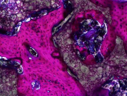
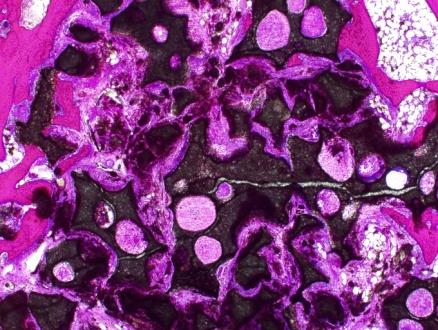
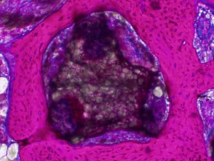
5 Group 6 Week Histology 12 Week Histology BioSphere (4x top; 20x bottom) Novabone (4x top; 20x bottom) Actifuse (4x top; 20x bottom) Figure 3. Six and twelve week histology images for BioSphere, Novabone, and Actifuse 5
6 The 12-week Actifuse histology also showed increased bone formation. However, there were still a few specimens that had voids in the central area of the defect. Similar to the 6 week results, bone formation on the surface of the particles was limited to a few contact points and there was no sign of a bioactive layer. Based on the 6 and 12 week results, Actifuse did not appear to be bioactive. Histomorphometry The results of the histomorphometric analysis (HMA) confirmed the qualitative observations seen with the microct and histology analysis (Figure 4). The results showed that the BioSphere Group had more bone than Novabone and Actifuse at both the 6 and 12 week timepoints. At 6 weeks, the BioSphere group showed a 40% average bone area compared to 24% for the Novabone group and 33% for the Actifuse group. This represented a 67% increase in bone over Novabone and a 21% increase over Actifuse. The 6 week results for the control groups showed 1% bone area for the empty defect. 60% 50% 40% 30% 20% 10% 0% 6 Weeks 12 Weeks Empty Actifuse Novabone BioSphere Figure 4. Histomorphometry data showing average bone % for both groups at 6 and 12 weeks By 12 weeks, the average bone % area increased in the BioSphere group to 48% while Novabone and Actifuse increased to 37%. Similar to the 6 weeks results, the BioSphere showed an increase in bone formation against both Novabone and Actifuse (30% increase). The empty defect showed an increase in bone with an average bone area of 10%. However, the defect still remained largely unfilled and was still considered critically sized. DISCUSSION The current bioactive glass products on the market are effective bone graft materials due to the bioactive and osteostimulative properties of the glass. However, the bone healing capacity of these products has not been fully realized due to the use of irregular bioactive glass particles and a broad particle size range. Although this shape and size has been well characterized over the years, it does not take into account the recent data showing the importance of bioactive glass ion release on bone formation. BioSphere represents the first bioactive glass product specifically designed to maximize the bone healing properties of 45S5 bioactive glass. Data from the study demonstrated that a sphere based bioactive glass putty provided better bone healing than a putty composed of irregular bioactive glass particles or a silicated version of hydroxyapatite. The radiographic and histological analysis of the BioSphere showed that the defects were fully filled in with bone at 6 weeks. By 12 weeks, the bone continued to form and advanced remodeling was evident. For the Novabone and Actifuse groups, bone formation was seen at the periphery of the defect at 6 weeks, but had not yet reached the center. By 12 weeks, additional bone formation was seen; however, a central void was still seen in some specimens. Histomorphometric analysis confirmed the qualitative observations and showed more bone with the BioSphere group at both timepoints. Compared to Novabone, BioSphere utilizes the exact same 45S5 bioactive glass composition. However, the results clearly showed improved bone formation with BioSphere. The difference was attributed to the shape and optimized size of the BioSphere particles. During the development of BioSphere, various sphere sizes were evaluated and the bimodal distribution of small and large spheres proved to be the best combination. It was found that the use of spherical particles resulted in a number of advantages over irregular particles. Unlike irregular particles which have highly variable particle geometry, the sphere shape is uniform and provides better control over the dissolution of the 6

![Scanning electron micrographs of particles from the BioSphere (top) and Novabone (bottom) [100X] The sphere shape allows the bioactive glass particles to dissolve in a uniform and repeatable manner.](/docs-images/89/97848553/images/7-1.jpg "In addition, glass dissolution can be further controlled by using narrow size ranges. Dissolution testing has shown that smaller particles dissolve at a faster rate due to increased surface area.")
7 glass. This is shown in the electron microscope image in Figure 5. Figure 5. Scanning electron micrographs of particles from the BioSphere (top) and Novabone (bottom) [100X] The sphere shape allows the bioactive glass particles to dissolve in a uniform and repeatable manner. In addition, glass dissolution can be further controlled by using narrow size ranges. Dissolution testing has shown that smaller particles dissolve at a faster rate due to increased surface area. Therefore, changes to the size of the particle led to changes in the dissolution rate of the glass. This versatility allowed the bone healing properties of the spherical particles to be optimized during the development of BioSphere. Additionally, the use of a narrow size range of spherical particles also allows for improved particle packing in a bone defect. Once implanted, the putty carrier is quickly dissolved in a few days. Therefore, the spacing of the particles within the bone defect results in the formation of a pore network that allows for bone in-growth. Due to the sphere shape, the particles maintain uniform spacing and an open porosity. With irregular particles, the packing density is higher, and the porosity and pore size is reduced. Due to the flat surface on the irregular particles, the particles can pack into areas with little to no porosity. In addition, the smaller irregular particles tend to fill in the spaces between the larger particles resulting in smaller pores and a higher packing density. This may explain the peripheral bone growth seen in the 6 and 12 week Novabone specimens. Due to the small pore size and tight packing, bone growth through the Novabone implant may been delayed until the small particles were resorbed. Compared to Actifuse, BioSphere also showed improved bone formation. Histological analysis showed that new bone formed over the entire surface of the BioSphere particles while the Actifuse particles showed only partial bone contact. The Actifuse data also showed limited bone growth in the central implant area at both 6 and 12 weeks. The difference in bone formation between BioSphere and Actifuse may be attributed to the composition of the particles. BioSphere particles are composed of the well-characterized 45S5 bioactive glass formula while Actifuse is composed of hydroxyapatite with a 0.8% silica substitution. According to the manufacturer of Actifuse, the addition was silica was done to improve the bone healing properties of hydroxyapatite. However, the resultant material is not bioactive (as seen by the results of this study) and resulted in less bone than BioSphere. CONCLUSION The in vivo data from the rabbit femur study showed that changes to the shape and size of 45S5 bioactive glass particles had a substantial effect on improving bone healing. Femoral defects filled with BioSphere resulted in faster and more robust bone healing compared to defects filled with a bioactive glass putty containing irregular particles or a putty containing silicated hydroxyapatite. Although bioactive glass is a highly characterized material and a well established bone graft material, it was evident that modifying particle geometry could improve bone healing. This was attributed to changes in the glass dissolution, ion release profile, and particle packing. Based on these results, a sphere based bioactive glass putty may result in faster healing and improved clinical outcomes in orthopaedic surgery. 7
8 REFERENCES 1 Oonishi et al. Particulate bioactive glass compared with hydroxyapatite as a bone graft substitute. Clin Orthop Rel Res, 334: (1997). 2 Hench et al. Bonding mechanisms at the interface of ceramic prosthetic materials. J Biomed Mater Res, 5(6): (1971). 3 Hench et al. Bonding mechanisms at the interface of ceramic prosthetic materials. J Biomed Mater Res, 5(6): (1971). 4 Hench. The story of bioactive glass J Mater Sci Mater Med, 17(11): (2006). 5 Xynos et al. bioactive glass 45S5 stimulates osteoblast turnover and enhances bone formation in vitro: implications and applications for bone tissue engineering. Calcif Tissue Inter, 67(4): (2000). 6 Xynos et al. Gene expression profiling of human osteoblasts following treatment with the ionic products of bioactive glass 45S5 dissolution. J Biomed Mater Res, 55(2): (2001). 7 Jell et al. Gene activation by bioactive glasses. J Mater Sci: Mater Med, 17: (2006). 8 Bosetti et al. Type I collagen production by osteoblast-like cells in contact with different bioactive glasses. J Biomed Mater Res A, 64(1): (2003). 9 Gao et al. Silica-based bioactive glasses modulate expression of bone morphogenetic protein-2 in SAOS-2 osteoblasts in vitro. Biomaterials, 22(12): (2001). 10 Vogel et al. "In vivo comparison of bioactive glass particles in rabbits." Biomaterials, 22(4): (2001). 11 Mushipe et al. "Cancellous bone repair using bovine trabecular bone matrix particulates." Biomaterials, 23(2): (2002). 12 Voor et al. "Is hydroxyapatite cement an alternative for allograft bone chips in bone grafting procedures? A mechanical and histological study in a rabbit cancellous bone defect model." J Biomed Mater Res B Appl Biomater, 71(2): (2004). MKT-BPY-001 Rev B 8
BIOACTIVE SYNTHETIC GRAFT
 B U I L D S T R O N G B O N E F A S T Putty Particulate Morsels A BIOACTIVE SYNTHETIC BONE FOR FASTER HEALING NovaBone is a 100% bioactive synthetic material composed from elements that occur naturally
B U I L D S T R O N G B O N E F A S T
 B U I L D S T R O N G B O N E F A S T Putty MIS Particulate Morsels A BIOACTIVE SYNTHETIC BONE FOR FASTER HEALING NovaBone is a 100% bioactive synthetic material composed from elements that occur naturally
Inion BioRestore. Bone Graft Substitute. Product Overview
 Inion BioRestore Bone Graft Substitute Product Overview Inion BioRestore Introduction Inion BioRestore is a synthetic bone graft substitute, which remodels into bone and is easy to use. Inion BioRestore
Bioactive Glass Biphasic β-tcp & HA Granules Alkylene Oxide Polymer Carrier. MEDLINEUNITE Bioactive Bone Graft
 Bioactive Glass Biphasic β-tcp & HA Granules Alkylene Oxide Polymer Carrier Principles of Bone Healing Reparative Phase Healing Cascade 1. Cellular infiltration and migration to site (fibroblasts, macrophages,
BIOACTIVE BASICS. Bioactive materials elicit a controlled action and reaction in the physiological environment.
 BIOACTIVE BASICS Bioactive materials elicit a controlled action and reaction in the physiological environment. Bioglass is a glass ceramic composed of silicon dioxide (SiO 2 ), sodium oxide (Na 2 O), calcium
ctive Bone Bonding Bone Regeneration Osteostimulation*
 www.bonalive.com ctive Bone Bonding Bone Regeneration Osteostimulation* A revolution in bone grafting Osteoconductive bone graft substitute with osteostimulative* properties *non-osteoinductive Before
SalvinOss Xenograft Bone Graft Material In Vivo Testing Summary
 SalvinOss Xenograft Bone Graft Material In Vivo Testing Summary Summary of In Vivo Use Of Bioresorbable Xenograft Bone Graft Materials In The Treatment Of One-Walled Intrabony Defects In A Canine Model
PRO-STIM Injectable Inductive Graft TECHNICAL MONOGRAPH
 PRO-STIM Injectable Inductive Graft TECHNICAL MONOGRAPH PRO-STIM Injectable Inductive Graft TECHNICAL MONOGRAPH Headline Contents Headline Appendix 4 5 7 8 13 14 Clinical Benefits Self-Forming Porous Scaffold
Bioactive Bone Glass Substitute
 Bioactive Bone Glass Substitute Injectable Putty BIOMATERIALS IMPROVING THE BONE REGENERATION Noraker has been involved in biomaterial development since 2005. It s today an innovative manufacturer of medical
Gene Activation for Excellent Bone Remodeling
 Osteostimulative bone regeneration granules Gene Activation for Excellent Bone Remodeling Injectable Putty Granules BIOMATERIALS STIMULATING THE BONE REGENERATION Noraker has been involved in biomaterial
Comparative Animal Study of OssiMend and Healos 13,14
 Comparative Animal Study of OssiMend and Healos 13,14 Objective This study was conducted to evaluate the use of OssiMend as compared with Healos in combination with autologous bone marrow as a bone grafting
Product Information. MIS Corporation. All Rights Reserved.
 Product Information MIS Corporation. All Rights Reserved. MIS Warranty: MIS exercises great care and effort in maintaining the superior quality of its products. All MIS products are guaranteed to be free
Biomaterials Line. MIS Corporation. All Rights Reserved.
 7. Biomaterials Line MIS Corporation. All Rights Reserved. 6. MIS s Quality System complies with international quality standards: ISO 13485: 2003 - Quality Management System for Medical Devices, ISO 9001:
Versatile grafting Solutions
 Versatile grafting Solutions A Canadian company serving Canadian dentists since 1997 Here s why your colleagues are calling us for their bone regeneration needs Founded in 1997 Citagenix has been providing
Surgical Technique Guide. Impact & Inject Formulations
 Surgical Technique Guide Impact & Inject Formulations System Overview OsteoVation QWIK is a bone void filler consisting of a proprietary composite of calcium phosphate and calcium sulfate granules. The
Calcium Phosphate Cement
 Calcium Phosphate Cement Fast-Setting Bone Graft and AutoGraft Extender. * Ossilix is a high performance next generation calcium phosphate cement indicated for filling bony defects in cancellous bone.
ORS 2015 Annual Meeting (Orthopedic Research Society)
") ORS 2015 Annual Meeting (Orthopedic Research Society) Poster No: 1045 CERAMENT Bone Void Filler Impregnated with Gentamicin Increases Bone Formation and Decreases the Rate of Detectable Infection after
ORS 2015 Annual Meeting (Orthopedic Research Society) Poster No: 1045 CERAMENT Bone Void Filler Impregnated with Gentamicin Increases Bone Formation and Decreases the Rate of Detectable Infection after
chronos Bone Void Filler. Beta-Tricalcium Phosphate ( β-tcp) bone graft substitute.
 bone graft substitute.") chronos Bone Void Filler. Beta-Tricalcium Phosphate ( β-tcp) bone graft substitute. Osteoconductive Resorbable Synthetic chronos Bone Void Filler chronos granules and preforms are synthetic, porous, osteoconductive,
chronos Bone Void Filler. Beta-Tricalcium Phosphate ( β-tcp) bone graft substitute. Osteoconductive Resorbable Synthetic chronos Bone Void Filler chronos granules and preforms are synthetic, porous, osteoconductive,
Innovative Range of Regenerative Solutions
 TM Innovative Range of Regenerative Solutions MIS Implant Technologies Ltd. All rights reserved. Optimal volumes and quality of hard and soft tissue are required to satisfy the goals of oral rehabilitation
GRAFTON DEMINERALIZED BONE: FIBER TECHNOLOGY AND PERFORMANCE IMPLICATIONS
 GRAFTON DEMINERALIZED BONE: FIBER TECHNOLOGY AND PERFORMANCE IMPLICATIONS Based Upon the Published Source: Martin GJ, Jr., Boden SD, Titus L, Scarborough NL. New formulations of demineralized bone matrix
AttraX. Portfolio. Surface Drives Performance
 Portfolio Surface Drives Performance Advanced Biomaterial Microarchitecture Drives Bone Formation Traditional calcium phosphate materials generally do not give rise to bone formation when implanted in
Symbios Xenograft Granules Porcine Bone Graft Material
 Symbios Xenograft Granules Porcine Bone Graft Material 32671122-USX-1607_Symbios Highlight Brochure.indd 1 2016-09-27 12:12 Symbios Xenograft Granules Porcine bone graft material Symbios Xenograft Granules
ADVANCED BONE GRAFT SYSTEM OVERVIEW
 ADVANCED BONE GRAFT SYSTEM OVERVIEW NANOSS BIOACTIVE ADVANCED BONE GRAFT Table Of Contents INTRODUCTION System Overview... 1 NANOSS BIOACTIVE COMPONENTS Comparison of nanoss Bioactive and Human Bone...
Regeneration Bone Grafting & Soft Tissue Management
 Regeneration Bone Grafting & Soft Tissue Management Human Histology OSTEON II 26.5 months Regeneration Products information Table of contents Bone Graft Material OSTEON II OSTEON OSTEON II Collagen OSTEON
OssiMend Bone Graft Matrix
 OssiMend All-Natural Mineral-Collagen Bone Grafting Matrix Anorganic bone mineral and type I collagen Highly purified, biocompatible matrix Osteoconductive Osteoinductive and Osteogenic in conjunction
Properties of GENESYS TM Biocomposite Material Compared to Pure Polylactide (PLA)
") Properties of GENESYS TM Biocomposite Material Compared to Pure Polylactide (PLA) Purpose: To compare the bone in-growth of GENESYS biocomposite material (96L/4D PLA copolymer mixed with β-tcp micro-particles)
Properties of GENESYS TM Biocomposite Material Compared to Pure Polylactide (PLA) Purpose: To compare the bone in-growth of GENESYS biocomposite material (96L/4D PLA copolymer mixed with β-tcp micro-particles)
Bone Void Filler. Callos. The Next Generation in Calcium Phosphate Cement A COLSON ASSOCIATE
 Callos Bone Void Filler The Next Generation in Calcium Phosphate Cement A COLSON ASSOCIATE Callos Calcium Phosphate Cement Callos is a high performance next generation calcium phosphate cement indicated
botiss biomaterials bone & tissue regeneration collacone max Innovative composite matrix socket preservation form-fitting resorbable composite
 bone & tissue regeneration botiss biomaterials socket preservation Innovative composite matrix form-fitting resorbable composite 1 Socket preservation safeguarding your sockets collacone.. max flexbone*
"The Evaluation Nano Calcium Silicate Cements Performance for Palpation"
 "The Evaluation Nano Calcium Silicate Cements Performance for Palpation" Delaram Amini* Bachelor Student, Department of Medical Islamic Azad University Of Tehran Medical Branch Tehran,Iran Maryam Chenani
botiss dental bone & tissue regeneration biomaterials collacone max Innovative composite matrix socket preservation form-fitting resorbable composite
 dental bone & tissue regeneration botiss biomaterials socket preservation Innovative composite matrix form-fitting resorbable composite 1 botiss regeneration system maxresorb flexbone* collacone.. max
THE NEXT FRONTIER OF BONE REGENERATION. where Technology meets Nature
 THE NEXT FRONTIER OF BONE REGENERATION where Technology meets Nature SmartBone is a new hybrid bioactive bone substitute specifically developed for bone regeneration in reconstructive surgery. SmartBone
Radiological and histological analysis of synthetic bone grafts in recurring giant cell tumour of bone: a retrospective study
 Journal of Orthopaedic Surgery 2010;18(1):63-7 Radiological and histological analysis of synthetic bone grafts in recurring giant cell tumour of bone: a retrospective study Hiroyuki Hattori, Hiroaki Matsuoka,
Regeneration Bone Grafting & Soft Tissue Management
 Regeneration Bone Grafting & Soft Tissue Management 1 Regeneration Bone Grafting & Soft Tissue Management Table of contents Bone Graft Material OSTEON II OSTEON OSTEON II Collagen OSTEON Collagen 5 6
BONE void FILLER. Beta-tricalcium phosphate ( b-tcp) bone graft substitute. OvERvIEW BROCHURE
 bone graft substitute. OvERvIEW BROCHURE") chronos BONE void FILLER Beta-tricalcium phosphate ( b-tcp) bone graft substitute OvERvIEW BROCHURE chronos STRIP chronos Strip is a synthetic bone void filler manufactured from chronos Beta-Tricalcium
chronos BONE void FILLER Beta-tricalcium phosphate ( b-tcp) bone graft substitute OvERvIEW BROCHURE chronos STRIP chronos Strip is a synthetic bone void filler manufactured from chronos Beta-Tricalcium
Evaluation of different grafting materials in three-wall intra-bony defects around dental implants in beagle dogs
 Current Applied Physics 5 (2005) 507 511 www.elsevier.com/locate/cap Evaluation of different grafting materials in three-wall intra-bony defects around dental implants in beagle dogs Ui-Won Jung a, Hee-Il
More than bone regeneration. A total solution.
 More than bone regeneration. A total solution. More than a dental implant company. A total solution. When it comes to treatment options, your patients want positive results both functionally and esthetically.
THE NEXT FRONTIER OF BONE REGENERATION. where Technology meets Nature
 THE NEXT FRONTIER OF BONE REGENERATION where Technology meets Nature SmartBone is a new hybrid bioactive bone substitute specifically developed for bone regeneration in reconstructive surgery. SmartBone
Indications. Neuro Surgery. Plastic Surgery. ENT Surgery. Oral Maxillofacial Surgery
 Indications Neuro Surgery Cranioplasty/Craniectomy Bur Hole Filling Cranial Cuts Onlay & Inlay grafting Plastic Surgery Cranial Reconstruction Orbital Reconstruction Onlay Grafting ENT Surgery Frontal
( ) 2009;28(2):89-94
 2009;28(2):89-94") ( ) 2009;28(2):89-94 Osseointegration is important in the functional aspect, however, esthetics is also important, especially in the maxillary anterior region. An adequate surgical technique is necessary
( ) 2009;28(2):89-94 Osseointegration is important in the functional aspect, however, esthetics is also important, especially in the maxillary anterior region. An adequate surgical technique is necessary
synthetic CANCELLOUS BONE technical monograph Presented by Barbara Blum, Ph.D.
 C E L L P L E X TCP synthetic CANCELLOUS BONE technical monograph Presented by Barbara Blum, Ph.D. TECHNICAL MONOGRAPH CELLPLEX TCP SYNTHETIC CANCELLOUS BONE CELLPLEX TCP synthetic CANCELLOUS BONE introduction
A WIDE RANGE OF REGENERATIVE SOLUTIONS
 A WIDE RANGE OF REGENERATIVE SOLUTIONS INDICATIONS: 1/ SOCKET AND RIDGE PRESERVATION 2/ FILLING OF EXTRACTION SOCKETS Biomaterials offers portfolio of regenerative materials for implantology, aimed at
POWER TO RESTORE WITHOUT LEAVING A TRACE. The only remaining evidence of the trauma
 POWER TO RESTORE WITHOUT LEAVING A TRACE The only remaining evidence of the trauma Perfect partner for your trauma and non-unions Your choice of synthetic bone graft not only influences the efficiency
BioVin Collagen Membrane
 BioVin Collagen Membrane Resorbable cross-linked collagen membrane BioVin Bovine Bone Bovine Bone Substitute OToss Synthetic Bone Synthetic Resorbable Biphasic Calcium Phosphate OToss Synthetic Bone Inject
CERAMENT BONE VOID FILLER
 CERAMENT BONE VOID FILLER Rapid and complete bone remodeling TM CERAMENT BONE VOID FILLER CERAMENT is an injectable, moldable, drillable and radiopaque bone substitute which provides rapid and complete
Evaluation of an osteostimulative putty in the sheep spine
 DOI 10.1007/s10856-010-4175-5 Evaluation of an osteostimulative putty in the sheep spine Zhen Wang Bin Lu Lei Chen Jiang Chang Received: 6 May 2010 / Accepted: 22 October 2010 Ó Springer Science+Business
BonAlive Clinical Cases
 BonAlive Clinical Cases Inhibition of bacterial growth Osteostimulation* Bioactive bone bonding *Non-osteoinduction BonAlive mechanism of action BonAlive composition: 53% SiO 2, 23% Na 2 O, 20% CaO, 4%
CRANIAL RECONSTRUCTION SOLUTIONS
 CRANIAL RECONSTRUCTION SOLUTIONS CRANIAL RECONSTRUCTION SOLUTIONS Your partner of CHOiCE at depuy Synthes CMF, we are dedicated to providing solutions for your individual patient needs. We do this through
In search of the optimal material for dental bone grafting
 2 EDI An overview of dental bone graft materials In search of the optimal material for dental bone grafting Sean Aiken, Keele, and Dr Anthony Bendkowski, Maidstone/England The range of dental bone graft
Regeneration Bone Grafting & Soft Tissue Management
 Regeneration Bone Grafting & Soft Tissue Management OSTEON TM II Table of Contents Bone Graft Material OSTEON TM II Collagen 04 OSTEON TM Collagen 06 OSTEON TM II 08 OSTEON TM 12 ORTHOPEDIC OSTEON TM 14
BEGO BIOMATERIALS When the result counts
 BEGO BIOMATERIALS When the result counts Partners in Progress INTRO Challenge what exists get the right answers We practise systematic thinking with a passion and we are never satisfied with the status
THE NEW STANDARD OF EXCELLENCE IN BIOMATERIALS. Collagenated heterologous cortico-cancellous bone mix + TSV Gel GTO I N S P I R E D B Y N A T U R E
 GTO THE NEW STANDARD OF EXCELLENCE IN BIOMATERIALS Collagenated heterologous cortico-cancellous bone mix + TSV Gel R E G E N E R A T I O N S C I E N C E I N S P I R E D B Y N A T U R E A unique biotechnology
Resorbable bilayer synthetic membrane Biomimetic tissue-engineered matrix for GBR and GTR
 Resorbable bilayer synthetic membrane Biomimetic tissue-engineered matrix for GBR and GTR Patented Jet-Spraying technology: Full barrier effect during 4 weeks, and complete resorption in 6 months PATENTED
Basics of Cartilage Restoration Introduction of TruFit
 Basics of Cartilage Restoration Introduction of TruFit Philip A. Davidson, MD Heiden Orthopaedics Park City, Utah USA Smith & Nephew Seminar London, UK October 2008 Cartilage Restoration A wide realm between..
PEEK-OPTIMA HA Enhanced: The Latest Material Development for Interbody Fusion
 PEEK-OPTIMA HA Enhanced: The Latest Material Development for Interbody Fusion Ovine Cervical Fusion Study Finds Performance Advantages with PEEK-OPTIMA HA Enhanced Polymer. 1 Introduction For more than
Bone modeling and remodeling How does a bone form without augmentation?
 Overview on Bone formation,bone grafts materials, and Augma Biomaterials bone cements Bone modeling and remodeling How does a bone form without augmentation? Bone Modeling Initial formation of a bone A
DRIVING THE FUTURE. About HOSPITAL INNOVATIONS. Strong commitment to healthcare innovation
 idente Catalogue About HOSPITAL INNOVATIONS Since 2008, Hospital Innovations has been supplying a growing range of specialist products for use in orthopaedic and corrective surgery; with an emphasis on
BONE AUGMENTATION AND GRAFTING
 1 A Computer-Guided Bone Block Harvesting Procedure: A Proof-of-Principle Case Report and Technical Notes Effectiveness of Lateral Bone Augmentation on the Alveolar Crest Dimension: A Systematic Review
Ankle and subtalar arthrodesis
 OSTEOAMP Allogeneic Morphogenetic Proteins Ankle Nonunions OSTEOAMP Case Report ANKLE NONUNIONS Dr. Jason George DeVries Orthopedic & Sports Medicine, Bay Care Clinic, 501 N. 10th Street, Manitowoc, WI
REGENERATION BY DESIGN
 REGENERATION BY DESIGN ALLOGRAFT, XENOGRAFT, PUTT Y & BIOACTIVE GLASS FOR BONE REGENERATION INSTRUMENTS FOR COLLECTING BONE BARRIER MEMBRANES FOR GUIDED REGENERATION GUIDED IMPLANT TREATMENT Wide Selection
The Essential Choice. With the Benefits of Biologic Predictability
 The Essential Choice With the Benefits of Biologic Predictability Documented, Reliable, Experienced is the essential choice for your daily regenerative needs. Throughout our long history and dedication
Bone Grafting and Bone Graft Substitutes. Original Author: James Krieg, MD Revision Author: David Hak, MD Last Revision May 2010
 Bone Grafting and Bone Graft Substitutes Original Author: James Krieg, MD Revision Author: David Hak, MD Last Revision May 2010 Bone Graft Function Structural support of articular fracture Tibial plateau
Procedure Manual and Catalog
 Procedure Manual and Catalog TM Why SynthoGraft? SynthoGraft offers a unique structure which provides stability, while its micro-porosity allows for rapid vascularization and subsequent resorption. Although
SPINE BIOMATERIALS Crop Bleed
 A comprehensive guide to biomaterials offered by Posterolateral DBX PUTTY DBX STRIP DBX MIX DBX INJECT chronos Strip chronos Granules chronos Preforms PROCURE CHOICE Marrow Aspiration Kit Bone Marrow Aspiration
Synthetic and absorbable autograft substitute with osteoinduction
 putty Synthetic and absorbable autograft substitute with osteoinduction Quality made in Germany Synthetic and absorbable autograft substitute with osteoinduction Information on the synthetic bone grafting
Callos Bone Void Filler
 Surgical Technique Callos Bone Void Filler Acumed is a global leader of innovative orthopaedic and medical solutions. We are dedicated to developing products, service methods, and approaches that improve
Bone Tissue Biology & The Application of Synthetic Compounds for the Facilitation of Bone Tissue Healing
 Bone Tissue Biology & The Application of Synthetic Compounds for the Facilitation of Bone Tissue Healing Ryan T. Jones Western Michigan University May 2011 Introduction Bone has unique properties: Tensile
Type of evidence. Design rationale. Economic analysis
 Bone&JointScience Our Innovation in Focus Vol 06 No 02 December 2016 HEALICOIL REGENESORB Suture Anchor: Comparison of bone ingrowth at 18 months with a solid body anchor implant in an ovine reconstruction
The Essential Choice. With the Benefits of Biologic Predictability
 The Essential Choice With the Benefits of Biologic Predictability Documented, Reliable, Experienced is the essential choice for your daily regenerative needs. Throughout our long history and dedication
Bone augmentation with maxgraft
 Patient information bone & tissue regeneration botiss biomaterials Bone augmentation with maxgraft established safe X100 natural Implantation stability is crucial for success Atrophy of the jaw bone loss
Micro-CT evaluation on osteoconductivity of DCPD-coated β-tcp granule using experimental rats
 Journal of Physics: Conference Series PAPER OPEN ACCESS Micro-CT evaluation on osteoconductivity of DCPD-coated β-tcp granule using experimental rats To cite this article: Khairul Anuar Shariff et al 2018
The Outcome of Bone Substitute Wedges in Medial Opening High Tibial Osteotomy
 Send Orders for Reprints to reprints@benthamscience.net The Open Orthopaedics Journal, 2013, 7, 373-377 373 Open Access The Outcome of Bone Substitute Wedges in Medial Opening High Tibial Osteotomy N.M.
Cerasorb M DENTAL. O:\Zulassung\Cerasorb Dental Kanada 2013\Texte\Cerasorb M Dental final IFU docx
 Cerasorb M DENTAL Resorbable, pure-phase beta-tricalcium phosphate matrix with interconnecting porosity for bone regeneration for use in dental and maxillofacial surgery DESCRIPTION: Cerasorb M DENTAL
Limited bone availability makes implant placement challenging
 Bone Grafting: Essential Indications and Techniques in Implant Dentistry Limited bone availability makes implant placement challenging and sometimes unpredictable. Candidates for implant therapy must have
P105 Predictable Bone Grafting for Site Preparation for Implants and Prosthetics Workshop JAMES GRISDALE, DDS THURSDAY, FEBRUARY 26
 P105 Predictable Bone Grafting for Site Preparation for Implants and Prosthetics Workshop JAMES GRISDALE, DDS THURSDAY, FEBRUARY 26 Please complete the speaker evaluation form in the Midwinter Meeting
Biology. Dr. Khalida Ibrahim
 Biology Dr. Khalida Ibrahim BONE TISSUE Bone tissue is a specialized form of connective tissue and is the main element of the skeletal tissues. It is composed of cells and an extracellular matrix in which
In the last decade of the 20 th century, special emphasis was put on an emerging field of science: Tissue engineering,which combines the state of the
 In the last decade of the 20 th century, special emphasis was put on an emerging field of science: Tissue engineering,which combines the state of the art materials science with concepts from the life sciences.
Bone augmentation with biomaterials
 Patient information dental bone & tissue regeneration botiss biomaterials Bone augmentation with biomaterials established safe natural X100 Implantation stability is crucial for success Atrophy of the
THE NEXT GENERATION OF REGENERATION
 THE NEXT GENERATION OF REGENERATION Why SynthoGraft? SynthoGraft offers a unique structure which provides stability, while its micro-porosity allows for rapid vascularization and subsequent resorption.
HOSPITAL INNOVATIONS
 idente Catalogue About HOSPITAL INNOVATIONS Since 2008, Hospital Innovations has been supplying a growing range of specialist products for use in orthopaedic and corrective surgery; with an emphasis on
THE ESSENTIALS IN BONE GRAFTING BRIDGING MEDICAL GAPS
 THE ESSENTIALS IN BONE GRAFTING BRIDGING MEDICAL GAPS FAST FACTS Clean and sterile, free of human, animal and chemical material. Products can be custom ordered, ranging from pastes and cements to granulations
Chapter 4 THE OSTEOGENICITY OF AUTOLOGOUS BONE TRANSPLANTS IN
 Proefschrift_Moyo_Kruyt.book Page 39 Wednesday, September 10, 2003 1:03 PM The osteogenicity of autologous bone transplants in the goat THE OSTEOGENICITY OF AUTOLOGOUS BONE TRANSPLANTS IN THE GOAT 4 Summary
Accelerated Neutral Atom Beam Processing Improves PEEK In Vivo Osseointegration
 Accelerated Neutral Atom Beam Processing Improves PEEK In Vivo Osseointegration Joseph Khoury, PhD 1, Cathy Tkaczyk, PhD 2, Art Kurz 1, James Bachand 1, Richard Svrluga 1, Michel Assad, PhD 2. 1 Exogenesis
Advice sheet for Surgeons and Radiologists. Radiographic appearance of CERAMENT
 Advice sheet for Surgeons and Radiologists Radiographic appearance of CERAMENT The structure and function of CERAMENT CERAMENT is an osteoconductive bone graft substitute which consists of hydroxyapatite
REASONS TO USE R.T.R.
 3 REASONS TO USE R.T.R. AFTER EACH EXTRACTION Fully resorbable ß-TCP material RTR 3raisons 120x280.indd 1 16/06/15 10:52 1AVOID SPONTANEOUS RIDGE RESORPTION After tooth extraction, spontaneous healing
OSSEODENSIFICATION: A Paradigm Shift In Implant Osteotomy Preparation. Introducing Densah Bur Technology
 OSSEODENSIFICATION: A Paradigm Shift In Implant Osteotomy Preparation Introducing Densah Bur Technology Densah Bur Technology * The Innovation That Makes Osseodensification Possible A New Paradigm In Dental
Talos -C (HA) and the PEEK-OPTIMA HA Enhanced Technology
 and the PEEK-OPTIMA HA Enhanced Technology") v Talos -C (HA) and the HA Enhanced Technology The innovative Talos -C (HA) IBF Implant made with polymer is fully integrated with the well known material Hydroxyapatite (HA). The Talos -C (HA) IBF Implant
v Talos -C (HA) and the HA Enhanced Technology The innovative Talos -C (HA) IBF Implant made with polymer is fully integrated with the well known material Hydroxyapatite (HA). The Talos -C (HA) IBF Implant
Bone Grafting for Socket Preservation
 Bone Grafting for Socket Preservation Dr. Karl R. Koerner Normal extraction facial bone loss. Excessive force. Commonly the thickness of facial bone. Hussain, A. et al. Ridge preservation comparing a nonresorbable
Allograft Bone and Super Critical Fluid (SCF) Technology
 Technology") Allograft Bone and Super Critical Fluid (SCF) Technology Summary: 1. Supercritical Fluids (SCF) have the properties of a gas and a liquid when it is in its supercritical state; 2. SCF treatment provides
Allograft Bone and Super Critical Fluid (SCF) Technology Summary: 1. Supercritical Fluids (SCF) have the properties of a gas and a liquid when it is in its supercritical state; 2. SCF treatment provides
Orthopedic & Sports Medicine, Bay Care Clinic, 501 N. 10th Street, Manitowoc, WI Procedure. Subtalar arthrodesis
 OSTEOAMP Allogeneic Morphogenetic Proteins Subtalar Nonunions OSTEOAMP Case Report SUBTALAR NONUNIONS Dr. Jason George DeVries and Dr. Brandon M. Scharer Orthopedic & Sports Medicine, Bay Care Clinic,
Guided Tissue and Bone Regeneration
 Guided Tissue and Bone Regeneration One toolbox for your needs! Tefguide 9000 742 401 12x24 mm pc 1 Osgide 9000 701 520 15 x 20 mm pc 1 Osbone 9000 800 255 250-1000 μm 0.25 cc 5 Product Specifications
FDBA(Freeze Dried Bone Allograft) SureOss Cortical Bone - Powder / Chip. OsteOss Cortical with Cancellous Bone - Powder / Chip
 SureOss Cortical Bone - Powder / Chip. OsteOss Cortical with Cancellous Bone - Powder / Chip") PERIODONTAL Bone & Skin Allograft Products FDBA(Freeze Dried Bone Allograft) SureOss - Powder / Chip OsteOss Cortical with - Powder / Chip DFDBA(Demineralized Freeze Dried Bone Allograft) SureOss -D Demineralized
PERIODONTAL Bone & Skin Allograft Products FDBA(Freeze Dried Bone Allograft) SureOss - Powder / Chip OsteOss Cortical with - Powder / Chip DFDBA(Demineralized Freeze Dried Bone Allograft) SureOss -D Demineralized
1 Rua Santa Adélia, 166, Bangu, Santo André SP, Brazil
 Evaluation of the bioactivity behavior of a 48 wt %SiO 2 bioglass through experiments in simulated body fluid Roger Borges 1, a, Antônio Carlos da Silva 2,b and Juliana Marchi 1,c 1 Rua Santa Adélia, 166,
Ethan M. Braunstein, M.D. 1, Steven A. Goldstein, Ph.D. 2, Janet Ku, M.S. 2, Patrick Smith, M.D. 2, and Larry S. Matthews, M.D. 2
 Skeletal Radiol (1986) 15:27-31 Skeletal Radiology Computed tomography and plain radiography in experimental fracture healing Ethan M. Braunstein, M.D. 1, Steven A. Goldstein, Ph.D. 2, Janet Ku, M.S. 2,
Research Article Evaluation of Amniotic-Derived Membrane Biomaterial as an Adjunct for Repair of Critical Sized Bone Defects
 Advances in Orthopedic Surgery, Article ID 572586, 4 pages http://dx.doi.org/10.1155/2014/572586 Research Article Evaluation of Amniotic-Derived Membrane Biomaterial as an Adjunct for Repair of Critical
The injectable, self-setting calcium phosphate bone graft substitute.
 The injectable, self-setting calcium phosphate bone graft substitute. With a porous architecture similar to natural bone. STRUCSURE CP Bone Graft Substitute Because simplicity matters STRUCSURE CP Bone
Graftys Quickset & Graftys HBS use in plateau tibial fracture
 Graftys Quickset & Graftys HBS use in plateau tibial fracture Introduction& Patient Profile 35 years-old women A complex plateau tibial fracture Pre-operative Scan & Radiography Pre-operative AP Scan Surgical
Optimum implant geometry
 Design Rationale Optimum implant geometry Extending the proven Tri-Lock heritage The original Tri-Lock was introduced in 1981. This implant was the first proximally coated tapered-wedge hip stem available
THE NEXT FRONTIER OF BONE REGENERATION
 THE NEXT FRONTIER OF BONE REGENERATION where Technology meets Nature swiss made SmartBone is new composite bone substitute specifically developed for bone regeneration in oral and maxillofacial reconstructive
Citagenix Catalogue 2017 ALLOGRAFTS BONE SUBSTITUTES BONE GRANULES & BLOCKS RESORBABLE MEMBRANES
 Citagenix Catalogue 2017 ALLOGRAFTS BONE SUBSTITUTES BONE GRANULES & BLOCKS RESORBABLE MEMBRANES 100% Allograft Hydratable Inductive Matrix NEW Create your customized osteoinductive graft by combining
Synthesis and Characterization of Bone Ingrowth Associated with Bone Cement Co-Polymerized with a Biodegradable Material
 Biomaterials Research (2005) 9(2) : 71-76 Biomaterials Research C The Korean Society for Biomaterials Synthesis and Characterization of Bone Ingrowth Associated with Bone Cement Co-Polymerized with a Biodegradable