High-definition optical coherence tomography: adapted algorithmic method for pattern analysis of inflammatory skin diseases: a pilot study
|
|
|
- Lindsey Logan
- 5 years ago
- Views:
Transcription
1 DOI /s ORIGINAL PAPER High-definition optical coherence tomography: adapted algorithmic method for pattern analysis of inflammatory skin diseases: a pilot study Marc Boone Sarah Norrenberg Gregor Jemec Véronique Del Marmol Received: 17 September 2012 / Revised: 12 December 2012 / Accepted: 14 December 2012 Ó The Author(s) This article is published with open access at Springerlink.com Abstract High-definition optical coherence tomography (HD-OCT) is a non-invasive technique for morphological investigation of tissue with cellular resolution filling the imaging gap between reflectance confocal microscopy and conventional optical coherence tomography. The aim of this study is first to correlate dermatopathologic descriptors of inflammatory skin conditions with epidermal alteration to features observed by HD-OCT. Secondly, to assess the discriminative accuracy of common inflammatory reaction patterns with epidermal alteration using HD-OCT by applying Ackerman s algorithmic method of pattern recognition. The generated HD-OCT images of 160 patients presenting an inflammatory skin disease were analyzed with respect to the following criteria: visualization of individual cells in the epidermis and dermis and morphology of dermo-epidermal junction, papillary dermis and reticular dermis. A set of morphological features corresponding to dermatopathological descriptors are obtained and the discriminative accuracy of HD-OCT of inflammatory reaction patterns could be demonstrated. These patterns are spongiotic dermatitis, psoriasiform dermatitis, interface dermatitis and ballooning dermatitis. Additional studies to test the sensitivity and specificity of the proposed M. Boone (&) Department of Dermatology, Hôpital Erasme, Université Libre de Bruxelles, 128 Kardinaal Sterckxlaan, 1860 Meise, Belgium dr.boone@scarlet.be M. Boone S. Norrenberg V. Del Marmol Department of Dermatology, Hôpital Erasme, Université Libre de Bruxelles, 808 Route de Lennik, 1070 Bruxelles, Belgium G. Jemec Department of Dermatology, Health Sciences Faculty, Roskilde Hospital, University of Copenhagen, Copenhagen, Denmark algorithm for pattern analysis are essential. The other categories of Ackerman s pattern recognition need to be evaluated. This study provides a set of morphological features generated by HD-OCT imaging very similar to those described for reflectance confocal microscopy but with the advantages not only to visualize individual cells up to a depth of 570 lm but also in both slice and en face mode. An adapted algorithmic method for pattern analysis of common inflammatory skin diseases could be proposed. This new technique appears to be a promising method for non-invasive diagnosis, evaluation and management of common inflammatory skin diseases. Keywords High-definition optical coherence tomography Reflectance confocal microscopy Inflammatory skin diseases Ackerman s algorithmic method of pattern recognition Introduction Most dermatological diseases were originally defined morphologically, and terms such as papules and pustules are still important when diagnosing skin disease. Later dermatopathology evolved with its own special vocabulary of terms used to describe histological skin alterations, allowing dermatopathologists to classify skin diseases in great detail [21]. In dermatopathology, the initial approach is often to classify the changes broadly according to whether a process is predominantly inflammatory, predominantly proliferative/neoplastic, both inflammatory and proliferative or non-inflammatory/non-proliferative. In general, the majority of pathologic processes can be classified into one of these groups, although challenging exceptions occur,
2 particularly when overlapping features appear [8]. Inflammatory diseases are dynamic, and knowledge of the point of time when a dermatitis is biopsied is essential to optimal microscopic evaluation. Following the first sorting, more detailed diagnosis is then produced by identifying the specific qualitative and quantitative characteristics. Specific stains and biomarkers aid this process, but it has been suggested that overall pattern analysis is both specific and sufficient in the practical management of many diseases. Ackerman and colleagues [1] thus defined eight basic patterns of inflammatory diseases of the skin, where the pattern strongly indicates diagnosis prior to more detailed studies. One of the challenges of skin imaging is diagnostic accuracy in comparison to the gold standard of histopathology. Because of the optical complexity of skin, which contains numerous reflective elements, two essential options exist for in vivo imaging: either sufficient optical resolution is achieved in a sufficient volume of skin to allow diagnosis, with accuracy comparable to that of histopathology, or in vivo methods have to rely on pattern recognition at a lower magnification. Combining in vivo imaging with the conceptual approach by Ackerman and colleagues may therefore be a significant step forward for skin imaging. Reflectance confocal microscopy imaging of inflammatory skin conditions with epidermal alterations has already been studied in detail. These studies provide a set of welldescribed morphological criteria with obvious diagnostic impact [2 6, 14 17, 19, 20, 22, 23, 25, 26]. One limitation of this technique is that the assessment of microanatomic structures can be performed only to a depth of maximum 250 lm. A broader field of view and a greater penetration of the tissue can be achieved with OCT at the cost of resolution. High-definition optical coherence tomography (HD-OCT) is a new non-invasive technique for morphological investigation of tissue with cellular resolution filling the imaging gap between reflectance confocal microscopy and conventional optical coherence tomography [9 11]. The aim of this study was therefore first to correlate dermatopathologic descriptors of inflammatory skin conditions with features observed by HD-OCT. Secondly, to evaluate the discriminative accuracy of common inflammatory reaction patterns with epidermal alteration using HD-OCT by applying Ackerman s algorithmic method of pattern recognition. Materials and methods Subjects A total of 160 fair-skinned (Fitzpatrick types II and III, 99 females and 61 males) subjects with an age range of years (Table 1) with an inflammatory skin disease participated in this comparative study. All provided informed consent. Of these, 93 had allergic contact dermatitis (1 histologically verified and 93 positive patch tests) and 30 atopic dermatitis (fulfilling the diagnostic criteria of Hanifin and Rajka, 2 histologically verified), 9 lichen planus (histologically verified), 2 erythema multiforme (histologically verified), 3 discoid lupus erythematosus (histologically verified) and 23 chronic plaque psoriasis (3 histologically verified). High-definition optical coherence tomography High-definition OCT is based on the principle of conventional OCT, with 3 lm resolution in both transversal and axial directions, to visualize individual cells (Skintell Ò, Agfa Healthcare). Moreover, the system is capable of Table 1 Characteristics of 160 patients according different subgroups of perivascular dermatitis Spongiotic dermatitis (n = ) Allergic contact dermatitis (n = 93) Atopic dermatitis (n = 30) Acute (patch testing) Subacute Chronic Subacute & chronic F M F M F M F M years years years 19 & 66 years years years years years Interface dermatitis (n = 12) Ballooning dermatitis (n = 2) Psoriasiform dermatitis (n = 23) Lichenoid Vacuolar F M F M F M F M years years 46 years 32 & 53 years 34 years 46 years years years
3 capturing a slice image and an en face image in real time, as well as fast 3D acquisition. The tissue penetration depth goes up to 570 lm. Further technical details are discussed elsewhere [9 11]. Intermediate (109) magnification The standard light microscope is characterized by an adjustable lateral resolution of 4 lm up to 0.1 lm depending on the objective and a fixed axial resolution of 5 lm. The circular field of view is 20 mm up to 0.2 mm in diameter depending on the objective used. HD-OCT is characterized by a fixed lateral and axial resolution of 3 lm. The field of view is fixed at mm. The intermediate (109) magnification of the standard light microscope (characterized by a circular field of view of 4 mm and a lateral resolution of 3 lm) corresponds with the magnification obtained with HD-OCT. Method HD-OCT was done on the lesional and unaffected, clinically normal looking paralesional or contralateral skin of 160 patients with a well-established diagnosis based on history and clinical examination by a board-certified specialist of dermatology. If needed to clarify the diagnosis cultures, patch testing and any other procedures including biopsies were taken. HD-OCT imaging was done prior to any procedure. The generated pictures were analyzed with respect to the following criteria: visualization of individual cells in the stratum corneum, stratum granulosum, stratum spinosum and morphology of dermo-epidermal junction, papillary dermis and reticular dermis. Measurements of epidermal thickness have been performed using the measurement toolbar of the Skintell Ò program. Each voxel of the 3-dimensional HD-OCT image is defined by a unique set of x, y, z coordinates. These coordinates permit to measure with accuracy the epidermal thickness. Measurements were carried out in a climate-controlled room, at 24 C and 40 % relative humidity. Hematoxylin and eosin (H&E)-stained histologic vertical sections were correlated with the corresponding crosssectional HD-OCT images but also with the en face images. First, descriptive dermatopathological terms derived from reflectance confocal microscopy were applied to HD-OCT image (Table 2). Secondly, to evaluate relevant inflammatory reaction patterns in HD-OCT images, Ackerman s algorithmic method of pattern recognition in the perivascular dermatitis group was applied (Table 3). Results Systematic description of inflammatory changes as seen in HD-OCT (Tables 2, 3) A set of morphological features generated by HD-OCT imaging very similar to those described for reflectance confocal microscopy are summarized in Table 3. By looking at the slice image, one can identify the exact position (stratum corneum, stratum granulosum, stratum spinosum, stratum basale, papillary dermis) of the chosen en face image. The combination of both modes permits the exact micro-anatomic localization. This particular feature is only possible with the HD-OCT. This makes it very easy to distinguish, e.g., parakeratosis from hypergranulosis. Spongiotic dermatitis HD-OCT imaging of these skin conditions clearly showed the presence of spongiosis or intercellular edema that stretches apart keratinocytes and sometimes results in the formation of intraepidermal vesicles. Sometimes exocytosis of inflammatory cells could be identified. Morphologic features correlating with the acute, subacute and chronic stages could be observed (Table 3). In the acute form (Fig. 1), there was no hyperkeratosis. Compared to normal skin [12, 13, 18, 24], a significant increase ([50 %) in epidermal thickness was observed due to spongiosis. Intercellular edema decreased the reflectivity from cellular layers. Intraepidermal microvesicles were present, sometimes with macrovesicles in case of severe allergic reactions. Migration of inflammatory cells into the epidermis (exocytosis) was presented. There was no epidermal hyperplasia (acanthosis). In moderate allergic reactions, no dermal papillary thickening was observed. Inflammatory edema in the dermis appeared to lower dermal reflectivity. In moderate forms, dilated signal-free cavities in the dermis corresponding to blood vessels and lymphatic vessels were noticed. In these forms, the dermal perivascular inflammation was marked with highly reflective inflammatory cells. In the subacute form (Fig. 2), no significant hyperkeratosis was observed. Small mounds of parakeratosis occured in the keratin layer over the spongiotic vesicles. Because of spongiosis, a moderate increase (±10 %) of epidermal thickness compared to the normal skin [12, 13, 18, 24] could be observed. Spongiotic vesicles were seen. Acanthosis was minimal with some elongation of rete ridges. The papillary dermal edema was minimal. There was no papillary dermal thickening. There was a clear dermal perivascular inflammation. In chronic spongiotic dermatitis (Fig. 3), hyperkeratotic orthokeratosis was noticed in which localized patches of parakeratosis could be observed. There was also localized patchy
4 Table 2 High-definition optical coherence tomography descriptors Histological terms Spongiosis Exocytosis Spongiotic blisters/ vesicles Acanthosis Hyperkeratosis Parakeratosis Papillomatosis Hypergranulosis Hypogranulosis Necrotic keratinocytes Keratotic follicular plugging Interface changes Perivascular inflammation Dilated blood vessels HD-OCT features En face: darker area relative to surrounding epithelium of stratum corneum. Intercellular spaces between keratinocytes larger than normal (Figs. 1, 2: yellow circle) En face: high reflective small dots corresponding to lymphocytes observed at the level of the stratum spinosum in single or in small aggregates (Figs. 1, 2: white arrow) Cross-sectional and en face: dark round to poly-lobulated areas could be observed (Fig. 1: yellow arrow) Cross-sectional: thickening (?10 to ±300 %) of the epidermis compared to normal [12, 13, 18, 24], mainly of the stratum spinosum. Often associated with hyperkeratosis. The epidermis can be uniformly thickened (Figs. 3, 4: green double arrow) or a disproportionate expansion of rete ridges can be observed (Figs. 6, 7) En face: dark zones between entrance signal and stratum granulosum (Figs. 3, 6, 7: red arrow). Cross-sectional: the thickness of the stratum corneum was measured. A total thickness of [20 lm was measured (Figs. 3, 6, 7) In both en face and cross-sectional: highly reflective nucleated structures in stratum corneum (Fig. 4: violet arrow) En face: increased number and density of dermal papillary rings at the dermo-epidermal junction (Figs. 3, 4: orangecolored circle and arrow) In cross-sectional and en face: increase in thickness of stratum granulosum (normally 1 or 2 layers thick). This is usually seen in association with acanthosis and orthokeratotic hyperkeratosis (Figs. 6, 7: light-green arrow) In cross-sectional and en face: the granular layer is reduced or absent. This is always an abnormal condition and very often associated with parakeratotic hyperkeratosis (Figs. 4, 5) In cross-sectional and en face: at the level of stratum spinosum as single units and at suprabasal layer as aggregates. They appeared as bright, polygonal structures, larger than the surrounding keratinocytes (Figs. 6, 8) In cross-sectional and en face: plugging of the dilated openings of hair follicles by masses of keratin. A feature of a limited number of skin conditions such as discoid lupus erythematosus (Fig. 7) In cross-sectional and en face: morphological alteration at the junction or interface between the epidermis and dermis. Specifically one could observe high reflective small dots corresponding to lymphocytes at the level of the junction, as singles or clusters associated with total (band like) or partial (patchy) obliteration of the papillary rings (Figs. 6, 7) In cross-sectional and en face mode: high reflective small dots corresponding to lymphocytes clustered around blood vessels (Fig. 2: violet circle) In cross-sectional and en face mode: prominent round or linear dark canalicular structures within papillary dermis (Fig. 3: violet arrow) hypergranulosis (Table 3). Moderate (±10 %) to marked (±30 %) epidermal acanthosis was also observed. Spongiosis was minimal to absent and no intraepidermal microvessel was observed. The papillary dermal thickening was pronounced with papillomatosis. There was dermal perivascular inflammation. Psoriasiform dermatitis Using chronic plaque psoriasis as the morphological prototype, a number of features could be observed in HD-OCT images (Fig. 4). Hyperkeratosis was pronounced. Hyperreflective structures in the stratum corneum were corresponding to parakeratosis. Absence or reduction of the granular cell layer was regularly observed. The honeycomb pattern of the epidermis was conserved, but acanthosis with a more regular elongation and broadening of the rete ridges was present. At the top of the dermal papillae, a thinner epidermis was frequently noticed. Between the enlarged rete ridges, the dermal papillae were swollen due to edema and contained prominent dilated capillaries. A disappearing of the brighter papillary rings was often observed. The more pronounced the inflammation, the more dim the papillary rings became. The dermo-epidermal junction was also less well defined due to psoriasiform papillomatosis with an increased number and density of papillary rings. The reflectivity in the papillary dermis was lower than that in healthy skin suggesting an important papillary edema. Dilated blood vessels are visible surrounded by an important perivascular inflammatory infiltrate. Munro s micro abscesses could be demonstrated in two patients (Fig. 5). Interface dermatitis A consistent HD-OCT feature of Lichen planus (Fig. 6) as a prototype of lichenoid-interface dermatitis appeared to be hyperkeratosis coupled with rare or absent parakeratosis. Irregular thickening of granular layer and acanthosis, with irregular lengthening of rete ridge was observed. Moderate spongiosis could be observed. Degeneration of epidermal basal layer with necrotic keratinocytes visualized as total obliteration of the ring-like structures around the dermal papillae. Dermal inflammatory cell infiltrate, usually heavy, was diffuse and closely associated to the DEJ.
5 Table 3 Discriminative accuracy of high-definition optical coherence tomography of common inflammatory reaction patterns Spongiotic Interface dermatitis dermatitis Ac? Chr Vacuolar Lichenoid Ballooning dermatitis Psoriasiform dermatitis Spongiosis (?)???? -/? -/? -/? -/? Exocytosis (?)????????? Spongiotic blisters/ vesicles (?)????/ Acanthosis -? (?)??????? (irregular) Epidermal atrophy -?? - (chronic)????? (irregular)????? (irregular)??? (regular) Papillomatosis -??(?) - - -?? Hyperkeratosis -? (?)?????? Parakeratosis -?? - (rare) - (rare) - (rare)?? Hypergranulosis Hypogranulosis -?? /? Acantholysis Necrotic Keratinocyte?/-? -??? (ballooning) - Dyskeratosis?/-? -??? - Honeycomb pattern -/?? (?)? - - -?? preserved Epidermal disarray Infundibula: keratotic plug?/-?? Interface dermatitis -??? (focal)??? (diffuse)??? (focal) - (swollen, edema DP) Dermal edema?????????? Dilated bloodvessels (?)??? (??)????/-??/??? (in dermal papilla) Perivascular inflammation (?)??? (??)?????????? Adnexial inflammation -?/ Inflammatory cells were also present in the epidermis. Sparse dilated vessels and sparse dermal sclerosis were seen. The infundibula were normal. In contrast, HD-OCT features of discoid lupus erythematosus (Fig. 7) as a prototype of vacuolar-interface dermatitis without ballooning showed dilated hyperkeratotic infundibula. Interface changes appeared here with high reflective cells at the level of the dermo-epidermal junction in singles or as clusters. This was associated with a partial (focal) obliteration of ring-like structures around the dermal papillae. Irregular thickening of the epidermis with hypergranulosis and acanthosis could be seen, and necrotic keratinocytes were present. Inflammatory cells were also present in the epidermis. There was a clear dermal inflammation with dilated vessels. In more chronic forms, the epidermal was more atrophic together with a thickened basal membrane area. Erythema multiforme as prototype of ballooning dermatitis HD-OCT features of ballooning dermatitis (Fig. 8) as seen in drug-induced erythema multiforme included variable degrees of epidermal necrosis. Necrotic keratinocytes were present at all epidermal levels. Ballooning of spinous cells was also seen with vacuolar alteration of the basilar epidermis (basal vacuolization). At the junction between the epidermis and dermis, small, discrete vacuoles and characteristic melanophages were seen. Papillary dermis was edematous with dilated capillaries. There was a perivascular infiltrate. An algorithm based on pattern analysis at intermediate (109) magnification (Fig. 9) Step one: Determination of the basic pattern formed by infiltrates of cells. The perivascular dermatitis by far the most common group was selected for this study. The other patterns defined by Ackerman et al. [1] have been excluded. The inflammatory infiltrate around venules of the superficial plexus in the papillary dermis and around venules of the upper part of the reticular dermis could be observed. An infiltrate deeper than 570 lm could not be detected by the HD-OCT. Step two: Division of the perivascular dermatitis pattern: This group can be separated into perivascular alone with no apparent epidermal involvement or perivascular with
 and en face with z values] clearly shows the presence of spongiosis or intercellular edema (yellow circle) that stretches apart keratinocytes and results in the")
6 Fig. 1 Acute allergic contact dermatitis: (a) patch testing graded??? for ethylenediamine hydrochloride. HD-OCT imaging [crosssectional (b) and en face with z values] clearly shows the presence of spongiosis or intercellular edema (yellow circle) that stretches apart keratinocytes and results in the formation of intraepidermal involvement of the epidermis. This study has been focused only on the perivascular dermatitis group with apparent epidermal involvement. Four main criteria have been found leading to specific subgroup diagnosis: A: spongiosis with or without spongiotic vesicles, B: orthokeratosis or parakeratosis, C: interface changes with partial or total obliteration of papillary rings and D: ballooning degeneration. The secondary criteria of selection were: pronounced dermal edema, exocytosis, regular or irregular acanthosis, papillomatosis, (macro)vesicles (yellow arrows). Exocytosis (white arrows) of inflammatory cells can be demonstrated. There is no hyperkeratosis. Intercellular edema decreased the reflectivity from cellular layers. z values indicate the depth of the en face image (in lm) swollen dermal papillae, dilated blood vessels in dermal papillae, epidermal atrophy, thickened basal membrane area and dilated hyperkeratotic infundibula. The combination of main criteria and secondary criteria could lead to a more specific subgroup diagnosis (Fig. 9). Acute spongiotic dermatitis is characterized by a combination of spongiosis with spongiotic vesicles, exocytosis and dermal edema. The chronic form presented a mixture of orthokeratosis, spongiosis without spongiotic
, a small increase of epidermal thickness compared to the normal skin can be observed.")
7 Fig. 2 Subacute lesion in atopic dermatitis patient. Cross-sectional (a) and en face HD-OCT images (z values indicated). No significant hyperkeratosis is observed. Because of spongiosis (yellow circles), a small increase of epidermal thickness compared to the normal skin can be observed. Exocytosis (white arrow) of inflammatory cells can vesicles, exocytosis and regular acanthosis. Psoriasiform dermatitis is characterized by a combination of parakeratosis, regular acanthosis, no obliteration but hyporeflectivity of papillary rings, papillomatosis, swollen dermal papillae and dilated blood vessels in dermal papillae. The lichenoid variant of interface dermatitis presented orthohyperkeratosis, irregular acanthosis and total (diffuse) obliteration of papillary rings. The vacuolar variant of interface dermatitis in this study was discoid lupus be demonstrated epidermal acanthosis is minimal with some elongation of rete ridges. The papillary dermal edema is minimal. There is no papillary dermal thickening. There is a clear dermal perivascular inflammation (violet circles) erythematosus and presented irregular acanthosis, partial (focal) obliteration of papillary rings and dilated hyperkeratotic infundibula. In more chronic forms, the epidermal atrophy was pronounced together with a thickened basal membrane area. A special variant of the perivascular dermatitis group is the ballooning dermatitis and was characterized by necrotic keratinocytes and ballooned spinous cells in combination with a focal obliteration of papillary rings.
.")
8 Fig. 3 Chronic lesion in atopic dermatitis patient. Cross-sectional and en face HD-OCT images (z values indicated). Hyperkeratotic orthokeratosis is noticed (red arrows). Moderate epidermal acanthosis (green double arrow). Spongiosis is minimal and no intraepidermal Step three: Identification of the inflammatory cells that make up the infiltrate such as lymphocytes and granulocytes. Specific types of inflammatory cells often can be recognized for what they are as follows: lymphocytes (diameter 7 8 lm) are high reflective small dots that seem to be about equidistant from one another because they have a solitary uniform nucleus. Neutrophils (diameter lm) having a smaller multilobed nucleus are somewhat less reflective but larger dots (Fig. 5). microvesicles are observed. The papillary dermal thickening is pronounced with papillomatosis (orange circle and arrow). There is vascular dilatation (violet arrows) and dermal perivascular inflammation (violet circle) Discussion Skin imaging potentially allows much more than the possibility of non-invasive diagnosis in a clinical setting. It may also provide a dynamic method for the study of how lesions evolve and thereby contribute to our basic understanding of skin biology. One important approach to our understanding of lesional evolution is, fortunately, through diagnostic studies. As stated, two main options exist for
. Acanthosis with a more regular elongation and broadening of the rete ridges is presented (green double arrow).")
9 Fig. 4 Leg, chronic plaque psoriasis. Cross-sectional and en face HD-OCT images (z values indicated). Hyperkeratosis is pronounced. Refractile structures in the stratum corneum correspond to parakeratosis (violet arrows). Acanthosis with a more regular elongation and broadening of the rete ridges is presented (green double arrow). in vivo imaging: either to develop a method offering sufficient optical resolution or to establish reliable pattern recognition at a lower magnification. Over the years, a series of imaging techniques have been developed with varying degrees of resolution and penetration. Currently, RCM has the best resolution, albeit at the cost of a very limited depth of imaging. In consequence, RCM criteria for a number of diagnoses have been Between these enlarged rete ridges (yellow double arrow), the dermal papillae are swollen by edema. Papillary rings are less brighter than normal. The dermo-epidermal junction is also less defined due to psoriasiform papillomatosis with an increased number and density of papillary rings (orange circle and arrow) established comparing the method with histology [2 6, 14 17, 19, 20, 22, 23, 25, 26]. HD-OCT is a promising highresolution imaging technique which offers comparable resolution to RCM and improved penetration [9 11]. This study suggests that HD-OCT features of inflammatory skin conditions with epidermal alteration correlate well with dermatopathologic descriptors as defined in RCM. The morphological features imaged by HD-OCT were very

10 Fig. 5 Chronic palmar psoriasis lesion. Cross-sectional and en face HD-OCT images. Collections of neutrophils are found in the stratum corneum (pink circles). These lesions correspond to Munro microabscesses. z values are indicated similar to those described for RCM. Furthermore, it was possible to apply pattern recognition derived from intermediate magnification histopathology meaningfully to the HD-OCT images. HD-OCT allowed visualization of cellular infiltrates, although identification of the inflammatory cells that make up the infiltrate such as lymphocytes and granulocytes remains difficult for both RCM and HD-OCT. Cytological changes such as ballooning are however easily seen. One of the restrictions of RCM in evaluating inflammatory skin diseases is the limited penetration depth [2 6, 14 17, 19, 20, 22, 23, 25, 26]. With HD-OCT not only the visualization and assessment of individual cells but also microanatomic structures can be performed to a depth of 570 lm. This allows the method to identify specific patterns such as spongiosis, interface dermatitis and acanthosis. The method therefore appears to be able to identify the components needed for pattern analysis. Images of common inflammatory skin diseases were therefore evaluated by applying Ackerman s algorithmic method of pattern recognition at intermediate (109) magnification. Of Ackerman s eight essential patterns of
 and acanthosis (green double arrows), with irregular lengthening of rete ridge is observed.")
11 Fig. 6 Lichen planus: lesion on the calf. Cross-sectional and en face HD-OCT images. Hyperkeratosis (red arrows) is present without parakeratosis. Irregular thickening of granular layer (light-green arrows) and acanthosis (green double arrows), with irregular lengthening of rete ridge is observed. Moderate spongiosis can be observed (yellow circle). Degeneration of epidermal basal layer with necrotic keratinocytes visualized as total obliteration of the ring-like inflammatory skin diseases, perivascular dermatitis is the most common by far. Most of the common inflammatory skin diseases are perivascular dermatitides among those being allergic contact dermatitis, lichen planus and psoriasis. A perivascular dermatitis is identified by the presence of inflammatory cells around venules situated either in the upper part of the reticular dermis (superficial plexus) or in structures around the dermal papillae. Heavy dermal inflammatory cell infiltrate is diffuse and closely applied to dermal-epidermal junction (dark-green circles). Inflammatory cells are also present in the epidermis (white arrows). The combination of irregular lengthening of rete ridges, basal layer destruction and upper dermal edema and inflammatory infiltrate, produces a saw-tooth appearance of rete ridges. z values are indicated both the upper and lower parts of the reticular dermis (superficial and deep vascular plexuses) [1]. Once a pattern of perivascular dermatitis has been identified, it is necessary to make an evaluation about whether the epidermis, including the dermo-epidermal junction, is involved in the process. If the epidermis is affected, the pattern may be one in which spongiosis is
 and acanthosis (green double arrow).")
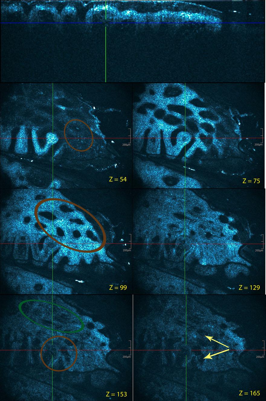
12 Fig. 7 Discoid lupus erythematosus: facial lesion. Cross-sectional and en face HD-OCT images. Dilated hyperkeratotic infundibula (light-magenta arrows). Irregular thickening of the epidermis with hypergranulosis (light-green arrows) and acanthosis (green double arrow). Necrotic keratinocytes and inflammatory cells (white arrows) discernible (spongiotic dermatitis), or the dermo-epidermal interface is obscured (interface dermatitis) or spinous cells are ballooned (ballooning dermatitis) or psoriasiform acanthosis is apparent (psoriasiform dermatitis). Just like RCM, HD-OCT allows discriminating between these inflammatory reaction patterns, the only difference appears to be the increased depth of the tissue that can be visualized by HD-OCT. are present in the epidermis. Interface changes with high reflective cells at the level of the dermo-epidermal junction as singles and clusters (dark-green circles). This is associated with a partial obliteration of ring-like structures around the dermal papillae. Z values are indicated Fig. 8 Drug-induced erythema multiforme: palm. Cross-sectionalc and en face HD-OCT images (z values indicated). Necrotic keratinocytes were generally present at all epidermal levels (brown encircled) (z = 54). Ballooning degeneration of spinous cells (brown circle) (z = 99). At the junction between the epidermis and dermis, small, discrete vacuoles are observed (brown circle) (z = 153). Partial obliteration of the ring-like structures around the dermal papillae (dark-green circle). Melanophages can also be observed in papillary dermis (light-yellow arrows)
13

14 Acute allergic spongiotic dermatitis is characterized by spongiosis, spongiotic vesicles and dermal edema in both slice and en face HD-OCT imaging. Hyperkeratosis without parakeratosis, acanthosis and the presence of papillary rings are typical features for chronic spongiotic dermatitis. Chronic plaque psoriasis is characterized by hyperkeratosis with parakeratosis, regular acanthosis, papillomatosis with swollen, edematous dermal papillae and dilated blood vessels in slice and en face HD-OCT imaging. An important finding for chronic plaque psoriasis is the disappearing of the brighter papillary rings especially when the inflammation was pronounced. The decreased melanin content of the keratinocytes of the basal cell layer is most probably responsible for this observation. a-msh is a well-known mediator of skin pigmentation. More recently, it has been shown that a-msh also exerts a strong anti-inflammatory and immunosuppressive activities [7]. The observed attenuation of the brightness of the papillary rings could be a morphologic correlate of the neuro-immunological abnormalities in chronic psoriatic lesions. HD-OCT features of interface changes are the presence of small reflective cells at the level of the dermo-epidermal junction in singles or clusters. This is also associated with total or partial obliteration of the ring-like structures around the dermal papillae. In ballooning dermatitis, such as erythema multiforme ballooning, degeneration of cells of the stratum spinosum is observed combined with focal interface changes. Typical HD-OCT features of discoid lupus erythematosus as prototype of vacuolar-interface dermatitis without ballooning are the focal interface changes and the dilated hyperkeratotic infundibula. Hyperkeratosis without parakeratosis, irregular acanthosis and almost total obliteration of papillary rings are strong HD-OCT descriptors for Lichen planus as prototype of lichenoid-interface dermatitis. Based on these findings, a HD-OCT-adapted algorithmic method for pattern analysis of common inflammatory skin diseases belonging to Ackerman s perivasculitis group [1] is proposed in Fig. 9. Studies are currently undertaken to evaluate the possibilities and limitations of HD-OCT imaging in Ackerman s seven other essential patterns of inflammatory skin diseases. Fig. 9 Adapted algorithmic method for pattern analysis of inflammatory skin diseases belonging to Ackerman s perivascular dermatitis group [3]
15 Histopathological assessment of inflammatory skin diseases remains the gold standard of diagnosis of inflammatory skin diseases. Although the discussed inflammatory skin diseases have more characteristic clinical findings, making utility of in vivo non-invasive imaging techniques limited, HD-OCT represents an interesting tool for rapid imaging of these skin diseases. Despite the above-mentioned limitations, HD-OCT imaging offers a unique opportunity to analyze, to guide the best location of a biopsy and to monitor over time inflammatory skin diseases non-invasively at a cellular resolution. Blind evaluations and inter-observer reproducibility are mandatory. Therefore, additional studies to test the sensitivity and specificity of the proposed algorithm for pattern analysis are essential to validate the findings of this pilot study. Conflict of interest None declared. Open Access This article is distributed under the terms of the Creative Commons Attribution License which permits any use, distribution, and reproduction in any medium, provided the original author(s) and the source are credited. References 1. Ackerman AB (2005) An algorithmic method for diagnosis that employs pattern analysis. In: Ackerman AB, Böer A, Bennin B, Gottlieb GJ (eds) Histologic diagnosis of inflammatory skin diseases, an algorithmic method based on pattern analysis, 3rd edn. Ardor Scribendi, New York, pp Ardigo M, Cota C, Berardesca E, González S (2009) Concordance between in vivo reflectance confocal microscopy and histology in the evaluation of plaque psoriasis. J Eur Acad Dermatol Venereol 23: Ardigò M, Maliszewski I, Cota C, Scope A, Sacerdoti G, Gonzalez S, Berardesca E (2007) Preliminary evaluation of in vivo reflectance confocal microscopy features of discoid lupus erythematosus. Br J Dermatol 156: Astner S, González S, Gonzalez E (2006) Noninvasive evaluation of allergic and irritant contact dermatitis by in vivo reflectance confocal microscopy. Dermatitis 17: Astner S, Gonzalez E, Cheung A, Rius-Diaz F, González S (2005) Pilot study on the sensitivity and specificity of in vivo reflectance confocal microscopy in the diagnosis of allergic contact dermatitis. J Am Acad Dermatol 53: Astner S, González E, Cheung AC, Rius-Díaz F, Doukas AG, William F, González S (2005) Non-invasive evaluation of the kinetics of allergic and irritant contact dermatitis. J Invest Dermatol 124: Auriemma M, Brzoska T, Klenner L, Kupas V, Goerge T, Voskort M, Zhao Z, Sparwasser T, Luger TA, Loser K (2012) a-msh-stimulated tolerogenic dendritic cells induce functional regulatory T cells and ameliorate ongoing skin inflammation. J Invest Dermatol 132: Barnhill RL, Jones RD (2010) Pattern recognition in dermatopathology. In: Barnhill RL, Crowson AN, Magro CM, Piepkorn MW (eds) Dermatopathology, 3rd edn. The McGraw-Hill Companies, New York, pp Boone M (2011) Optical coherence tomography: improved lateral and axial resolution: application in inflammatory skin diseases with epidermal alteration. In: eposter PO173 during 20th EADV congress, Lisbon, Portugal 10. Boone M, Jemec GB, Del Marmol V (2012) High-definition optical coherence tomography enables visualization of individual cells in healthy skin, comparison to reflectance confocal microscopy. Exp Dermatol 21: Boone M, Norrenberg S, Jemec GB, Del Marmol V (2012) Imaging of basal cell carcinoma by high-definition optical coherence tomography, histomorphologic correlation. A pilot study. Br J Dermatol 167: Gambichler T, Matip R, Moussa G, Altmeyer P, Hoffmann K (2006) In vivo data of epidermal thickness evaluated by optical coherence tomography: effects of age, gender, skin type, and anatomic site. J Dermatol Sci 44: Gambichler T, Boms S, Stücker M, Kreuter A, Moussa G, Sand M, Altmeyer P, Hoffmann K (2006) Epidermal thickness assessed by optical coherence tomography and routine histology: preliminary results of method comparison. J Eur Acad Dermatol Venereol 20: González S, Rajadhyaksha M, Rubinstein G, Anderson RR (1999) Characterization of psoriasis in vivo by reflectance confocal microscopy. J Med 30: González S, González E, White WM, Rajadhyaksha M, Anderson RR (1999) Allergic contact dermatitis: correlation of in vivo confocal imaging to routine histology. J Am Acad Dermatol 40: Hicks SP, Swindells KJ, Middelkamp-Hup MA, Sifakis MA, González E, González S (2003) Confocal histopathology of irritant contact dermatitis in vivo and the impact of skin color (black vs white). J Am Acad Dermatol 48: Koller S, Gerger A, Ahlgrimm-Siess V, Weger W, Smolle J, Hofmann-Wellenhof R (2003) In vivo reflectance confocal microscopy of erythematosquamous skin diseases. Exp Dermatol 18: Mogensen M, Morsy HA, Thrane L, Jemec GB (2008) Morphology and epidermal thickness of normal skin imaged by optical coherence tomography. Dermatology 217: Moscarella E, González S, Agozzino M, Sánchez-Mateos JL, Panetta C, Contaldo M, Ardigò M (2011) Pilot study on reflectance confocal microscopy imaging of lichen planus: a real-time, non-invasive aid for clinical diagnosis. J Eur Acad Dermatol Venereol doi. doi: /j x 20. Sakanashi EN, Matsumura M, Kikuchi K, Ikeda M, Miura H (2010) A comparative study of allergic contact dermatitis by patch test versus reflectance confocal laser microscopy, with nickel and cobalt. Eur J Dermatol 20: Stevens A, Wheater PR, Lowe JS (1989) Illustrated glossary of dermatopathological terms. In: Stevens A, Wheater PR, Lowe JS (eds) Clinical dermatopathology: a text and colour atlas, 1st edn. Churchill Livingstone, Edinburg, pp Suihko C, Serup J (2008) Fluorescence confocal laser scanning microscopy for in vivo imaging of epidermal reactions to two experimental irritants. Skin Res Technol 14: Swindells K, Burnett N, Rius-Diaz F, González E, Mihm MC, González S (2004) Reflectance confocal microscopy may differentiate acute allergic and irritant contact dermatitis in vivo. J Am Acad Dermatol 50: Weissman J, Hancewicz T, Kaplan P (2004) Optical coherence tomography of skin for measurement of epidermal thickness by shapelet-based image analysis. Opt Express 12: Wolberink EA, van Erp PE, de Boer-van Huizen RT, van de Kerkhof PC, Gerritsen MJ (2012) Reflectance confocal microscopy: an effective tool for monitoring UVB phototherapy in psoriasis. Br J Dermatol 167: Wolberink EA, van Erp PE, Teussink MM, van de Kerkhof PC, Gerritsen MJ (2011) Cellular features of psoriatic skin: imaging and quantification using in vivo reflectance confocal microscopy. Cytometry B Clin Cytom 80:
Differentiating allergic and irritant contact dermatitis by high-definition optical coherence tomography: a pilot study
 DOI 10.1007/s00403-014-1492-4 ORIGINAL PAPER Differentiating allergic and irritant contact dermatitis by high-definition optical coherence tomography: a pilot study Marc A. L. M. Boone Gregor B. E. Jemec
HIGH-RESOLUTION OPTICAL COHERENCE TOMOGRAPHY FOR THE DIAGNOSIS OF ACTINIC KERATOSIS
 Romanian Reports in Physics XX, XYZ (2018) HIGH-RESOLUTION OPTICAL COHERENCE TOMOGRAPHY FOR THE DIAGNOSIS OF ACTINIC KERATOSIS A.G. PEHOIU 1, I. POPESCU 2, C. GIURCANEANU 1,2, A.M. FORSEA 1,2 1 Carol Davila
insight Exceptional SKINTELL in-depth, non-invasive dermatological solution HealthCare
 Optical Coherence Tomography insight Exceptional SKINTELL in-depth, non-invasive dermatological solution HealthCare From outside, exceptional insight inside SKINTELL from Agfa HealthCare enables you to
Original Article. Palmoplantar Psoriasis versus Eczema: Major Histopathologic Clues for Diagnosis
 Iranian Journal of Pathology (2014) 9 (4), 251-256 251 Original Article Palmoplantar Psoriasis versus Eczema: Major Histopathologic Clues for Diagnosis Kambiz Kamyab Hesari, Zahra Safaei Naraghi, Azita
Histopathology: skin pathology
 Histopathology: skin pathology These presentations are to help you identify, and to test yourself on identifying, basic histopathological features. They do not contain the additional factual information
Pathology of the skin. 2nd Department of Pathology, Semmelweis University
 Pathology of the skin 2nd Department of Pathology, Semmelweis University Histology of the skin Epidermis: Stratum corneum Stratum granulosum Stratum spinosum Stratum basale Dermis: papillary and reticular
Benign and malignant epithelial lesions: Seborrheic keratosis: A common benign pigmented epidermal tumor occur in middle-aged or older persons more
 Benign and malignant epithelial lesions: Seborrheic keratosis: A common benign pigmented epidermal tumor occur in middle-aged or older persons more common on the trunk; but extremities, head and neck are
BJD. Summary. British Journal of Dermatology DERMATOPATHOLOGY
 DERMATOPATHOLOGY BJD British Journal of Dermatology Imaging of basal cell carcinoma by high-definition optical coherence tomography: histomorphological correlation. A pilot study M.A.L.M. Boone, 1 S. Norrenberg,
Actinic keratosis (AK): Dr Sarma s simple guide
: Dr Sarma s simple guide") Actinic keratosis (AK): Dr Sarma s simple guide Actinic keratosis is a very common lesion that you will see in your day-to-day practice. First, let me explain the name Actinic keratosis. It means keratosis
Actinic keratosis (AK): Dr Sarma s simple guide Actinic keratosis is a very common lesion that you will see in your day-to-day practice. First, let me explain the name Actinic keratosis. It means keratosis
Citation The Journal of Dermatology, 37(8), available at
, available at") NAOSITE: Nagasaki University's Ac Title Two cases of blaschkitis with promi Author(s) Utani, Atsushi Citation The Journal of Dermatology, 37(8), Issue Date 2010-08 URL Right http://hdl.handle.net/10069/25634
NAOSITE: Nagasaki University's Ac Title Two cases of blaschkitis with promi Author(s) Utani, Atsushi Citation The Journal of Dermatology, 37(8), Issue Date 2010-08 URL Right http://hdl.handle.net/10069/25634
Inflammatory skin disease I Jade Wititsuwannakul, MD Chulalongkorn University, Thailand
 Inflammatory skin disease I Jade Wititsuwannakul, MD Chulalongkorn University, Thailand Superficial Perivascular Dermatitis Interface Dermatitis Vacuolar Dermatitis Lichenoid Dermatitis Barnhill Textbook
Morphologic Features of Melanocytes, Pigmented Keratinocytes, and Melanophages by In Vivo Confocal Scanning Laser Microscopy
 Morphologic Features of Melanocytes, Pigmented Keratinocytes, and Melanophages by In Vivo Confocal Scanning Laser Microscopy Klaus J. Busam, M.D., Carlos Charles, M.D., Grace Lee, M.D., Allan C Halpern,
EXPERIMENTAL THERMAL BURNS I. A study of the immediate and delayed histopathological changes of the skin.
 EXPERIMENTAL THERMAL BURNS I A study of the immediate and delayed histopathological changes of the skin. RJ Brennan, M.D. and B. Rovatti M.D. The purpose of this study was to determine the progressive
Retrospective 10 years review of 100 patients with psoriasis in the Kingdom of Saudi Arabia (KSA)
") Retrospective 10 years review of 100 patients with psoriasis in the Kingdom of Saudi Arabia (KSA) Ahmed Abdullah Alhumidi King saud university, Riyadh, kingdom of Saudi Arabia Abstract Background: This
Retrospective 10 years review of 100 patients with psoriasis in the Kingdom of Saudi Arabia (KSA) Ahmed Abdullah Alhumidi King saud university, Riyadh, kingdom of Saudi Arabia Abstract Background: This
Actinic keratosis (AK) is the most common precancerous
 is the most common precancerous") Confocal laser microscopic imaging of actinic keratoses in vivo: A preliminary report David Aghassi, MD, R. Rox Anderson, MD, and Salvador González, MD Boston, Massachusetts Background: Real-time near-infrared
Confocal laser microscopic imaging of actinic keratoses in vivo: A preliminary report David Aghassi, MD, R. Rox Anderson, MD, and Salvador González, MD Boston, Massachusetts Background: Real-time near-infrared
Antonella Tosti Fredric Brandt Endowed Professor of Dermatology & Cutaneous Surgery
 Dermoscopy in the evaluation and treatment of hair loss Antonella Tosti Fredric Brandt Endowed Professor of Dermatology & Cutaneous Surgery DISCLOSURE OF RELATIONSHIPS WITH INDUSTRY Antonella Tosti, MD
Guttate psoriasis =ﻒدﺼﻠا ﻲﻄﻘﻨﻠا
 1 / 69 Psoriasis Psoriasis may be divided into psoriasis vulgaris, generalized pustular psoriasis, and localized pustular ps Psoriasis Vulgaris Clinical Features 2 / 69 Psoriasis vulgaris is a common chronic
Psoraisis = ﻒدﺼﻠا 1 / 84
 1 / 84 2 / 84 3 / 84 4 / 84 5 / 84 6 / 84 Psoriasis Psoriasis may be divided into psoriasis vulgaris, generalized pustular psoriasis, and localized pustular ps Psoriasis Vulgaris Clinical Features 7 /
BJD. Summary. British Journal of Dermatology DERMATOPATHOLOGY
 DERMATOPATHOLOGY BJD British Journal of Dermatology Actinic keratosis in the en-face and slice imaging mode of high-definition optical coherence tomography and comparison with histology T. Maier, 1 M.
Basics in Dermoscopy
 Basics in Dermoscopy Manal Bosseila Professor of Dermatology, Cairo University Member of European Academy Dermatology & Venereology EADV Member of International Dermoscopy Society IDS Member of Aesthetic
Observations on the Pathology of Lesions Associated with Stephanofilaria dinniki Round, 1964 from the Black Rhinoceros (Diceros bicornis)
") Journal of Helminthology, ~ol. XXXVIII, Nos. 1/2, 1964, pp. 171-174. Observations on the Pathology of Lesions Associated with Stephanofilaria dinniki Round, 1964 from the Black Rhinoceros (Diceros bicornis)
Journal of Helminthology, ~ol. XXXVIII, Nos. 1/2, 1964, pp. 171-174. Observations on the Pathology of Lesions Associated with Stephanofilaria dinniki Round, 1964 from the Black Rhinoceros (Diceros bicornis)
SPECIAL TOPIC. Institut National de la Sante et de la Recherche Medicale (INSERM U895), Nice, France c
, Nice, France c") November 2011 1260 Volume 10 Issue 1i Copyright 2011 ORIGINAL ARTICLES Journal of Drugs in Dermatology SPECIAL TOPIC A Pilot Study Using Reflectance Confocal Microscopy (RCM) in the Assessment of a Novel
November 2011 1260 Volume 10 Issue 1i Copyright 2011 ORIGINAL ARTICLES Journal of Drugs in Dermatology SPECIAL TOPIC A Pilot Study Using Reflectance Confocal Microscopy (RCM) in the Assessment of a Novel
A Histopathologic Study of Papulosquamous Lesions of Skin
 Original 404 Article Indian Journal of Pathology: Research and Practice Volume 6 Number 2, April - June 2017 (Part 2) DOI: http://dx.doi.org/10.21088/ijprp.2278.148x.6217.11 A Histopathologic Study of
STUDY. Morphologic Features of Melanophages Under In Vivo Reflectance Confocal Microscopy
 STUDY Morphologic Features of Melanophages Under In Vivo Reflectance Confocal Microscopy Pascale Guitera, MD; Ling-Xi L. Li, MD, PhD; Richard A. Scolyer, MD; Scott W. Menzies, MS, PhD Objectives: To determine
Discoid Lupus Erythematosus
 S023 Hair and Scalp Dermoscopy Discoid Lupus Erythematosus Bruna Duque Estrada, M.D. Instituto de Dermatologia Prof. Rubem David Azulay Rio de Janeiro, Brazil. Disclosure of Relationship with Industry
Diagnosis of Lentigo Maligna Melanoma. Steven Q. Wang, M.D. Memorial Sloan-Kettering Cancer Center Basking Ridge, NJ
 Diagnosis of Lentigo Maligna Melanoma Steven Q. Wang, M.D. Memorial Sloan-Kettering Cancer Center Basking Ridge, NJ Conflict of Interest: None Topics Epidemiology and Natural History Clinical and Histologic
Dermoscopy: Recognizing Top Five Common In- Office Diagnoses
 Dermoscopy: Recognizing Top Five Common In- Office Diagnoses Vu A. Ngo, DO Department of Family Medicine and Dermatology Choctaw Nation Health Services Authority Learning Objectives Introduction to dermoscopy
EARLY ONLINE RELEASE
 EARLY ONLINE RELEASE Note: This article was posted on the Archives Web site as an Early Online Release. Early Online Release articles have been peer reviewed, copyedited, and reviewed by the authors. Additional
Benign Lichenoid Keratosis
 Benign Lichenoid Keratosis ALAN F. FRIGY, M.D. AND PHILIP H. COOPER, M.D. The microscopic spectrum of benign lichenoid keratosis (BLK) was studied by examination of 30 examples. BLK consists of a segment
Reflectance-Mode Confocal Microscopy for the In Vivo Characterization of Pagetoid Melanocytosis in Melanomas and Nevi
 See related Commentary on page vii Reflectance-Mode Confocal Microscopy for the In Vivo Characterization of Pagetoid Melanocytosis in Melanomas and Nevi Giovanni Pellacani, Anna Maria Cesinaro,w and Stefania
ISPUB.COM. A Case of Actinic Lichen Planus. K Choi, H Kim, H Kim, Y Park INTRODUCTION CASE REPORT
 ISPUB.COM The Internet Journal of Dermatology Volume 8 Number K Choi, H Kim, H Kim, Y Park Citation K Choi, H Kim, H Kim, Y Park.. The Internet Journal of Dermatology. 2009 Volume 8 Number. Abstract The
ISPUB.COM. Seborrheic Keratosis: A Pictorial Review of the Histopathologic Variations. D Sarma, S Repertinger
 ISPUB.COM The Internet Journal of Dermatology Volume 7 Number 2 Seborrheic Keratosis: A Pictorial Review of the Histopathologic Variations D Sarma, S Repertinger Citation D Sarma, S Repertinger.. The Internet
Pathology of the skin. Dr Fónyad László, 1sz. Patológiai és Kísérleti Rákkutató Intézet, SE
 Pathology of the skin Dr Fónyad László, 1sz. Patológiai és Kísérleti Rákkutató Intézet, SE The skin Biggest organ Kb. 1.8 nm Kb. 10 kg Most frequent site for tumor development (BCC) Pathology of the skin
Pimples and Boils!! Dr Nathan Harvey Anatomical Pathology, PathWest
 Pimples and Boils!! Dr Nathan Harvey Anatomical Pathology, PathWest Overview & Learning Objectives Review the cardinal signs/symptoms of acute inflammation Review the histological features of acute inflammation
Diagnostic Applicability of In Vivo Confocal Laser Scanning Microscopy in Melanocytic Skin Tumors
 See related Commentaries on pages v, vi and viii Diagnostic Applicability of In Vivo Confocal Laser Scanning Microscopy in Melanocytic Skin Tumors Armin Gerger, Silvia Koller, Thomas Kern, Cesare Massone,
Mucinoses Diverse group of disorders which have in common deposition of basophilic, finely granular and stringy material in the connective tissues of
 Cutaneous Mucinoses Nathan C. Walk, M.D. Mucinoses Diverse group of disorders which have in common deposition of basophilic, finely granular and stringy material in the connective tissues of the dermis.
Chapter 6 Squamous Cell Carcinoma: Variants and Challenges
 Chapter 6 Squamous Cell Carcinoma: Variants and Challenges Michael B. Morgan EPIDEMIOLOGY: Second most common skin cancer, rare in the dark-skinned races. ETIOLOGY: Ultraviolet light, HPV infection. PATHOGENESIS:
MECHANISMS OF HUMAN DISEASE: LABORATORY SESSION PATHOLOGY OF THE SKIN LAB. Friday, February 12, :30 am 11:00 am
 MECHANISMS OF HUMAN DISEASE: LABORATORY SESSION PATHOLOGY OF THE SKIN LAB Friday, February 12, 2012 9:30 am 11:00 am FACULTY COPY GOALS: Describe the basic clinical and morphologic features of various
CD30 + cells in benign inflammatory infiltrate of some dermatological diseases. Abstract. Latef M. El Balshy. Benha University-Benha, Egypt.
 CD30 + cells in benign inflammatory infiltrate of some dermatological diseases 1 Asmaa M. El Refaeie, 1 Osama H. Abdel Salam, 1 Sherine H.Abd EL-Rahman and 2 Abdel Latef M. El Balshy. 1 Dermatology & Andrology
Integumentary System
 Integumentary System Overview Functions 1. Protection 2. Excretion of wastes 3. Maintenance of T b 4. Synthesis of vitamin D 3 5. Storage of lipids 6. Detection of sensory stimuli Epidermis Tissue types
Assisting diagnosis of melanoma through the noninvasive biopsy of skin lesions
 Assisting diagnosis of melanoma through the noninvasive biopsy of skin lesions Symon D Oyly Cotton Ela Claridge School of Computer Science, The University of Birmingham Birmingham B15 2TT, UK Per Hall
A Descriptive Study on Patients of Papulosquamous Lesion at Tertiary Care Institute
 MVP Journal of Medical Sciences, Vol 1(1), 30 35, January 2014 A Descriptive Study on Patients of Papulosquamous Lesion at Tertiary Care Institute S. D. Chavhan 1*, S. V. Mahajan 2 and A. J. Vankudre 3
Lichenoid Tissue Reaction in Malignant Melanoma A Potential Diagnostic Pitfall
 natomic Pathology / LICHENOID TISSUE RECTION IN MLIGNNT MELNOM Lichenoid Tissue Reaction in Malignant Melanoma Potential Diagnostic Pitfall CPT Scott R. Dalton, MC, US, 1,3 Capt Matt. aptista, USF, MC,
22/04/2015. Dermoscopy of Melanoma. Ilsphi Browne. Overview
 Dermoscopy of Melanoma Ilsphi Browne Overview The device Dermoscopic criteria (terminology) Colour Patterns Global features Local features Approach to diagnosing pigmented lesions Other uses in general
Dermatopathology: The tumor is composed of keratinocytes which show atypia, increase mitoses and abnormal mitoses.
 Squamous cell carcinoma (SCC): A common malignant tumor of keratinocytes arising in the epidermis, usually from a precancerous condition: 1- UV induced actinic keratosis, usually of low grade malignancy.
Histopathological spectrum of non-infectious erythematous, papulo-squamous lesions
 Histopathological spectrum of non-infectious erythematous, papulo-squamous lesions B. Rajasekhar Reddy 1*, Nalini Krishna.M 2 1 Associate Professor, Department of Pathology, Mediciti Institute of Medical
Histopathology of Melanoma
 THE YALE JOURNAL OF BIOLOGY AND MEDICINE 48, 409-416 (1975) Histopathology of Melanoma G. J. WALKER SMITH Department ofpathology, Yale University School ofmedicine, 333 Cedar Street, New Haven, Connecticut
Conflicts. Objectives. University of Texas Health Science Center at San Antonio. Pediatrics Grand Rounds 24 August Pediatric Dermatology 101
 Pediatric Dermatology 101 John C. Browning, MD, FAAD, FAAP Conflicts Investigator: ViroXis Advisor: ViroXis Advisory Board: TopMD Speaker: Galderma Objectives Understand the meaning and importance of cutaneous
VivoSight Imaging for Dermatologists
 VivoSight Imaging for Dermatologists VivoSight: The new real time imaging tool VivoSight Image of Skin Stratum corneum 160 µm Stratum granulosum Stratum spinosum Epidermis-dermis junction Papillary dermis
Gastrooesophageal reflux disease. Jera Jeruc Institute of pathology, Faculty of Medicine, Ljubljana, Slovenia
 Gastrooesophageal reflux disease Jera Jeruc Institute of pathology, Faculty of Medicine, Ljubljana, Slovenia Reflux esophagitis (RE) GERD: a spectrum of clinical conditions and histologic alterations resulting
Disseminated epidermolytic acanthoma probably related to trauma
 Disseminated epidermolytic acanthoma probably related to trauma I. Sánchez-Carpintero, A. España and M.A. Idoate* Departments of Dermatology and *Pathology, University Clinic of Navarra, School of Medicine,
Some skin conditions
 Some skin conditions Some skin conditions Acute Inflammatory Dermatoses Chronic Inflammatory Dermatoses Blistering (Bullous) Diseases Panniculitis Disorders of Epidermal Appendages -Urticaria -Acute eczematous
The Integumentary System: An Overview
 The Integumentary System: An Overview Functions: Protective covering Helps regulate body temperature Retards water loss from deeper tissues Houses sensory receptors Synthesizes biochemicals Excretes small
Disclosure. Objectives. PAFP CME Conference Lou Mancano MD, FAAFP Reading Health System November 18, 2016
 PAFP CME Conference Lou Mancano MD, FAAFP Reading Health System November 18, 2016 1 Disclosure The speaker has no conflict of interest, financial agreement, or working affiliation with any group or organization.
F006 Imaging in Dermatology Melanocytic Neoplasia Clinical-Confocal-Pathological-Correlations
 F006 Imaging in Dermatology Melanocytic Neoplasia Clinical-Confocal-Pathological-Correlations Melissa Gill, MD SkinMedical Research and Diagnostics Dobbs Ferry, NY, USA Department of Pathology SUNY Downstate
04/09/2018. Squamous Cell Neoplasia and Precursor Lesions. Agenda. Squamous Dysplasia. Squamo-proliferative lesions. Architectural features
 Squamous Cell Neoplasia and Precursor Lesions Jennifer L. Hunt, MD, MEd Aubrey J. Hough Jr, MD, Endowed Professor of Pathology Chair of Pathology and Laboratory Medicine University of Arkansas for Medical
Confocalist. Why this is important? No Relevant Conflict of Interest Dermpath Lab
 Confocal Application in Practice Everyday (CAPE) AAD F109: Imaging in San Diego 2/18/2018 Jane M. Grant-Kels, MD Founding Chair Emeritus Department of Dermatology Professor of Dermatology, Pathology, &
Case No. 5; Slide No. B13/8956/2
 Interface diseases Case No. 5; Slide No. B13/8956/2 Histological findings Severe hydropic vacuolation of epidermal and follicular basal cells/ interface dermatitis Multifocally apoptotic keratinocytes
Chronology of lichen planus-like keratosis features by dermoscopy: a summary of 17 cases
 DERMATOLOGY PRACTICAL & CONCEPTUAL www.derm101.com Chronology of lichen planus-like keratosis features by dermoscopy: a summary of 17 cases Soko Watanabe 1, Mizuki Sawada 1, Itaru Dekio 1, Sumiko Ishizaki
4. Pityriasis lichenoides
 Go Back to the Top To Order, Visit the Purchasing Page for Details usually more than 5 cm in diameter and accompanied by poikiloderma. Some but not all patients may develop mycosis fungoides (Fig. 22.35).
Papulosquamous: clinicopathological
 International Journal of Research in Medical Sciences Narayankar SL et al. Int J Res Med Sci. 2018 Jan;6(1):309-316 www.msjonline.org pissn 2320-6071 eissn 2320-6012 Original Research Article DOI: http://dx.doi.org/10.18203/2320-6012.ijrms20175740
MECHANISMS OF HUMAN DISEASE: LABORATORY SESSION PATHOLOGY OF THE SKIN LAB. Friday, February 13, :30 am 11:00 am
 MECHANISMS OF HUMAN DISEASE: LABORATORY SESSION PATHOLOGY OF THE SKIN LAB Friday, February 13, 2009 9:30 am 11:00 am FACULTY COPY GOALS: Describe the basic clinical and morphologic features of various
LARYNGEAL DYSPLASIA. Tomas Fernandez M; 3 rd year ENT resident, Son Espases University Hospital
 LARYNGEAL DYSPLASIA Tomas Fernandez M; 3 rd year ENT resident, Son Espases University Hospital INTRODUCTION Laryngeal cancer constitutes 1-2% of all malignancies diagnosed worldwide Survival is related
Spongiotic Dermatitis
 Prepared by Kurt Schaberg Introduction to Inflammatory Dermpath Spongiotic Dermatitis intraepidermal intercellular edema (spongiosis) - presence of widened intercellular spaces between keratinocytes, with
INTEGUMENTARY 1-Epidermis, 2-Dermis, Structure of thick and thin skin I- Epidermis . Stratum basale
 INTEGUMENTARY The skin (integument, cutis ) and its derivatives constitute the integumentary system. It form the external covering of the body and is the largest organ of the body. The skin consists of
Appendix : Dermoscopy
 Go Back to the Top To Order, Visit the Purchasing Page for Details APP Appendix : Dermoscopy Dermoscopy, also known as dermatoscopy, epiluminoscopy and epiluminescent microscopy, is an effective non-invasive
Original Research Article
 CLINICOPATHOLOGICAL STUDY OF PAPULOSQUAMOUS SKIN LESIONS Chowdari Balaji 1, Metta Parvathi 2, Seeram Satish Kumar 3, Gunta Divya Lekha 4, Latchupatula Lavanya 5, Mantripragada Vidya Soundarya Lahari 6,
Dermoscopic patterns in active and regressive lichen planus and lichen planus variants: a morphological study
 DERMTOLOGY PRCTICL & CONCEPTUL www.derm101.com Dermoscopic patterns in active and regressive lichen planus and lichen planus variants: a morphological study Şule Güngör 1, Ilteriş O. Topal 1, Emek K. Göncü
STUDY. Reflectance Confocal Microscopy and Features of Melanocytic Lesions. An Internet-Based Study of the Reproducibility of Terminology
 STUDY Reflectance Confocal Microscopy and Features of Melanocytic Lesions An Internet-Based Study of the Reproducibility of Terminology Giovanni Pellacani, MD; Marco Vinceti, MD; Sara Bassoli, MD; Ralph
4 Skin and Body Membranes Study Guide
 Name: SKIN AND BODY MEMBRANES: 4 Skin and Body Membranes Study Guide Period: Body membranes, which cover body surfaces, line its cavities, and form protective sheets around organs, fall into two major
Supplementary Online Content
 Supplementary Online Content Ross NA, Chung H-J, Li Q, Andrews JP, Keller MS, Uitto J. Pityriasis rubra pilaris: a case series of patients. Published online March 9, 26. JAMA Dermatol. doi:./jamadermatol.26.9.
Basal cell carcinoma 5/28/2011
 Goal of this Presentation A practical approach to the diagnosis of cutaneous carcinomas and their mimics Thaddeus Mully, MD University of California San Francisco To review common non-melanoma skin cancers
Squamous Cell Neoplasia and Precursor Lesions
 Squamous Cell Neoplasia and Precursor Lesions Jennifer L. Hunt, MD, MEd Aubrey J. Hough Jr, MD, Endowed Professor of Pathology Chair of Pathology and Laboratory Medicine University of Arkansas for Medical
Skin (Integumentary System) Wheater, Chap. 9
 Wheater, Chap. 9") Skin (Integumentary System) Wheater, Chap. 9 Skin (Integument) Consists of skin and associated derivatives Largest organ of body (21 ft 2 ; 9 lbs.; has 11 miles of blood vessels) Functions: Protection
Skin (Integumentary System) Wheater, Chap. 9 Skin (Integument) Consists of skin and associated derivatives Largest organ of body (21 ft 2 ; 9 lbs.; has 11 miles of blood vessels) Functions: Protection
From the Standpoint of Dermatology
 [ Document Identification Number : DIN01022812 ] Digital Color Imaging in Biomedicine, 67-72, 2001.02.28 Toshihiko NUMAHARA *1 (numahara@kms.ac.jp)
Unit 4 - The Skin and Body Membranes 1
 Unit 4 - The Skin and Body Membranes 1 I. Unit 4: Skin and Body Membranes A. Body Membranes 1. Function of body membranes a) Cover body surfaces b) Line body cavities c) Form protective sheets around organs
B. Incorrect! The ectoderm does not produce the dermis. C. Incorrect! The dermis is derived from the mesoderm.
 Human Anatomy - Problem Drill 04: The Integumentary System Question No. 1 of 10 Instructions: (1) Read the problem and answer choices carefully, (2) Work the problems on paper as 1. From the inner cell
Microscopic study of the normal skin in cases of mycosis fungoides
 Microscopic study of the normal skin in cases of mycosis fungoides M. El Darouti, S. Marzook, M. Bosseila, O. Abu Zeid, O. El- Safouri, A. Zayed, A. El-Ramly Egyptian Dermatology Online Journal 2 (1):
Growth rate of melanoma in vivo and correlation with dermatoscopic and dermatopathologic findings
 Dermatology Practical & Conceptual www.derm101.com Growth rate of melanoma in vivo and correlation with dermatoscopic and dermatopathologic findings Jürgen Beer, M.D. 1, Lina Xu, M.D. 1, Philipp Tschandl,
Abrupt Intralesional Color Change on Dermoscopy as a New Indicator of Early Superficial Spreading Melanoma in a Japanese Woman
 Published online: June 24, 2015 1662 6567/15/0072 0123$39.50/0 This is an Open Access article licensed under the terms of the Creative Commons Attribution-NonCommercial 3.0 Unported license (CC BY-NC)
JAM ACAD DERMATOL VOLUME 76, NUMBER 2. Research Letters 351
 JAM ACAD DERMATOL Research Letters 351 Standard step sectioning of skin biopsy specimens diagnosed as superficial basal cell carcinoma frequently yields deeper and more aggressive subtypes To the Editor:
CINtec p16 INK4a Staining Atlas
 CINtec p16 INK4a Staining Atlas Rating Rating Positive The rating positive will be assigned if the p16 INK4a -stained slide shows a continuous staining of cells of the basal and parabasal cell layers of
Confocalist. Why this is important? 7/17/2017. No Relevant Conflict of Interest. Dermpath Lab
 Confocal Application in Practice Everyday (CAPE) AAD NYC 7/2017 Jane M. Grant-Kels, MD Founding Chair Emeritus Department of Dermatology Professor of Dermatology, Pathology, & Pediatrics Director of Cutaneous
Principles of Anatomy and Physiology
 Principles of Anatomy and Physiology 14 th Edition CHAPTER 5 The Integumentary System Introduction The organs of the integumentary system include the skin and its accessory structures including hair, nails,
Skin and Body Membranes Body Membranes Function of body membranes Cover body surfaces Line body cavities Form protective sheets around organs
 Skin and Body Membranes Body Membranes Function of body membranes Cover body surfaces Line body cavities Form protective sheets around organs Classification of Body Membranes Epithelial membranes Cutaneous
THERE IS A GROUP OF PAtients. Defining Urticarial Dermatitis. A Subset of Dermal Hypersensitivity Reaction Pattern
 STUDY Defining Urticarial Dermatitis A Subset of Dermal Hypersensitivity Reaction Pattern Steven Kossard, FACD; Ian Hamann, FACD; Barbara Wilkinson, BSc Background: Urticarial dermatitis may represent
Skin and Body Membranes
 4 Skin and Body Membranes PowerPoint Lecture Slide Presentation by Jerry L. Cook, Sam Houston University ESSENTIALS OF HUMAN ANATOMY & PHYSIOLOGY EIGHTH EDITION ELAINE N. MARIEB Skin and Body Membranes
This section covers the basic knowledge of normal skin structure and function required to help understand how skin diseases occur.
 Background Knowledge Functions of normal skin Background Knowledge This section covers the basic knowledge of normal skin structure and function required to help understand how skin diseases occur. Learning
CH 05 THE INTEGUMENTARY SYSTEM
 CH 05 THE INTEGUMENTARY SYSTEM This system consists of skin and its derivatives. The skin is one of the largest organs of the body in terms of surface area. The functions of the integumentary system include:
JMSCR Vol 05 Issue 10 Page October 2017
 www.jmscr.igmpublication.org Impact Factor 5.84 Index Copernicus Value: 71.58 ISSN (e)-2347-176x ISSN (p) 2455-0450 DOI: https://dx.doi.org/10.18535/jmscr/v5i10.125 Histomorphological Study of Lichen Planus
Evaluation of Epidermal Reaction Pattern and Assessment of Histopathological Findings of Various Skin Disorders
 ORIGINAL RESEARCH www.ijcmr.com Evaluation of Epidermal Reaction Pattern and Assessment of Histopathological Findings of Various Skin Disorders Shweta Sharma 1, Dhara P Trivedi 2, Ronak Vyas 3 ABSTRACT
How to decipher a pathology report for alopecia
 How to decipher a pathology report for alopecia DISCLOSURE OF RELATIONSHIPS WITH INDUSTRY Lynne J. Goldberg, MD S063-Hair Disorders Made Easier DISCLOSURES I do not have any relationships with industry
Ch. 4: Skin and Body Membranes
 Ch. 4: Skin and Body Membranes I. Body Membranes A. Function of body membranes 1. Cover body surfaces 2. Line body cavities 3. Form protective sheets around organs II. Classification of Body Membranes
The histopathological spectrum of acute generalized exanthematous pustulosis (AGEP) and its differentiation from generalized pustular psoriasis
 and its differentiation from generalized pustular psoriasis") J Cutan Pathol 2010: 37: 1220 1229 doi: 10.1111/j.1600-0560.2010.01612.x John Wiley & Sons. Printed in Singapore Copyright 2010 John Wiley & Sons A/S Journal of Cutaneous Pathology The histopathological
J Cutan Pathol 2010: 37: 1220 1229 doi: 10.1111/j.1600-0560.2010.01612.x John Wiley & Sons. Printed in Singapore Copyright 2010 John Wiley & Sons A/S Journal of Cutaneous Pathology The histopathological
the upper part of the stratum mueosum Blum (1) and Laymon and Murphy (6) THE HISTOLOGY OF ICHTHYOSIS*
 and Laymon and Murphy (6) THE HISTOLOGY OF ICHTHYOSIS*") Ten JOURNAL OF NVESTGATVE DERMATOLOGY Copyright 966 by The williams & wilkins Co. vol. 6, No. & Printed in U.S.A. Descriptions of histological characteristics in ichthyosis differ on the significance of
Ten JOURNAL OF NVESTGATVE DERMATOLOGY Copyright 966 by The williams & wilkins Co. vol. 6, No. & Printed in U.S.A. Descriptions of histological characteristics in ichthyosis differ on the significance of
Integumentary System (Script) Slide 1: Integumentary System. Slide 2: An overview of the integumentary system
 Slide 1: Integumentary System. Slide 2: An overview of the integumentary system") Integumentary System (Script) Slide 1: Integumentary System Slide 2: An overview of the integumentary system Skin is the body s largest and heaviest organ making up 15% of body weight. Most skin is 1 to
Integumentary System (Script) Slide 1: Integumentary System Slide 2: An overview of the integumentary system Skin is the body s largest and heaviest organ making up 15% of body weight. Most skin is 1 to
Ex. 7: Integumentary
 Collin County Community College BIOL. 2401 Ex. 7: Integumentary. Skin or Integument Consists of three major regions Epidermis outermost superficial region Dermis middle region Hypodermis (superficial fascia)
PowerPoint Lecture Slide Presentation by Patty Bostwick-Taylor, Florence-Darlington Technical College Skin and Body Membranes
 PowerPoint Lecture Slide Presentation by Patty Bostwick-Taylor, Florence-Darlington Technical College Skin and Body Membranes 4 Body Membranes Function of body membranes Cover body surfaces Line body cavities
Effect of Propecia on the Hair Follicle in Male Androgenetic Alopecia: A Confocal Laser Scanning Microscopy and Video Imaging Study
 Effect of Propecia on the Hair Follicle in Male Androgenetic Alopecia: A Confocal Laser Scanning Microscopy and Video Imaging Study Investigators: Department of Dermatology, University of Minnesota, Minneapolis,