Fast Facts: Skin Cancer
|
|
|
- Dorcas Thompson
- 5 years ago
- Views:
Transcription
1


2 Fast Facts Fast Facts: Skin Cancer Second edition Karen L Agnew MBChB FRACP Consultant Dermatologist Auckland City and Starship Children s Hospitals Auckland, New Zealand Christopher B Bunker MA MD FRCP Consultant Dermatologist University College and Chelsea and Westminster Hospitals, and Professor of Dermatology University College and Imperial College London, UK Sarah T Arron MD PhD Assistant Professor of Dermatology Director, High Risk Skin Cancer Program Chief of Mohs Micrographic Surgery, San Francisco Veterans Administration Medical Center University of California, San Francisco CA, USA Declaration of Independence This book is as balanced and as practical as we can make it. Ideas for improvement are always welcome: feedback@fastfacts.com
3 Fast Facts: Skin Cancer First published August 2005; reprinted 2007 Second edition September 2013 Text 2013 Karen L Agnew, Christopher B Bunker, Sarah T Arron 2013 in this edition Health Press Limited Health Press Limited, Elizabeth House, Queen Street, Abingdon, Oxford OX14 3LN, UK Tel: +44 (0) Fax: +44 (0) Book orders can be placed by telephone or via the website. For regional distributors or to order via the website, please go to: fastfacts.com For telephone orders, please call (UK, Europe and Asia Pacific), (USA, toll free) or (Americas). Fast Facts is a trademark of Health Press Limited. All rights reserved. No part of this publication may be reproduced, stored in a retrieval system, or transmitted in any form or by any means, electronic, mechanical, photocopying, recording or otherwise, without the express permission of the publisher. The rights of Karen L Agnew, Christopher B Bunker and Sarah T Arron to be identified as the authors of this work have been asserted in accordance with the Copyright, Designs & Patents Act 1988 Sections 77 and 78. The photographs in this book are reproduced courtesy of Medical Illustration UK Ltd, Chelsea and Westminster Hospital, London, UK, and Aleks Itkin MD, Scripps Clinic, La Jolla, CA, USA. The publisher and the authors have made every effort to ensure the accuracy of this book, but cannot accept responsibility for any errors or omissions. For all drugs, please consult the product labeling approved in your country for prescribing information. Registered names, trademarks, etc. used in this book, even when not marked as such, are not to be considered unprotected by law. A CIP record for this title is available from the British Library. ISBN Agnew, K (Karen) Fast Facts: Skin Cancer/ Karen L Agnew, Christopher B Bunker, Sarah T Arron Medical illustrations by Annamaria Dutto, Withernsea, UK. Typesetting and page layout by Zed, Oxford, UK. Printed by Latimer Trend & Company Limited, Plymouth, UK. Text printed on biodegradable and recyclable paper manufactured using elemental chlorine free (ECF) wood pulp from well-managed forests.
4 Glossary 4 Introduction 7 Epidemiology 9 Pathogenesis 18 Clinical features and diagnosis 28 Management 69 Prognosis 94 Prevention 101 Future trends 107 Useful resources 111 Index 113
5 Glossary Actinic keratosis (solar keratosis): a sun-induced precancerous cutaneous lesion, presenting as a scaly patch or plaque, comprising atypical keratinocytes microscopically 5-ALA: 5-aminolevulinic acid a naturally occurring substance in the human body that is converted to protoporphyrin IX, a photosensitizer, especially in actively growing cells; often present in precancers or cancers Basal cell carcinoma (BCC): a lowgrade skin cancer; the most common human malignancy, composed microscopically of basaloid cells, which are locally invasive and rarely metastasize Bowen s disease: a scaly erythematous plaque that is a type of in situ squamous cell carcinoma Breslow thickness: cutaneous melanoma thickness, measured histologically from the top of the viable epidermis (granular layer) to the deepest tumor cell in the skin; an important prognostic indicator Chondrodermatitis nodularis helicis: a small, benign but painful papule on the helix of the ear caused by inflammation of the ear cartilage Clark s level of invasion: a grading system for the level of invasion of primary cutaneous melanoma. Like Breslow thickness, it correlates with risk of metastasis, with a worse prognosis for the higher levels: Level I is confined to the epidermis Level II extends to the papillary dermis past the basement membrane Level III fills the papillary dermis and compresses the reticular dermis Level IV invades the reticular dermis Level V involves subcutaneous tissue Cryosurgery: a dermatologic treatment in which a very cold substance or cryogen (usually liquid nitrogen) is applied to the skin to freeze cutaneous lesions and cause controlled necrosis Dermascope/dermatoscope: a handheld magnifying instrument that assists with the examination of cutaneous lesions Epidermolysis bullosa dystrophica: an inherited blistering disease, characterized by atrophy of blistered areas, severe scarring and nail changes that occur after separation of the epidermis Erythema ab igne: a red-brown hyperpigmentation of the skin caused by chronic local exposure to heat Erythroplasia of Queyrat: squamous cell carcinoma in situ of unkeratinized genital epithelium, i.e. uncircumcised men Gorlin s syndrome: an inherited disease in which a mutated gene (PTCH1) predisposes to development of tens to hundreds of BCC and to certain other developmental anomalies (also known as basal cell nevus syndrome and nevoid basal cell carcinoma syndrome) 4
6 Glossary Hedgehog signaling pathway: a cascade of signaling molecules that influence embryologic development and later cell division, in which mutations can lead to BCC and other malignancies HPV: human papillomavirus, responsible for warts; only certain subtypes are related to the development of cancer Hyperkeratosis: thickening of the outer layer of skin In situ: in place a cancer that has not spread to invade neighboring tissues Keratinocyte: a cell of the epidermis characterized by keratin production Keratoacanthoma: a rapidly growing epidermal tumor comprising welldifferentiated atypical keratinocytes; the tumor usually regresses spontaneously; more aggressive forms are often difficult to differentiate from squamous cell carcinoma. It is probably best regarded as a variant of squamous carcinoma Lentigo (plural: lentigines): a dark spot with more pigment cells than normal skin; a sign of sun damage but lacking malignant potential Lentigo maligna (Hutchinson s melanotic freckle): an irregularly pigmented macule most commonly found on the face and/or neck of older people representing in situ melanoma with the potential to progress into invasive malignant melanoma (lentigo maligna melanoma) MAL: methyl aminolevulinate used as a photosensitizer in photodynamic therapy Melanocyte: a melanin-producing cell situated in the basal layer of the epidermis; the normal counterpart of a melanoma cell Melanoma: a cutaneous tumor with strong metastatic potential, comprising malignant melanocytes Mohs micrographic surgery: a procedure in which a cutaneous neoplasm is excised and the margins frozen-sectioned and assessed histologically in stages as the tumor is removed; the wound is only repaired when there is histological confirmation of complete excision Nevomelanocyte: precursor of a melanocytic nevus cell derived from either epidermal melanoblasts or dermal Schwann cells Nevus: developmental abnormality. A melanocytic nevus (commonly a mole); a cluster of benign pigment cells that usually appear in the first decades of life ( bathing trunk nevi are congenital lesions that cover a large area of the body) Parakeratosis: retention of nuclei in the upper layers of the epidermis Photodynamic therapy (PDT): a treatment in which a photosensitizer such as 5-ALA or MAL is applied to the tissue and then activated by a source of visible light, resulting in cell destruction Punch biopsy: a small skin specimen for histological assessment obtained by using a circular blade attached to a handle Mammillated: having nipple-like projections 5
7 Fast Facts: Skin Cancer PUVA: administration of psoralen (a phototoxic drug), which acts as a skin sensitizer, followed by exposure to ultraviolet A (UVA) light; used to treat psoriasis, vitiligo and other skin diseases Seborrheic keratosis: a benign pigmented, often papillomatous, cutaneous lesion generally seen in older individuals; also called stucco keratosis Squamous cell carcinoma (SCC): a malignant cutaneous neoplasm, derived from keratinocytes, that usually presents as an enlarging nodule on sun-exposed sites; these tumors have metastatic potential Sunburn cells: keratinocytes undergoing apoptosis as a result of UV irradiation UPF: ultraviolet protection factor; the degree of UV protectiveness of a fabric, calculated by measuring the transmission of UVA and UVB through given fabrics with a spectrophotometer. Fabrics with a tight weave, a dark color and a heavy weight have a higher UPF (e.g. denim=1700) and are more protective UV: ultraviolet; the portion of sunlight energy responsible for sunburn, tanning and cancer Xeroderma pigmentosum: a rare autosomal-recessive disorder of defective DNA repair characterized by more than a thousandfold risk of UVinduced skin cancer Sun protection factor (SPF): a number that quantifies the degree of protection given by a sunscreen from the erythemogenic wavelengths (primarily UVB). The SPF value is obtained by dividing the exposure time required to develop barely detectable erythema (sunburn) for sunscreen-protected skin by that for unprotected skin 6
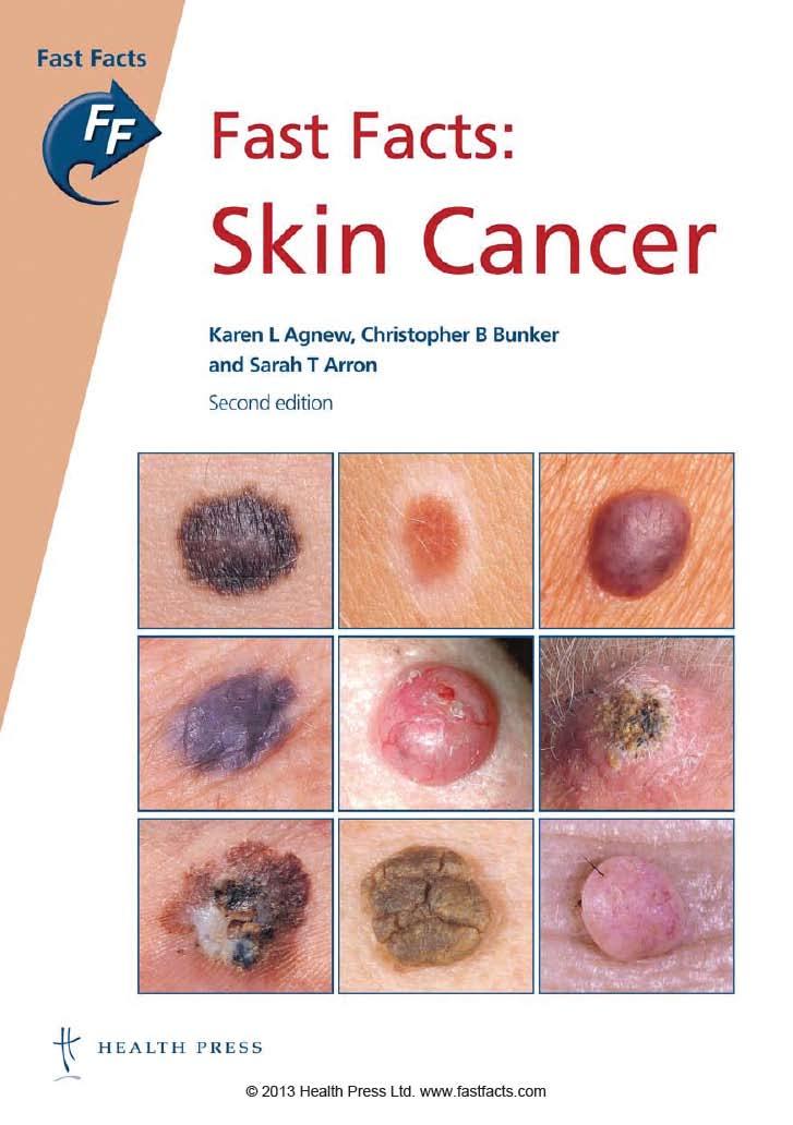
8 Introduction Skin cancer is the most common human cancer worldwide, and the incidence continues to increase. However, most skin cancers are preventable and treatable. It is of crucial importance for all of us to understand how we can prevent, diagnose and treat these common cancers. Fast Facts: Skin Cancer provides essential information that will help healthcare professionals, medical students and the public to understand this important and rapidly evolving area of medicine. There are three main types of skin cancer: malignant melanoma and the two non-melanoma skin cancers, basal cell carcinoma (BCC) and squamous cell carcinoma (SCC). Most skin malignancies are curable if they are diagnosed early enough, but both melanoma, in particular, and SCC can kill. Theoretically, skin cancer can be prevented by educating people about recreational habits, clothing, sunscreens and sunbeds (primary prevention), but this approach could take a generation or more before it has a sizeable effect. Meanwhile, emerging evidence surrounding the role of vitamin D means that physicians need to consider supplementation for individuals adhering to strict sun avoidance practices. When skin cancer does occur, early recognition and specialist referral saves lives. Serious morbidity and mortality can be prevented or reduced by educating patients and physicians alike, so that at-risk individuals and early lesions are identified with accuracy. There are nine tumors on the cover of this book, and three of them are malignant cancers can you identify them? This book will show you how. Surgery is usually the treatment of choice for skin cancer, although there are other options. Although relatively simple surgery suffices for most cancers in most patients, some require multidisciplinary input from a dermatologist, surgeon, pathologist, radiotherapist or radiation oncologist, and medical oncologist. Mohs micrographic surgery has resulted in significantly improved cure rates for selected skin cancers, particularly the non-melanoma skin cancers BCC and SCC. Yet, 7
9 Fast Facts: Skin Cancer despite recent advances in the treatment of disseminated disease, our methods remain inaccurate and mortality of metastatic skin cancer is high. In this second edition of Fast Facts: Skin Cancer we present updated facts about the epidemiology and causation of skin cancer, alongside a commonsense approach to identification and management. Our aim is to provide an evidence-based, practical resource that will answer common questions, and reflect the national and international consensus guidelines that are constantly evolving. It will assist practice, education, training, audit and research. Fast Facts: Skin Cancer is written by dermatologists to provide you with the information that your dermatologist wants you to know. We have designed an easy-to-read, fact-filled and practical primer to skin cancer. Whether you are a physician, nurse, medical student or other healthcare professional, or an interested patient or curious layman, this book is for you. Read on and join the effort to prevent and treat skin cancer! Acknowledgment: We would like to thank Dr Barbara A Gilchrest, Professor of Dermatology, Boston University School of Medicine, for her contribution to the first edition of this handbook. 8

10 1 Epidemiology Basal cell carcinoma Basal cell carcinoma (BCC) is the most common malignant neoplasm in white individuals. Its incidence has increased in recent decades: the highest rates are in Australia, where it affects over 2% of men. In the UK, USA, Australia and New Zealand these basic data are still not collected centrally and estimates vary. Extrapolated data reported in a recent paper suggest that, in 2010, approximately patients were treated surgically for BCCs in the UK. The tumor most commonly arises on the head and neck (Figure 1.1) and, overall, the incidence is higher in men than in women. Risk factors. The risk of BCC is increased in those who: had episodes of severe childhood sunburn have red hair tan poorly. The association with sunlight is not well understood. Cumulative ultraviolet (UV) radiation was thought to be the most important risk factor. However, exposure to the sun during childhood and Figure 1.1 Basal cell carcinoma most commonly arises on the head and neck. 9
.")
11 Fast Facts: Skin Cancer adolescence particularly intense intermittent exposure may be of greater importance in the development of BCC than previously thought. Other risk factors for BCC include: North European ancestry a positive family history immunosuppression exposure to arsenic previous radiotherapy. BCC can develop in a sebaceous nevus (Figure 1.2). It is also associated with a number of conditions, including albinism, xeroderma pigmentosum (a rare autosomal-recessive disorder that affects DNA repair), nevoid BCC syndrome (Gorlin s syndrome) and HIV infection. Table 1.1 summarizes the risk factors for BCC compared with squamous cell carcinoma (SCC) and melanoma. Squamous cell carcinoma Cutaneous SCC is the second most common skin cancer in white individuals; SCC represents roughly 20% of all non-melanoma skin carcinomas. The incidence of non-melanoma skin cancer (BCC and SCC together) is approximately fold greater than that of melanoma. SCC is most commonly seen in the elderly and is three times more common in men than women. The incidence is rising, and is highest 10 Figure 1.2 Sebaceous nevus on scalp basal cell carcinoma and squamous cell carcinoma can develop from this type of birthmark.
12 Epidemiology TABLE 1.1 Risk factors for skin cancer Basal cell carcinoma Squamous cell carcinoma Melanoma Sex Male > female Male > female Male > female Relationship to sun exposure Childhood; intermittent Chronic Childhood; intermittent Sunburn history Severe sunburn in childhood Episode of severe sunburn Episode of severe sunburn Tanning ability Non-tanners Non-tanners Non-tanners Skin color Fair skin/freckles Fair skin/freckles Fair skin/ freckles Hair color Red Red/blond Red/blond Eye color Light eye color Light eye color Blue Precursor lesions Actinic keratosis Actinic keratosis Nevi in countries with high sun exposure. Non-melanoma skin cancer morbidity and mortality statistics are not registered in the UK, USA, New Zealand or Australia. The American Cancer Society estimates that 3.5 million cases of non-melanoma skin cancer were diagnosed in In Australia, the total number of non-melanoma skin cancer treatments rose to in 2010, and it has been suggested that UK figures are likely to be similar. Lesions occur on sun-exposed sites, predominantly the head and neck (Figure 1.3). Risk factors. UV radiation is the strongest etiologic factor. In particular, sun exposure, both recent and cumulative, is implicated in SCC development. This malignancy is more common in fair-skinned individuals with red or blond hair (see Table 1.1). 11
 (e.g.")
13 Fast Facts: Skin Cancer Figure 1.3 Squamous cell carcinoma on the lip these lesions predominantly occur on sun-exposed sites, particularly the head and neck. 12 The acquired risk factors for SCC include: a history of severe sunburn sunbed use 200+ treatments with psoralen and UVA phototherapy (PUVA) (e.g. for psoriasis) smoking chronic immunosuppression (as occurs with alcoholism, HIV infection, chronic lymphatic leukemia and organ transplantation). Much of the research identifying the association between immunosuppression and SCC has been performed in the organ transplant population. The risk of developing SCC is increased in organ transplant recipients, and the ratio of BCC to SCC incidence is reversed in this population. Other predisposing factors for SCC include previous exposure to polycyclic hydrocarbons or radiation (Figure 1.4), exposure to arsenic, and infection with the human papillomavirus (HPV). Further genetic risk factors include conditions with P53 (also known as TP53) gene mutation, xeroderma pigmentosum and albinism. In addition, invasive SCC is recognized to develop from actinic keratosis and in situ SCC, as well as at sites of chronic injury, such as ulceration, infection, scars and epidermolysis bullosa
14 3 Clinical features and diagnosis Clinical assessment is fundamental to the diagnostic process and begins with the patient history. A story of changing, enlarging and irritating lesions should alert suspicion. Patients who present with a skin cancer invariably describe a growth that is changing. It is reassuring to hear that a lesion is longstanding and is not changing. Rigorous systematic clinical examination is mandatory to elicit key morphologic features (size, shape, symmetry, surface, margin, color, texture) of benign or malignant skin tumors. Primary care providers should consider early referral to a dermatologist for evaluation of any concerning lesion. A clear diagnosis of a skin tumor is usually attained after clinical assessment, but histology is regarded as the gold standard. The dermascope/dermatoscope, or surface microscope, is a handheld instrument that assists with the examination of cutaneous lesions. Generally, this light magnification system has a fixed magnification of 10. Oil is usually applied to the skin lesion to reduce light refraction; however, newer systems with cross-polarized light enable examination without oil. The role of the dermascope is still evolving, but is of growing importance as the sensitivity and specificity of individual signs and patterns are described. Biopsy and histology Biopsy and histology are integral to the diagnosis of skin cancer. Shave biopsy. When the skin lesion is pedunculated or only appears to involve the epidermis and upper dermis, a superficial slice of skin can be removed with a scalpel or razor blade. Sutures are not usually required and electrodesiccation or aluminum chloride may be applied to achieve hemostasis. The procedure leaves only a small scab that heals in 2 4 weeks. 28 Punch biopsy. Dermatologists are enthusiasts for this biopsy, and use it for suspected skin dermatoses and cancers. A circular blade attached
15 Clinical features and diagnosis to a handle is rotated through the skin, capturing a core of tissue from the epidermis to the subcutaneous fat. The core is removed and sent to the laboratory for histological assessment. Once the biopsy has been taken, the residual skin defect can be closed with sutures or adhesive strips. Alternatively, the wound can be left to heal on its own without stitches. A range of skin biopsy diameters is available, but in most situations a punch biopsy diameter of 3 6 mm is appropriate. Excisional biopsy involves the removal of the entire lesion. The excision is usually elliptical. Excisional biopsy is the favored investigation for suspected melanomas in the UK, New Zealand and Australia. This technique allows the pathologist to evaluate the entire lesion. Incisional biopsy involves removing an ellipse of tissue using a surgical blade, so that a proportion of the lesion can be sent for histological assessment. The remaining defect is usually repaired with sutures. This procedure is used when an excisional biopsy is technically challenging, such as in a large lentigo maligna melanoma. Benign lesions Acquired melanocytic nevi usually become apparent after the age of 12, and the numbers increase over the next 20 years before they start to dissipate. There are several types of acquired nevi: junctional compound intradermal halo pigmented spindle cell nevus of Reed Spitz blue. Junctional nevi usually develop in adolescents, compound nevi in young adults and intradermal nevi in older adults. This observation has led to the hypothesis that throughout life the nevomelanocyte composition progresses from a junctional nevus to a compound nevus and then on to an intradermal nevus. 29


. It tends to have a uniform color in the range of tan to dark brown.")
16 Fast Facts: Skin Cancer (a) (b) Figure 3.1 Junctional nevus: a flat, pigmented lesion, usually < 6 mm in diameter. Junctional nevus most commonly arises in childhood and early adulthood. It is a flat sharply demarcated pigmented lesion, usually with a diameter of less than 6 mm (Figure 3.1). Junctional nevi often develop on palms and soles and tend to maintain the normal skin markings. Histologically, discrete nests of nevomelanocytes can be found in contact with the basal layer of the epidermis. Compound nevus is a slightly elevated well-delineated pigmented lesion, most commonly found in adults (Figure 3.2). It tends to have a uniform color in the range of tan to dark brown. A proportion of the nevomelanocytes are free within the papillary dermis, whereas others are in contact with the overlying epidermal basal layer. (a) (b) (c) Figure 3.2 Compound nevus: an elevated well-delineated pigmented lesion, which tends to have a uniform color, ranging from tan to dark brown. 30
Skin Cancer. All You Need to Keep up to Speed. by Karen L Agnew, Barbara A Gilchrest and Christopher B Bunker. Epidemiology 9.
 Skin Cancer All You Need to Keep up to Speed by Karen L Agnew, Barbara A Gilchrest and Christopher B Bunker Epidemiology 9 Pathogenesis 17 Clinical features and diagnosis 26 Management 64 Prognosis 86
Dermatopathology: The tumor is composed of keratinocytes which show atypia, increase mitoses and abnormal mitoses.
 Squamous cell carcinoma (SCC): A common malignant tumor of keratinocytes arising in the epidermis, usually from a precancerous condition: 1- UV induced actinic keratosis, usually of low grade malignancy.
Clinical characteristics
 Skin Cancer Fernando Vega, MD Seattle Healing Arts Clinical characteristics Precancerous lesions Common skin cancers ACTINIC KERATOSIS Precancerous skin lesions Actinic keratoses Dysplastic melanocytic
Glenn D. Goldman, MD. University of Vermont Medical Center. University of Vermont College of Medicine
 Glenn D. Goldman, MD University of Vermont Medical Center University of Vermont College of Medicine Recognize and identify the main types of skin cancer and their precursors Identify and understand new
IT S FUNDAMENTAL MY DEAR WATSON! A SHERLOCKIAN APPROACH TO DERMATOLOGY
 IT S FUNDAMENTAL MY DEAR WATSON! A SHERLOCKIAN APPROACH TO DERMATOLOGY Skin, Bones, and other Private Parts Symposium Dermatology Lectures by Debra Shelby, PhD, DNP, FNP-BC, FADNP, FAANP Debra Shelby,
Benign and malignant epithelial lesions: Seborrheic keratosis: A common benign pigmented epidermal tumor occur in middle-aged or older persons more
 Benign and malignant epithelial lesions: Seborrheic keratosis: A common benign pigmented epidermal tumor occur in middle-aged or older persons more common on the trunk; but extremities, head and neck are
Learning Objectives. Tanning. The Skin. Classic Features. Sun Reactive Skin Type Classification. Skin Cancers: Preventing, Screening and Treating
 Learning Objectives Skin Cancers: Preventing, Screening and Treating Robert A. Baldor, MD, FAAFP Professor, Family Medicine & Community Health University of Massachusetts Medical School Distinguish the
Identifying Skin Cancer. Mary S. Stone MD Professor of Dermatology and Pathology University of Iowa Carver College of Medicine March, 2018
 Identifying Skin Cancer Mary S. Stone MD Professor of Dermatology and Pathology University of Iowa Carver College of Medicine March, 2018 American Cancer Society web site Skin Cancer Melanoma Non-Melanoma
Know who is at risk: LOOK! for ABCDs, rapidly changing lesions, do a biopsy when indicated
 Lindy P. Fox, MD Assistant Professor Director, Hospital Consultation Service Department of Dermatology University of California, San Francisco Applies to adults without history of malignancy or premalignant
MECHANISMS OF HUMAN DISEASE: LABORATORY SESSION PATHOLOGY OF THE SKIN LAB. Friday, February 12, :30 am 11:00 am
 MECHANISMS OF HUMAN DISEASE: LABORATORY SESSION PATHOLOGY OF THE SKIN LAB Friday, February 12, 2012 9:30 am 11:00 am FACULTY COPY GOALS: Describe the basic clinical and morphologic features of various
Pathology of the skin. 2nd Department of Pathology, Semmelweis University
 Pathology of the skin 2nd Department of Pathology, Semmelweis University Histology of the skin Epidermis: Stratum corneum Stratum granulosum Stratum spinosum Stratum basale Dermis: papillary and reticular
Know who is at risk: LOOK! for ABCDs, rapidly changing lesions, do a biopsy when indicated
 Lindy P. Fox, MD Associate Professor Director, Hospital Consultation Service Department of Dermatology University of California, San Francisco Applies to adults without history of malignancy or premalignant
Cutaneous Malignancies: A Primer COPYRIGHT. Marissa Heller, M.D.
 Cutaneous Malignancies: A Primer Marissa Heller, M.D. Associate Director of Dermatologic Surgery Department of Dermatology Beth Israel Deaconess Medical Center December 10, 2016 Skin Cancer Non-melanoma
Periocular Malignancies
 Periocular Malignancies Andrew Gurwood, O.D., F.A.A.O., Dipl. Marc Myers, O.D., F.A.A.O. Drs. Myers and Gurwood have no financial interests to disclose. Course Description Discussion of the most common
Skin Cancer - Non-Melanoma
 Skin Cancer - Non-Melanoma Introduction Each year, millions of people find out that they have skin cancer. Skin cancer is almost 100% curable if found early and treated right away. It is possible to prevent
Skin Cancer. 5 Warning Signs. American Osteopathic College of Occupational and Preventive Medicine OMED 2012, San Diego, Monday, October 8, 2012 C-1
 Skin Cancer AMERICAN OSTEOPATHIC COLLEGE OF OCCUPATIONAL & PREVENTIVE MEDICINE OMED 2012 October 8, 2012 E. Robert Wanat II, D.O., M.P.H. Learning Objectives: Identify the 3 Basic Types of Skin Cancer
Large majority caused by sun exposure Often sun exposure before age 20 Persons who burn easily and tan poorly are at greatest risk.
 Basics of Skin Cancer Detection and Treatment of Non- Melanoma Skin Cancers Large majority caused by sun exposure Often sun exposure before age 20 Persons who burn easily and tan poorly are at greatest
Glenn D. Goldman, MD. Fletcher Allen Health Care. University of Vermont College of Medicine
 Glenn D. Goldman, MD Fletcher Allen Health Care University of Vermont College of Medicine Recognize and identify the main types of skin cancer Understand how and why Mohs surgery is utilized for the treatment
Benign versus Cancerous Lesions How to tell the difference FMF 2014 Christie Freeman MD, CCFP, DipPDerm, MSc
 1 Benign versus Cancerous Lesions How to tell the difference FMF 2014 Christie Freeman MD, CCFP, DipPDerm, MSc Benign lesions Seborrheic Keratoses: Warty, stuck-on Genetics and birthdays Can start in late
General information about skin cancer
 Skin Cancer General information about skin cancer Key points Skin cancer is a disease in which malignant (cancer) cells form in the tissues of the skin. There are different types of cancer that start in
MECHANISMS OF HUMAN DISEASE: LABORATORY SESSION PATHOLOGY OF THE SKIN LAB. Friday, February 13, :30 am 11:00 am
 MECHANISMS OF HUMAN DISEASE: LABORATORY SESSION PATHOLOGY OF THE SKIN LAB Friday, February 13, 2009 9:30 am 11:00 am FACULTY COPY GOALS: Describe the basic clinical and morphologic features of various
Living Beyond Cancer Skin Cancer Detection and Prevention
 Living Beyond Cancer Skin Cancer Detection and Prevention Cutaneous Skin Cancers Identification Diagnosis Treatment options Prevention What is the most common cancer in people? What is the most common
 Page 1 of 15 Title Authored By Course No Contact Hours 2 Skin Cancer the Real Picture for Early Detection and Treatment Cheryl Sommer RN, MSN, ARNP SC120604 Purpose The purpose of this course is to provide
Skin Malignancies Non - Melanoma & Melanoma Marilyn Ng, MD Dept. of Surgery M&M Conference Downstate Medical Center July 19, 2012
 Skin Malignancies Non - Melanoma & Melanoma Marilyn Ng, MD Dept. of Surgery M&M Conference Downstate Medical Center July 19, 2012 Case Presentation 57 yo man with 3 month hx of a nonhealing < 1 cm right
Dermatology for the PCP Deanna G. Brown, MD, FAAD Susong Dermatology Consulting Staff at CHI Memorial
 Dermatology for the PCP Deanna G. Brown, MD, FAAD Susong Dermatology Consulting Staff at CHI Memorial Cutaneous Oncology for the PCP Deanna G. Brown, MD, FAAD Susong Dermatology Consulting Staff at CHI
Regeneron and Sanofi are financial supporters of The Skin Cancer Foundation and collaborated in the development of this article. US-ONC /2018
 A D E E P E R L O O K When detected early, most cases of local cutaneous squamous cell carcinoma are easily treated and usually cured. But when they become more advanced, this second most common form of
Dermatological Manifestations in the Elderly. Sanjay Siddha Staff Dermatologist UHN & MSH
 Dermatological Manifestations in the Elderly Sanjay Siddha Staff Dermatologist UHN & MSH Disclosure No actual or potential conflicts of interest or commercial relationships to declare Objectives Recognize
Malignant Melanoma Early Stage. A guide for patients
 This melanoma patient brochure is designed to help educate melanoma patients and their caregivers. It was developed under the guidance of Dr. Michael Smylie, Professor, Department of Oncology, University
Actinic keratosis (AK): Dr Sarma s simple guide
: Dr Sarma s simple guide") Actinic keratosis (AK): Dr Sarma s simple guide Actinic keratosis is a very common lesion that you will see in your day-to-day practice. First, let me explain the name Actinic keratosis. It means keratosis
Actinic keratosis (AK): Dr Sarma s simple guide Actinic keratosis is a very common lesion that you will see in your day-to-day practice. First, let me explain the name Actinic keratosis. It means keratosis
Fast Facts: Multiple Sclerosis
 Fast Facts Fast Facts: Multiple Sclerosis George D Perkin and Jerry S Wolinsky Second edition 2006 Health Press Ltd. www.fastfacts.com Fast Facts Fast Facts: Multiple Sclerosis Second edition George D
Melanoma: The Basics. What is a melanocyte?
 Melanoma: The Basics What is a melanocyte? A melanocyte is a normal cell, found in the skin, which produces melanin. Melanin is a black or dark brown pigment that is seen in the skin, hair, and parts of
Skin Cancer 101: Diagnosis and Management of the Most Common Cancer
 Skin Cancer 101: Diagnosis and Management of the Most Common Cancer Sarah Patton, PA-C, MSHS Skin Surgery Center www.skinsurgerycenter.com Seattle/Bellevue, WA Skin cancer Skin cancer is by far the most
Premalignant skin tumours
 Chapter 14: Premalignant skin tumours page: 434 Premalignant skin tumours page: 435 Solar keratoses (senile keratoses) Raised red and well-defined plaques with a rough surface covered in scales of varying
LUMPS AND BUMPS: AN ORGANIZED APPROACH TO DIAGNOSIS AND MANAGEMENT
 LUMPS AND BUMPS: AN ORGANIZED APPROACH TO DIAGNOSIS AND MANAGEMENT Tammy P. Than, M.S., O.D., F.A.A.O. The University of Alabama at Birmingham / School of Optometry 1716 University Blvd. Birmingham, AL
SQUAMOUS CELL CARCINOMA
 SQUAMOUS CELL CARCINOMA What are the aims of this leaflet? This leaflet has been written to help you understand more about squamous cell carcinomas of the skin. It tells you what they are, what causes
Basal cell carcinoma 5/28/2011
 Goal of this Presentation A practical approach to the diagnosis of cutaneous carcinomas and their mimics Thaddeus Mully, MD University of California San Francisco To review common non-melanoma skin cancers
Skin Cancer. Dr Elizabeth Ogden Associate Specialist in Dermatology East and North Herts Dr Elizabeth Ogden
 Skin Cancer Dr Elizabeth Ogden Associate Specialist in Dermatology East and North Herts 13.10.16 Skin Cancer Melanoma mole cancer - is a true cancer which can metastasize and kill Non Melanoma skin cancer
SKIN CANCER. Most common cancer diagnosis 40% of all cancers
 SKIN CANCER Most common cancer diagnosis 40% of all cancers OBJECTIVES Review common and uncommon cancers of the skin. Special emphasis on melanoma and dysplastic nevus Review pathology/tnm/staging, which
I have a skin lump doc! What s next? 12 th August 2017 Dr. Sue-Ann Ho Ju Ee
 I have a skin lump doc! What s next? 12 th August 2017 Dr. Sue-Ann Ho Ju Ee Some thoughts Is this skin cancer? How common is this? How likely is this in this patient? What happens next if it s something
Fast Facts: Ovarian Cancer
 Fast Facts Fast Facts: Ovarian Cancer Christina Fotopoulou MD PhD Consultant Gynaecological Oncologist Queen Charlotte s and Chelsea Hospital London, UK Thomas J Herzog MD Professor of Obstetrics and Gynecology
Principles of Anatomy and Physiology
 Principles of Anatomy and Physiology 14 th Edition CHAPTER 5 The Integumentary System Introduction The organs of the integumentary system include the skin and its accessory structures including hair, nails,
Oral and Maxillofacial Surgery Department
 Oral and Maxillofacial Surgery Department This leaflet explains: Lentigo Maligna What are the aims of this leaflet? This leaflet has been written to help you understand more about lentigo maligna and melanoma
MELANOMA. Some people are more likely to get a m Melanoma than others:
 MELANOMA This leaflet has been written to help you understand more about Melanoma. It tells you what is it, what causes it, what can be done about it, how it can be prevented, and where you can find out
Integumentary System-Skin and Body Coverings
 Integumentary System-Skin and Body Coverings List the four types of epithelial or connective membranes. The epithelial cutaneous includes your and is exposed to the. Its function is to. An example is..
David B. Troxel, MD. Common Medicolegal Situations: Misdiagnosis of Melanoma
 Common Medicolegal Situations: Misdiagnosis of Melanoma David B. Troxel, MD Medical Director, The Doctors Company, Napa, California Clinical Professor Emeritus, University of California at Berkeley Past
Periocular skin cancer
 Periocular skin cancer Information for patients Skin cancer involving the skin of the eyelid or around the eye is called a periocular skin cancer. Eyelid skin cancers occur most often on the lower eyelid,
MELANOMA. 4 Fitzroy Square, London W1T 5HQ Tel: Fax: Registered Charity No.
 MELANOMA This leaflet had been written to help you understand more about melanoma. It tells you what it is, what causes it, what can be done about it, how it can be prevented, and where you can find out
Dermoscopy: Recognizing Top Five Common In- Office Diagnoses
 Dermoscopy: Recognizing Top Five Common In- Office Diagnoses Vu A. Ngo, DO Department of Family Medicine and Dermatology Choctaw Nation Health Services Authority Learning Objectives Introduction to dermoscopy
Disclosures. I have no conflicts of interest to disclose
 Disclosures I have no conflicts of interest to disclose Lindy P. Fox, MD Associate Professor Director, Hospital Consultation Service Department of Dermatology University of California, San Francisco 2
SKIN. 3. How is the skin structured around the finger joints to allow for flexible movement of the fingers?
 SKIN Objectives for Exam #1: 1. List various skin structures and describe their functions. 2. Describe skin responses to increases and decreases in body temperature. 3. Provide examples of various skin
Wellness Along the Cancer Journey: Cancer Types Revised October 2015 Chapter 7: Skin Cancer
 Wellness Along the Cancer Journey: Cancer Types Revised October 2015 Chapter 7: Skin Cancer Cancer Types Rev. 10.20.15 Page 56 Skin Cancer Group Discussion True False Not Sure 1. People with darker skin
Common Benign Lesions and Skin Cancers. 22nd May 2015 Dr Mark Foley
 Common Benign Lesions and Skin Cancers 22nd May 2015 Dr Mark Foley Thank you for downloading this file. This intended to supplement the presentation given at the NZ Wound Care Conference, it is not intended
VACAVILLE DERMATOLOGY
 Connecting the Dots on those Spots NANDAN V. KAMATH, M.D. VACAVILLE DERMATOLOGY Sources All of the photos were taken with permission from the Dermnet NZ website - Dermnet New Zealand after communicating
Skin Malignancies. Presented by Dr. Douglas Paauw
 Skin Malignancies Presented by Dr. Douglas Paauw Disclosure: Dr. Paauw has no significant financial interest in any of the products or manufacturers mentioned. How Common Is Skin Cancer? *½ of all White
Histopathology: skin pathology
 Histopathology: skin pathology These presentations are to help you identify, and to test yourself on identifying, basic histopathological features. They do not contain the additional factual information
Skin Cancer. There are many types of diseases. From a simple cold to the deadly disease
 Skin Cancer Skin Cancer 1 There are many types of diseases. From a simple cold to the deadly disease Mesothelioma. Some diseases are almost harmless and some can kill you in less than a year. There are
Nonmelanoma skin cancers
 Skin cancer Philip Clarke Nonmelanoma skin cancers Treatment options Background Australia has one of the highest skin cancer rates in the world. Early detection and treatment of skin cancer is vital to
Mohs. Micrographic Surgery. For Treating Skin Cancer
 Mohs Micrographic Surgery For Treating Skin Cancer Skin Cancer Basics Skin cancer is common. Over the past three decades, more people have had skin cancer than all other cancers combined. Each year in
LENTIGO SIMPLEX. Epidemiology
 LENTIGO SIMPLEX Epidemiology The frequency of lentigo simplex in children and adults has not been determined. There does not appear to be a racial or gender predilection. Lentigo simplex is the most common
Non-melanoma Skin Cancer
 Non-melanoma Skin Cancer Understanding your diagnosis 1 888 939-3333 cancer.ca Non-melanoma Skin Cancer Understanding your diagnosis When you first hear that you have cancer, you may feel alone and afraid.
Skin Cancers Emerging Trends and Treatment Approaches
 Skin Cancers Emerging Trends and Treatment Approaches Andrei Metelitsa, MD, FRCPC, FAAD Clinical Associate Professor, Dermatology, U of C Co-Director, Institute for Skin Advancement Copyright 2017 by Sea
Pathology. Skin Tumor. Bayan N. Mohammad 15/10/2015. Mohammad al-orjani. Page 0 of 23
 #7 35 Pathology Skin Tumor Bayan N. Mohammad 15/10/2015 Mohammad al-orjani Page 0 of 23 بسم هللا الرحمن الرحيم GREETINGS This lecture is about skin tumors, all the slides are included and every slide will
Diagnostics guidance Published: 11 November 2015 nice.org.uk/guidance/dg19
 VivaScope 1500 and 3000 imaging systems for detecting skin cancer lesions Diagnostics guidance Published: 11 November 2015 nice.org.uk/guidance/dg19 NICE 2018. All rights reserved. Subject to Notice of
Technicians & Nurses Program
 ASCRS ASOA Symposium & Congress Technicians & Nurses Program May 6-10, 2016 New Orleans Evaluation and Treatment of Eyelid Malignancies Richard C. Allen MD PhD FACS Professor Section of Ophthalmology Dept.
Identifying Benign and Malignant Skin Lesions. No Disclosures. Common Benign Lesions. Benign Lesions 2/25/2018. Stucco Keratoses.
 Dermatology in Primary Care Identifying Benign and Malignant Skin Lesions Christy Quire Baker, APRN, FNP-BC, DCNP Dermatology Certified Nurse Practitioner No Disclosures Common Benign Lesions Seborrheic
Basal cell carcinoma
 Basal cell carcinoma Delivering the best in care UHB is a no smoking Trust To see all of our current patient information leaflets please visit www.uhb.nhs.uk/patient-information-leaflets.htm What are basal
Melanoma and Dermoscopy. Disclosure Statement: ABCDE's of melanoma. Co-President, Usatine Media
 Melanoma and Dermoscopy Richard P. Usatine, MD, FAAFP Professor, Family and Community Medicine Professor, Dermatology and Cutaneous Surgery Medical Director, University Skin Clinic University of Texas
Describe the functions of the vertebrate integumentary system. Discuss the structure of the skin and how it relates to function.
 Chapter 5 Describe the functions of the vertebrate integumentary system. Discuss the structure of the skin and how it relates to function. Explain the basis for different skin colors. Describe the structure
Malignant tumors of melanocytes: Part 1. Deba P Sarma, MD., Omaha
 Malignant tumors of melanocytes: Part 1 Deba P Sarma, MD., Omaha The melanocytic tumor is one of the most difficult and confusing areas in Dematopathology. It is true that most (95%) of such lesions are
2004 Health Press Ltd.
 Eczema and Contact Dermatitis John Berth-Jones Consultant Dermatologist University Hospitals Coventry and Warwickshire NHS Trust, Coventry, UK Eunice Tan Specialist Registrar in Dermatology Norfolk and
Disclosures. Melanoma and Non melanoma Skin Cancer: What You Need to Know. I have no conflicts of interest to disclose
 Disclosures Melanoma and Non melanoma Skin Cancer: What You Need to Know I have no conflicts of interest to disclose Lindy P. Fox, MD Associate Professor Director, Hospital Consultation Service Department
MOHS MICROGRAPHIC SURGERY: AN OVERVIEW
 MOHS MICROGRAPHIC SURGERY: AN OVERVIEW SKIN CANCER: Skin cancer is far and away the most common malignant tumor found in humans. The most frequent types of skin cancer are basal cell carcinoma, squamous
Skin lesions The Good and the Bad. Dr Virginia Hubbard Ipswich Hospital NHS Trust Barts and the London School of Medicine and Dentistry
 Skin lesions The Good and the Bad Dr Virginia Hubbard Ipswich Hospital NHS Trust Barts and the London School of Medicine and Dentistry Case 1 32 year old woman Australian Lesion on back New hair growing
Interesting Case Series. Aggressive Tumor of the Midface
 Interesting Case Series Aggressive Tumor of the Midface Adrian Frunza, MD, Dragos Slavescu, MD, and Ioan Lascar, MD, PhD Bucharest Emergency Clinical Hospital, Bucharest University School of Medicine,
Have a Voice in Your Choice!
 Have a Voice in Your Choice! BLU-U Blue Light Photodynamic Therapy The LEVULAN KERASTICK for Topical Solution plus blue light illumination using the BLU-U Blue Light Photodynamic Therapy Illuminator is
Human Anatomy & Physiology
 PowerPoint Lecture Slides prepared by Barbara Heard, Atlantic Cape Community College Ninth Edition Human Anatomy & Physiology C H A P T E R 5 Annie Leibovitz/Contact Press Images 2013 Pearson Education,
Squamous Cell Skin Cancer
 Please complete our online survey at NCCN GUIDELINES FOR PATIENTS NCCN.org/patients/survey 2019 Squamous Cell Skin Cancer Presented with support from: Available online at NCCN.org/patients Ü Squamous Cell
SKIN HISTOLOGY AND FUNCTION
 SKIN HISTOLOGY AND FUNCTION THREE LAYERS : EPIDERMIS BASEMENT MEMBRANE DERMIS EPIDERMIS : COMPOSED OF KERATINOCYTES NO MATRIX DEEP BASAL LAYER MITOTICALLY ACTIVE SPINOUS LAYER MATURE HYALIN HORNY LAYER
11/8/2012. Chapter 6 Part 1 Objectives: Skin = Integument = Cutaneous Membrane. The Structure of Skin. Epidermis
 Chapter 6 Part 1 Objectives: Define organ, and associate the skin as an organ of the integumentary system. List the general functions of the skin. Describe the structure of the layers of the skin. Summarize
Actinic Keratoses and Bowen s disease
 Actinic Keratoses and Bowen s disease Delivering the best in care UHB is a no smoking Trust To see all of our current patient information leaflets please visit www.uhb.nhs.uk/patient-information-leaflets.htm
Integumentary System
 Integumentary System The integumentary system is commonly known as the Skin Largest organ of human body 10% total body weight and would cover over 20 square feet Functions of Skin 1. Protection Barrier
Melanocytic Tumors
 Melanocytic Tumors 803-808-7387 www.gracepets.com These notes are provided to help you understand the diagnosis or possible diagnosis of cancer in your pet. For general information on cancer in pets ask
An Overview of Melanoma. Harriet Kluger, M.D. Associate Professor Section of Medical Oncology Yale Cancer Center
 An Overview of Melanoma Harriet Kluger, M.D. Associate Professor Section of Medical Oncology Yale Cancer Center Melanoma Statistics Median age at presentation 45-55 55 years Incidence: 2003 54,200 cases
Gastroenterology Second edition
 Clinical drawings for your patients Patient Pictures books are: PATI ENT PICTU RES Patient Pictures time-saving books for doctors and nurses. Use them when describing medical conditions and treatments
Skin Cancer Awareness
 Skin Cancer Awareness Presented by BHS Call: 800-327-2251 Visit: www.bhsonline.com 2016 BHS. All rights reserved. 1 Training Summary More than 3.5 million new cases of skin cancer will be diagnosed in
Dual Wavelength Phototherapy System
 Dual Wavelength Phototherapy System The AKLARUS Blue and Red Combination System is an effective, drugfree alternative for treating acne & photodamaged skin. The non-invasive Aklarus treatment has been
Skin Tumors in Children
 AAD San Diego S021 2018 Skin Tumors in Children Jane M. Grant-Kels, MD,FAAD grant@uchc.edu Founding Chair Emeritus, Derm Dept, UCONN Vice Chair Dept of Dermatology Professor of Dermatology, Pathology and
Springer Healthcare. Staging and Diagnosing Cutaneous Melanoma. Concise Reference. Dirk Schadendorf, Corinna Kochs, Elisabeth Livingstone
 Concise Reference Staging and Diagnosing Cutaneous Melanoma Dirk Schadendorf, Corinna Kochs, Elisabeth Livingstone Extracted from Handbook of Cutaneous Melanoma: A Guide to Diagnosis and Treatment Published
Moh's Surgery Information Packet
 Moh's Surgery Information Packet BE SURE TO BRING THE FOLLOWING TO YOUR APPOINTMENT Insurance card Insurance referral (if required by your insurance) Name and address of your primary care provider as well
See spot change: Lesion identification and management in primary care ERIN HENNESSEY DNP, APRN, FNP-C
 See spot change: Lesion identification and management in primary care ERIN HENNESSEY DNP, APRN, FNP-C Learning objectives Discuss malignant skin lesions commonly seen in primary care. Identify common treatments
Atypical Nevi When to Re-excise. Catherine Barry, DO Dermatopathologist
 Atypical Nevi When to Re-excise Catherine Barry, DO Dermatopathologist Why talk about skin cancer? Because it s the most common type of cancer! Non-melanoma Skin Cancers Basal Cell Carcinoma Squamous Cell
Toby Maurer, MD University of California, San Francisco. Lifetime risk of an American developing melanoma
 Distinguishing Pigmented Skin Lesions and Melanoma Toby Maurer, MD University of California, San Francisco Epidemiology of Melanoma Lifetime risk of an American developing melanoma 1935: 1 in 1500 1980:
Alcohol should be avoided for 3 days prior to surgery and 2 days after the procedure.
 Mohs Surgery Information Packet Be sure to bring the following to your appointment: Insurance Card Insurance Referral ( If required by your insurance) Name and address of your primary care provider as
Toby Maurer, MD University of California, San Francisco. Lifetime risk of an American developing melanoma
 Distinguishing Pigmented Skin Lesions and Melanoma Toby Maurer, MD University of California, San Francisco Epidemiology of Melanoma Lifetime risk of an American developing melanoma 1935: 1 in 1500 1980:
WHAT DOES THE PATHOLOGY REPORT MEAN?
 Melanoma WHAT IS MELANOMA? Melanoma is a type of cancer that affects cells called melanocytes. These cells are found mainly in skin but also in the lining of other areas such as nose and rectum, and also
المركب النموذج--- سبيتز وحمة = Type Spitz's Nevus, Compound SPITZ NEVUS 1 / 7
 SPITZ NEVUS 1 / 7 Epidemiology An annual incidence rate of 1.4 cases of Spitz nevus per 100,000 individuals has been estimated in Australia, compared with 25.4 per 100,000 individuals for cutaneous melanoma
Multiple Primary Melanoma in a Thai Male: A Case Report
 Case Report Multiple Primary Melanoma in a Thai Male: A Case Report J Med Assoc Thai 2014; 97 (Suppl. 2): S234-S238 Full text. e-journal: http://www.jmatonline.com Kittisak Payapvipapong MD*, Pinyapat
DERMATOLOGY ROTATION: COMPETENCY-BASED GOALS AND OBJECTIVES
 UNC DIVISION OF PLASTIC AND RECONSTRUCTIVE SURGERY DERMATOLOGY ROTATION: COMPETENCY-BASED GOALS AND OBJECTIVES MEDICAL KNOWLEDGE A. Anatomy/Physiology/Embryology Goal: The resident will have knowledge
SUN, SAVVY AND SKIN : A REVIEW OF SKIN CANCER IN SOUTH AFRICA AND BEYOND
 SUN, SAVVY AND SKIN : A REVIEW OF SKIN CANCER IN SOUTH AFRICA AND BEYOND Dr Lester M. Davids Redox Laboratory, Dept of Human Biology University of Cape Town Crafting a Road Map for Research on Sun Exposure