Financial Disclosure. Maximizing OCT in Diagnosis and Management of Glaucoma. Case 1: Dennis, 65yo WM DIAGNOSIS: CASES 1 2 7/29/2016
|
|
|
- Jacob Higgins
- 6 years ago
- Views:
Transcription
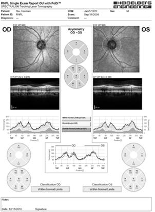
1 Financial Disclosure Maximizing OCT in Diagnosis and Management of Glaucoma Aerie Pharmaceuticals Alcon Laboratories Allergan Carl Zeiss Meditec Danica J. Marrelli, OD, FAAO AAO Diplomate, Glaucoma University of Houston College of Optometry Case 1: Dennis, 65yo WM DIAGNOSIS: CASES 1 2 IS THIS GLAUCOMA? POH: ( ) injury, ( ) surgery PMH: (+) DM2, (+) Systemic HTN FH: ( ) glaucoma Meds: metformin, HCTZ All: None Case 1: Dennis, 65yo WM Case 1: Dennis, 65yo WM BVA: 20/25 OD 20/20 OS Pupils: 3mm, 3+ D/C OD/OS, ( ) RAPD CVF: FTFC OD, OS Slit lamp: mild nuclear sclerosis OD>OS IOP: 31mmHg OD, 30mmHg OS See ONH and VF 1

2 Case 1: Dennis, 65yo WM Case 1: Is this glaucoma? A. Yes B. No C. I need additional information Case 2: Maria, 45 yo Hispanic female CC: referred for glaucoma evaluation POH: unremarkable, no trauma, no surgery PMH: unremarkable, no vascular disease FH: no known glaucoma, most family in Mexico Meds: None All: None Case 2: Maria, 45 yo Hispanic female BVA: 20/20 OD, OS Pupils: 3mm, 3+ D/C OD/OS, ( ) RAPD Slit Lamp: normal, no secondary signs, open angles Gonioscopy: open to CB, no secondary findings IOP: 24mmHg OD 23mmHg OS See ONH and VF Case 2: Maria, 45 yo Hispanic female Case 2: Maria, 45 yo Hispanic female 2

 Optic disc hemorrhage Peripapillary atrophy Why is disc size important?")
3 Case 2: Is this glaucoma? A. Yes B. No C. I need additional information Is this glaucoma? If there is characteristic optic nerve damage Yes If there are no characteristic optic nerve or VF changes Usually No Glaucoma Basics Glaucoma is a disease of ganglion cells Damage occurs at level of the lamina cribrosa Selective damage to superior and inferior poles of the optic nerve/rnfl Asymmetry between sup/inf poles as well as OD/OS asymmetry Characteristic Optic Nerve Changes Large C/D ratio FOR THE SIZE OF THE OPTIC NERVE Focal or diffuse rim thinning Focal or diffuse RNFL loss (discuss later) Optic disc hemorrhage Peripapillary atrophy Why is disc size important? Disc Size Algorithm Vertical Height (mm) C/D Ratio 2SD 1SD Mean +1SD +2SD Glaucoma Handbook, Litwak 3


 Defects in RNFL may precede glaucomatous")

4 Rim Thinning ISNT Rule Disc Hemorrhage & Peripapillary Atrophy Glaucoma Handbook, Litwak Case 1: Dennis, 65yo WM Case 2: Maria, 45 yo Hispanic female Case 2: Maria, 45 yo Hispanic female EVALUATION OF RETINAL NERVE FIBER LAYER (RNFL) Defects in RNFL may precede glaucomatous visual field loss and structural changes in ONH Can help to differentiate physiologic cupping from glaucomatous cupping 4


: slits and wedges")
5 ANATOMY OF THE NERVE FIBER LAYER Retinal Nerve Fiber Layer RNFL is thickest (and brightest) in superior and inferior arcades RNFL is thinner (dimmer) in papillomacular bundle and nasal bundles BRIGHT DIMMMER BRIGHT pattern RNFL Drop Out Diffuse (More Common) RNFL Drop Out Focal Loss (less common): slits and wedges RNFL Drop Out Focal Loss: slits and wedges 5


6 Glaucoma Detection Imaging ONH/RNFL photography Scanning laser ophthalmoscopy Scanning laser polarimetry Optical Coherence Tomography Time Domain Spectral Domain 1990s 2013 Quantifiable/Objective Imaging Diagnostic capability : Good for early glaucoma Excellent for moderate to severe glaucoma Improved when more than one parameter is evaluated Many of us rely on imaging devices to identify glaucoma (and progression) Has imaging become the answer? 6


7 Is automated imaging the answer? What Information Do the Instruments Give Us? Optic Nerve Parameters Disc Size Rim Area Rim Volume Cup Volume Retinal Nerve Fiber Layer Parameters TSNIT curves Average RNFL thickness Sectoral RNFL thickness Macular Thickness Ganglion Cell Complex Inner Retina Total Macular Thickness Quality/Signal Quantitative Parameters Thickness Map Deviation Map Thickness Profiles Quadrant/Clock Hour RNFL Thickness 7


 Small: <1.63 Med 1.63 1.")

8 Quality Signal Strength Circle Placement Systematic Strategy Thickness Map Systematic Strategy Systematic Strategy Quantitative Parameters Average RNFL Measures average thickness around calculation circle Affected by blood vessels, astrocytes, glial cells Global measure (will miss focal loss) Tipping Point = 75µm RNFL Symmetry Systematic Strategy Quantitative Parameters Rim Area Uses Bruch s membrane as edge of disc Range mm (avg 1.31) Disc Area Range mm2 (avg 1.83) Small: <1.63 Med Large >1.97 C/D ratio Cup Volume Systematic Strategy Thickness Profiles Compared to normative data Quadrant and Clock Hour RNFL analysis Systematic Strategy 8
")

9 Case 3: Victor, 61 yo HM Referred for glaucoma evaluation ( big C/D ) POH: unremarkable PMH: hypothyroid disease, pre diabetes Fam Hx: No glaucoma Meds: thyroid replacement hormone Allergies: NKDA Case 3: Victor, 61 yo HM BCVA: 20/20 OD, OS Pupils, EOMs, CVF: normal OU Slit lamp: normal, ( ) secondary signs Gonioscopy: open to CB, normal IOP: 18mmHg OD 15mmHg OS See ONH and VF Quality: Excellent Thickness Map: Lots of thick colors Quantitative Parameters: Average RNFL: VERY THICK! RNFL Symmetry: High Rim Area: Thicker than average Disc Area: VERY LARGE C/D: Large (but large disc) 9


10 Thickness Profiles Neuro retinal Rim? RNFL: Super thick! Is this glaucoma? Pitfalls of Imaging #1: Database Normative Database REFERENCE DATABASE Reference population WITHOUT DISEASE in question, to which an individual s data will be compared Can be utilized to classify the patient s data as normal or abnormal Can be utilized to measure reproducibility Problem: normal and healthy are often used interchangeably Does abnormal mean unhealthy? Database Considerations Inclusion/exclusion criteria Cohort should be representative of the population from which the suspects are drawn, except free of the disease in question How do you define un diseased? Exclusion criteria should be minimized to avoid a hyper normal database Include co morbidities that are often seen in population, so long as they don t interfere with the data in question Database Considerations Covariates: Some covariates are known to affect measurements (age, refractive error, axial length, etc) When the effect of the covariate is known and is potentially large, adjustment for that covariate is preferred. Database Size: Sufficient to adequately characterize the reference population, including covariates OCT Database Information RTVue: 600 eyes (Largest database) Age Disc Size Ethnic Group (African, Chinese, Japanese, Caucasian, Hispanic, Indian, other ) Cirrus: 284 eyes Image quality 6 or above Age RE: 12D to +8D Ethnic Groups: Causasian (43%), Asian (24%), AA (18%), Hispanic (12%); Spectralis: 201 patients All Caucasian Age RE 7D to +5D 10


")


11 Normative Distribution Back to Case 3 White = upper 5% normal (Cirrus) Green = middle 95% (Cirrus) or upper 95% (Spectralis, RTVue) Yellow = Lower 5% Red= Lowest 1% Gray = not in database Back to Case 3 11

 myopic LASIK 2002 OU Fam")


12 CASE 4: Allison 50 year old Caucasian female referred for glaucoma evaluation due to ONH appearance PMH: hypothyroidism; Sjogren syndrome POH: (+) myopic LASIK 2002 OU Fam Hx: Brother recently diagnosed with glaucoma Meds: Levoxyl, Restasis, AT Case 4: Allison BCVA: 20/25+ OD, OS Pupils, EOMS, CVF: normal SLE: Dry eye, mild meibomian gland dysfunction LASIK flaps visible No K spindle, TIDs, PXmaterial IOP: OD OS Pachymetry: 478 OD 457 OS 12


13 Pitfalls of OCT#2: Non glaucoma NAION Retinal Dystrophies Vein Occlusion (hemi/branch) Optic Neuritis Toxic/nutritional/infectious optic neuropathies Pitfalls of OCT #3: Operator Error Circle Placement Segmentation Errors Misc. Errors Dry Eye Misc Errors Floater Misc. Errors Blink Misc Errors Motion Artifact 13


14 Key Points Regarding ONH/RNFL Scans Need clinical correlation NEVER interpret OCT printout in isolation In situations where the disc is anomalous (e.g. very large, very small, tilted), the normative/reference database will classify the patient as abnormal, when they may be normal Can still use these scans for progression/change over time Newest Addition to Glaucoma Diagnosis Arsenal: Macular Imaging 1998: Zeimer et al reported on macular thickness loss in patients with known glaucomatous damage 2003: Greenfield reported correlation between total macular thickness and MD on VF in glaucoma patients (time domain OCT) Newer instruments can segment retinal thickness into specific areas RT Vue RT Vue FLV% quantifies the graphical representation Quantifies level of Irregularities 14
 Looked at conversion and progression rates at 6 years When baseline GCC FLV was within normal limits 10% of suspects convert to VF defects 30% of glaucoma patients convert to VF defects When")
 Baseline FD OCT Risk Factors for Glaucomatous Visual Field Conversion in the Advanced Imaging for Glaucoma Study.")

15 FLV% and GLV% Why are these numbers so powerful? Reported at the recent AGS conference by the AIG study group ( Looked at conversion and progression rates at 6 years When baseline GCC FLV was within normal limits 10% of suspects convert to VF defects 30% of glaucoma patients convert to VF defects When baseline GCC FLV was borderline or outside normal limits 41% of suspects convert to VF defects 60% of glaucoma patients convert to VF defects Cirrus OCT Huang, D. (28 February 2014) Baseline FD OCT Risk Factors for Glaucomatous Visual Field Conversion in the Advanced Imaging for Glaucoma Study. Presentation at the American Glaucoma Society Meeting, Washington, DC. Our eyes are designed to catch loss of symmetry Spectralis macula Heidelberg Engineering, Inc. All Rights Reserved. 87 Advantages of Macular Analysis More reproducible measure (if not using retinal nerve fiber layer) than peripapillary RNFL Fewer blood vessels an other cell components Better superior/inferior symmetry and symmetry between eyes than peripapillary RNFL Disadvantages of Macular Imaging Macular imaging is not helpful in glaucoma cases in which patients have concurrent macular disease AMD ERM CME DME Macular hole 15
 DM2 and HTN FOH: Unremarkable VA: 20/20 OD, OS SLE: Normal OU,")


16 Case: Leo 71yo AAM Referral for glaucoma suspicion, based on age/race/iop POH: Unremarkable PMH: (+) DM2 and HTN FOH: Unremarkable VA: 20/20 OD, OS SLE: Normal OU, mild cataract OU IOP: 23mmHg OD, OS CCT: 587 microns OD 582microns OS What about the 10 2 VF? Central 8 degrees from the center of the foveal contains more than 30% of retinal ganglion cells 24 2 and 30 2 test strategies use a 6 degree test grid pattern; these points fall outside of the densist region of ganglion cells 10 2 test strategy uses a 2 degree test grid Recent research has shown that in some patients with small regions of macular gangion cell loss, 10 2 testing may be better able to detect VF loss 16


17 Glaucoma Progression Once the diagnosis of glaucoma has been made, the MOST IMPORTANT remaining question is whether the disease is stable and the therapy/compliance are sufficient, or whether the disease is progressive and the therapy in relation to the life expectancy has to be intensified. Progression of Glaucoma, World Glaucoma Association, 2011 Kugler Publications Progression of Glaucoma Although most glaucoma patients will show some evidence of progression if followed long enough, the rate of deterioration can be highly variable among them. While most patients progress slowly, others have aggressive disease with fast deterioration which can eventually result in blindness or substantial impairment unless appropriate interventions take place. WGA Consensus Statements on Structure & Function Both ON structure and function should be evaluated for detection of progression Currently, no specific test can be regarded as the perfect standard for determination of progression Progression detected by functional means will not always be corroborated using structural tests, and vice versa WGA Consensus Statements The use of standard automated perimetry as the sole method for detection of change may result in failure to detect or underestimation of progression in eyes with early glaucoma damage. Progressive structural changes are often but not always predictive of future development of or progression of functional deficits in glaucoma. 17
: change over a designated time period using regression analysis.")

18 Detection & Measurement of Change (Structure & Function) Event Analysis (EA): change that exceeds a certain predefined threshold compared to the baseline value; generally determined by measurement reproducibility Trend Analysis (TA): change over a designated time period using regression analysis. Generally takes more exams to obtain a reliable slope Something to Consider There is normal age related attrition of RNFL Positive correlation between baseline RNFL thickness and rate of loss with age Cross sectional and longitudinal studies differ in the amount of loss per year Changes seen over time may be age related, related to diseases other than glaucoma, or true glaucoma progression Structural Progression: WGA Consensus Structural Progression: WGA Consensus Serial optic disc photography and RNFL photography are valuable and enduring methods for monitoring structural progression Subjective estimate of C/D is insufficient Color fundus imaging is the preferred modality to identify disc hemorrhage & change in PPA Critically evaluate for: Narrowing of NRR/notching Enlargement of cupping Disc hemorrhage Serial optic disc photography and RNFL photography are valuable and enduring methods for monitoring structural progression Subjective estimate of C/D is insufficient Color fundus imaging is the preferred modality to identify disc hemorrhage & change in PPA Critically evaluate for: Narrowing of NRR/notching Enlargement of cupping Disc hemorrhage 18


19 Structural Changes WGA The agreement for progression among ONH, RNFL, and macular parameters is poor The rates of change vary considering within and between glaucoma patients Differences in technologies and scan protocols could influence the detection of progression Structural Changes Several imaging instruments provide reproducible measurements & quantitative assessment of ONH and RNFL changes Image quality is crucial** Multiple baseline exams facilitate progression analysis Cirrus RNFL and ONH Report shows RNFL and ONH for both eyes, using the Optic Disc Cube 200x200 scan Cirrus GPA Analysis Using the Cirrus HD OCT, we can identify progression in RNFL loss through event analysis and trend analysis. Event analysis assesses changes that are beyond an expected variability at certain points compared to normative data. If a patient falls outside this area, it is identified as progression. Trend analysis looks at the rate of change over time, using linear regression to determine whether or not the trend is outside the expected rate of RNFL loss. 19


 RNFL Thickness Profiles Progression")
20 Cirrus GPA Analysis Cirrus GPA Analysis RNFL Thickness Maps provide a topographical display of RNFL for each exam. RNFL Thickness Change Maps demonstrate change in RNFL between exams. Up to 6 progression maps are compared to baseline. Areas of statistically significant change are color-coded yellow when first noted and then red when the change is sustained over consecutive visits. GPA case example courtesy of David Leung, MD Cirrus GPA Analysis Cirrus GPA Analysis TSNIT values from baseline and current exams are plotted. Areas of statistically significant change are color-coded yellow when first noted and then red when the change is sustained over consecutive visits. Average RNFL Thickness values are plotted for each exam. Yellow marker denotes change from both baseline exams. Red marker denotes change sustained over consecutive visits. Rate and significance of change are shown in text Cirrus GPA Analysis RNFL Summary Updated Guided Progression Analysis (GPA ) Page 1 Page 2 Legend summarizes GPA analyses and indicates with a check mark if there is possible or likely loss of RNFL RNFL Thickness Map Progression (best for focal change) RNFL Thickness Profiles Progression (best for broader focal change) Average RNFL Thickness Progression (best for diffuse change)


21 Heidelberg Spectralis Baseline Scan First Follow-Up Second Follow-Up 21


22 Optovue Trend Analysis Structural Changes WGA If structural progression is detected, it needs to be VERIFIED as to whether it is true vs artifact, AND whether it is typical for glaucoma (progressive rim thinning, notch, RNFL defects, disc hemorrhage) Example 1 22


23 Let s VERIFY Example 2 23
24 Summary OCT has significantly added to our ability to diagnose glaucoma and detect progression OCT technology is not perfect, and the clinician must be aware of pitfalls and caveats OCT should be considered one of many tools in the toolbox of the glaucoma practitioner. Thank you for your attention! Questions? me: 24
Financial Disclosure. Visual Field Interpretation RELIABILITY VISUAL FIELD INTERPRETATION IN GLAUCOMA METHODS OF DATA PRESENTATION
 VISUAL FIELD INTERPRETATION IN GLAUCOMA Danica J. Marrelli, OD, FAAO University of Houston College of Optometry Financial Disclosure I have received speaking and/or consulting fees from: Aerie Pharmaceutical
Advances in OCT Murray Fingeret, OD
 Disclosures Advances in OCT Murray Fingeret, OD Consultant Alcon, Allergan, Bausch & Lomb, Carl Zeiss Meditec, Diopsys, Heidelberg Engineering, Reichert, Topcon Currently Approved OCT Devices OCT Devices
Mark Dunbar: Disclosure
 Important Things to Understand About OCT Mark T. Dunbar, O.D., F.A.A.O. Bascom Palmer Eye Institute University of Miami, School of Medicine Mark Dunbar: Disclosure Optometry Advisory Board for: Allergan
How to Be Efficient and Effective. Disclosure. Topics CASE CM. Case JF 2007 OHTN / POAG? How to Be Efficient and Effective with. with New Technology
 How to Be Efficient and Effective with Disclosure COPE Course ID: 40750 GL Michael Chaglasian has the following disclosures: 1. Advisory Board: Allergan, Inc., Alcon Labs, B+L Carl Zeiss Meditec 2. Research:
Is this glaucoma? Leo Semes, OD Michael Chaglasian, OD Danica Marrelli, OD. Optometry s Meeting 2015 Seattle, WA
 Is this glaucoma? Leo Semes, OD Michael Chaglasian, OD Danica Marrelli, OD Optometry s Meeting 2015 Seattle, WA Case 1. 54 WM Engineer is referred to UAB Eye Care as a glaucoma suspect. Mild myopic refractive
CHAPTER 13 CLINICAL CASES INTRODUCTION
 2 CHAPTER 3 CLINICAL CASES INTRODUCTION The previous chapters of this book have systematically presented various aspects of visual field testing and is now put into a clinical context. In this chapter,
Macular Ganglion Cell Complex Measurement Using Spectral Domain Optical Coherence Tomography in Glaucoma
 Med. J. Cairo Univ., Vol. 83, No. 2, September: 67-72, 2015 www.medicaljournalofcairouniversity.net Macular Ganglion Cell Complex Measurement Using Spectral Domain Optical Coherence Tomography in Glaucoma
Evolving glaucoma management True diagnostic integration for the preservation of vision
 Evolving glaucoma management True diagnostic integration for the preservation of vision // GLAUCOMA MANAGEMENT MADE BY ZEISS The moment you are certain it is glaucoma. This is the moment we work for. There
Optical Coherence Tomography (OCT)
") Understanding and Interpreting OCT Mark Dunbar: Disclosure The Swiss Army Pocket Knife of Eye Care Mark T. Dunbar, O.D., F.A.A.O. Bascom Palmer Eye Institute University of Miami, School of Medicine Consultant
Understanding and Interpreting OCT Mark Dunbar: Disclosure The Swiss Army Pocket Knife of Eye Care Mark T. Dunbar, O.D., F.A.A.O. Bascom Palmer Eye Institute University of Miami, School of Medicine Consultant
Il contributo dell'angio-oct: valutazione integrata della componente nervosa e vascolare della malattia glaucomatosa
 SIMPOSIO G.O.A.L. - LE NUOVE FRONTIERE DIAGNOSTICHE E LE LINEE DI INDIRIZZO AMBULATORIALI DEL GLAUCOMA Coordinatore e moderatore: D. Mazzacane Presidente: L. Rossetti Il contributo dell'angio-oct: valutazione
8/6/17. Disclosures Aerie Pharmaceuticals Alcon BioTissue Diopsys Optovue Shire
 Nathan Lighthizer, O.D., F.A.A.O. Associate Professor Assistant Dean for Clinical Care Director of Continuing Education Chief of Specialty Care Clinics Oklahoma College of Optometry Tahlequah, OK lighthiz@nsuok.edu
OCT in the Diagnosis and Follow-up of Glaucoma
 OCT in the Diagnosis and Follow-up of Glaucoma Karim A Raafat MD. Professor Of Ophthalmology Cairo University Hmmmm! Do I have Glaucoma or not?! 1 Visual Function 100% - N Gl Structure : - 5000 axon /
Glaucoma Pearls and Grand Rounds Vision Expo East 2016
 Glaucoma Pearls and Grand Rounds Vision Expo East 2016 Murray Fingeret, Ben Gaddie, Richard Madonna Disclosures Murray Fingeret - Consultant Alcon, Allergan, Bausch & Lomb, Carl Zeiss Meditec, Dyopsys,
STRUCTURE & FUNCTION An Integrated Approach for the Detection and Follow-up of Glaucoma. Module 3a GDx
 STRUCTURE & FUNCTION An Integrated Approach for the Detection and Follow-up of Glaucoma Module 3a GDx Educational Slide Deck Carl Zeiss Meditec, Inc. November 2005 1 Structure & Function Modules Module
The Optic Nerve Head In Glaucoma. Clinical Pearl #1. Characteristics of Normal Disk 9/26/2017. Initial detectable damage Structure vs function
 The Optic Nerve Head In Glaucoma Clinical Pearl #1 Eric E. Schmidt, O.D., F.A.A.O. Omni Eye Specialists Wilmington,NC schmidtyvision@msn.com Glaucoma is an optic neuropathy Initial detectable damage Structure
Structural examina.on: Imaging
 ManaMa: Glaucoma Structural examina.on: Imaging Luís Abegão Pinto, MD, PhD Department of Ophthalmology CHLC Lisbon Faculty of Medicine, Lisbon University 1 11-10- 2013 Structural changes Qualitative changes
Cirrus TM HD-OCT. Details defi ne your decisions
 Cirrus TM HD-OCT Details defi ne your decisions 2 With high-defi nition OCT Carl Zeiss Meditec takes you beyond standard spectral domain Built on 10 years experience at the vanguard of innovation, Carl
Visual Field Interpretation Anthony B. Litwak, OD, FAAO VA Medical Center Baltimore, MD
 Visual Field Interpretation Anthony B. Litwak, OD, FAAO VA Medical Center Baltimore, MD Dr. Litwak is on the speaker bureau and advisory panel for Alcon and Zeiss Meditek o Confirms Glaucoma Diagnosis!
Beyond the C/D Ratio: Evaluating a Glaucomatous Optic Nerve. Marcus Gonzales, OD, FAAO Cedar Springs Eye Clinic COPE ID#: GL
 Beyond the C/D Ratio: Evaluating a Glaucomatous Optic Nerve Marcus Gonzales, OD, FAAO Cedar Springs Eye Clinic COPE ID#: 27809-GL Points to Remember Glaucoma affects the ONH in characteristic patterns
Glaucoma Evaluation. OCT Pearls for Glaucoma. OCT: Retinal Nerve Fiber Layer. Financial Disclosures. OCT: Macula. Case Example
 OCT Pearls for Glaucoma using OCT of the macula for glaucoma Glaucoma Evaluation Right eye Visual Acuity 20/25 20/25 IOP 13 13 Central corneal 530 530 thickness Anterior exam Normal with PCIOL Normal with
University Hospital Basel. Optical Coherence Tomography Emerging Role in the Assessment of MS PD Dr. Konstantin Gugleta
 University Hospital Basel Optical Coherence Tomography Emerging Role in the Assessment of MS PD Dr. Konstantin Gugleta 15th State of the Art SMSS, Lucerne January 2013 Retinal Nerve Fiber Layer 1.200.000
Ganglion cell analysis by optical coherence tomography (OCT) Jonathan A. Micieli, MD Valérie Biousse, MD
 Jonathan A. Micieli, MD Valérie Biousse, MD") Ganglion cell analysis by optical coherence tomography (OCT) Jonathan A. Micieli, MD Valérie Biousse, MD Figure 1. Normal OCT of the macula (cross section through the line indicated on the fundus photo)
Ganglion cell analysis by optical coherence tomography (OCT) Jonathan A. Micieli, MD Valérie Biousse, MD Figure 1. Normal OCT of the macula (cross section through the line indicated on the fundus photo)
Retinal Nerve Fiber Layer Measurements in Myopia Using Optical Coherence Tomography
 Original Article Philippine Journal of OPHTHALMOLOGY Retinal Nerve Fiber Layer Measurements in Myopia Using Optical Coherence Tomography Dennis L. del Rosario, MD and Mario M. Yatco, MD University of Santo
How Strongly Do You Feel That This Patient Has Glaucoma? % % % % %
 My Favorite Cases Anthony B. Litwak, OD, FAAO VA Medical Center Baltimore, Maryland Dr. Litwak is a speaker and on advisory boards for Alcon and Zeiss Meditek CASE CR 35 yohf Neg PMH +FOH mother and grandmother
Noel de Jesus Atienza, MD, MSc and Joseph Anthony Tumbocon, MD
 Original Article Philippine Journal of OPHTHALMOLOGY Diagnostic Accuracy of the Optical Coherence Tomography in Assessing Glaucoma Among Filipinos. Part 1: Categorical Outcomes Based on a Normative Database
My Favourite Cases Anthony B. Litwak, OD, FAAO VA Medical Center Baltimore, MD
 My Favourite Cases Anthony B. Litwak, OD, FAAO VA Medical Center Baltimore, MD Dr. Litwak is a speaker and on advisory boards for Alcon and Zeiss Meditek CASE CR! 35 YOHF! Neg PMH! +FOH mother and grandmother
OtticaFisiopatologica
 Anno quindicesimo dicembre 2010 How to assess the retinal nerve fiber layer thickness Antonio Ferreras Miguel Servet University Hospital, Zaragoza. Aragón Health Sciences Institute University of Zaragoza
Intro to Glaucoma/2006
 Intro to Glaucoma/2006 Managing Patients with Glaucoma is Exciting Interesting Challenging But can often be frustrating! Clinical Challenges To identify patients with risk factors for possible glaucoma.
tracking progression we can better manage our patients. Like any tool, any instrument you ve got to
 EIYESS ALBEINUTI, MD 1 As we know OCT has become very instrumental in taking care of glaucoma patients whether we have the ability to objectively image the RNFL and therefore pickup earlier signs of damage
The Role of the RNFL in the Diagnosis of Glaucoma
 Chapter 1. The Role of the RNFL in the Diagnosis of Glaucoma Introduction Glaucoma is an optic neuropathy characterized by a loss of of retinal ganglion cells and their axons, the Retinal Nerve Fiber Layer
Early Detection Of Glaucoma Clinical Clues. Points To Live By. Glaucoma Risk Factors 10/3/2014
 Early Detection Of Glaucoma Clinical Clues Eric E. Schmidt, O.D. Omni Eye Specialists Wilmington, NC schmidtyvision@msn.com Points To Live By 25% of G pxs NEVER have IOP >21mm 50% of G pxs have trough
A Review Of Risk Factors. Early Detection Of Glaucoma Clinical Clues. A risk factor analysis is critical. Points To Live By
 A Review Of Risk Factors Early Detection Of Glaucoma Clinical Clues Eric E. Schmidt, O.D. Omni Eye Specialists Wilmington, NC schmidtyvision@msn.com FINDACAR Family history IOP Nearsightedness Diabetes/Vascular
Moving forward with a different perspective
 Moving forward with a different perspective The Leader In Vision Diagnostics Offers A New Perspective Marco has served the eyecare community by offering exceptional lane products and automated high tech
C a t a r a c t G l a u c o m a R e t i n a R e f r a c t i v e. The GDxVCC Early answers and ongoing assessment for glaucoma
 C a t a r a c t G l a u c o m a R e t i n a R e f r a c t i v e The GDxVCC Early answers and ongoing assessment for glaucoma The quantifiable approach to quality care Only Humphrey GPA software Early insight
Ganglion cell complex scan in the early prediction of glaucoma
 Original article in the early prediction of glaucoma Ganekal S Nayana Super Specialty Eye Hospital and Research Center, Davangere, Karnataka, India Abstract Objective: To compare the macular ganglion cell
Cirrus TM HD-OCT. Details define your decisions
 Cirrus TM HD-OCT Details define your decisions 2 With high-definition OCT Carl Zeiss Meditec takes you beyond standard spectral domain Built on 10 years experience at the vanguard of innovation, Carl Zeiss
Translating data and measurements from stratus to cirrus OCT in glaucoma patients and healthy subjects
 Romanian Journal of Ophthalmology, Volume 60, Issue 3, July-September 2016. pp:158-164 GENERAL ARTICLE Translating data and measurements from stratus to cirrus OCT in glaucoma patients and healthy subjects
PRIMUS 200 from ZEISS The essential OCT
 PRIMUS 200 from ZEISS The essential OCT Seeing beyond the surface. ZEISS PRIMUS 200 // INNOVATION MADE BY ZEISS Clear Visualization. Advanced Technology. Reliability. Essential elements of your first OCT.
Retinal nerve fiber layer thickness in Indian eyes with optical coherence tomography
 Original articles in Indian eyes with optical coherence tomography Malik A, Singh M, Arya SK, Sood S, Ichhpujani P Department of Ophthalmology Government Medical College and Hospital, Sector 32, Chandigarh,
PRIMUS 200 from ZEISS The essential OCT
 EN 00_00I The contents of the brochure may differ from the current status of approval of the product in your country. Please contact your regional representative for more information. Subject to change
The Measure of Confidence
 Heidelberg_936357.qxd:Layout 1 5/9/08 12:01 PM 12:02 Page 1 (Cyan (Magenta (Yellow (Black (UV Five Powerful Solutions to Fit Your Practice PowerCheck Glaucoma FastCheck+ GPS Software and Retina Edema Index
SOUTH-EAST EUROPEAN JOURNAL of OPHTHALMOLOGY 2015; 1 (1) 34 40
 34 40") Review article SOUTH-EAST EUROPEAN JOURNAL of OPHTHALMOLOGY 2015; 1 (1) 34 40 Retinal nerve fiber layer versus peripapillary capillary density assessment A powerful tool for detecting optic nerve head
Review article SOUTH-EAST EUROPEAN JOURNAL of OPHTHALMOLOGY 2015; 1 (1) 34 40 Retinal nerve fiber layer versus peripapillary capillary density assessment A powerful tool for detecting optic nerve head
53 year old woman attends your practice for routine exam. She has no past medical history or family history of note.
 Case 1 Normal Tension Glaucoma 53 year old woman attends your practice for routine exam. She has no past medical history or family history of note. Table 1. Right Eye Left Eye Visual acuity 6/6 6/6 Ishihara
Comparative evaluation of time domain and spectral domain optical coherence tomography in retinal nerve fiber layer thickness measurements
 Original article Comparative evaluation of time domain and spectral domain optical coherence tomography in retinal nerve fiber layer thickness measurements Dewang Angmo, 1 Shibal Bhartiya, 1 Sanjay K Mishra,
1/25/2019 OCT & OCTA RETINAL IMAGING: HOW TO PREVENT RAGING GLAUCOMA! THE ORIGINAL RAGING GLAUCOMA OCT RETINAL IMAGING OPTIC NERVE HEAD EXAMINATION
 OCT & OCTA RETINAL IMAGING: HOW TO PREVENT RAGING GLAUCOMA! Craig Thomas, O.D. 3900 West Wheatland Road Dallas, Texas 75237 972-780-7199 thpckc@yahoo.com THE ORIGINAL RAGING GLAUCOMA 47-year-old Black
Glaucoma Diagnosis. Definition of Glaucoma. Diagnosing Glaucoma. Vision Institute Annual Fall Conference
 Glaucoma Diagnosis Vision Institute Annual Fall Conference Mitchell W. Dul, OD, MS, FAAO mdul@sunyopt.edu Richard J. Madonna, MA, OD, FAAO rmadonna@sunyopt.edu Definition of Glaucoma Glaucoma can be regarded
History/principles of the OCT What does the normal retinal OCT look like Vitreal disorders Retinal/RPE disorders Choroidal disorders
 Nathan Lighthizer, O.D., F.A.A.O. Assistant Professor Assistant Dean for Clinical Care Director of Continuing Education Chief of Specialty Care Clinics Chief of Electrodiagnostics Clinic Oklahoma College
Evaluation of ONH Pallor in Glaucoma Patients and Suspects. Leticia Rousso, O.D. Joseph Sowka, O.D
 Evaluation of ONH Pallor in Glaucoma Patients and Suspects Leticia Rousso, O.D Joseph Sowka, O.D I. Abstract This case report will evaluate a young glaucoma suspect with unilateral sectoral optic nerve
Method for comparing visual field defects to local RNFL and RGC damage seen on frequency domain OCT in patients with glaucoma.
 Method for comparing visual field defects to local RNFL and RGC damage seen on frequency domain OCT in patients with glaucoma. Donald C. Hood 1,2,* and Ali S. Raza 1 1 Department of Psychology, Columbia
Course # Getting to Know Your OCT
 Course # 140 Getting to Know Your OCT Course Title: Lecturer: Getting to Know Your OCT Brad Sutton, OD, FAAO IU School of Optometry Financial Disclosures No financial disclosures Optical Coherence Tomography-OCT
Glaucoma: a disease of the macula?
 Glaucoma: a disease of the macula? Derek MacDonald, OD, FAAO Waterloo, Ontario, Canada Please silence all mobile devices and remove items from chairs so others can sit. Unauthorized recording of this session
Research Article The Pattern of Retinal Nerve Fiber Layer and Macular Ganglion Cell-Inner Plexiform Layer Thickness Changes in Glaucoma
 Hindawi Ophthalmology Volume 2017, Article ID 78365, 8 pages https://doi.org/10.1155/2017/78365 Research Article The Pattern of Retinal Nerve Fiber Layer and Macular Ganglion Cell-Inner Plexiform Layer
Glaucoma Diagnosis & Tracking with Optical Coherence Tomography
 Glaucoma Diagnosis & Tracking with Optical Coherence Tomography David Huang, MD, PhD Charles C. Manger III, MD Chair of Corneal Laser Surgery Assoc. Prof. of Ophthalmology & Biomedical Engineering Doheny
Automated Visual Field Analysis for Glaucoma
 Island of Vision Automated Visual Field Analysis for Glaucoma Normal Visual Field Parameters 60 superior 60 nasal 75 inferior 100 temporal Macula the central 13 Fovea the central 3 Visual field is limited
Goals. Glaucoma PARA PEARL TO DO. Vision Loss with Glaucoma
 Glaucoma Janet R. Fett, OD Drs. Kincaid, Fett and Tharp So Sioux City, NE eyewear21@hotmail.com Goals Understand Glaucoma Disease process Understand how your data (objective and subjective) assists in
Available online at Pelagia Research Library. Advances in Applied Science Research, 2013, 4(6):
:") Available online at www.pelagiaresearchlibrary.com Advances in Applied Science Research, 2013, 4(6):201-206 ISSN: 0976-8610 CODEN (USA): AASRFC Comparison of glaucoma diagnostic ability of retinal nerve
Available online at www.pelagiaresearchlibrary.com Advances in Applied Science Research, 2013, 4(6):201-206 ISSN: 0976-8610 CODEN (USA): AASRFC Comparison of glaucoma diagnostic ability of retinal nerve
Seiji T. Takagi, Yoshiyuki Kita, Asuka Takeyama, and Goji Tomita. 1. Introduction. 2. Subjects and Methods
 Ophthalmology Volume 2011, Article ID 914250, 5 pages doi:10.1155/2011/914250 Clinical Study Macular Retinal Ganglion Cell Complex Thickness and Its Relationship to the Optic Nerve Head Topography in Glaucomatous
DISCLOSURE: What to do? 2/22/2016
 DISCLOSURE: Dr. Joseph Sowka is a member of the Speakers Bureau for Alcon Laboratories, and Carl Zeiss Meditec. He is on the advisory boards for Alcon, Zeiss, and Allergan. He is a consultant for Alcon.
Differences between Non-arteritic Anterior Ischemic Optic Neuropathy and Open Angle Glaucoma with Altitudinal Visual Field Defect
 pissn: 1011-8942 eissn: 2092-9382 Korean J Ophthalmol 2015;29(6):418-423 http://dx.doi.org/10.3341/kjo.2015.29.6.418 Original Article Differences between Non-arteritic Anterior Ischemic Optic Neuropathy
Comparison of Retinal Nerve Fiber Layer Thickness between Stratus and Spectralis OCT
 pissn: 1011-8942 eissn: 2092-9382 Korean J Ophthalmol 2011;25(3):166-173 DOI: 10.3341/kjo.2011.25.3.166 Original Article Comparison of Retinal Nerve Fiber Layer Thickness between Stratus and Spectralis
New Concepts in Glaucoma Ben Gaddie, OD Moderator Murray Fingeret, OD Louis Pasquale, MD
 New Concepts in Glaucoma Ben Gaddie, OD Moderator Murray Fingeret, OD Louis Pasquale, MD New Concepts in Glaucoma Optical Coherence Tomography: Is it necessary and needed to diagnose and monitor glaucoma?
Jong Chul Han, Da Ye Choi, and Changwon Kee. 1. Introduction
 Ophthalmology Volume 2015, Article ID 641204, 7 pages http://dx.doi.org/10.1155/2015/641204 Clinical Study The Different Characteristics of Cirrus Optical Coherence Tomography between Superior Segmental
Optical coherence tomography (OCT) is a relatively new noninvasive. The Use of Optical Coherence Tomography in Neurology DIAGNOSTIC UPDATE
 is a relatively new noninvasive. The Use of Optical Coherence Tomography in Neurology DIAGNOSTIC UPDATE") DIAGNOSTIC UPDATE The Use of Optical Coherence Tomography in Neurology Cédric Lamirel, MD,* Nancy Newman, MD,* Valérie Biousse, MD* Departments of *Ophthalmology, Neurology, and Neurological Surgery, Emory
DIAGNOSTIC UPDATE The Use of Optical Coherence Tomography in Neurology Cédric Lamirel, MD,* Nancy Newman, MD,* Valérie Biousse, MD* Departments of *Ophthalmology, Neurology, and Neurological Surgery, Emory
To assess the glaucoma diagnostic ability of Fourier Domain Optical Coherence Tomography
 American Journal of Engineering Research (AJER) e-issn : 2320-0847 p-issn : 2320-0936 Volume-02, Issue-11, pp-104-110 www.ajer.org Research Paper Open Access To assess the glaucoma diagnostic ability of
Introducing ANGIOVUE ESSENTIAL. Built on the Avanti Widefield OCT Platform. OCT Angiography for Primary Eye Care
 Introducing ANGIOVUE ESSENTIAL Built on the Avanti Widefield OCT Platform OCT Angiography for Primary Eye Care Transform Your View of the Retina OCT Angiography (OCTA) is a quick non-invasive test that
A Formula to Predict Spectral Domain Optical Coherence Tomography (OCT) Retinal Nerve Fiber Layer Measurements Based on Time Domain OCT Measurements
 Retinal Nerve Fiber Layer Measurements Based on Time Domain OCT Measurements") pissn: 1011-8942 eissn: 2092-9382 Korean J Ophthalmol 2012;26(5):369-377 http://dx.doi.org/10.3341/kjo.2012.26.5.369 Original Article A Formula to Predict Spectral Domain Optical Coherence Tomography (OCT)
pissn: 1011-8942 eissn: 2092-9382 Korean J Ophthalmol 2012;26(5):369-377 http://dx.doi.org/10.3341/kjo.2012.26.5.369 Original Article A Formula to Predict Spectral Domain Optical Coherence Tomography (OCT)
International Journal of Scientific & Engineering Research, Volume 4, Issue 12, December ISSN
 International Journal of Scientific & Engineering Research, Volume 4, Issue 12, December-2013 108 Name of Chief and Corresponding Author : Dr Chandrima Paul TITLE : Comparison of glaucoma diagnostic ability
Incorporating OCT Angiography Into Patient Care
 Incorporating OCT Angiography Into Patient Care Beth A. Steele, OD, FAAO OCT A: Introduction Isolates microvascular circulation from OCT image data Axial resolution = 5 microns (i.e. fine capillaries visible)
CLINICAL SCIENCES. Felipe A. Medeiros, MD; Linda M. Zangwill, PhD; Christopher Bowd, PhD; Robert N. Weinreb, MD
 CLINICAL SCIENCES Comparison of the GDx VCC Scanning Laser Polarimeter, HRT II Confocal Scanning Laser Ophthalmoscope, and Stratus OCT Optical Coherence Tomograph for the Detection of Glaucoma Felipe A.
Two Pits in a Pod: Using EDI-OCT to evaluate the lamina cribrosa in a patient with openangle glaucoma and multiple optic pits
 Two Pits in a Pod: Using EDI-OCT to evaluate the lamina cribrosa in a patient with openangle glaucoma and multiple optic pits Abstract: EDI-OCT imaging is used to evaluate glaucoma by examining the thinning
Clinical and OCT features of different types and stages of diabetic optic neuropathy
 Clinical and OCT features of different types and stages of diabetic optic neuropathy P.A. Bezditko 1, Dr Sc (Med), Prof.; M.A. Karliychuk 2, Cand Sc (Med) 1 Kharkiv National Medical University ; Kharkiv
10/3/2018. Case: 63 year old white female 6/11/2012 visit. Glaucoma Update for the Primary Care OD CHRISTOPHER WOLFE, OD, FAAO, DIPL ABO
 DISCLOSURE STATEMENT Ihave no direct financial or proprietary interest in any companies, products or services mentioned in this presentation. GLAUCOMA UPDATE FOR THE PRIMARY CARE OD Please silence all
New Technologies in Glaucoma Management: From ERG to OCT
 What s New and What s Next in Glaucoma New Technologies in Glaucoma Management: From ERG to OCT Ben Gaddie, OD FAAO Murray Fingeret, OD FAAO IOP 24- Hour IOP Role of hysteresis in glaucoma risk Cerebrospinal
OCT Interpretation in Retinal Disease
 OCT Interpretation in Retinal Disease Jay M. Haynie, OD, FAAO Financial Disclosure I have received honoraria or am on the advisory board for the following companies: Carl Zeiss Meditec Advanced Ocular
Research Article Repeatability of Perimacular Ganglion Cell Complex Analysis with Spectral-Domain Optical Coherence Tomography
 Ophthalmology Volume 2015, Article ID 605940, 5 pages http://dx.doi.org/10.1155/2015/605940 Research Article Repeatability of Perimacular Ganglion Cell Complex Analysis with Spectral-Domain Optical Coherence
Title: OCT Analysis Workshop: Interpretation of OCT printouts
 Title: OCT Analysis Workshop: Interpretation of OCT printouts Authors: David Yang, OD, FAAO Staff Optometrist, VA Palo Alto Health Care System Associate Clinical Professor, UC Berkeley School of Optometry
GLAUCOMA SUMMARY BENCHMARKS FOR PREFERRED PRACTICE PATTERN GUIDELINES
 SUMMARY BENCHMARKS FOR PREFERRED PRACTICE PATTERN GUIDELINES Introduction These are summary benchmarks for the Academy s Preferred Practice Pattern (PPP) guidelines. The Preferred Practice Pattern series
Collaboration in the care of glaucoma patients and glaucoma suspects. Barry Emara MD FRCS(C) Nico Ristorante November 29, 2012
 Nico Ristorante November 29, 2012") Collaboration in the care of glaucoma patients and glaucoma suspects Barry Emara MD FRCS(C) Nico Ristorante November 29, 2012 Goals of Collaboration Patient-centred and evidence based approach Timely access
Collaboration in the care of glaucoma patients and glaucoma suspects Barry Emara MD FRCS(C) Nico Ristorante November 29, 2012 Goals of Collaboration Patient-centred and evidence based approach Timely access
Patterns of Subsequent Progression of Localized Retinal Nerve Fiber Layer Defects on Red-free Fundus Photographs in Normal-tension Glaucoma
 pissn: 1011-8942 eissn: 2092-9382 Korean J Ophthalmol 2014;28(4):330-336 http://dx.doi.org/10.3341/kjo.2014.28.4.330 Original Article Patterns of Subsequent Progression of Localized Retinal Nerve Fiber
R&M Solutions
 Mohamed Hosny El-Bradey, MD., Assistant Professor of Ophthalmology, Tanta University. Wael El Haig, MD., Professor of Ophthalmology. Zagazeeg University. 1 Myopic CNV is considered the most common vision
Targeting Intraocular Pressure in Glaucoma: a Teaching Case Report 1
 Targeting Intraocular Pressure in Glaucoma: a Teaching Case Report 1 By: Andrew Kemp, OD, Marcus Gonzales, OD, FAAO, Joe DeLoach, OD, FAAO, and Zanna Kruoch, OD FAAO Background Glaucoma is a range of conditions
OCT Angiography in Primary Eye Care
 OCT Angiography in Primary Eye Care An Image Interpretation Primer Julie Rodman, OD, MS, FAAO and Nadia Waheed, MD, MPH Table of Contents Diabetic Retinopathy 3-6 Choroidal Neovascularization 7-9 Central
Linking structure and function in glaucoma
 CET CONTINUING Sponsored by 1 CET POINT Linking structure and function in glaucoma 50 Dr Samantha McGinnigle PhD, BSc (Hons), MCOptom, AHEA This article will give an overview of the latest imaging technology
Glaucoma Progression. Murray Fingeret, OD
 Glaucoma Progression Murray Fingeret, OD Managing glaucoma or glaucoma suspects Summary For patients diagnosed or at risk of glaucoma Stage disease Risk assessment Asses for progression Treatment plan
Relationship between the GDx VCC and Stratus OCT in Primary Open Angle Glaucoma
 Relationship between the GDx VCC and Stratus OCT in Primary Open Angle Glaucoma Reza Zarei, MD 1 Mohammad Soleimani, MD 2 Sasan Moghimi, MD 3 Mohammad Yaser Kiarudi, MD 2 Mahmoud Jabbarvand, MD 1 Yadollah
Do You See What I See!!! Shane R. Kannarr, OD
 Do You See What I See!!! Shane R. Kannarr, OD skannarr@kannarreyecare.com Define Specialty Testing Additional Test to: Prove/Disprove Diagnosis To monitor progression of a condition To document a condition
Corporate Medical Policy
 Corporate Medical Policy Glaucoma, Evaluation by Ophthalmologic Techniques File Name: Origination: Last CAP Review: Next CAP Review: Last Review: glaucoma_evaluation_by_ophthalmologic_techniques 3/2001
What Is O.C.T. and Why Should I Give A Rip? OCT & Me How Optical Coherence Tomography Changed the Life of a Small Town Optometrist 5/19/2014
 OCT & Me How Optical Coherence Tomography Changed the Life of a Small Town Optometrist Email: myoder@wcoil.com Mark A. Yoder, O.D. 107 N. Main Street PO Box 123 Bluffton, OH 45817 @yoderod 115.02 Histoplasma
Glaucoma: Diagnostic Modalities
 Glaucoma: Diagnostic Modalities - Dr. Barun Kumar Nayak, Dr. Sarika Ramugade Glaucoma is a leading cause of blindness in the world, especially in older people. Early detection and treatment by ophthalmologist
Optic Disc Evaluation: Is the Optic Disc Glaucomatous and Has it Progressed?
 Optic Disc Evaluation: Is the Optic Disc Glaucomatous and Has it Progressed? Jody Piltz-Seymour, M.D. Clinical Professor Perelman School of Medicine University of Pennsylvania Wills Glaucoma Service Valley
The Effect of Pupil Dilation on Scanning Laser Polarimetry With Variable Corneal Compensation
 C L I N I C A L S C I E N C E The Effect of Pupil Dilation on Scanning Laser Polarimetry With Variable Corneal Compensation Amjad Horani, MD; Shahar Frenkel, MD, PhD; Eytan Z. Blumenthal, MD BACKGROUND
Non-arteritic anterior ischemic optic neuropathy (NAION) with segmental optic disc edema. Jonathan A. Micieli, MD Valérie Biousse, MD
 with segmental optic disc edema. Jonathan A. Micieli, MD Valérie Biousse, MD") Non-arteritic anterior ischemic optic neuropathy (NAION) with segmental optic disc edema Jonathan A. Micieli, MD Valérie Biousse, MD A 75 year old white woman lost vision in the inferior part of her visual
Non-arteritic anterior ischemic optic neuropathy (NAION) with segmental optic disc edema Jonathan A. Micieli, MD Valérie Biousse, MD A 75 year old white woman lost vision in the inferior part of her visual
Factors Associated With Visual Field Progression in Cirrus Optical Coherence Tomography-guided Progression Analysis: A Topographic Approach
 ORIGINAL STUDY Factors Associated With Visual Field Progression in Cirrus Optical Coherence Tomography-guided Progression Analysis: A Topographic Approach Joong Won Shin, MD, Kyung Rim Sung, MD, PhD, Jiyun
Peripapillary Retinal Thickness. Maps in the Evaluation of Glaucoma Patients: A Novel Concept.
 Peripapillary Retinal Thickness Maps in the Evaluation of Glaucoma Patients: A Novel Concept The Harvard community has made this article openly available. Please share how this access benefits you. Your
EXPERIMENTAL AND THERAPEUTIC MEDICINE 6: , 2013
 268 Comparison of optic nerve morphology in eyes with glaucoma and eyes with non-arteritic anterior ischemic optic neuropathy by Fourier domain optical coherence tomography YUXIN YANG 1, HAITAO ZHANG 1,
Snellen VA 2 IOP 3 Ocular Examination * Only dilate if repeat photos required
 Page 1 of 7 Visit Date: Check Completed Modules Page Number Snellen VA 2 IOP 3 Ocular Examination * Only dilate if repeat photos required Humphrey VFs (if required) 5 Disc Photography (if required) 6 OCT
7/25/2018. You talk about glaucoma in cup-to-disc ratios ASSESSING THE OPTIC DISC: IS PHOTOGRAPHY STILL NECESSARY IN THE OCT ERA?
 MAXIMIZING YOUR DIAGNOSTIC TECHNOLOGIES: SOMETHING OLD, NEW, BORROWED AND BLUE Greg Caldwell, OD Joseph Sowka, OD Jessica Steen, OD ASSESSING THE OPTIC DISC: IS PHOTOGRAPHY STILL NECESSARY IN THE OCT ERA?
CLINICAL SCIENCES. Comparison of Glaucoma Diagnostic Capabilities of Cirrus HD and Stratus Optical Coherence Tomography
 CLINICAL SCIENCES Comparison of Glaucoma Diagnostic Capabilities of Cirrus HD and Stratus Optical Coherence Tomography Seong Bae Park, MD; Kyung Rim Sung, MD, PhD; Sung Yong Kang, MD; Kyung Ri Kim, BS;
Optical Coherence Tomograpic Features in Idiopathic Retinitis, Vasculitis, Aneurysms and Neuroretinitis (IRVAN)
") Columbia International Publishing Journal of Ophthalmic Research (2014) Research Article Optical Coherence Tomograpic Features in Idiopathic Retinitis, Vasculitis, Aneurysms and Neuroretinitis (IRVAN)
Columbia International Publishing Journal of Ophthalmic Research (2014) Research Article Optical Coherence Tomograpic Features in Idiopathic Retinitis, Vasculitis, Aneurysms and Neuroretinitis (IRVAN)
3/16/2018. Perimetry
 Perimetry The normal visual field extends further away from fixation temporally and inferiorly than superiorly and nasally. From the center of the retina this sensitivity decreases towards the periphery,