History/principles of the OCT What does the normal retinal OCT look like Vitreal disorders Retinal/RPE disorders Choroidal disorders
|
|
|
- Marilynn Miller
- 5 years ago
- Views:
Transcription
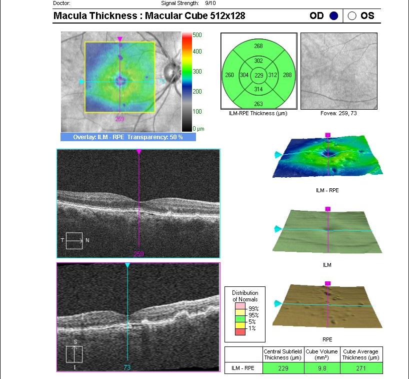
1 Nathan Lighthizer, O.D., F.A.A.O. Assistant Professor Assistant Dean for Clinical Care Director of Continuing Education Chief of Specialty Care Clinics Chief of Electrodiagnostics Clinic Oklahoma College of Optometry Tahlequah, OK Beyond (Retina) First History/principles of the OCT What does the normal retinal OCT look like Vitreal disorders Retinal/RPE disorders Choroidal disorders Glaucoma What does the normal ONH OCT look like rnfl GCA ONH disorders 1
: 1178-1181. Original Founders: David Huang, M.D., PhD Dr.")

2 1991: 1 st scientific description of the OCT Huang et al, Science. 1991; 254 (5035): Original Founders: David Huang, M.D., PhD Dr. James Fujimoto, PhD Eric Swanson, MS Carmen Puliafito, M.D. Joel Schulman, M.D. Introduced commercially in the mid-1990 s 1995 OCT1 debuted at 100 axial scans per second with a resolution of 20 microns 2


3 Stratus OCT 2002 Time domain 500 axial scans/second 10 micron resolution Spectral-Domain OCT 2007 Fourier-Domain 27,000 40,000 axial scans/second Analyzes data using a spectrometer Does not use a moving mirror Very fast acquisition speed 65x greater acquisition speed 3-D imaging *** micron resolution *** 3
4 Carl Zeiss: Cirrus OptiVue: RtVue and the ivue Heidelberg: Spectralis Topcon Optos Made by OPKO Carl Zeiss: Cirrus OptiVue: RtVue and the ivue Heidelberg: Spectralis Topcon Optos Made by OPKO 4

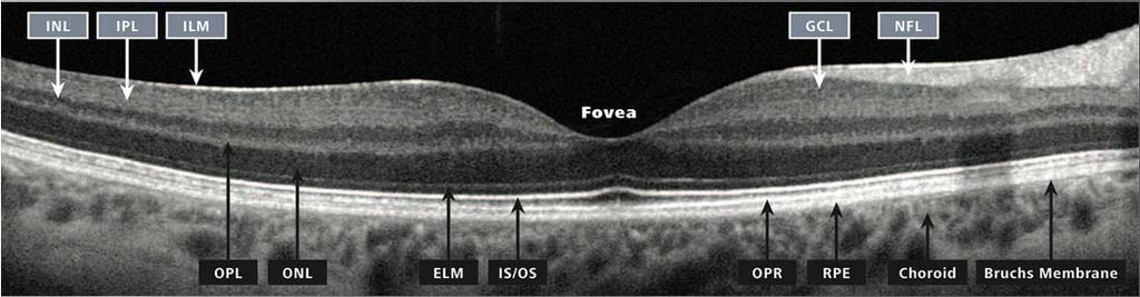
5 Choroid 10 layers of the retina RPE Photoreceptors ELM Outer nuclear layer Outer plexiform layer Inner nuclear layer Inner plexiform layer Ganglion cell layer Nerve fiber layer ILM Vitreous 5

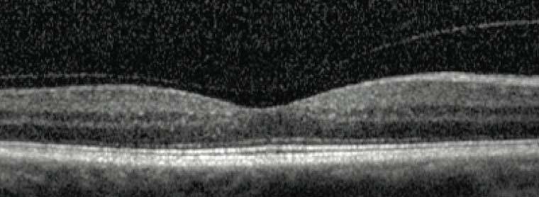
6 Vitreous Neuro-Sensory Retina RPE Choroid 6


7 Epiretinal Membrane (ERM) Epiretinal Membrane (ERM) 7
8 8

9 9
 Watzke-Allen sign")
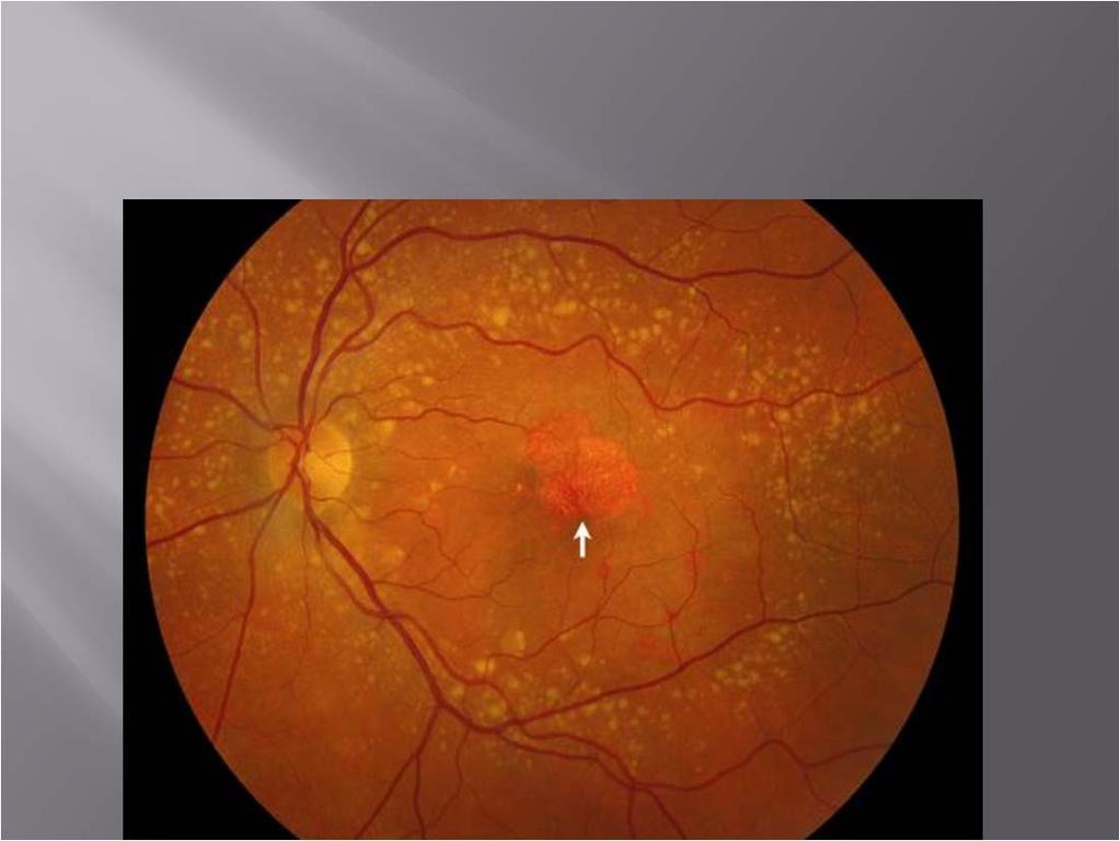
10 Unilateral, decreased vision Often in year old women Anyone w/ a history of trauma Symptoms: Decreased vision, metamorphopsia 20/200 for full thickness holes Signs: Red hole in the macula (+) Watzke-Allen sign 10


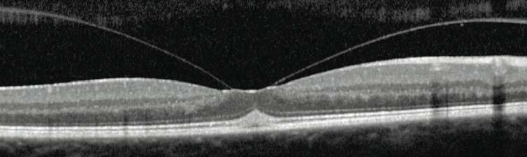
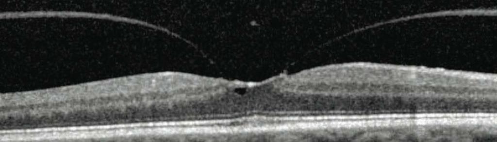
11 Stages Stage 1a -> impending hole. Normal foveal depression with yellow spot/dot in fovea. Stage 1b -> Abnormal foveal depression with yellow ring. Stage 1b macular hole Stages Stage 2 -> Small full-thickness hole. 20/80-20/400. Stage 3 -> Full-thickness hole w/ cuff of SRF. No PVD Stage 4 -> Full-thickness hole with cuff of SRF, with complete PVD. Stage 2 macular hole 11


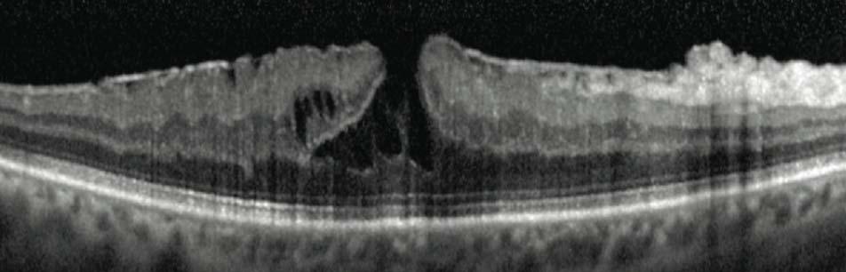
12 Stages Stage 2 -> Small full-thickness hole. 20/80-20/400. Stage 3 -> Full-thickness hole w/ cuff of SRF. No PVD Stage 4 -> Full-thickness hole with cuff of SRF, with complete PVD. Stage 3 Macular hole Stage 4 macular hole 12


13 Small FTMH w/o traction 154 microns 237 microns 13


14 Medium FTMH w/o traction microns Large FTMH with traction > 400 microns 14
 Vision 20/40 or worse How long has the")

15 Treatment: Stage 2 holes or beyond (full thickness macular holes) Vision 20/40 or worse How long has the hole been there??? Vitrectomy & membrane peel Face down??? Prognosis: 20/40 or better in up to 65% of cases 15

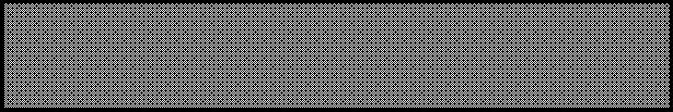
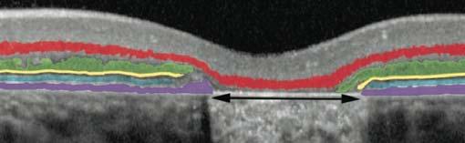
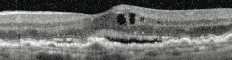
16 Partial thickness macular holes Aborted macular holes Upside down anvil anvil-like VA -> usually 20/40 or better 4 characteristics 1. Irregular foveal contour 2. Break in inner fovea 3. Intraretinal split 4. Intact foveal photoreceptors 16

17 Partial thickness macular holes Aborted macular holes Upside down anvil anvil-like VA -> usually 20/40 or better 4 characteristics 1. Irregular foveal contour 2. Break in inner fovea 3. Intraretinal split 4. Intact foveal photoreceptors False hole Simulates macular hole w/o actual tissue dehiscence Full thickness retinal tissue is still present Not an anvil VA Usually 20/20 20/30 unless significant ERM is present 17
18 Vitreous Neuro-Sensory Retina RPE Choroid 18
19 19
20 20


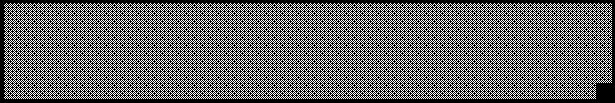
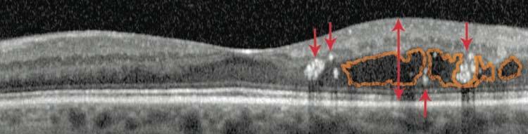
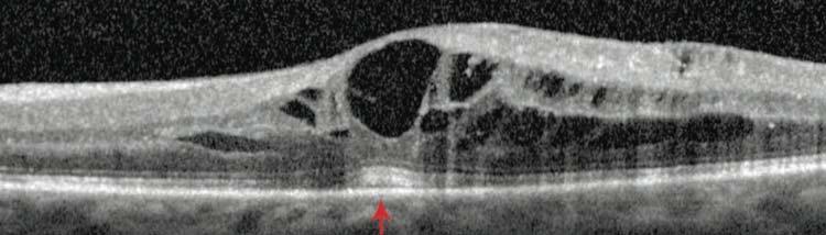
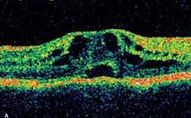
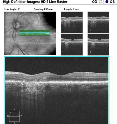
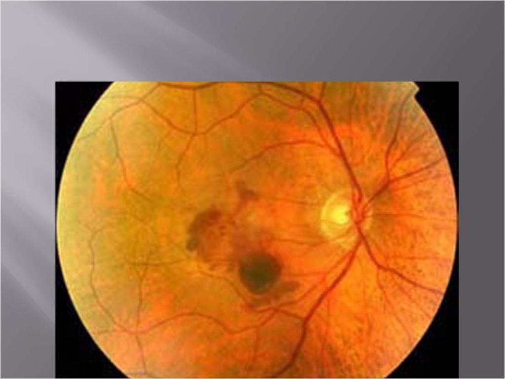
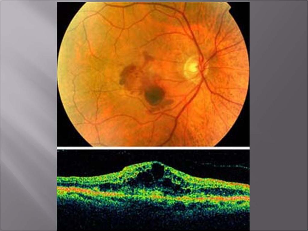
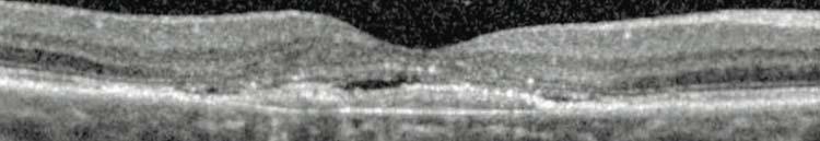
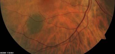
21 Demographics year old men, stressed/type A personalities Symptoms Unilateral, blurred vision VA -> usually 20/20 20/80 Metamorphopsia Signs Localized serous detachment of the neurosensory retina in the macula 21


22 DDx: Optic disc pit CNVM 22


23 Med associations: Steroids Nasal sprays, steroid creams, oral, injectable Ephedra Ephedrine & pseudoephedrine Treatment: Observation/lifestyle change D/C steroid if possible Possible laser therapy Vitreous Neuro-Sensory Retina RPE Choroid 23
24 24
25 25

26 26
27 27
28 28
29 29
30 30
31 31

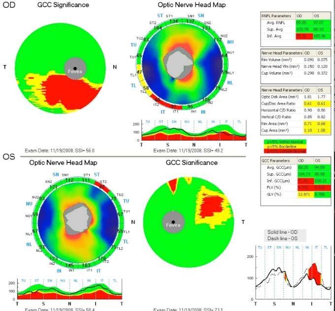
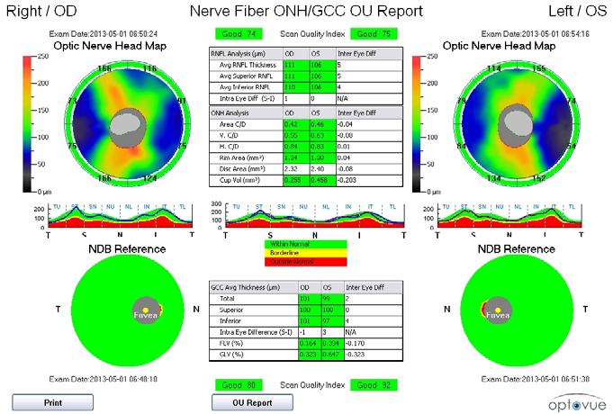
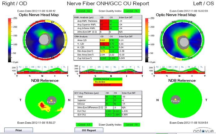
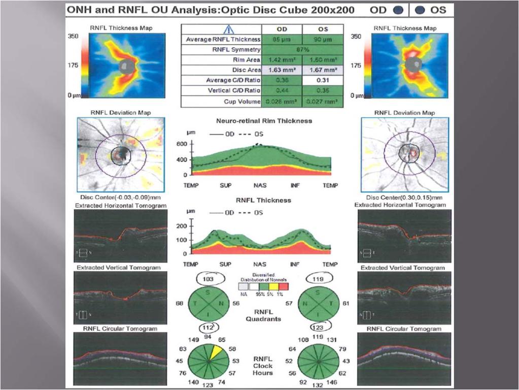
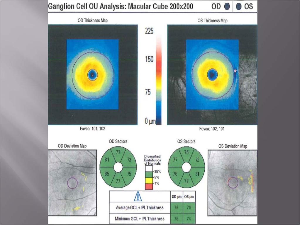
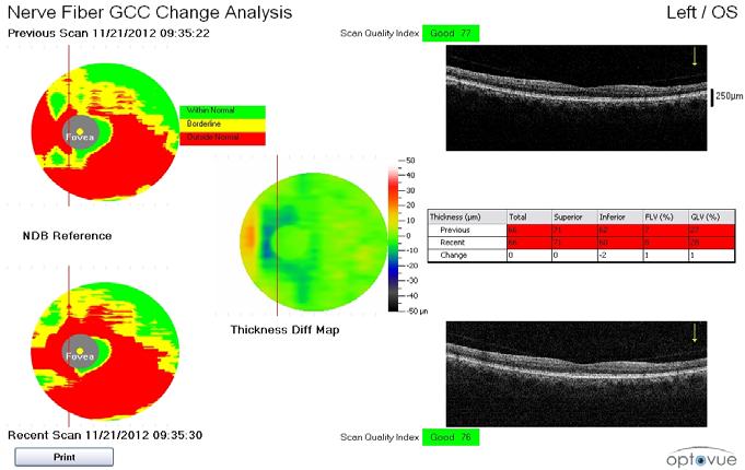
32 RNFL GCC 32

33 33
34 34
35 35


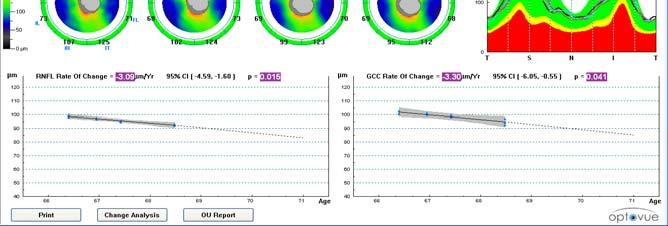
36 Assess change in RNFL and GCC thickness over time. RNFL Rate of Change = µ/Year GCC Rate of Change = µ/Year 36
37 37

38 75 38
8/6/17. Disclosures Aerie Pharmaceuticals Alcon BioTissue Diopsys Optovue Shire
 Nathan Lighthizer, O.D., F.A.A.O. Associate Professor Assistant Dean for Clinical Care Director of Continuing Education Chief of Specialty Care Clinics Oklahoma College of Optometry Tahlequah, OK lighthiz@nsuok.edu
Course # Getting to Know Your OCT
 Course # 140 Getting to Know Your OCT Course Title: Lecturer: Getting to Know Your OCT Brad Sutton, OD, FAAO IU School of Optometry Financial Disclosures No financial disclosures Optical Coherence Tomography-OCT
OCT Interpretation in Retinal Disease
 OCT Interpretation in Retinal Disease Jay M. Haynie, OD, FAAO Financial Disclosure I have received honoraria or am on the advisory board for the following companies: Carl Zeiss Meditec Advanced Ocular
Advances in OCT Murray Fingeret, OD
 Disclosures Advances in OCT Murray Fingeret, OD Consultant Alcon, Allergan, Bausch & Lomb, Carl Zeiss Meditec, Diopsys, Heidelberg Engineering, Reichert, Topcon Currently Approved OCT Devices OCT Devices
Optical Coherence Tomography (OCT)
") Understanding and Interpreting OCT Mark Dunbar: Disclosure The Swiss Army Pocket Knife of Eye Care Mark T. Dunbar, O.D., F.A.A.O. Bascom Palmer Eye Institute University of Miami, School of Medicine Consultant
Understanding and Interpreting OCT Mark Dunbar: Disclosure The Swiss Army Pocket Knife of Eye Care Mark T. Dunbar, O.D., F.A.A.O. Bascom Palmer Eye Institute University of Miami, School of Medicine Consultant
Mark Dunbar: Disclosure
 Important Things to Understand About OCT Mark T. Dunbar, O.D., F.A.A.O. Bascom Palmer Eye Institute University of Miami, School of Medicine Mark Dunbar: Disclosure Optometry Advisory Board for: Allergan
OCT Interpretation. Financial Disclosure. Jay M. Haynie, OD, FAAO. OCT Image Layers 7/21/2014
 OCT Interpretation Jay M. Haynie, OD, FAAO Financial Disclosure I have received honoraria or am on the advisory board for the following companies: Olympia Tacoma Renton Kennewick - Washington Carl Zeiss
OPTIC DISC PIT Pathogenesis and Management OPTIC DISC PIT
 OPTIC DISC PIT Pathogenesis and Management Abdel-Latif Siam Ain Shams University Cairo Egypt OPTIC DISC PIT Congenital pit is an atypical coloboma usually located on the temporal edge of the disc, associated
Cirrus TM HD-OCT. Details define your decisions
 Cirrus TM HD-OCT Details define your decisions 2 With high-definition OCT Carl Zeiss Meditec takes you beyond standard spectral domain Built on 10 years experience at the vanguard of innovation, Carl Zeiss
Cirrus TM HD-OCT. Details defi ne your decisions
 Cirrus TM HD-OCT Details defi ne your decisions 2 With high-defi nition OCT Carl Zeiss Meditec takes you beyond standard spectral domain Built on 10 years experience at the vanguard of innovation, Carl
Optical Coherence Tomography in Diabetic Retinopathy. Mrs Samantha Mann Consultant Ophthalmologist Clinical Lead of SEL-DESP
 Optical Coherence Tomography in Diabetic Retinopathy Mrs Samantha Mann Consultant Ophthalmologist Clinical Lead of SEL-DESP Content OCT imaging Retinal layers OCT features in Diabetes Some NON DR features
What Is O.C.T. and Why Should I Give A Rip? OCT & Me How Optical Coherence Tomography Changed the Life of a Small Town Optometrist 5/19/2014
 OCT & Me How Optical Coherence Tomography Changed the Life of a Small Town Optometrist Email: myoder@wcoil.com Mark A. Yoder, O.D. 107 N. Main Street PO Box 123 Bluffton, OH 45817 @yoderod 115.02 Histoplasma
Re(nal and OCT Grand Rounds
 Op#cal Coherence Tomography Op(cal: Light- based Re(nal and OCT Grand Rounds Steven Ferrucci, OD, FAAO Chief, Optometry Sepulveda VA Professor, SCCO/MBKU Coherence: property of light waves in which the
Vitreomacular interface disorders. Ghanbari MD 1393:10:25
 Vitreomacular interface disorders Ghanbari MD 1393:10:25 Human vitreous after dissection of the sclera, choroid, and retina. Lamellar structure of the posterior vitreous cortex (PVC) in the monkey. V =
Incorporating OCT Angiography Into Patient Care
 Incorporating OCT Angiography Into Patient Care Beth A. Steele, OD, FAAO OCT A: Introduction Isolates microvascular circulation from OCT image data Axial resolution = 5 microns (i.e. fine capillaries visible)
Title: OCT Analysis Workshop: Interpretation of OCT printouts
 Title: OCT Analysis Workshop: Interpretation of OCT printouts Authors: David Yang, OD, FAAO Staff Optometrist, VA Palo Alto Health Care System Associate Clinical Professor, UC Berkeley School of Optometry
Optical Coherence Tomography (OCT) in Uveitis Piergiorgio Neri, BMedSc, MD, PhD Head Ocular Immunology Unit
 in Uveitis Piergiorgio Neri, BMedSc, MD, PhD Head Ocular Immunology Unit") The Eye Clinic Polytechnic University of Marche Head: Prof Alfonso Giovannini November, 1991 Optical Coherence Tomography (OCT) in Uveitis Piergiorgio Neri, BMedSc, MD, PhD Head Ocular Immunology Unit
The Eye Clinic Polytechnic University of Marche Head: Prof Alfonso Giovannini November, 1991 Optical Coherence Tomography (OCT) in Uveitis Piergiorgio Neri, BMedSc, MD, PhD Head Ocular Immunology Unit
VITREOMACULAR UPDATE FOR THE PRIMARY CARE OD
 VITREOMACULAR UPDATE FOR THE PRIMARY CARE OD VITREOMACULAR UPDATE FOR THE PRIMARY CARE OD 1 2 DISCLOSURE STATEMENT I have received lecture honoraria from TearScience. I have no direct financial or proprietary
Managing the Vitreomacular Interface
 Managing the Vitreomacular Interface A Guide to VMA, VMT, Holes and ERM Anna K. Bedwell, OD, FAAO Indiana University School of Optometry Please silence all mobile devices and remove items from chairs so
EPIRETINAL MEMBRANE & VITREOMACULAR TRACTION
 EPIRETINAL MEMBRANE & VITREOMACULAR TRACTION Management of ERM and VMT K.V.Chalam,MD,PhD,MBA,FACS Professor and Director of Retina Loma Linda Eye Institute Los Angeles, USA REVIEW ANATOMY The vitreous
An A to Z guide on Epiretinal Membranes (ERMs) Paris Tranos PhD,ICO,FRCS OPHTHALMICA Vitreoretinal & Uveitis Department
 Paris Tranos PhD,ICO,FRCS OPHTHALMICA Vitreoretinal & Uveitis Department") An A to Z guide on Epiretinal Membranes (ERMs) Paris Tranos PhD,ICO,FRCS OPHTHALMICA Vitreoretinal & Uveitis Department Types of ERM Natural history OCT prognostic factors ERM with co-existing pathology
An A to Z guide on Epiretinal Membranes (ERMs) Paris Tranos PhD,ICO,FRCS OPHTHALMICA Vitreoretinal & Uveitis Department Types of ERM Natural history OCT prognostic factors ERM with co-existing pathology
The Quick Guide to OCT Mastery 50 Real Cases with Expert Analysis
 OPTICAL COHERENCE TOMOGRAPHY The Quick Guide to OCT Mastery 50 Real Cases with Expert Analysis VOL 1 Sanjay Sharma, MD, FRCS, MSc (Epid), MBA Ophthalmologist, Epidemiologist Queen s University, Canada
Ganglion cell analysis by optical coherence tomography (OCT) Jonathan A. Micieli, MD Valérie Biousse, MD
 Jonathan A. Micieli, MD Valérie Biousse, MD") Ganglion cell analysis by optical coherence tomography (OCT) Jonathan A. Micieli, MD Valérie Biousse, MD Figure 1. Normal OCT of the macula (cross section through the line indicated on the fundus photo)
Ganglion cell analysis by optical coherence tomography (OCT) Jonathan A. Micieli, MD Valérie Biousse, MD Figure 1. Normal OCT of the macula (cross section through the line indicated on the fundus photo)
OCT in Diabetic Macular Edema and its Correlation with Flourescein Angiography
 Uvea OCT in Diabetic Macular Edema and its Correlation with Flourescein Angiography Kirti Jaisingh MS Kirti Jaisingh MS, Yashpal Goel* MS, Kshitij Aditya** DO * Guru Nanak Eye Centre, New Delhi ** Baba
Optical coherence tomography of the vitreoretinal interface in macular hole formation
 1092 St Thomas s Hospital, London V Tanner D S Chauhan T L Jackson T H Williamson Correspondence to: Mr V Tanner, Royal Berkshire Hospital, London Road, Reading RG1 5AN, UK tannerone@aol.com Accepted for
R&M Solutions
 Mohamed Hosny El-Bradey, MD., Assistant Professor of Ophthalmology, Tanta University. Wael El Haig, MD., Professor of Ophthalmology. Zagazeeg University. 1 Myopic CNV is considered the most common vision
Clinical Study Spectral Domain OCT: An Aid to Diagnosis and Surgical Planning of Retinal Detachments
 Ophthalmology Volume 2011, Article ID 725362, 4 pages doi:10.1155/2011/725362 Clinical Study Spectral Domain OCT: An Aid to Diagnosis and Surgical Planning of Retinal Detachments Graham Auger and Stephen
Method for comparing visual field defects to local RNFL and RGC damage seen on frequency domain OCT in patients with glaucoma.
 Method for comparing visual field defects to local RNFL and RGC damage seen on frequency domain OCT in patients with glaucoma. Donald C. Hood 1,2,* and Ali S. Raza 1 1 Department of Psychology, Columbia
Objective Assessment of Macula and Optic Nerve
 Objective Assessment of Macula and Optic Nerve Jerry Sherman Disclosure: Dr. Sherman has lectured and received honorarium from Carl Zeiss Meditec, Topcon, Optovue, Optos, and PHP, Diopsys, Eye Solutions,Quantel,
IN NICU OCT UTILIZES A CONCEPT KNOWN AS INTERFEROMETRY APPLICATIONS FOR OCT THE PRIMARY USE IN THE EYE - RETINA
 2016 25 YEARS OF OPTICAL COHERENCE TOMOGRAPHY OPTICAL COHERENCE TOMOGRAPHY IN NICU Marcin Stopa, MD, PhD, FEBO Department of Ophthalmology, Chair of Ophthalmology and Optometry. Poznan University of Medical
Structure WGA. Structure WGA. Structural Assessment in Glaucoma. What s New
 New Developments in the Structural and Functional Assessment of the Glaucomas John G. Flanagan PhD, MCOptom, FAAO Professor, School of Optometry, University of Waterloo Professor, Dept of Ophthalmol &
Dehiscence of detached internal limiting membrane in eyes with myopic traction maculopathy with spontaneous resolution
 Hirota et al. BMC Ophthalmology 2014, 14:39 RESEARCH ARTICLE Open Access Dehiscence of detached internal limiting membrane in eyes with myopic traction maculopathy with spontaneous resolution Kazunari
Optical Coherence Tomograpic Features in Idiopathic Retinitis, Vasculitis, Aneurysms and Neuroretinitis (IRVAN)
") Columbia International Publishing Journal of Ophthalmic Research (2014) Research Article Optical Coherence Tomograpic Features in Idiopathic Retinitis, Vasculitis, Aneurysms and Neuroretinitis (IRVAN)
Columbia International Publishing Journal of Ophthalmic Research (2014) Research Article Optical Coherence Tomograpic Features in Idiopathic Retinitis, Vasculitis, Aneurysms and Neuroretinitis (IRVAN)
Case report 12/10/2014. Delphine Lam ; Dr Mayer Srour Service d ophtalmologie Professeur E.Souied Université Paris Est
 Case report 12/10/2014 Delphine Lam ; Dr Mayer Srour Service d ophtalmologie Professeur E.Souied Medical history Man, 75 years old Complaint: Vision loss in left eye in June 2014 Past ophthalmologic history:
OCT Angiography in Primary Eye Care
 OCT Angiography in Primary Eye Care An Image Interpretation Primer Julie Rodman, OD, MS, FAAO and Nadia Waheed, MD, MPH Table of Contents Diabetic Retinopathy 3-6 Choroidal Neovascularization 7-9 Central
How to Be Efficient and Effective. Disclosure. Topics CASE CM. Case JF 2007 OHTN / POAG? How to Be Efficient and Effective with. with New Technology
 How to Be Efficient and Effective with Disclosure COPE Course ID: 40750 GL Michael Chaglasian has the following disclosures: 1. Advisory Board: Allergan, Inc., Alcon Labs, B+L Carl Zeiss Meditec 2. Research:
Optical coherence tomography (OCT) is a relatively new noninvasive. The Use of Optical Coherence Tomography in Neurology DIAGNOSTIC UPDATE
 is a relatively new noninvasive. The Use of Optical Coherence Tomography in Neurology DIAGNOSTIC UPDATE") DIAGNOSTIC UPDATE The Use of Optical Coherence Tomography in Neurology Cédric Lamirel, MD,* Nancy Newman, MD,* Valérie Biousse, MD* Departments of *Ophthalmology, Neurology, and Neurological Surgery, Emory
DIAGNOSTIC UPDATE The Use of Optical Coherence Tomography in Neurology Cédric Lamirel, MD,* Nancy Newman, MD,* Valérie Biousse, MD* Departments of *Ophthalmology, Neurology, and Neurological Surgery, Emory
Often asymptomatic but can cause a reduction in BCVA and distortion of vision.
 Christopher Wolfe, OD, FAAO, Dipl. ABO Epiretinal Membrane (ERM) and Vitreomacular Traction (VMT) Epiretinal membrane (macular pucker, cellophane maculopathy, premacular fibrosis) consists of a layer of
Disclosures. Definitions. Goals. Imaging and glaucoma 3/22/2016
 Pinakin Davey OD, PhD, FAAO Professor and Director of Research Disclosures Principal investigator for ivue OCT trial Principal investigator Topcon FDA trials for Maestro and OCT 2000 Consultant for Topcon
Structural examina.on: Imaging
 ManaMa: Glaucoma Structural examina.on: Imaging Luís Abegão Pinto, MD, PhD Department of Ophthalmology CHLC Lisbon Faculty of Medicine, Lisbon University 1 11-10- 2013 Structural changes Qualitative changes
Do You See What I See!!! Shane R. Kannarr, OD
 Do You See What I See!!! Shane R. Kannarr, OD skannarr@kannarreyecare.com Define Specialty Testing Additional Test to: Prove/Disprove Diagnosis To monitor progression of a condition To document a condition
ZEISS AngioPlex OCT Angiography. Clinical Case Reports
 Clinical Case Reports Proliferative Diabetic Retinopathy (PDR) Case Report 969 PROLIFERATIVE DIABETIC RETINOPATHY 1 1-year-old diabetic female presents for follow-up of proliferative diabetic retinopathy
AperTO - Archivio Istituzionale Open Access dell'università di Torino
 AperTO - Archivio Istituzionale Open Access dell'università di Torino Artifacts in automatic retinal segmentation using different optical coherence tomography instruments. This is the author's manuscript
1. AGENCY USE ONLY (Leave blank) 2. REPORT DATE 3. REPORT TYPE AND DATES COVERED FINAL 01 May 93 TO 30 Apr 95. mwwin i ii ^.. IIM«-»- "«'» ' ' i» 1
 2. REPORT DATE 3. REPORT TYPE AND DATES COVERED FINAL 01 May 93 TO 30 Apr 95. mwwin i ii ^.. IIM«-»- «'» ' ' i» 1") REPORT DOCUMENTATION PAGI AFOSR-TR- OiOl Public reoortma burden for this colleaion of information is estimated to average i nour per respor aathenna and maintaining the data needed, and completing and
REPORT DOCUMENTATION PAGI AFOSR-TR- OiOl Public reoortma burden for this colleaion of information is estimated to average i nour per respor aathenna and maintaining the data needed, and completing and
Moving forward with a different perspective
 Moving forward with a different perspective The Leader In Vision Diagnostics Offers A New Perspective Marco has served the eyecare community by offering exceptional lane products and automated high tech
Optical Coherence Tomography: Pearls for the Anterior Segment Surgeon Basic Science Michael Stewart, M.D.
 Optical Coherence Tomography: Pearls for the Anterior Segment Surgeon Basic Science Michael Stewart, M.D. Disclosure OCT Optical Coherence Tomography No relevant financial relationships I will refer to
VMA at the macula resulting in VMT
 Ocriplasmina for pharmacologic treatment in VMT Teresio Avitabile 1 Introduction PVD is a normal, physiologic process that occurs with aging; however, in some cases, PVD is incomplete Incomplete PVD localized
Vitreomacular Traction: Management
 Miscellaneous Refractive Surgery Vitreomacular Traction: Management Raji K. MS, DNB Raji K. MS, DNB, A.K. Upadhyay MS, S. Waikar MS, DNB, P. Tiwari MBBS Department of Ophthalmology, Command Hospital (WC)
Re)nal and OCT Grand Rounds. What's new in OCT? Principles of AngioVue OCTA. Vascular Imaging No Referral Needed 3/9/18. Spectral Domain: Many Op3ons
nal and OCT Grand Rounds. What's new in OCT? Principles of AngioVue OCTA. Vascular Imaging No Referral Needed 3/9/18. Spectral Domain: Many Op3ons") Spectral Domain: Many Op3ons Re)nal and OCT Grand Rounds Steven Ferrucci, OD, FAAO Chief, Optometry Sepulveda VA Professor, SCCO/MBKU Ease of use Customer support Integra)on of other technology FAF Color
Spectral Domain: Many Op3ons Re)nal and OCT Grand Rounds Steven Ferrucci, OD, FAAO Chief, Optometry Sepulveda VA Professor, SCCO/MBKU Ease of use Customer support Integra)on of other technology FAF Color
Consulting Fee: Alcon Laboratories
 Consulting Fee: Alcon Laboratories Pre-Op EMM Post PPV, Forceps EMM & ILM Peeling 25/27 Gauge, Trans-Conjunctival, Sutureless PPV Inside-Out, End-Grasping Forceps Peeling w/ Alcon 25G End-Grasping DSP
OCT in the Diagnosis and Follow-up of Glaucoma
 OCT in the Diagnosis and Follow-up of Glaucoma Karim A Raafat MD. Professor Of Ophthalmology Cairo University Hmmmm! Do I have Glaucoma or not?! 1 Visual Function 100% - N Gl Structure : - 5000 axon /
Ophthalmic Imager Role
 MASTER OCT Ophthalmic Photographers Society October 18, 2014 Chicago, IL James B Soque, CRA COA Pamela A Weber, MD Island Retina Shirley, New York Commack, New York Financial Disclosure Genentech Ophthotech
University Hospital Basel. Optical Coherence Tomography Emerging Role in the Assessment of MS PD Dr. Konstantin Gugleta
 University Hospital Basel Optical Coherence Tomography Emerging Role in the Assessment of MS PD Dr. Konstantin Gugleta 15th State of the Art SMSS, Lucerne January 2013 Retinal Nerve Fiber Layer 1.200.000
Reproducibility of Choroidal Thickness Measurements Across Three Spectral Domain Optical Coherence Tomography Systems
 Reproducibility of Choroidal Thickness Measurements Across Three Spectral Domain Optical Coherence Tomography Systems The MIT Faculty has made this article openly available. Please share how this access
Measuring of the fovea and foveola using line scans and 3D Macular scans obtained with spectral domain optical coherent tomography.
 Measuring of the fovea and foveola using line scans and 3D Macular scans obtained with spectral domain optical coherent tomography. Vakhrameeva O.A. 1, Moiseenko G.A. 1, Maltsev D.S. 2, Sukhinin M.V. 2,
Optical Coherence Tomography Findings in Highly Myopic Eyes following Cataract Surgery
 Optical Coherence Tomography Findings in Highly Myopic Eyes following Cataract Surgery Fedra Hajizadeh, MD 1 Mohammad Riazi Esfahani, MD 1,2 Hooshang Faghihi, MD 3 Mehdi Khanlari, MD 4 Abstract Purpose:
Diabetic Retinopathy Clinical Research Network
 Diabetic Retinopathy Clinical Research Network Comparison of Time Domain OCT and Spectral Domain OCT Retinal Thickness Measurement in Diabetic Macular Edema Version 1.0 June 16, 2009 comparison of td vs
Ganglion cell complex scan in the early prediction of glaucoma
 Original article in the early prediction of glaucoma Ganekal S Nayana Super Specialty Eye Hospital and Research Center, Davangere, Karnataka, India Abstract Objective: To compare the macular ganglion cell
ATLAS OF OCT. Retinal Anatomy in Health & Pathology by Neal A. Adams, MD. Provided to you by:
 ATLAS OF OCT Retinal Anatomy in Health & Pathology by Neal A. Adams, MD Provided to you by: Atlas of OCT The OCT Atlas is written by Neal A. Adams, MD, and produced by Heidelberg Engineering, Inc. to help
NIH Public Access Author Manuscript Retin Cases Brief Rep. Author manuscript; available in PMC 2012 January 1.
 NIH Public Access Author Manuscript Published in final edited form as: Retin Cases Brief Rep. 2011 ; 5(1): 46 48. doi:10.1097/icb.0b013e3181cafc49. Intact Retinal Tissue and Retinal Pigment Epithelium
Diagnosis in AMD. Managing your AMD Patients
 Managing your AMD Patients Robert W. Dunphy, O.D., F.A.A.O. Diagnosis in AMD Have suspicion Identify relative risk Conduct surveillance Biometry Utilize technology to facilitate detection of change / stability
OCT Assessment of the Vitreoretinal Relationship in CSME
 December 2007 Sonia Rani John et al. - IFIS 375 ORIGINAL ARTICLE OCT Assessment of the Vitreoretinal Relationship in CSME Dr. Manoj S. DNB FRCS, Dr. Unnikrishnan Nair MS DO FRCS, Dr. Gargi Sathish MS Introduction
Spectral- and time-domain optical coherence tomography measurements of macular thickness in young myopic eyes
 Liu et al. Diagnostic Pathology 2014, 9:38 RESEARCH Open Access Spectral- and time-domain optical coherence tomography measurements of macular thickness in young myopic eyes Lin Liu, Jun Zou *, Lili Jia,
Il contributo dell'angio-oct: valutazione integrata della componente nervosa e vascolare della malattia glaucomatosa
 SIMPOSIO G.O.A.L. - LE NUOVE FRONTIERE DIAGNOSTICHE E LE LINEE DI INDIRIZZO AMBULATORIALI DEL GLAUCOMA Coordinatore e moderatore: D. Mazzacane Presidente: L. Rossetti Il contributo dell'angio-oct: valutazione
Χειρουργική Ωχράσ Κηλίδασ. Γ. Γ. Παππάς, Βεληδέιεηο ΓΝ
 Χειρουργική Ωχράσ Κηλίδασ Γ. Γ. Παππάς, Βεληδέιεηο ΓΝ FREQUENCY OF MACULAR SURGERY Incidence= 8 in 100000 (McCannel 2009) Prevalence = between 0.2 (Mitchell 2007) and 3.3 (Baltimore Eye Survey 1996) 2nd
We are IntechOpen, the world s leading publisher of Open Access books Built by scientists, for scientists. International authors and editors
 We are IntechOpen, the world s leading publisher of Open Access books Built by scientists, for scientists 3,900 116,000 120M Open access books available International authors and editors Downloads Our
Early diagnosis and treatment of VMT with single Intravitreal Injection of Pharmacologic Vitreolysis. Stratos Gotzaridis MD Athens
 Early diagnosis and treatment of VMT with single Intravitreal Injection of Pharmacologic Vitreolysis Stratos Gotzaridis MD Athens The Vitreous Body Gel composed of 98-99% water 1% macromolecules Glycoproteins
case profile: Macular Holes
 Early Detection Saves Sight 2012 ISSUE 2 newsletter for optometrists I m pleased to let you know about a new series of resources for optometrists that is being produced by the team at CFEH. The Clinical
OCT Image Analysis System for Grading and Diagnosis of Retinal Diseases and its Integration in i-hospital
 Progress Report for1 st Quarter, May-July 2017 OCT Image Analysis System for Grading and Diagnosis of Retinal Diseases and its Integration in i-hospital Milestone 1: Designing Annotation tool extraction
Analysis of Peripapillary Atrophy Using Spectral Domain Optical Coherence Tomography
 Analysis of Peripapillary Atrophy Using Spectral Domain Optical Coherence Tomography The MIT Faculty has made this article openly available. Please share how this access benefits you. Your story matters.
Steven Ferrucci, OD. FAAO; Jeffry Gerson, OD, FAAO; Robert Prouty, OD, FAAO; Leo semes OD, FAAO
 PARDON THE OBJECTION: RETINA Steven Ferrucci, OD. FAAO; Jeffry Gerson, OD, FAAO; Robert Prouty, OD, FAAO; Leo semes OD, FAAO 1. Introductions/Disclosures (Ferrucci) 2. The genetics of AMD (Gerson) a. Background
Comparison of Spectral/Fourier Domain Optical Coherence Tomography Instruments for Assessment of Normal Macular Thickness
 Comparison of Spectral/Fourier Domain Optical Coherence Tomography Instruments for Assessment of Normal Macular Thickness The MIT Faculty has made this article openly available. Please share how this access
Clinical spectrum of lamellar macular defects including pseudoholes and pseudocysts defined by optical coherence tomography
 Clinical spectrum of lamellar macular defects including pseudoholes and pseudocysts defined by optical coherence tomography J C Chen, 1,2 L R Lee 1,3 1 City Eye Centre, Brisbane, Australia; 2 Institute
Optical Coherence Tomography of the Retina. New Technology - New Insights
 Bahrain Medical Bulletin, Vol. 27, No. 3, September 2005 Optical Coherence Tomography of the Retina. New Technology - New Insights Mohinder Singh, FRCS,FRCOphth* Optical imaging is an emerging new technology,
New Technologies in Glaucoma Management: From ERG to OCT
 What s New and What s Next in Glaucoma New Technologies in Glaucoma Management: From ERG to OCT Ben Gaddie, OD FAAO Murray Fingeret, OD FAAO IOP 24- Hour IOP Role of hysteresis in glaucoma risk Cerebrospinal
Macular Holes. The Retina
 Macular Holes This information leaflet aims to give you some information about the condition of Macular Holes. It is a common causes of poor vision amongst retired people. So What is a Macular Hole? This
12/2/16. Ways to differentiate:
 Nate Lighthizer, O.D., F.A.A.O. Assistant Dean for Clinical Care Services Director of CE Chief of Specialty Care Clinics Chief of Electrodiagnostics Clinic Oklahoma College of Optometry lighthiz@nsuok.edu
Citation BioMed Research International, 2015, v. 2015, article no Creative Commons: Attribution 3.0 Hong Kong License
 Title Relationship between Outer Retinal Layers Thickness and Visual Acuity in Diabetic Macular Edema Author(s) Wong, RLM; Lee, JWY; Yau, GSK; Wong, IYH Citation BioMed Research International, 2015, v.
Normative spectral domain optical coherence tomography data on macular and retinal nerve fiber layer thickness in Indians
 AOP*** 1 Original Article Normative spectral domain optical coherence tomography data on macular and retinal nerve fiber layer thickness in Indians Bindu Appukuttan, Anantharaman Giridhar, Mahesh Gopalakrishnan,
Introducing ANGIOVUE ESSENTIAL. Built on the Avanti Widefield OCT Platform. OCT Angiography for Primary Eye Care
 Introducing ANGIOVUE ESSENTIAL Built on the Avanti Widefield OCT Platform OCT Angiography for Primary Eye Care Transform Your View of the Retina OCT Angiography (OCTA) is a quick non-invasive test that
3/6/2014. Hoda MH Mostafa MD Associate Professor of Ophthalmology Cairo University. The author has no proprietary interest. Today s Objectives
 Hoda MH Mostafa MD Associate Professor of Ophthalmology Cairo University The author has no proprietary interest Today s Objectives Identify the CLINICAL SCENARIOS IN MACULAR EDEMA where OCT plays a MAJOR
Yasser R. Serag, MD Tamer Wasfi, MD El- Saied El-Dessoukey, MD Magdi S. Moussa, MD Anselm Kampik, MD
 Microperimetric Evaluation of Brilliant Blue G- assisted Internal Limiting Membrane Peeling By Yasser R. Serag, MD Tamer Wasfi, MD El- Saied El-Dessoukey, MD Magdi S. Moussa, MD Anselm Kampik, MD The internal
We are IntechOpen, the world s leading publisher of Open Access books Built by scientists, for scientists. International authors and editors
 We are IntechOpen, the world s leading publisher of Open Access books Built by scientists, for scientists 3,500 108,000 1.7 M Open access books available International authors and editors Downloads Our
Learn Connect Succeed. JCAHPO Regional Meetings 2016
 Learn Connect Succeed JCAHPO Regional Meetings 2016 pearls and pitfalls of ophthalmic imaging JCHAPO 2016 Conference Vikas Chopra, M.D. Medical Director, UCLA Doheny Eye Centers Pasadena Principal Investigator,
Vitreo-retinal interface pathologies and fibrinolytic treatment approaches
 Vitreo-retinal interface pathologies and fibrinolytic treatment approaches Constantin J. Pournaras Memorial A. de Rothschild Clinical Research Group La Colline Ophthalmology Center Vitreoretinal Interface
The Future of Retinal Imaging Has Arrived!
 Financial Disclosure The Future of Retinal Imaging Has Arrived! Joseph J. Pizzimenti, OD, FAAO Carlo Pelino, OD, FAAO! Pizzimenti:! Honoraria! Kemin! Nicox! Review of Optometry! Optometric Management!
HOW TO MAKE THE MOST OF A NEW OCT. with Kelly Kerksick, OD
 HOW TO MAKE THE MOST OF A NEW OCT with Kelly Kerksick, OD 3 How to Make the Most of a New OCT Kelly Kerksick, OD, graduated from Southern College of Optometry and immediately started her own private practice
OCT Angiography The Next Frontier
 Choroid Retina avascular 5/13/2017 OCT Angiography The Next Frontier Pierce Kenworthy OD, FAAO June 9, 2017 OCT Angiography (OCTA) 2016 Non-invasive, motion contrast imaging Represents erythrocyte movement
Re)nal and OCT Grand Rounds
nal and OCT Grand Rounds") Disclosures Re)nal and OCT Grand Rounds Steven Ferrucci, OD, FAAO Chief, Optometry Sepulveda VA Professor, SCCO/MBKU Speakers bureau and/or Advisory Board for: Alcon Allergan AutoGenomics B&L Heidelberg
Disclosures Re)nal and OCT Grand Rounds Steven Ferrucci, OD, FAAO Chief, Optometry Sepulveda VA Professor, SCCO/MBKU Speakers bureau and/or Advisory Board for: Alcon Allergan AutoGenomics B&L Heidelberg
Automated Visual Field Analysis for Glaucoma
 Island of Vision Automated Visual Field Analysis for Glaucoma Normal Visual Field Parameters 60 superior 60 nasal 75 inferior 100 temporal Macula the central 13 Fovea the central 3 Visual field is limited
Early surgery preserves more vision for patients with Epiretinal Membranes
 Early surgery preserves more vision for patients with Epiretinal Membranes Rahman R 1, Stephenson J 2 KEYWORDS: Epiretinal membrane, Combined phakovitrectomy, OCT. Addresses: 1 Ms Rubina Rahman*, CalderdaleRoyalHospital,
Visual and Anatomical Outcomes of Vitreous Surgery for Large Macular Holes
 March 2009 Raju K.V. et al. - Closed Globe Injuries 31 ORIGINAL ARTICLE Visual and Anatomical Outcomes of Vitreous Surgery for Large Macular Holes Dr. Mahesh G. MS DO DNB FRCSEd, Dr. A. Giridhar MS, Dr.
Macular Thickness Measurement via Heidelberg Spectralis SD-OCT in Pediatric Patients
 Ophthalmology Research: An International Journal 2(6): 384-390, 2014, Article no. OR.2014.6.013 SCIENCEDOMAIN international www.sciencedomain.org Macular Thickness Measurement via Heidelberg Spectralis
Fundus Autofluorescence. Jonathan A. Micieli, MD Valérie Biousse, MD
 Fundus Autofluorescence Jonathan A. Micieli, MD Valérie Biousse, MD The retinal pigment epithelium (RPE) has many important functions including phagocytosis of the photoreceptor outer segments Cone Rod
Overview. Macular OCT Artifact Study
 Imaging Artifacts Sarah Moyer, CRA, OCT-C Director, Ophthalmic Imaging Kittner Eye Center University of North Carolina Chapel Hill, NC Disclose financial interest now Overview Sarah s Thoughts on Artifacts
PRIMUS 200 from ZEISS The essential OCT
 EN 00_00I The contents of the brochure may differ from the current status of approval of the product in your country. Please contact your regional representative for more information. Subject to change
Case History. Legends of the Posterior Segment CASE 1 4/22/2018
 Legends of the Posterior Segment Blair Lonsberry, MS, OD, MEd., FAAO Professor of Optometry Pacific University College of Optometry blonsberry@pacificu.edu CASE 1 Case History 38 black male, complaining
New Concepts in Glaucoma Ben Gaddie, OD Moderator Murray Fingeret, OD Louis Pasquale, MD
 New Concepts in Glaucoma Ben Gaddie, OD Moderator Murray Fingeret, OD Louis Pasquale, MD New Concepts in Glaucoma Optical Coherence Tomography: Is it necessary and needed to diagnose and monitor glaucoma?
Re)nal and OCT Grand Rounds
nal and OCT Grand Rounds") Op#cal Coherence Tomography Op)cal: Light- based Re)nal and OCT Grand Rounds Steven Ferrucci, OD, FAAO Chief, Optometry Sepulveda VA Professor, SCCO/MBKU Coherence: property of light waves in which the
Op#cal Coherence Tomography Op)cal: Light- based Re)nal and OCT Grand Rounds Steven Ferrucci, OD, FAAO Chief, Optometry Sepulveda VA Professor, SCCO/MBKU Coherence: property of light waves in which the
FALSE! True or False: Back to Basics. Key to Retinal Assessment: Making Visible what is Invisible Jerome Sherman
 Key to Retinal Assessment: Making Visible what is Invisible Jerome Sherman Disclosure: Dr. Sherman has lectured and received honorarium from Carl Zeiss Meditec, Topcon, Optovue, Optos, Eye Solutions, PHP,