Outline and Introduction. SECTIONS 1. Orbit 2. Eyelid 3. Conjunctiva 4. Cornea 5. Uvea 6. Lens 7. Retina/Vitreous 8. Optic Nerve/Glaucoma
|
|
|
- Harold Gregory
- 5 years ago
- Views:
Transcription


1 Pathology of the
2 Outline and Introduction SECTIONS 1. Orbit 2. Eyelid 3. Conjunctiva 4. Cornea 5. Uvea 6. Lens 7. Retina/Vitreous 8. Optic Nerve/Glaucoma

3 Intro - Basic Anatomy

4 THE ORBIT Anatomy Thyroid Orbitopathy Tumors Inflammation/Infection Trauma

5 Orbit - Anatomy Bones of the orbit Sphenoid Maxillary Ethmoid Lacrimal Zygoma Palatine Frontal


6 Orbit - Osteology

7 Orbit Posterior Contents The ANNULUS OF ZINN is the tendonring that encircles the ON and acts as an origin for the muscles.
8 Orbit Anterior Boundary The ORBITAL SEPTUM is the anterior fascial boundary to the orbit
9 Thyroid-Related Orbitopathy (Graves Disease) Autoimmune condition, triggered by TSH-R Antibodies, with lymphocytic infiltration, FIBROSIS, and ENLARGEMENT of extraocular muscles. Proptosis, strabismus/muscle-restriction, exposure problems (dry-eye), and compressive optic neuropathy. Treated with steroids, radiation therapy, or surgical decompression (opening the orbital walls into the sinuses)


10 Orbit Thyroid-Related Orbitopathy
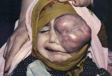
11 Orbit - Tumors Wide variety of lacrimal, lymphoid, neural, vascular, meningeal origin tumors, and metastatic tumors Children rhabdomyosarcoma is the most common primary malignancy of orbit. neuroblastoma is most common metastatic tumor

12 Orbit - Inflammation Orbital Cellulits frequently extends from adjacent sinus infections, or periocular trauma. A life and sight threatening emergency! Can extend into the cavernous sinus, and brain. Pre-Septal vs. Post-Septal can be distinguished by involvement of intraorbital structures


13 Orbit - Inflammation


14 Orbit - Trauma Blow out fractures occur when blunt trauma to the eye causes the orbit to rupture Orbital Floor fractures can cause restricted upgaze if there is muscle entrapment

15 LIDS - Anatomy LAYERS: Skin Orbicularis Tarsal plate Meibomian glands Palpebral conjunctiva

16 LIDS - Histology

17 Malignant Basal cell carcinoma -most common Squamous Melanoma Sebaceous cell carcinoma Benign Chalazion vs. Hordeolum Papillomas/Verrucae Epidermal inclusion cysts Many others LIDS - Tumors


18 LIDS - Tumors Chalazion a cyst of the meibomian gland Hordeolum an inflammed cyst of the MG (foreign body granuloma)
19 Conjunctiva Thin, non-keratinized skin covering the sclera (bulbar) or the inner surface of the lid (palpebral) Rich in goblet cells, which secret the mucinous components of the tear film


20 Conjunctiva The bulbar layer is continous with the palpebral layer
, bacterial, or allergic cause.")
21 Conjunctiva Pathologic conditions Conjunctivitis ( pink-eye ) is an inflammation of the conjunctiva due to a viral (Adenovirus), bacterial, or allergic cause. MALT, germinal centers


22 Conjunctivitis A rare granulomatous variety - Cat-scratch Fever! - Bartonella henselae
23 Conjunctiva Degenerative conditions Pinguecula on the conj only Pterygium encroaching onto cornea Histologically identical Both involve elastotic degeneration of the conjunctiva, usually due to chronic ultraviolet exposure.


24 Conjunctiva Degenerative conditions Small pinguecula Pterygium
")
25 Conjunctiva - tumors Conjunctival intraepithelial neoplasia (CIN) Squamous Cell Melanoma Lymphoid - arising from mucosa-associated lymphoid tissue (MALT) Lymphoma
, HPV")
26 Conjunctiva CIN (squamous cell), HPV 16/18
27 Cornea The cornea is a unique transparent and avascular tissue that is the most important refractive structure of the eye. Anatomy Inflammation/Infection Dystrophy/Ectasia
28 Cornea - Anatomy 5 Layers: 1. Epithelium Continuous with conj, richly innervated by CN-V 1 2. Bowman s Membrane 3. Stroma The thickest central portion (90%). This is where LASIK/Refractive surgery happens! Primarily made up of Type 1 Collagen in uniformly-spaced lamellar bundles. 4. Descemet s membrane 5. Endothelium pumps the water out of the cornea and keeps it clear

29 Cornea The uniform spacing of the stromal collagen bundles at a distance of approx ¼ wavelength light allows transparency.


30 Cornea - Refractive Surgery Excimer Laser is applied to the stromal bed, underneath a reflected corneal flap (LASIK). The tissue is ablated precisely to counteract the refractive error of the eye.


31 LASIK Traumatic Flap Dislocation
32 Cornea Inflammation/Infection Keratitis inflammation of cornea Bacterial ulcer Frequent in contact lens users, Pseudomonas most common Viral Herpes (HSV) is a frequent etiology Autoimmune, Syphilis, Fungal, ameobic, and many other types


33 Cornea - HSV Keratitis Epithelial dendritic Keratitis Stromal Keratits (note the vessels and clouding)

34 Cornea - Bacterial Ulcer Epithelial defect, infiltrate of white cells into the cornea, and a layered leukocyte collection in the AC (Hypopyon)
35 Cornea Stromal Dystrophy Dystrophy a heritable disorder resulting in abnormal tissue morphology, function, or abnormal depositions of material into the cornea. MANY types, affecting each specific layer.


36 Cornea Stromal Dystrophy Granular Dystrophy Hyaline material deposited in stroma


37 Cornea Stromal Dystrophy Lattice Dystrophy Amyloid deposition with apple-green birefringence. Stains with Congo Red


38 Cornea - Ectasia Progressive deformation of cornea is an ectasia. Keratoconus is the most common ectatic dystrophy. Ectasia can also be a complication of refractive surgery

39 Cornea Transplant

40 THE UVEA

41 The Uvea The uvea is: 1. The Iris 2. The Ciliary body 3. The Choroid Each has a function 1. Iris is a diaphragm for light 2. Ciliary body suspends and flexes the lens, and makes the aqueous humor 3. The choroid helps nourish the outer retina

42 The Uvea - Angle The angle is a special region of the uvea where the iris meets the cornea Regulates the outflow of Aqueous humor through the Canal of Schlem Determines the Intraocular pressure (Important in Glaucoma)
43 The Uvea - Inflammation Uveitis is inflamation of any combination of the iris, ciliary body, or choroid. Many etiologies (autoimmune, syphilis, sacrcoid, TB, HLA-B27, infectious, idiopathic, etc ) Many names (iritis, anterior uveitis, iridocylitis, choroiditis, etc ) depending on the location Sometimes associated with SERIOUS systemic inflamatory diseases (eg. arthritic diseases), inflamatory bowel disease, and vasculitis.


44 The Uvea Anterior Uveitis Anterior uveitis/iritis WBCs floating in the aqueous

45 Uvea Posterior Uveitis Active Toxoplasmosis Choroiditis, and old scar (above)
46 The Uvea - Tumors The Choroid is a highly perfused vascular net feeding the outer retina It is a potential target site for metastasis for carcinoma, such as breast and lung.

47 The Uvea - Tumors The uvea (especially choroid) is also richly pigmented, and primary melanocytic tumors are common. Nevi and malignant melanomas are both relatively common, and can be difficult to distinguish, clinically. Tumors with spindle-b or epithelioid histologic patterns are malignant

48 THE LENS
49 The Lens A transparent, avascular structure consisting of concentric cellular fibers Highest protein content of the body (Crystallins), which account for a high refractive index Interaction of the ciliary body muscle, through the zonular fibers, cause dynamic shape changes. In concert with the cornea, helps to focus light on the retina.


50 The Lens Entire structure encapsulated Lens cells migrate and elongate into fibers The deepest fibers are the oldest ones The lens continues to fatten throughout life Central fibers become sclerotic and opaque with time


51 The Lens - Cataract Opacities of the lens develop with time, or insult UV light, steroids, and inflammation are pathogenic factors

52 The Lens Cataract surgery A opening into the lens capsule is made The cataract is emulsified with ultrasound energy, and aspirated out of the eye


53 The Lens Cataract surgery The dense, cloudy crystalline lens is removed, and replaced with an optical implant.

54 The Retina
55 The Retina Anatomy Detachment Vascular disease/ischemic retinopathy Microvascular (Diabetes) Vascular occlusion (Vein occlusion/arterial Occlusion) Macular degeneration Tumors

56 The Retina - Anatomy Cell types (overview) Photoreceptors (detect light signal) Bipolars transmit/modulate signal to ganglion cells Ganglion cells send signal by long axons through optic nerve and into visual pathways of the brain Other cell types

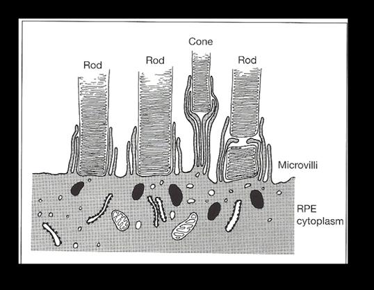
57 The Retina - Anatomy Layers (inside to out): 1. Inner limiting membrane 2. Nerve Fiber Layer 3. Ganglion Cell Layer 4. Inner plexiform layer 5. Inner nuclear layer 6. Outer plexiform layer 7. Outer nuclear layer 8. Photoreceptor segments 9. Retinal Pigment Epithelium 10. Bruch s Membrane (Choroid) (Sclera)
58 Retina Anatomy Pathologic conditions of layers 1) Retinal detachment: Separation between RPE and photoreceptor segments 2) Macular degeneration: Bruch s membrane damaged by deposition of drusen, allowing leaky choroidal vessels to grow into retina (exudative type).


59 The Retina - Detachment Retinal tears are the most frequent causes of detachment (rhegmatogenous RD) Tears can be spot welded with laser to prevent detachment


60 Retinal Detachment


61 The Retina Macular degeneration Clinical appearance of drusen in Macular degeneration
62 The Retina Vasculopathy Microvascular (small vessel disease) Diabetes Sickle Cell Radiation Macrovascular (large vessel occlusions) Central retinal vein occlusion (CRVO) Branch retinal vein occlusion (BRVO) Central retinal artery occlusion (CRAO) Branch retinal artery occlusion (BRAO)

63 The Retina Diabetic Retinopathy Microvascular dysfunction leads to tissue ischemia Thickened and Leaky Capillary basement membranes Loss of pericytes Microaneurysms Nonperfusion
64 The Retina Diabetic Retinopathy Ischemia leads to vascular endothelial growth factor (VEGF) production from injured tissues Promotes Neovascularization (abnormal blood vessel growth) of the retina, optic nerve, or iris. Abnormal vessels can cause edema or tractional retinal detachments VEGF implicated in other ischemic eye diseases, like Retinopathy of Prematurity


65 The Retina Diabetic Retinopathy Microaneurysms! Capillary dropout and Nonperfusion! Neovascularization!


66 The Retina Diabetic Retinopathy Retinal neovascularization
67 The Retina MACROvascular Disease CRVO/BRVO variety of anatomical prothrombotic predispositions CRAO/BRAO watch out for carotid/cardiac embolic disease, or vasculitis.


68 The Retina Macrovascular disease CRVO Hemorrhage, congestion, ischemia

69 The Retina Macrovascular disease Ischemic CRVO led to VEGF production, which caused neovascularization of iris.
70 The Retina - Tumors Retinoblastoma Classic pediatric tumor of retina Hereditary or Sporadic Requires two gene mutations (Knudsen s two-hit hypothesis) Classic histologic features of Flexner- Wintersteiner Rosettes, and fleurettes

71

72 OPTIC NERVE
 causes are most important Papilledema swelling due to increased")
73 Pathologic Conditions of the Optic Nerve Ischemic Neuropathy due to arteritic (Giant Cell Arteritis) or non-arteritic causes. Optic Neuritis Many causes, but demyelinating (Multiple Sclerosis) causes are most important Papilledema swelling due to increased intracranial pressure
74 Optic Nerve Pathologic conditions of Glaucoma progressive injury of optic nerve, frequently associated with elevated intraocular pressure - Characteristic cupping of nerve - Loss of retinal nerve fiber layer - Advancing peripheral visual field loss


75 Optic Nerve - Glaucoma Normal Cupped Advanced Glaucoma Loss of rim correlates to loss of axons from ganglion cells in retina (Nerve fiber layer). Regions of lost ganglion cells/axons cause visual field loss.

76 This Concludes Eye Pathology
77 Summary of *key* topics: Thyroid orbitopathy Ditzels on the front of the eye Corneal layers Uveitis as a manifestation of systemic disease Lens and cataract Diabetic Retinopathy Retinal Detachment Glaucoma

78 Final discussion points?
Ocular Pathology. I. Congenital and/or developmental. A. Trisomy 21. Hypertelorism (widely spaced eyes) Keratoconus (cone shaped cornea)
 Keratoconus (cone shaped cornea)") I. Congenital and/or developmental Robbins Pathologic Basis of Disease, 6 th Ed. A. Trisomy 21 Hypertelorism (widely spaced eyes) Keratoconus (cone shaped cornea) Focal hypoplasia of iris Cataracts frequently
I. Congenital and/or developmental Robbins Pathologic Basis of Disease, 6 th Ed. A. Trisomy 21 Hypertelorism (widely spaced eyes) Keratoconus (cone shaped cornea) Focal hypoplasia of iris Cataracts frequently
The Orbit. The Orbit OCULAR ANATOMY AND DISSECTION 9/25/2014. The eye is a 23 mm organ...how difficult can this be? Openings in the orbit
 The eye is a 23 mm organ...how difficult can this be? OCULAR ANATOMY AND DISSECTION JEFFREY M. GAMBLE, OD COLUMBIA EYE CONSULTANTS OPTOMETRY & UNIVERSITY OF MISSOURI DEPARTMENT OF OPHTHALMOLOGY CLINICAL
Ocular Anatomy for the Paraoptometric
 Ocular Anatomy for the Paraoptometric Minnesota Optometric Association Paraoptometric CE Friday September 30, 2016 Lindsay A. Sicks, OD, FAAO Assistant Professor, Illinois College of Optometry lsicks@ico.edu
The Eye. The Orbit. The EYE What a Trip!!! - The Anterior Segment 5/12/2015. Jill J Luebbert, CPOT, ABOC
 The EYE What a Trip!!! - The Anterior Segment Jill J Luebbert, CPOT, ABOC The Eye The Orbit Bony socket containing the eye and most of its accessory organs consisting of 7 bones 1 The Seven Bones of the
THE EYE: RETINA AND GLOBE
 Neuroanatomy Suzanne Stensaas February 24, 2011, 10:00-12:00 p.m. Reading: Waxman Ch. 15. Your histology and gross anatomy books should be useful. Reading: Histology of the Eye from any histology book
Histology of the Eye
 Histology of the Eye Objectives By the end of this lecture, the student should be able to describe: The general structure of the eye. The microscopic structure of:»cornea.»retina. EYE BULB Three coats
Unit VIII Problem 8 Anatomy: Orbit and Eyeball
 Unit VIII Problem 8 Anatomy: Orbit and Eyeball - The bony orbit: it is protecting our eyeball and resembling a pyramid: With a base directed: anterolaterally. And an apex directed: posteromedially. Notes:
Eye Care for Animals Micki Armour VMD DACVO THE CORNEA
 Eye Care for Animals Micki Armour VMD DACVO THE CORNEA ANATOMY 0.5-0.6mm thick 4 primary layers Epithelium (5-7 cell layers) Stroma (90% total thickness) Descemet s membrane Endothelium (1 layer) ANATOMY-
Dr. D. Y. Patil Medical College, Pimpri, Pune
 Dr. D. Y. Patil Medical College, Pimpri, Pune - 411 018 Period : 04/July/16 to 22/September/16 Semester : 7 th Semester Department : Ophthalmology Lecture Lesson Plan Sr No Date Topic Learning objectives
Department of Ophthalmology
 Period : 03/July/17 to 07/September/17 Semester : 7 th Semester Department of Ophthalmology Lecture Lesson Plan Sr 1 03.07.17 Uvea-Anatomy, Uvea-Anatomy, Classification of Uveitis Dr R Paranjpe Classification
Vision I. Steven McLoon Department of Neuroscience University of Minnesota
 Vision I Steven McLoon Department of Neuroscience University of Minnesota 1 Eye Cornea Sclera Conjunctiva 2 Eye The conjunctiva lines the inner surface of the eyelids and outer surface of the sclera. 3
Around The Globe in 60 Minutes
 Around The Globe in 60 Minutes Around the GLOBE in Sixty Minutes Basic Ocular Anatomy, Examination, and Diagnostic Techniques Introduction Focusing on canine and feline ocular anatomy and basic examination
Ophthalmology. Juliette Stenz, MD
 Ophthalmology Juliette Stenz, MD Required Slide Disclosures NO SIGNIFICANT FINANCIAL, GENERAL, OR OBLIGATION INTERESTS TO REPORT Required Slide At the end of this session, students will be able to: 1.
UNDERSTAND MORE ABOUT UVEITIS UVEITIS
 UNDERSTAND MORE ABOUT UVEITIS UVEITIS Uveitis What is uveitis? Uveitis is inflammation of the uvea, the middle layer of your eye. The eye is shaped much like a tennis ball, with three different layers
Table of Contents 1 Orbit 3 2 Eyelids 7
 Table of Contents Preface, x List of abbreviations xi Glossary xii Section I Atlas 1 1 Orbit 3 Clinical signs associated with orbital neoplasia 3 Clinical signs associated with orbital cellulitis 3 Enophthalmos
Department of Ophthalmology
 Department of Ophthalmology Period : 02/July/18 to 30/August/18 Semester : 7 th Semester Lecture Lesson Plan Sr. Date Topic Lesson plan Name of Faculty No. 1 02.07.18 Lens- Lens-Anatomy, Classification
ASSESSING THE EYES. Structures. Eyelids Extraocularmuscles Eyelashes Lacrimal glands: Lacrimal ducts Cornea Conjunctiva Sclera Pupils Iris.
 ASSESSING THE EYES Structures External Eyelids Extraocularmuscles Eyelashes Lacrimal glands: Lacrimal ducts Cornea Conjunctiva Sclera Pupils Iris 1 2 Structures Internal Optic disc Physiological cup Retinal
Reviewing Codes 1200 accommodation and presbyopia, in vivo and in vitro - VI 1210 adaptive optics: imaging science - VI 1220 adaptive optics:
 2018 - Reviewing Codes 1200 accommodation and presbyopia, in vivo and in vitro - VI 1210 adaptive optics: imaging science - VI 1220 adaptive optics: visual function and performance - VI 1230 allergic conjunctivitis
1 Eyelids. Lacrimal Apparatus. Orbital Region. 3 The Orbit. The Eye
 1 1 Eyelids Orbital Region 2 Lacrimal Apparatus 3 The Orbit 4 The Eye 2 Eyelids The eyelids protect the eye from injury and excessive light by their closure. The upper eyelid is larger and more mobile
Scrub In. What is the function of vitreous humor? What does the pupil do when exposed to bright light? a. Maintain eye shape and provide color vision
 Scrub In What is the function of vitreous humor? a. Maintain eye shape and provide color vision b. Maintain eye shape and refract light rays c. Provide night vision and color vision d. Provide night vision
02/03/2014. Average Length: 23mm (Infant ~16mm) Approximately the size of a quarter Volume: ~5mL
 Approximately the size of a quarter Volume: ~5mL") Identify the anatomy of the eye. Explain the basic physiology of the parts of the eye. Briefly discuss various surgeries related to different parts of the anatomy. Average Length: 23mm (Infant ~16mm) Approximately
Identify the anatomy of the eye. Explain the basic physiology of the parts of the eye. Briefly discuss various surgeries related to different parts of the anatomy. Average Length: 23mm (Infant ~16mm) Approximately
Pediatric Ocular Sonography
 Pediatric Ocular Sonography Cicero J Torres A Silva, MD Associate Professor of Radiology 2016 SPR Pediatric Ultrasound Course Yale University School of Medicine None Disclosures Objectives of Presentation
CORNEAL CONDITIONS CORNEAL TRANSPLANTATION
 GENERAL INFORMATION CORNEAL CONDITIONS CORNEAL TRANSPLANTATION WHAT ARE CORNEAL CONDITIONS? The cornea is the clear outer layer of the eye. Shaped like a dome, it helps to protect the eye from foreign
Subject Index. Atopic keratoconjunctivitis (AKC) management 16 overview 15
 management 16 overview 15") Subject Index Acanthamoeba keratitis, see Infective keratitis Acute allergic conjunctivitis AKC, see Atopic keratoconjunctivitis Allergy acute allergic conjunctivitis 15 atopic keratoconjunctivitis 15
Subject Index Acanthamoeba keratitis, see Infective keratitis Acute allergic conjunctivitis AKC, see Atopic keratoconjunctivitis Allergy acute allergic conjunctivitis 15 atopic keratoconjunctivitis 15
Test Bank for Medical Surgical Nursing An Integrated Approach 3rd Edition by White
 Test Bank for Medical Surgical Nursing An Integrated Approach 3rd Edition by White Link full download : http://testbankair.com/download/test-bank-for-medical-surgical-nursing-anintegrated-approach-3rd-edition-by-white/
CENTRAL MERSEY LOCAL OPTICAL COMMITTEE
 CENTRAL MERSEY LOCAL OPTICAL COMMITTEE OPTOMETRIC REFERRAL GUIDELINES The ocular conditions listed in this document are intended to reflect those that might be encountered in optometric practice and this
The Special Senses: Part A
 PowerPoint Lecture Slides prepared by Janice Meeking, Mount Royal College CHAPTER 15 The Special Senses: Part A Warm Up What is the function of the eyeball? List any structures of the eyeball that you
Optic Nerve Disorders: Structure and Function and Causes
 Optic Nerve Disorders: Structure and Function and Causes Using Visual Fields, OCT and B-scan Ultrasound to Diagnose and Follow Optic Nerve Visual Losses Ohio Ophthalmological Society and Ophthalmic Tech
Disclosure Ocular Anatomy and Motility
 Disclosure Ocular Anatomy and Motility Jenean Carlton BA, ABOC, NCLC President, Carlton & Associates, LLC Carlton and Associates, LLC provides communications and educational materials for the optical industry
Degenerations. Conditions with cloudy cornea at birth or in infancy
 Dermoids The lesions are choristomas, which are congenital masses of tissue that have been dislocated from their normal position Limbal dermoids--overlapping the cornea and sclera, often inferotemporally
Medical School Histology Basics. VIBS 289 lab. Eye
 Medical School Histology Basics VIBS 289 lab Eye Larry Johnson Texas A&M University Aqueous humor OUTLINE OVERVIEW CELLULAR STRUCTURES THROUGH WHICH LIGHT PASSES A. CORNEA B. LENS C. RETINA STRUCTURES
Meet Libby. Corneal Dysgenesis, Degeneration, and Dystrophies Definitions. Dr. Victor Malinovsky
 Meet Libby Corneal Dysgenesis, Degeneration, and Dystrophies 2006 Dr. Victor Malinovsky Definitions Dysgenesis: (congenital anomalies) A development disorder that results in a congenital malformation of
By Darlene Jones, Nurse. May 2017
 By Darlene Jones, Nurse May 2017 Disclosure of potential conflict of interest Darlene Jones, Nurse I have no conflict of interest Course objectives Become familiar with the different pathologies in ophthalmology
PRECISION PROGRAM. Injection Technique Quick-Reference Guide. Companion booklet for the Video Guide to Injection Technique
 Injection Technique Quick-Reference Guide PRECISION PROGRAM Companion booklet for the Video Guide to Injection Technique Available at www.ozurdexprecisionprogram.com Provides step-by-step directions with
NEW YORK UNIVERSITY SCHOOL OF MEDICINE DEPARTMENT OF OPHTHALMOLOGY EDUCATIONAL OBJECTIVES AND GOALS
 NEW YORK UNIVERSITY SCHOOL OF MEDICINE DEPARTMENT OF OPHTHALMOLOGY EDUCATIONAL OBJECTIVES AND GOALS Revision Date: 6/30/06 Distribution Date: 7/6/06 The Department of Ophthalmology at the NYU Medical Center
DNB Question Paper. December
 DNB Question Paper December PAPER - I 1. Discuss clinical features, pathogenesis and management of normal tension glaucoma. (3+3+4) 2. Describe clinical manifestations, pathology, differential diagnosis
XUE HUI Department of Histology& Embryology, Basic Medicine College of Jilin University
 SENSE ORGAN XUE HUI Department of Histology& Embryology, Basic Medicine College of Jilin University EYE fibrous globe lens photosensitive cells a system of cells and nerves concentric layers the sclera
10 EYE EMERGENCIES. Who goes, who you better not send! Brant Slomovic, MD, FRCPC University Health Network
 10 EYE EMERGENCIES Who goes, who you better not send! Brant Slomovic, MD, FRCPC University Health Network DISCLOSURES I have none PVD CASE 1 WHAT IS A PVD? a process of aging (45-55) liquefaction of vitreous
Ocular Emergencies. What is an emergency to the patient is not necessarily an emergency to the staff
 OCULAR EMERGENCIES Ophthalmic Photographers Society November 15, 2013 Michael A. DellaVecchia MD PhD FACS Wills Eye Emergency Department Philadelphia PA Ocular Emergencies What is an emergency to the patient
Corneal specimens that influence clinical decisions
 Corneal specimens that influence clinical decisions Refractive surgery Corneal dystrophies Microbial infections J. Douglas Cameron, MD Chief, Ophthalmic Pathology Division Neuropathology Department Armed
Ocular Neoplasia What s Common? What s New? Richard R Dubielzig
 Ocular Neoplasia What s Common? What s New? Richard R Dubielzig Orbit 288 6% Tumors of the globe make up 3225 out of 6110 total neoplasms = 53%. Tumors of the conjunctiva make up 1192 out of 6110 total
Paediatric acute ophthalmology. Harry Bradshaw
 Paediatric acute ophthalmology Harry Bradshaw Approach Red eye Leukocoria Neurological Trauma Visual loss Red eye Orbital Eyelid Conjunctiva Cornea Uvea Orbital Orbit fixed volume Contiguous with sinuses,
Work Sheet And Course Hand Out
 Work Sheet And Course Hand Out This course provides the primary care health professional with a basic understanding of the eye, its function and the assessment of common sight- and non-sight threatening
CLINICAL PEARLS IN OCULAR ONCOLOGY
 CLINICAL PEARLS IN OCULAR ONCOLOGY IRIS NEVUS - Two kinds circumscribed and diffuse - Photodocumentation important to monitor growth - Risk Factors for iris nevus growth to melanoma (ABCDEF) A Age (young),
What Is O.C.T. and Why Should I Give A Rip? OCT & Me How Optical Coherence Tomography Changed the Life of a Small Town Optometrist 5/19/2014
 OCT & Me How Optical Coherence Tomography Changed the Life of a Small Town Optometrist Email: myoder@wcoil.com Mark A. Yoder, O.D. 107 N. Main Street PO Box 123 Bluffton, OH 45817 @yoderod 115.02 Histoplasma
Ocular Lecture. Sue Bednar NP Ali Atwater PA-C
 Ocular Lecture Sue Bednar NP Ali Atwater PA-C Triaging Ocular Complaints Painful Eye/Red eye +/-blurry vision +/-visual loss +/-floaters +/-fevers If any of the above findings exist, pt is likely to have
Special Senses: The Eye
 Unit 4 Special Senses: The Eye ESSENTIALS OF HUMAN ANATOMY & PHYSIOLOGY The Senses General senses of touch Temperature Pressure Pain Special senses Smell Taste Sight Hearing Equilibrium The Eye and Vision
4/22/16. Eye. External Anatomy of Eye. Accessory Structures. Bio 40B Dr. Kandula
 Eye Bio 40B Dr. Kandula External Anatomy of Eye Accessory Structures l Eyebrows l Levator Palpebrae Superioris - opens eye l Eyelashes l Ciliary glands modified sweat glands l Small sebaceous glands l
n Corneal epithelium is derived from surface ectoderm n Composed of stratified squamous epith. n 5% of total corneal thickness (50-90micro m thick)
") Cornea overview Dr. Sarita Tuladhar MD, Ophthalmology Gandaki Medical College Embryology CORNEA: n Corneal epithelium is derived from surface ectoderm n Corneal stroma, descement memb, bowman s layer,
Cornea overview Dr. Sarita Tuladhar MD, Ophthalmology Gandaki Medical College Embryology CORNEA: n Corneal epithelium is derived from surface ectoderm n Corneal stroma, descement memb, bowman s layer,
2009 REIMBURSEMENT GUIDE, VISUCAM and VISUCAM NM/FA
 2009 REIMBURSEMENT GUIDE FF 450 PLUS PRO NM, VISUCAM and VISUCAM NM/FA Zeiss Fundus Cameras INTRODUCTION The following guide provides an overview of billing and reimbursement for procedures performed with
PAINFUL PAINLESS Contact lens user BOV
 Common Causes Allergies Infections Ocular Cornea, uveitis, endophthalmitis Orbital Orbital cellulitis Inflammation Uveitis Scleritis / episcleritis Glaucomas Trauma Foreign bodies Chemical injuries History
Sense of Vision. Chapter 8. The Eye and Vision. The Eye Orbit. Eyebrows, Eyelids, Eyelashes. Accessory Organs 5/3/2016.
 Sense of Vision Chapter 8 Special Senses The Eye and Vision 70 percent of all sensory receptors are in the eyes Each eye has over 1 million nerve fibers Protection for the eye Most of the eye is enclosed
Sepideh Tara Rousta, MD FAAO Robert Wood Johnson University Hospital Saint Peter s University Hospital Wills Eye Hospital
 Sepideh Tara Rousta, MD FAAO Robert Wood Johnson University Hospital Saint Peter s University Hospital Wills Eye Hospital 14 mo old w R eye cross (parents) 9 mo old R eye crossing getting worse for past
OPHTHALMOLOGY REFERRAL GUIDE FOR GPS
 OPHTHALMOLOGY REFERRAL GUIDE FOR GPS A guidebook to support general practitioners in the management and referral of a range of common eye problems. Contents 3 Introduction 4 Ophthalmic Workup 6 Acute Visual
Abstracts. R Bhola. Visual Fields. K Golnik. C List different methods of assessing peripheral. C Describe relevant anatomy leading to different
 Abstracts Wednesday, July 8, 2015 Infection ontrol in Ophthalmology This course reviews infection control processes for the ophthalmic nurse and technician. Standard precautions, universal precautions,
The Pathology and Pathogenesis of Acute Glaucoma in Dogs. Richard R Dubielzig
 The Pathology and Pathogenesis of Acute Glaucoma in Dogs Richard R Dubielzig Overview of Glaucoma Intraocular Pressure too High to Support a Healthy Optic Nerve Terminology used in the classification of
OUR EYES & HOW WE SEE
 OUR EYES & HOW WE SEE UNDERSTAND MORE ABOUT OUR EYES & HOW WE SEE Our Eyes & How We See The eye is our visual gateway to the world. Within it, an array of delicate components labour away to give us the
Note: This is an outcome measure and can be calculated solely using registry data.
 Measure #191 (NQF 0565): Cataracts: 20/40 or Better Visual Acuity within 90 Days Following Cataract Surgery -- National Quality Strategy Domain: Effective Clinical Care DESCRIPTION: Percentage of patients
Moncef Khairallah, MD
 Moncef Khairallah, MD Department of Ophthalmology, Fattouma Bourguiba University Hospital Faculty of Medicine, University of Monastir Monastir, Tunisia INTRODUCTION IU: anatomic form of uveitis involving
Aging & Ophthalmology
 Aging & Ophthalmology Pr Jean-Marie Rakic Dr Denis Malaise January 2018 Major ocular diseases 1. Cataract 2. Age-related macular degeneration 3. Ischemic optic neuropathy 4. Horton arteritis 5. Glaucoma
JINNAH SINDH MEDICAL UNIVERSITY STUDY GUIDE- OPHTHALMOLOGY YEAR 4,
 INTRODUCTION Pakistan, the 7th most populous country in the world, has an urban population of 38.8% and rural dwellers of 61.2%. The country has faced challenges with vision impairment and blindness as
Index. C Canalicular system, 4 Carbonic anhydrase inhibitors, 29 30
 A Acanthamoeba keratitis (AK), 82, 83 Acute angle-closure crisis, 156 Acute angle-closure glaucoma (AACG), 121, 141, 284 causes of, 122 clinical presentation, 153 evaluation, 156 157 management/treatment,
Aristotle University Thessaloniki Medical School I. & II. Departments of Ophthalmology 90 DIAGNOSTIC & THERAPEUTIC APPROACHES IN OPHTHALMOLOGY
 Aristotle University Thessaloniki Medical School I. & II. Departments of Ophthalmology 90 DIAGNOSTIC & THERAPEUTIC APPROACHES IN OPHTHALMOLOGY The medical student should be able to... I. Pathophysiology
Vision is the most dominant sense, about 70% of all sensory receptors in the body are in the eyes Accessory Structures of the eye : Eyelashes :
 Sight By Jess Kapp Vision is the most dominant sense, about 70% of all sensory receptors in the body are in the eyes Accessory Structures of the eye : Eyelashes : Protect eye from debris and bacteria Eyebrows
 measure of your overall performance. An isolated glucose test is helpful to let you know what your sugar level is at one moment, but it doesn t tell you whether or not your diabetes is under adequate control
OCT Angiography The Next Frontier
 Choroid Retina avascular 5/13/2017 OCT Angiography The Next Frontier Pierce Kenworthy OD, FAAO June 9, 2017 OCT Angiography (OCTA) 2016 Non-invasive, motion contrast imaging Represents erythrocyte movement
THE SPECIAL SENSES. Introduction Vision
 THE SPECIAL SENSES Introduction Vision RECEPTORS Structures designed to respond to stimuli Variable complexity RECEPTORS: GENERAL PROPERTIES Transducers Receptor Potential Generator Potential RECEPTORS
Dr/ Marwa Abdellah EOS /16/2018. Dr/ Marwa Abdellah EOS When do you ask Fluorescein angiography for optic disc diseases???
 When do you ask Fluorescein angiography for optic disc diseases??? 1 NORMAL OPTIC DISC The normal optic disc on fluorescein angiography is fluorescent due to filling of vessels arising from the posterior
West Los Angeles VA Health Care Center
 West Los Angeles VA Health Care Center A review of the demographics of a group of general optometry patients seen recently (2015) at the main eye clinic in bldg. 304 yielded the following: Age range: 33-75
Ocular Urgencies and Emergencies
 Ocular Urgencies and Emergencies Pam Boyce, O.D., F.A.A.O. Boyce Family Eye Care, Ltd. 528 Devon Ave. Park Ridge, IL 60068 847-518-0303 Somebody s going to lose an eye Epidemiology 2.4 million ocular and
Patient Symptoms- What They Might Mean. Sarah Dougherty Wood, OD, MS, FAAO Heart of America, February 2011 Paraoptometric Lecture
 Patient Symptoms- What They Might Mean Sarah Dougherty Wood, OD, MS, FAAO Heart of America, February 2011 Paraoptometric Lecture Basic ocular anatomy and physiology Movie projector analogy Blur at near/eye
INTRODUCTION: ****************************************************************************************************
 BIOLOGY 211: HUMAN ANATOMY & PHYSIOLOGY **************************************************************************************************** EYES AND VISION ****************************************************************************************************
PART 1: GENERAL RETINAL ANATOMY
 PART 1: GENERAL RETINAL ANATOMY General Anatomy At Ora Serrata At Optic Nerve Head Fundoscopic View Of Normal Retina What Is So Special About Diabetic Retinopathy? The WHO definition of blindness is
Year 2 MBChB Clinical Skills Session Ophthalmoscopy. Reviewed & ratified by: Mr M Batterbury Consultant Ophthalmologist
 Year 2 MBChB Clinical Skills Session Ophthalmoscopy Reviewed & ratified by: o Mr M Batterbury Consultant Ophthalmologist Learning objectives o To understand the anatomy and physiology of the external and
Anterior Uveitis in Dogs
 Customer Name, Street Address, City, State, Zip code Phone number, Alt. phone number, Fax number, e-mail address, web site Anterior Uveitis in Dogs (Inflammation of the Front Part of the Eye, Including
ICD-10-CM Cornea. Type RT LT OU SINGLE CODE UNSPECIFIED. Acute atopic conjunctivitis H10.11 H10.12 H10.13 X H10.10
 ICD-10-CM Cornea Conjunctiva Acute atopic conjunctivitis H10.11 H10.12 H10.13 H10.10 Acute chemical conjunctivitis H10.211 H10.212 H10.213 H10.219 Acute conjunctivitis, unspecified H10.31 H10.32 H10.33
Mild NPDR. Moderate NPDR. Severe NPDR
 Diabetic retinopathy Diabetic retinopathy is the most common cause of blindness in adults aged 35-65 years-old. Hyperglycaemia is thought to cause increased retinal blood flow and abnormal metabolism in
Outline. Preventing & Treating Diabetes Related Blindness. Eye Care Center Doctors. Justin Kanoff, MD. Eye Care Center of Northern Colorado
 Outline Preventing & Treating Diabetes Related Blindness Justin Kanoff, MD Eye Care Center of Northern Colorado 303 974 4302 Introduction to Eye Care Center of Northern Colorado How the eye works Eye problems
MD (Ophthalmology) May 2007 Examination Paper I MD (Ophthalmology) May 2007 Examination Paper II
 May 2007 Examination Paper I MD (Ophthalmology) May 2007 Examination Paper II") All India Institute of Medical Science MD Ophthalmology Time: 3 hours Max. Marks: 100 Attempt all the questions briefly with labeled diagrams wherever possible Q1. Discuss the mechanisms of accommodation
All India Institute of Medical Science MD Ophthalmology Time: 3 hours Max. Marks: 100 Attempt all the questions briefly with labeled diagrams wherever possible Q1. Discuss the mechanisms of accommodation
Bony orbit Roof The orbital plate of the frontal bone Lateral wall: the zygomatic bone and the greater wing of the sphenoid
 Bony orbit Roof: Formed by: The orbital plate of the frontal bone, which separates the orbital cavity from the anterior cranial fossa and the frontal lobe of the cerebral hemisphere Lateral wall: Formed
Pearls, Pitfalls and Advances in Neuro-Ophthalmology
 Pearls, Pitfalls and Advances in Neuro-Ophthalmology Nancy J. Newman, MD Emory University Atlanta, GA Consultant for Gensight Biologics, Santhera Data Safety Monitoring Board for Quark AION Study Medical-legal
GNK485 The eye and related structures. Prof MC Bosman 2012
 GNK485 The eye and related structures Prof MC Bosman 2012 Surface anatomy Bony orbit Eyeball and Lacrimal apparatus Extra-ocular muscles Movements of the eye Innervation Arterial supply and venous drainage
Chapter 7, Section 1 Review Questions. Directions: Place the letter of the best definition next to each key term. Name PER Date
 Name PER Date Chapter 7, Section 1 Review Questions Directions: Place the letter of the best definition next to each key term. A. the middle layer of the wall of the eye B. the structure between the choroid
Special Senses PART A
 8 Special Senses PART A PowerPoint Lecture Slide Presentation by Jerry L. Cook, Sam Houston University ESSENTIALS OF HUMAN ANATOMY & PHYSIOLOGY EIGHTH EDITION ELAINE N. MARIEB The Senses General senses
Eye and Ocular Adnexa, Auditory Systems
 Eye and Ocular Adnexa, Auditory Systems CPT copyright 2011 American Medical Association. All rights reserved. Fee schedules, relative value units, conversion factors and/or related components are not assigned
Ocular Neoplasia CL Davis 9/08. Richard R Dubielzig
 Ocular Neoplasia CL Davis 9/08 Richard R Dubielzig 2135/5722 Canine Melanocytic Tumors Outside the Globe: 264 Conjunctival: 159 Eye Lid: 72 Skin: 33 Affecting the Globe: 1871 Anterior Uveal Melanocytoma:
OCCLUSIVE VASCULAR DISORDERS OF THE RETINA
 OCCLUSIVE VASCULAR DISORDERS OF THE RETINA Learning outcomes By the end of this lecture the students would be able to Classify occlusive vascular disorders (OVD) of the retina. Correlate the clinical features
ABSTRACT INDEX BY ELSEVIER SCIENCE INC. ALL RIGHTS RESERVED /99/$20.00 PII S (99)
") ABSTRACT INDEX A Acetazolamide Relationship between acetazolamide blood concentration and its side effects in glaucomatous patients, 404 Age groups Aged Dry eye and dry mouth in the elderly: a population-based
ABSTRACT INDEX A Acetazolamide Relationship between acetazolamide blood concentration and its side effects in glaucomatous patients, 404 Age groups Aged Dry eye and dry mouth in the elderly: a population-based
Head prof. MUDr. E. Vlková, CSc.
 MUDr. Karkanová Michala, Oční klinika LF MU a FN Brno Head prof. MUDr. E. Vlková, CSc. 3 parts: iris (iris) ciliary body (corpus ciliare) choroid (choroidea) Function: regulating the entry of light into
Dr Jo-Anne Pon. Dr Sean Every. 8:30-9:25 WS #70: Eye Essentials for GPs 9:35-10:30 WS #80: Eye Essentials for GPs (Repeated)
") Dr Sean Every Ophthalmologist Southern Eye Specialists Christchurch Dr Jo-Anne Pon Ophthalmologist Southern Eye Specialists, Christchurch Hospital, Christchurch 8:30-9:25 WS #70: Eye Essentials for GPs
Dr Sean Every Ophthalmologist Southern Eye Specialists Christchurch Dr Jo-Anne Pon Ophthalmologist Southern Eye Specialists, Christchurch Hospital, Christchurch 8:30-9:25 WS #70: Eye Essentials for GPs
Ocular and periocular trauma
 Ocular and periocular trauma No financial disclosures. Tina Rutar M.D. Assistant Professor of Clinical Ophthalmology and Pediatrics Director, Visual Center for the Child University of California San Francisco
A Case of Carotid-Cavernous Fistula
 A Case of Carotid-Cavernous Fistula By : Mohamed Elkhawaga 2 nd Year Resident of Ophthalmology Alexandria University A 19 year old male patient came to our outpatient clinic, complaining of : -Severe conjunctival
RETINAL CONDITIONS RETINAL CONDITIONS
 GENERAL INFORMATION RETINAL CONDITIONS RETINAL CONDITIONS WHAT ARE RETINAL CONDITIONS? Retinal conditions affect the light-sensitive tissue at the back of eye known as the retina. They include diseases
EYE INJURIES OBJECTIVES COMMON EYE EMERGENCIES 7/19/2017 IMPROVE ASSESSMENT OF EYE INJURIES
 EYE INJURIES BRITTA ANDERSON D.O. DMC PRIMARY CARE SPORTS MEDICINE ASSOCIATE TEAM PHYSICIAN DETROIT TIGERS OBJECTIVES IMPROVE ASSESSMENT OF EYE INJURIES UNDERSTAND WHAT IS CONSIDERED AN EMERGENCY DEVELOP
ICD-10-CM Are You Prepared? Part III Lids to Lens
 ICD-10-CM Are You Prepared? Part III Lids to Lens Rebecca H. Wartman OD March 2014 With contributions from Doug Morrow OD & Harvey Richman OD Overview This webinar will provide an introduction to ICD-10-CM
Special Senses. Unit 6.7 (6 th Edition) Chapter 7.7 (7 th Edition)
 Chapter 7.7 (7 th Edition)") Special Senses Unit 6.7 (6 th Edition) Chapter 7.7 (7 th Edition) 1 Learning Objectives Identify the five special senses. Identify the four general senses. Trace the pathway of light rays as they pass
Special Senses Unit 6.7 (6 th Edition) Chapter 7.7 (7 th Edition) 1 Learning Objectives Identify the five special senses. Identify the four general senses. Trace the pathway of light rays as they pass
C. Douglas Phillips MD FACR Director of Head and Neck Imaging Weill Cornell Medical College/NewYork-Presbyterian Hospital
 C. Douglas Phillips MD FACR Director of Head and Neck Imaging Weill Cornell Medical College/NewYork-Presbyterian Hospital Disclosures Neither I nor any family members have any pertinent financial relations
UVEITIS. Dr. Yılmaz ÖZYAZGAN
 UVEITIS Dr. Yılmaz ÖZYAZGAN UVEITIS DEFINITION BY STRICT DEFINITION, UVEITIS IS AN INFLAMMATION OF UVEAL TRACT. BUT IN PRACTICAL, IT IS GENERALLY NOT RESTRICTED TO THE UVEA AND INVOLVES OTHER ADJACENT