PART 1: GENERAL RETINAL ANATOMY
|
|
|
- Ethel Smith
- 5 years ago
- Views:
Transcription

1
2 PART 1: GENERAL RETINAL ANATOMY

3 General Anatomy

4 At Ora Serrata

5 At Optic Nerve Head

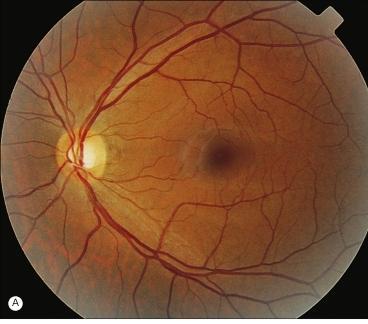
6 Fundoscopic View Of Normal Retina

7

8
9 What Is So Special About Diabetic Retinopathy? The WHO definition of blindness is a vision less than 3/60 in the better eye with best available spectacle correction. Diabetic Retinopathy is the most common cause of blindness amongst individuals of working age (20-65 years).
10 Part 2: Diabetic Retinopathy
11 Pathogenesis ALDOSE REDUCTASE PATHWAY CENTRAL ROLE OF VEGF MORPHOLOGICAL CHANGES IN PLATELETS BLOOD VISCOCITY
12 Aldose Reductase Pathway Aldose reductase converts glucose to sorbitol and galactose to galactitol. These sorbitol and galactitol are harmful for the eye in excess amount. Aldose reductase is present in high levels in: 1. Lens epithelial cells: Responsible for cataract formation. 2. Retinal cells: Responsible for Diabetic retinopathy. An effective aldose reductase inhibitor has not been developed yet.
13 Central Role Of VEGF VEGF NORMALLY INHIBITS THE GROWTH OF RETINAL EPITHELIAL CELLS. VEGF has a direct role in the proliferative retinal vascular abnormalities that are found in diabetes. The concentration of VEGF in aqueous and vitreous directly correlates with the severity of retinopathy.
14 Morphological Changes In Platelets & High Blood Viscosity They cause focal capillary occlusion and focal areas of ischemia in the retina which, in turn, contribute to the development of diabetic retinopathy.
15 Stages Of Diabetic Retinopathy 1. Nonproliferative Diabetic Retinopathy [NPDR]: Early and advanced. 2. Proliferative Diabetic Retinopathy [PDR].

16 NPDR and PDR. At a glance.
17 Characteristic Features Of MICROANEURYSMS: Early NPDR SMALL RED DOTS IN THE CENTRAL RETINAL LAYERS. IF THE WALL OF MICROANEURYSM IS WEAK ENOUGH, IT MAY LEAD TO INTRARETINAL HEMORRHAGES.
18 Microaneurysms
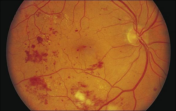
19 Intraretinal Hemorrhages
20 Macular Edema The intercellular edema fluid comes from leaking microaneurysms/ diffuse capillary incompetence. The edema causes scattering of light by the multiple interfaces it creates in the retina by separated retinal cells. This decreases the retina s translucency such that the normal retinal pigment epithelial and choroidal background pattern is blurred.
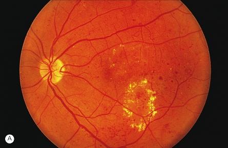
![Macular Edema [1] NPDR with](/docs-images/82/85805111/images/21-0.jpg "Macular Edema: Exudates: Yellow")
21 Macular Edema [1] NPDR with Macular Edema: Exudates: Yellow arrow

22 Macular Edema [2]
![Macular Edema [3] A. Fundus Exam. B.](/docs-images/82/85805111/images/23-0.jpg "Fluorescein Angiography. C.")
23 Macular Edema [3] A. Fundus Exam. B. Fluorescein Angiography. C. Optical Coherence Tomography [OCT].
24 Hard Exudates And Circinate Retinopathy If the leakage of fluid is severe enough, lipid accumulates and precipitates in the retina. In some cases, lipid is scattered through the macula. Then it is called. In others, it accumulates in a ring around a group of leaking microaneurysms/ around microaneurysms surrounding an area of capillary nonperfusion. This pattern is called.


25 Hard Exudates In The Macula
26 Circinate Retinopathy
27 Characteristic Features Of Advanced NPDR Due to increased retinal hypoxia, following changes are seen in the retina: 1. Intraretinal microvascular abnormalities (IRMA). 2. Cotton-wool spots. 3. Venous beading.
28 Intraretinal Microvascular Abnormalities (IRMA) They are dilated capillaries, which seem to function as collateral channels. Capillary hypoperfusion often surrounds IRMA.

29 Severe IRMA

30 Capillary Hypoperfusion Zone Due To IRMA
31 Cotton Wool Spots (Soft Exudates) The main cause of this feature is ischemic changes. Local ischemia causes effective obstruction of axoplasmic flow in the normally transparent nerve fiber layer AND, Subsequent swelling of the nerve fibers gives a characteristic white fluffy appearance to the cotton-wool spots. Fluorescein angiography shows no capillary perfusion in the area corresponding to a cottonwool spot.

32 Cotton Wool Spots [1]

33 Cotton Wool Spots [2]
34 Venous Beading Venous beading is an important sign of sluggish retinal circulation. It has an appearance of sausage shaped dilatation of retinal veins. They are nearly always adjacent to large areas of capillary nonperfusion.

35 Venous Beading [1]

36 Venous Beading [2]
37 PDR It is characterized by neovascularization [new blood vessel formation], which is of 2 types: 1. Neovascularization of the disc [NVD], 2. Neovascularization elsewhere [NVE]. NVD: New vessels arise within 1 disc diameter of optic nerve. NVE: New vessels arise from >1 disc diameter of optic nerve.
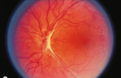
38 NVD
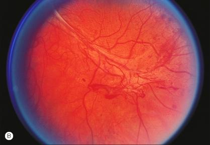
39 NVE
40 Vitreous Traction And Retinal Detachment The new vessels usually progress through a stage of further proliferation, with associated connective tissue formation. As PDR progresses, the fibrous component becomes more prominent. Vitreous traction is transmitted to the retina along these proliferations and may lead to traction retinal detachment. *. Davis et al. have stressed the role of the contracting vitreous in the production of vitreous hemorrhage, retinal breaks, and retinal detachment.
41 Types Of Diabetic Retinal Detachments Two types of diabetic retinal detachments occur: 1. Those that are caused by traction alone (nonrhegmatogenous) and, 2. Those caused by retinal break formation (rhegmatogenous). Optical coherence tomography (OCT) is used to describe/ determine those detachments.

 Up to")

42 Optical Coherence Tomography (OCT) Up to down: 1. Normal OCT, 2. Macular hole, 3. Macular edema.
43 Diagnosis Is Done By.. 1. Direct ophthalmoscopy. 2. Detection of systemic hyperglycemia: A. Fasting blood sugar testing, B. Glucose tolerance test, and C. Hemoglobin A 1c determinations. 3. Optical coherence tomography (OCT), where available.
44 Treatment Antiplatelet therapy. Antihypertensive drugs. Anti-VEGF agents. PRP. Vitrectomy.
45 Antiplatelet Therapy Aspirin 650 mg daily It does not influence the progression of retinopathy/ affect visual acuity/ influence the incidence of vitreous hemorrhages. But it reduces the incidence of stroke in diabetic patient.
46 ANTIHYPERTENSIVE DRUGS The Hypertension in Diabetes Study group has demonstrated that with better blood pressure control, a 37% risk reduction in microvascular changes can be achieved.
47 Anti VEGF Agents Anti-VEGF drugs are available for the treatment of macular degeneration. Recently, a protein kinase C inhibitor [PKCI] has been shown to reduce diabetes-induced hemodynamic abnormalities in patients with diabetic retinopathy and reduce the risk of vision loss in patients with macular edema.
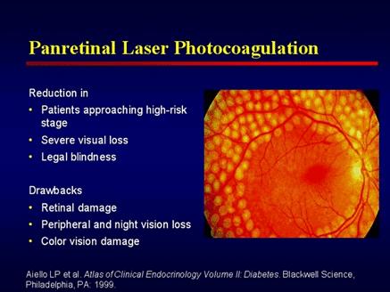
48 Pan Retinal Photocoagulation [PRP] Eyes with High Risk Characteristics [HRC] HRC is defined as presence of any of the following: 1. NVD (>1/4 th but <1/3 rd of the disc area). 2. NVD + vitreous hemorrhage. 3. NVE> ½ of the disc area + Vitreous/ Preretinal hemorrhage. The ETDRS [Early Treatment Diabetic Retinopathy Study] found that PRP lowers the risk of developing HRC by 50% in eyes with very severe NPDR and macular edema.
49
50 PRP Continued Mechanism of PRP [Proposed explanations]: 1. PRP decreases the production of vasoproliferative factors by eliminating some of the hypoxic retina. 2. PRP stimulates the release of antiangiogenic factors from the retinal pigment epithelium by thinning the retina. 3. PRP increases oxygenation of the remaining retina by allowing increased diffusion of oxygen from the choroid. 4. PRP leads to an increase in vasoinhibitors by directly stimulating the retinal pigment epithelium to produce inhibitors of vasoproliferation.

51 An Eye Treated With PRP

52 Treatment Of Macular Edema By PRP
53 Vitrectomy The major indications are of vitrectomy in diabetics are: 1. Macular-involving/ macular-threatening traction retinal detachment 2. Nonclearing vitreous hemorrhage and, 3. Combined traction-rhegmatogenous retinal detachment.
, and 4. To perform endophotocoagulation.")
54 Vitrectomy.Continued. The surgical objectives are: 1. To clear the media, 2. To release all anterior-posterior traction, 3. To release tangential traction via delamination or segmentation (cutting the fibrotic bridges between areas of tractional detachment), and 4. To perform endophotocoagulation. A possible cause of failure following an otherwise successful vitrectomy is
55 Thank You. Courtesy: 1. Parson s Disease Of The Eye. 2. Yanoff s Ophthalmology. 3. Michigan State University. 4. Atlas Of Clinical Endocrinology. 5. Emedicine Medscape. 6. Wikipedia. 7. Early Treatment Diabetic Retinopathy Study [ETDRS]. 8. Medline Plus Medical Encyclopedia.
Diabetic Retinopathy. Barry Emara MD FRCS(C) Giovanni Caboto Club October 3, 2012
 Giovanni Caboto Club October 3, 2012") Diabetic Retinopathy Barry Emara MD FRCS(C) Giovanni Caboto Club October 3, 2012 Outline Statistics Anatomy Categories Assessment Management Risk factors What do you need to do? Objectives Summarize the
Diabetic Retinopathy Barry Emara MD FRCS(C) Giovanni Caboto Club October 3, 2012 Outline Statistics Anatomy Categories Assessment Management Risk factors What do you need to do? Objectives Summarize the
Diabetic Retinopathy
 Diabetic Retinopathy Diabetes can be classified into type 1 diabetes mellitus and type 2 diabetes mellitus, formerly known as insulin-dependent diabetes mellitus, and non-insulin diabetes mellitus, respectively.
Diagnosis and treatment of diabetic retinopathy. Blake Cooper MD Ophthalmologist Vitreoretinal Surgeon Retina Associates Kansas City
 Diagnosis and treatment of diabetic retinopathy Blake Cooper MD Ophthalmologist Vitreoretinal Surgeon Retina Associates Kansas City Disclosures Consulted for Novo Nordisk 2017,2018. Will be discussing
The Human Eye. Cornea Iris. Pupil. Lens. Retina
 The Retina Thin layer of light-sensitive tissue at the back of the eye (the film of the camera). Light rays are focused on the retina then transmitted to the brain. The macula is the very small area in
INTRODUCTION AND SYMPTOMS
 CHAPTER 1 INTRODUCTION AND SYMPTOMS Introduction of Diabetic Retinopathy Diabetic retinopathy (DR) is a potentially blinding complication of diabetes. It is defined as presence of one or more definite
DIABETIC RETINOPATHY
 DIABETIC RETINOPATHY C. L. B. Canny, MD FRCSC Diabetic retinopathy is the most serious eye manifestation of diabetes and is responsible for most of the blindness caused by diabetes. Diabetic retinopathy
EyePACS Grading System (Part 3): Detecting Proliferative (Neovascular) Diabetic Retinopathy. George Bresnick MD MPA Jorge Cuadros OD PhD
: Detecting Proliferative (Neovascular) Diabetic Retinopathy. George Bresnick MD MPA Jorge Cuadros OD PhD") EyePACS Grading System (Part 3): Detecting Proliferative (Neovascular) Diabetic Retinopathy George Bresnick MD MPA Jorge Cuadros OD PhD Anatomy of the eye: 3 Normal Retina Retinal Arcades Macula Optic
EyePACS Grading System (Part 3): Detecting Proliferative (Neovascular) Diabetic Retinopathy George Bresnick MD MPA Jorge Cuadros OD PhD Anatomy of the eye: 3 Normal Retina Retinal Arcades Macula Optic
EyePACS Grading System (Part 2): Detecting Presence and Severity of Background (Non-Proliferative) Diabetic Retinopathy Lesion
: Detecting Presence and Severity of Background (Non-Proliferative) Diabetic Retinopathy Lesion") EyePACS Grading System (Part 2): Detecting Presence and Severity of Background (Non-Proliferative) Diabetic Retinopathy Lesion George Bresnick MD MPA Jorge Cuadros OD PhD Anatomy of the eye: 3 Normal Retina
EyePACS Grading System (Part 2): Detecting Presence and Severity of Background (Non-Proliferative) Diabetic Retinopathy Lesion George Bresnick MD MPA Jorge Cuadros OD PhD Anatomy of the eye: 3 Normal Retina
 measure of your overall performance. An isolated glucose test is helpful to let you know what your sugar level is at one moment, but it doesn t tell you whether or not your diabetes is under adequate control
Diabetic and the Eye: An Introduction
 Diabetic and the Eye: An Introduction Lawrence Iu FRCSEd (Ophth), FCOphthHK, FHKAM (Ophthalmology) Department of Ophthalmology, Grantham Hospital & Queen Mary Hospital Background Diabetes mellitus (DM)
Eyes on Diabetics: How to Avoid Blindness in Diabetic Patient
 Eyes on Diabetics: How to Avoid Blindness in Diabetic Patient Rova Virgana FK Unpad Pusat Mata Nasional RS Mata Cicendo Bandung Eye Center (Hospital and Clinic) PIT IDI Jabar 2018 Keys Facts from WHO
Clinically Significant Macular Edema (CSME)
") Clinically Significant Macular Edema (CSME) 1 Clinically Significant Macular Edema (CSME) Sadrina T. Shaw OMT I Student July 26, 2014 Advisor: Dr. Uwaydat Clinically Significant Macular Edema (CSME) 2
Clinically Significant Macular Edema (CSME) 1 Clinically Significant Macular Edema (CSME) Sadrina T. Shaw OMT I Student July 26, 2014 Advisor: Dr. Uwaydat Clinically Significant Macular Edema (CSME) 2
Diabetic Retinopathy
 Diabetic Retinopathy Diabetes mellitus is one of the leading causes of irreversible blindness worldwide. In the United States, it is the most common cause of blindness in people younger than 65 years.
Diabetic Retinopathy A Presentation for the Public
 Diabetic Retinopathy A Presentation for the Public Ray M. Balyeat, MD The Eye Institute Tulsa, Oklahoma The Healthy Eye Light rays enter the eye through the cornea, pupil and lens. These light rays are
New Developments in the treatment of Diabetic Retinopathy
 New Developments in the treatment of Diabetic Retinopathy B. Jeroen Klevering University Medical Centre Nijmegen - The Netherlands Topics Management of diabetic retinopathy Interventions a. primary (prevention)
ZEISS AngioPlex OCT Angiography. Clinical Case Reports
 Clinical Case Reports Proliferative Diabetic Retinopathy (PDR) Case Report 969 PROLIFERATIVE DIABETIC RETINOPATHY 1 1-year-old diabetic female presents for follow-up of proliferative diabetic retinopathy
FRANZCO, MD, MBBS. Royal Darwin Hospital
 Diabetes and Eye By Dr. Nishantha Wijesinghe FRANZCO, MD, MBBS Consultant Ophthalmologist Royal Darwin Hospital 98% of Diabetics do not need to suffer from severe visual loss Yet Diabetic eye disease is
Marcus Gonzales, OD, FAAO Cedar Springs Eye Clinic
 Marcus Gonzales, OD, FAAO Cedar Springs Eye Clinic 25.6 million adults 11.3% of the adult population 10.9 million adults 65 years and older 26.9% of this age population 79 million people are Pre-diabetic!!
Jay M. Haynie, O.D.; F.A.A.O. Olympia Tacoma Renton Kennewick Washington
 Jay M. Haynie, O.D.; F.A.A.O. Olympia Tacoma Renton Kennewick Washington I Jay M. Haynie, OD, FAAO have received honoraria from the following companies: Reichert Technologies Notal Vision Carl Zeiss Meditec
Guidelines for the Management of Diabetic Retinopathy for the Internist
 Visual Disorder Guidelines for the Management of Diabetic Retinopathy for the Internist JMAJ 45(1): 1 7, 2002 Sadao HORI Professor, Department of Ophthalmology, Tokyo Women s Medical University Abstract:
Diabetic Retinopatathy
 Diabetic Retinopatathy Jay M. Haynie, OD, FAAO Financial Disclosure I have received honoraria or am on the advisory board for the following companies: Carl Zeiss Meditec Arctic DX Macula Risk Advanced
Diabetic Management beyond traditional risk factors and LDL-C control: Can we improve macro and microvascular risks?
 Retinopathy Diabetes has a negative effect on eyes in many ways, increasing the risk of cataracts for example, but the most common and serious ocular complication of diabetes is retinopathy. Diabetic retinopathy
Outline. Preventing & Treating Diabetes Related Blindness. Eye Care Center Doctors. Justin Kanoff, MD. Eye Care Center of Northern Colorado
 Outline Preventing & Treating Diabetes Related Blindness Justin Kanoff, MD Eye Care Center of Northern Colorado 303 974 4302 Introduction to Eye Care Center of Northern Colorado How the eye works Eye problems
Diabetes & Your Eyes
 Diabetes & Your Eyes Diabetes is a disease that occurs when the pancreas does not secrete enough insulin or the body is unable to process it properly. Insulin is the hormone that regulates the level of
Is OCT-A Needed As An Investigative Tool During The Management Of Diabetic Macular Edema
 Is OCT-A Needed As An Investigative Tool During The Management Of Diabetic Macular Edema Ayman M Khattab MD, FRCS Professor of Ophthalmology Cairo University Diabetic Macular Edema (DME) Diabetic macular
OCT Angiography in Primary Eye Care
 OCT Angiography in Primary Eye Care An Image Interpretation Primer Julie Rodman, OD, MS, FAAO and Nadia Waheed, MD, MPH Table of Contents Diabetic Retinopathy 3-6 Choroidal Neovascularization 7-9 Central
A Patient s Guide to Diabetic Retinopathy
 Diabetic Retinopathy A Patient s Guide to Diabetic Retinopathy 840 Walnut Street, Philadelphia PA 19107 www.willseye.org Diabetic Retinopathy 1. Definition Diabetic retinopathy is a complication of diabetes
Diabesity A Public Health Crisis: AOA Evidence Based Translation to Care Series
 Diabesity A Public Health Crisis: AOA Evidence Based Translation to Care Series Joseph J. Pizzimenti, OD, FAAO Associate Professor Nova Southeastern University The Eye Care Institute pizzimen@nova.edu
Diabetic Retinopathy Screening in Hong Kong. Dr. Rita Gangwani M.S, FRCS (Ophth), FCOphth(HK), FHKAM Eye Institute, The University of Hong Kong
, FCOphth(HK), FHKAM Eye Institute, The University of Hong Kong") Diabetic Retinopathy Screening in Hong Kong Dr. Rita Gangwani M.S, FRCS (Ophth), FCOphth(HK), FHKAM Eye Institute, The University of Hong Kong Co-Investigators Prof. David Wong Prof. Sarah McGhee Dr. Wico
Diabetic Retinopathy Screening in Hong Kong Dr. Rita Gangwani M.S, FRCS (Ophth), FCOphth(HK), FHKAM Eye Institute, The University of Hong Kong Co-Investigators Prof. David Wong Prof. Sarah McGhee Dr. Wico
Understanding Diabetic Retinopathy
 Understanding Diabetic Retinopathy What Is Diabetic Retinopathy? Diabetes damages blood vessels in the rear of the eye. This condition is called diabetic retinopathy. It can lead to vision loss or blindness.
Diabetic Retinopathy
 Diabetic Retinopathy Introduction People with diabetes are more likely to have eye problems that can lead to blindness. Diabetic retinopathy is a disease of the eye s retina that is caused by diabetes.
Epidemiology and Pathophysiology of Diabetic Retinopathy
 Epidemiology and Pathophysiology of Diabetic Retinopathy Vincent Reppucci, MD Director, Retina Service Mt. Sinai St. Luke s-roosevelt Hospital Attending Physician, Retina Service New York Eye and Ear Infirmary
X-Plain Diabetic Retinopathy Reference Summary
 X-Plain Diabetic Retinopathy Reference Summary Introduction Patients with diabetes are more likely to have eye problems that can lead to blindness. Diabetic retinopathy is a disease of the eye s retina
Mild NPDR. Moderate NPDR. Severe NPDR
 Diabetic retinopathy Diabetic retinopathy is the most common cause of blindness in adults aged 35-65 years-old. Hyperglycaemia is thought to cause increased retinal blood flow and abnormal metabolism in
Brampton Hurontario Street Brampton, ON L6Y 0P6
 Diabetic Retinopathy What is Diabetic Retinopathy Diabetic retinopathy is one of the leading causes of blindness world-wide. Diabetes damages blood vessels in many organs of the body including the eyes.
DIABETIC RETINOPATHY (DR) PREFERRED PRACTICE PATTERNS (PPP) Philippines 2016
 PREFERRED PRACTICE PATTERNS (PPP) Philippines 2016") DIABETIC RETINOPATHY (DR) PREFERRED PRACTICE PATTERNS (PPP) Philippines 2016 The Diabetic Retinopathy (DR) Preferred Practice Patterns (PPP) Philippines: 2016 was prepared by the VitreoRetina Society of
DIABETIC RETINOPATHY (DR) PREFERRED PRACTICE PATTERNS (PPP) Philippines 2016 The Diabetic Retinopathy (DR) Preferred Practice Patterns (PPP) Philippines: 2016 was prepared by the VitreoRetina Society of
PROGRESSION OF DIABETIC RETINOPATHY FOLLOWING CATARACT SURGERY
 PROGRESSION OF DIABETIC RETINOPATHY FOLLOWING CATARACT SURGERY Yayan Heryanto, Iwan Sovani, Arief Kartasasmita, Erwin Iskandar, Djonggi Panggabean. Dept. of Ophthalmology Medical Faculty Unpad, Cicendo
Diabetic retinopathy damage to the blood vessels in the retina. Cataract clouding of the eye s lens. Cataracts develop at an earlier age in people
 Diabetic Retinopathy What is diabetic eye disease? Diabetic eye disease refers to a group of eye problems that people with diabetes may face as a complication of diabetes. All can cause severe vision loss
Dr/ Marwa Abdellah EOS /16/2018. Dr/ Marwa Abdellah EOS When do you ask Fluorescein angiography for optic disc diseases???
 When do you ask Fluorescein angiography for optic disc diseases??? 1 NORMAL OPTIC DISC The normal optic disc on fluorescein angiography is fluorescent due to filling of vessels arising from the posterior
Diabetes and Eye Health more than meets the eye Vision Initiative - in association with PSA
 Diabetes and Eye Health more than meets the eye Vision Initiative - in association with PSA Vision 2020 Australia Vision Initiative RANZCO & OAA (Vic) Proud members of Vision 2020 Australia Outline Vision
OCT Assessment of the Vitreoretinal Relationship in CSME
 December 2007 Sonia Rani John et al. - IFIS 375 ORIGINAL ARTICLE OCT Assessment of the Vitreoretinal Relationship in CSME Dr. Manoj S. DNB FRCS, Dr. Unnikrishnan Nair MS DO FRCS, Dr. Gargi Sathish MS Introduction
Medical Retina 2011 Nicholas Lee
 Medical Retina 2011 Nicholas Lee 1 Diabetic Retinopathy Epidemiology 1000 registered blind each year 2% diabetics registered as blind (8% of all Blind Registrations) 42% with Mild Background DR will progress
THE ROLE OF anti-vegf IN DIABETIC RETINOPATHY AND AGE RELATED MACULAR DEGENERATION
 THE ROLE OF anti-vegf IN DIABETIC RETINOPATHY AND AGE RELATED MACULAR DEGENERATION MOESTIDJAB DEPARTMENT OF OPHTHALMOLOGY SCHOOL OF MEDICINE AIRLANGGA UNIVERSITY DR SOETOMO HOSPITAL SURABAYA INTRODUCTION
Facts About Diabetic Eye Disease
 Facts About Diabetic Eye Disease Points to Remember 1. Diabetic eye disease comprises a group of eye conditions that affect people with diabetes. These conditions include diabetic retinopathy, diabetic
Clinical Case Presentation. Branch Retinal Vein Occlusion. Sarita M. Registered Nurse Whangarei Base Hospital
 Clinical Case Presentation on Branch Retinal Vein Occlusion Sarita M. Registered Nurse Whangarei Base Hospital Introduction Case Study Pathogenesis Clinical Features Investigations Treatment Follow-up
Diabetic Retinopathy WHAT IS DIABETIC RETINOPATHY? WHAT CAUSES DIABETIC RETINOPATHY? WHAT ARE THE STAGES OF DIABETIC RETINOPATHY?
 Diabetic Retinopathy WHAT IS DIABETIC RETINOPATHY? Diabetic retinopathy affects 8 million Americans with diabetes. A leading cause of blindness in American adults, it is caused by damage to the small blood
The Natural History of Diabetic Retinopathy and How Primary Care Makes A Difference
 The Natural History of Diabetic Retinopathy and How Primary Care Makes A Difference We will discuss - How exactly does blood sugar control affect retinopathy? - What are other factors that we measure in
Scott M. Pfahler D.O. Dayton Vitreo-Retinal Associates AOCOO-HNS Palm Springs, CA 2012
 Scott M. Pfahler D.O. Dayton Vitreo-Retinal Associates AOCOO-HNS Palm Springs, CA 2012 Proliferative Diabetic Retinopathy Laser Treatments Medical Treatment Surgical Treatment Diabetic Macular Edema Laser
Posterior Segment Update
 Posterior Segment Update Featured Speaker: Dr. Kyle Cheatham, FAAO, DIP ABO DISCLOSURE STATEMENT We have no direct financial or proprietary interest in any companies, products or services mentioned in
OCCLUSIVE VASCULAR DISORDERS OF THE RETINA
 OCCLUSIVE VASCULAR DISORDERS OF THE RETINA Learning outcomes By the end of this lecture the students would be able to Classify occlusive vascular disorders (OVD) of the retina. Correlate the clinical features
Measures have been taken, by the Utah Department of Health, Bureau of Health Promotions, to ensure no conflict of interest in this activity.
 Measures have been taken, by the Utah Department of Health, Bureau of Health Promotions, to ensure no conflict of interest in this activity. CNE/CPE/CEU s are available for this live webinar. You must
MANAGING DIABETIC RETINOPATHY. <Your Hospital Name> <Your Logo>
 MANAGING DIABETIC RETINOPATHY It s difficult living with Diabetes Mellitus. Ask any diabetic... Their lives are centered around meal plans, glucose levels, and insulin
Control of Systemic Factors Can Preserve Vision in Diabetic Retinopathy
 dmcjuly05_cme_dr 7/28/05 9:19 AM Page 38 Control of Systemic Factors Can Preserve Vision in Diabetic Retinopathy Jointly sponsored by The Dulaney Foundation and Diabetic Microvascular Complications Today.
SUPPLEMENTARY DATA. Supplementary Table 1. Characteristics of Subjects.
 Supplementary Table 1. Characteristics of Subjects. a includes one patient who had an aqueous sample taken from the same eye twice b includes one patients who had an aqueous sample taken from the same
Serious Eye diseases, New treatments. Mr. M. Usman Saeed MBBS, FRCS, FRCOphth Consultant Ophthalmologist
 Serious Eye diseases, New treatments Mr. M. Usman Saeed MBBS, FRCS, FRCOphth Consultant Ophthalmologist 5 major causes of loss of vision Cataracts Glaucoma Macular degeneration Retinal Vein occlusions
Documentation, Codebook, and Frequencies
 Documentation, Codebook, and Frequencies Ophthalmology Retinal Imaging Examination Survey Years: 2005 to 2006 SAS Transport File: OPXRET_D.XPT December 2008 NHANES 2005 2006 Data Documentation Exam Component:
Spontaneous Regression of Neovascularization at the Disc in Diabetic Retinopathy
 Korean J Ophthalmol Vol. 18:41-46, 2004 Spontaneous Regression of Neovascularization at the Disc in Diabetic Retinopathy Jae Ryong Han, MD, Won Kyung Ju, MD, In Won Park, MD Department of Ophthalmology,
RANZCO Screening and Referral Pathway for Diabetic Retinopathy #
 RANZCO Screening and Referral Pathway for Diabetic Retinopathy # Patient Presents a. Screen for Diabetic Retinopathy every 2 years b. Begin screening at diagnosis of Diabetes * Clinical Modifi ers Yearly
Diabetic Retinopathy in Primary Care
 Diabetic Retinopathy in Primary Care Epidemiology Diabetes is one of the most serious challenges to health care world rld-wide. According to recent projections it will affect 239 million people e by 2010-
Michael P. Blair, MD Retina Consultants, Ltd Libertyville/Des Plaines, Illinois Clinical Associate University of Chicago 17 October 2015
 Michael P. Blair, MD Retina Consultants, Ltd Libertyville/Des Plaines, Illinois Clinical Associate University of Chicago 17 October 2015 So What Parts of the Eye Retina are Affected by VHL Neural tissue
Diabetic retinopathy (DR) was first PROCEEDINGS DIABETIC RETINOPATHY * Ronald Klein, MD, MPH ABSTRACT
 was first PROCEEDINGS DIABETIC RETINOPATHY * Ronald Klein, MD, MPH ABSTRACT") DIABETIC RETINOPATHY * Ronald Klein, MD, MPH ABSTRACT Diabetic retinopathy (DR) is characterized by the development of retinal microaneurysms, hemorrhages, deposits of leaked lipoproteins (hard exudates),
DIABETIC RETINOPATHY * Ronald Klein, MD, MPH ABSTRACT Diabetic retinopathy (DR) is characterized by the development of retinal microaneurysms, hemorrhages, deposits of leaked lipoproteins (hard exudates),
Year 2 MBChB Clinical Skills Session Ophthalmoscopy. Reviewed & ratified by: Mr M Batterbury Consultant Ophthalmologist
 Year 2 MBChB Clinical Skills Session Ophthalmoscopy Reviewed & ratified by: o Mr M Batterbury Consultant Ophthalmologist Learning objectives o To understand the anatomy and physiology of the external and
Diabetic Retinopathy
 Diabetic Retinopathy Secretary for Quality of Care Anne L. Coleman, MD, PhD Academy Staff Nicholas P. Emptage, MAE Doris Mizuiri Shannon Kealey, MLS Flora C. Lum, MD Medical Editor: Design: Approved by:
Optical Coherence Tomography in Diabetic Retinopathy. Mrs Samantha Mann Consultant Ophthalmologist Clinical Lead of SEL-DESP
 Optical Coherence Tomography in Diabetic Retinopathy Mrs Samantha Mann Consultant Ophthalmologist Clinical Lead of SEL-DESP Content OCT imaging Retinal layers OCT features in Diabetes Some NON DR features
Fundus Fluorescein Angiography in Diabetic Retinopathy: Correlation of Angiographic Findings to the Clinical Maculopathy Abstract: Purpose:
 IOSR Journal of Dental and Medical Sciences (IOSR-JDMS) e-issn: 2279-0853, p-issn: 2279-0861.Volume 15, Issue 2 Ver. XII (Feb. 2016), PP 80-88 www.iosrjournals.org Fundus Fluorescein Angiography in Diabetic
MAGNITUDE OF DIABETIC EYE DISEASE IN INDIA
 Dear Doctor This booklet contains information about your role as a physician in preventing blindness in your diabetic patients. You are the first point of contact for your diabetic patients. You see them
DIABETES AND YOUR EYES. Presented by Dr. Andrea Hagler
 DIABETES AND YOUR EYES Presented by Dr. Andrea Hagler Tahlequah, OK Forest Grove, OR Brief Review of Diabetes The body s endocrine system is responsible for regulating growth, reproduction, and tissue
Leo Semes, OD, FAAO UAB Optometry
 Leo Semes, OD, FAAO UAB Optometry Safe; inert Has long track record - over 45 years Mixes with plasma and highlights blood vessel compromise Using specific exciting (490 nm)and absorption (510 nm) filters
Case Report: Indocyanine Green Dye Leakage from Retinal Artery in Branch Retinal Vein Occlusion
 Case Report: Indocyanine Green Dye Leakage from Retinal Artery in Branch Retinal Vein Occlusion Hiroki Fujita, Kyoko Ohno-Matsui, Soh Futagami and Takashi Tokoro Department of Visual Science, Tokyo Medical
Central Mersey Diabetic Retinopathy Screening Programme. Referring patients for Diabetic Retinopathy Screening
 Central Mersey Diabetic Retinopathy Screening Programme Referring patients for Diabetic Retinopathy Screening Information for GPs in Halton & St Helens, Knowsley and Warrington PCT Version: June 2008 Review
Age-Related Eye Diseases and Conditions. Jonathan M. Frantz, MD, F.A.C.S. Cataract and Refractive Surgeon
 Age-Related Eye Diseases and Conditions Jonathan M. Frantz, MD, F.A.C.S. Cataract and Refractive Surgeon Vision Changes Patients notice vision changes with aging. Many changes are common and can often
Clinical Trials in Diabetic Retinopathy. Harry W. Flynn Jr., M.D. Nidhi Relhan Batra, M.D.
 1 Clinical Trials in Diabetic Retinopathy 2018 Harry W. Flynn Jr., M.D. Nidhi Relhan Batra, M.D. Bascom Palmer Eye Institute 900 N.W. 17th Street Miami, FL 33136 Phone: (305) 326-6118 Fax: (305) 326-6417
The Quick Guide to OCT Mastery 50 Real Cases with Expert Analysis
 OPTICAL COHERENCE TOMOGRAPHY The Quick Guide to OCT Mastery 50 Real Cases with Expert Analysis VOL 1 Sanjay Sharma, MD, FRCS, MSc (Epid), MBA Ophthalmologist, Epidemiologist Queen s University, Canada
Clinical biomicroscopy versus fluorescein angiography: Effectiveness and sensitivity in detecting diabetic retinopathy
 European Journal of Ophthalmology / Vol. 17 no. 1, 2007 / pp. 84-88 Clinical biomicroscopy versus fluorescein angiography: Effectiveness and sensitivity in detecting diabetic retinopathy S.S. KHALAF, M.D.
Grand Rounds: Interesting and Exemplary Cases From Guanajuato and Djibouti
 Learning Community: January 25, 2015 Grand Rounds: Interesting and Exemplary Cases From Guanajuato and Djibouti JORGE CUADROS, OD, PHD EyePACS In Guanajuato Program started in 2007 Cameras go from clinic
Disease-Specific Fluorescein Angiography
 Ruth E. Picchiottino, CRA Disease-Specific Fluorescein Angiography 15 Disease-Specific Fluorescein Angiography Recommendations for tailoring retinal fluorescein angiography to diabetic retinopathy, macular
Study of clinical significance of optical coherence tomography in diagnosis & management of diabetic macular edema
 Original Research Article Study of clinical significance of optical coherence tomography in diagnosis & management of diabetic macular edema Neha Kantilal Desai 1,*, Somesh Vedprakash Aggarwal 2, Sonali
GENERAL INFORMATION DIABETIC EYE DISEASE
 GENERAL INFORMATION DIABETIC EYE DISEASE WHAT IS DIABETIC EYE DISEASE? Diabetic eye disease is a term used to describe the common eye complications seen in people with diabetes. It includes: Diabetic retinopathy
Amber Priority. Image Library
 Amber Priority Image Library Amber flag Diabetic Maculopathy (M1) Pre-proliferative Diabetic Retinopathy (R2) Old, treated and now inactive DR (R1/M0/P1or R0/M0/P1) Where only partial or incomplete images
Slide notes: The major chronic complications of diabetes mellitus are described here. Among these, microvascular complications have an important
 1 2 The major chronic complications of diabetes mellitus are described here. Among these, microvascular complications have an important role. They comprise microangiopathy, diabetic retinopathy, diabetic
Central venous occlusion
 Central venous occlusion Central venous occlusion (right eye) There are dark haemorrhages at the macula and all over the retina. Choroidal haemangioma A choroidal haemangioma has salmon pink colour. There
Vascular Disease Ocular Manifestations of Systemic Hypertension
 Vascular Disease Ocular Manifestations of Systemic Hypertension Maynard L. Pohl, OD, FAAO Pacific Cataract & Laser Institute 10500 NE 8 th Street, Suite 1650 Bellevue, WA 98004 USA 425-462-7664 Cerebrovascular
Moncef Khairallah, MD
 Moncef Khairallah, MD Department of Ophthalmology, Fattouma Bourguiba University Hospital Faculty of Medicine, University of Monastir Monastir, Tunisia INTRODUCTION IU: anatomic form of uveitis involving
Use of the Free Electron Laser for the Noninvasive Determination of Retinal Oxyhemoglobin Saturation by Near Infrared Reflectance Spectrophotometry
 Use of the Free Electron Laser for the Noninvasive Determination of Retinal Oxyhemoglobin Saturation by Near Infrared Reflectance Spectrophotometry Ref: Eye, M.C. Escher, 1946 Ref: Eye, M.C. Escher, 1946
Management of diabetic eye disease: an overview
 Peter Blows Photographic screening for diabetic eye disease can identify patients who will benefit from laser treatment. BOTSWANA MANAGEMENT Management of diabetic eye disease: an overview Laser for DR
ROLE OF LASER PHOTOCOAGULATION VERSUS INTRAVITREAL TRIAMCINOLONE ACETONIDE IN ANGIOGRAPHIC MACULAR EDEMA IN DIABETES MELLITUS
 ORIGINAL ARTICLE ROLE OF LASER PHOTOCOAGULATION VERSUS INTRAVITREAL TRIAMCINOLONE ACETONIDE IN ANGIOGRAPHIC MACULAR EDEMA IN DIABETES MELLITUS Aggarwal Somesh VP 1, Shah Sonali N 2, Bharwada Rekha M 3,
Sponsored by. Shared care and referral pathways. Part 2: diabetes screening leading from the front
 CET CONTINUING Sponsored by 1 CET POINT Shared care and referral pathways Part 2: diabetes screening leading from the front Chris Steele, BSc (Hons), FCOptom, DCLP, DipOC, DipTp(IP), FBCLA The alarming
NO FINANCIAL INTERESTS ARE DISCLOSED BY THE AUTHORS
 OCULAR FINDINGS IN APLASTIC ANEMIA: MULTICENTER STUDY & LITERATURE REVIEW Ahmad M Mansour1, MD, Jong Wook Lee, MD, PhD, Seung Ah Yahng, MD, Kyu Seop Kim, MD, Maha Shahin, MD, Nelson Hamerschlak, MD, PhD,
Diabetes Eye Q Quiz. 1) Diabetes is the leading cause of new blindness among adults in the US under the age of 74.
 Diabetes is the leading cause of new blindness among adults in the US under the age of 74.") Diabetes Eye Q Quiz From 1997 to 2011, the number of adults with diagnosed diabetes who reported visual impairment, that is, trouble seeing even with their glasses or contact lenses, increased from 2.7
Diabetes Eye Q Quiz From 1997 to 2011, the number of adults with diagnosed diabetes who reported visual impairment, that is, trouble seeing even with their glasses or contact lenses, increased from 2.7
EXUDATES DETECTION FROM DIGITAL FUNDUS IMAGE OF DIABETIC RETINOPATHY
 EXUDATES DETECTION FROM DIGITAL FUNDUS IMAGE OF DIABETIC RETINOPATHY Namrata 1 and Shaveta Arora 2 1 Department of EECE, ITM University, Gurgaon, Haryana, India. 2 Department of EECE, ITM University, Gurgaon,
evaluation of vitreoretinal adhesions in exudative AMD using optical coherence tomography
 evaluation of vitreoretinal adhesions in exudative AMD using optical coherence tomography Dr. Mahmoud Alaa Abouhusssein, FRCO Lecturer of ophthalmology, Alexandria university Dr. Amir Ramadan Gomaa, MD
10/17/2017. FDA Approved. Zeiss AngioPlex TM Optovue AngioVue TM
 Images retinal microvasculature without dye injection Displays structure and function from a single imaging system Standard of Care-2011 DFE, Fundus Photos, VF 10-2, SD-OCT, FAF, or mferg 2016-AAO Baseline
Case Report Inherent Challenges in Managing Long Standing Refractory Diabetic Macular Edema
 Cronicon OPEN ACCESS EC OPHTHALMOLOGY Case Report Inherent Challenges in Managing Long Standing Refractory Diabetic Macular Edema V Swetha E Jeganathan 1,2 * and Karen Madill 3 1 Department of Ophthalmology,
7.1 Grading Diabetic Retinopathy
 Chapter 7 DIABETIC RETINOPATHYGRADING -------------------------------------------------------------------------------------------------------------------------------------- A consistent approach to the
What Is O.C.T. and Why Should I Give A Rip? OCT & Me How Optical Coherence Tomography Changed the Life of a Small Town Optometrist 5/19/2014
 OCT & Me How Optical Coherence Tomography Changed the Life of a Small Town Optometrist Email: myoder@wcoil.com Mark A. Yoder, O.D. 107 N. Main Street PO Box 123 Bluffton, OH 45817 @yoderod 115.02 Histoplasma
NEPTUNE RED BANK BRICK
 NEPTUNE RED BANK BRICK Diabetes & The Eye Diabetics are more likely to develop Cataracts at a younger age. Diabetics are twice as likely to develop Glaucoma when compared to non-diabetics. The primary
The Era of anti- - - VEGF Kirk L. Halvorson, OD
 The Era of anti- - - VEGF Kirk L. Halvorson, OD Introduction: Anti- - - Vascular Endothelial Growth Factor (Anti- - - VEGF) medication is a relatively a new line of medications used in treating a variety
What is diabetes? Ocolusystemic Disease Essen6als. Statistics, cont. Statistics. Statistics. The Diabetes Epidemic 9/5/12
 What is diabetes? Ocolusystemic Disease Essen6als Steven Ferrucci, OD, FAAO Chief, Optometry Sepulveda VA Associate Professor, SCCO DM is a chronic disorder characterized by a lack of insulin or increased
Management of Diabetic Retinopathy. An Overview
 92 E.V. HORMONES GOTZARIDIS 2004, 3(2):92-99 ET AL Review Management of Diabetic Retinopathy. An Overview Eustratios V. Gotzaridis 1, Athina Markou 2, Zdenek Gregor 3 1 Eye Department, Athinaiki General
Venous Occlusive Diseases
 Venous Occlusive Diseases Bruce R. Saran, MD Adjunct Assistant Clinical Professor of Medicine Scheie Eye Institute University of Pennsylvania School of Medicine Philadelphia, PA -a division of: RVO Demographics