PEDIATRIC NEUROPATHOLOGY Part II: CNS MALFORMATIONS - AN UPDATE
|
|
|
- Pamela Lee
- 6 years ago
- Views:
Transcription


1 PEDIATRIC NEUROPATHOLOGY Part II: CNS MALFORMATIONS - AN UPDATE Dr. Kenneth M. Earle Memorial Neuropathology Review February 2016 Brian Harding DPhil FRCPath
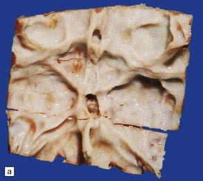
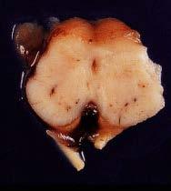
2 Malformations of Cerebral Cortex - Cortical development OUTLINE Lissencephaly Polymicrogyria Cortical dysplasia The smooth brain Heterotopia
3 TERMINOLOGY Macroscopic appearance of smooth brains: Completely smooth = agyria, lissencephaly Fewer coarser convolutions = macrogyria, pachygyria Cobblestone pattern Microscopic pattern: Lissencephaly type I.agyria,pachygyria w/wo 4 layers Lissencephaly type II cobblestone cortex/ cerebroocular dysplasia Polymicrogyria 2 or 4 layers Cortical Dysplasia with cytomegaly


4 Development of the cerebral cortex. Ellison & Love 2013, adapted from Hevner RF. J Neuropath Exp Neurol 2007; 66(2): )
5 Lissencephaly Neuronal migration disorder characterized by abnormal gyri Varies from agyria to pachygyria Severe mental retardation, hypotonia, intractable seizures Type 1 - cortex usually has 4 (instead of 6) layers, poorly organized

6

7
8 Disease CNS Gene Function of product Chromosome Lissencephaly (type I): autosomal recessive (Norman-Roberts type) Lissencephaly (type I): Miller- Dieker syndrome Dominant (haploinsufficiency) Lissencephaly (type I): isolated lissencephaly sequence (ILS) AD Lissencephaly (type I): X-linked isolated Lissencephaly (type I): X-linked (XLAG) Lissencephaly (type III) Lissencephaly with low sloping forehead and prominent nasal bridge Lissencephaly, cerebral heterotopias, facial dysmorphism RELN LIS1 and ε YWHAE ; (contiguous gene deletion) Reelin: 7q22 extracellular matrix protein produced by Cajal-Retzius cells required for neuronal migration LIS1: Non-catalytic subunit of brain platelet-activating factor acetyl hydrolase (PAFAH) Mouse model reeler mutant mouse causes cerebellar and cerebral cortical lamination anomalies 17p13.3 Targeted loss of function alleles of Pafah1b1 gene and ε Lissencephaly LIS1 deletion alone LIS1: as above 17p13.3 Targeted loss of function alleles of Pafah1b1 gene causes neuronal Lissencephaly with agenesis of corpus callosum in males; subcortical band heterotopia in females Lissencephaly with ambiguous genitalia Agyria, pachygyria or laminar heterotopia, abnormalities of corpus callosum, hippocampus, cerebellar vermis and brainstem DCX ARX Doublecortin: microtubuleassociated protein that interacts with non-receptor tyrosine kinases, including Abl Aristaless-related homeodomain transcription factor Xq22.3-q23 Xp22.13 TUBA1A Tubulin, alpha 1a 12q12-q14 suppression of doublecortin expression by RNAi inhibits neuronal migration in rat neocortex Targeted mutation of Arx

9 I Subarachnoid sp II III IV V VI

10 Layer I II III IV V VI Normal Mutations Lis1 DCX ARX TUBA1A wm 4 layer cortex with Posterior predominance 4 layer cortex with Anterior predominance 3 layer cortex 2 layer cortex Courtesy of Forman MS et al. J Neuropathol Exp Neurol 2005
11 Lissencephaly type I Miller-Dieker syndrome Microcephaly, bitemporal narrowing, vertical ridging in forehead, micrognathia Cryptorchidism, heart/kidney anomalies can be seen Contiguous gene deletion syndrome of 17p13.3 Most of the deletions are cytogenetically visible Lissencephaly due to deletion of LIS1 Facial features due to other genes on 17p13 More severe lissencephaly due to gene encoding ε YWHAE


12


13
14 Isolated Lissencephaly LIS1-17p13 - PAFAH1B1 (Platelet-activating factor acetylhydrolase, isoform 1B, alpha subunit) Isolated lissencephaly Autosomal dominant More severe occipital/posterior parietal DCX - Xq22 - Doublecortin X-linked dominant In males, isolated lissencephaly More severe anteriorly
15 Syndromic Lissencephaly RELN-7q22-Reelin Lissencephaly with severe ataxia Mental retardation, seizures Cerebellar hypoplasia Autosomal recessive
16 Syndromic Lissencephaly XLAG - ARX - Xp22 Lissencephaly with ambiguous genitalia Agenesis of the corpus callosum, severe seizures, temperature dysregulation, microcephaly Immature white matter Posterior-anterior gradient X-linked recessive Mutations also associated with infantile spasms, MR with dystonia


17
18 Lissencephaly II- Cobblestone Dysplasia AR cortex unlayered disorganized with cobblestone surface and thickened meninges can be partly polymicrogyric, variable muscular and ocular involvement with CNS disorder Dystroglycanopathies Walker-Warburg syndrome Muscle-eye-brain disease Fukuyama muscular dystrophy (Japan 3/10000
19 Disease CNS Gene Function of product Chromosome Mouse model Lissencephaly (type II): Fukuyama congenital muscular dystrophy Cobblestone lissencephaly, polymicrogyria FCMD Fukutin: gene interrupted by retrotransposon insertion. A secreted protein, which may function as a glycosyl transferase in the Golgi 9q31 Targeted mutation of FCMD gene causes muscular dystrophy and cortical dysplasia Lissencephaly (type II): muscle-eye-brain disease, type A, 5; type B, 5; type C, 5 Cobblestone lissencephaly, congenital myopia, glaucoma, retinal hypoplasia, mental retardation, hydrocephalus FKRP Protein targeted to the medial Golgi apparatus and necessary for posttranslational modification of dystroglycan 19q13.3 Lissencephaly (type II):Walker-Warburg syndrome Agyria, cobblestone lissencephaly, POMT1 cerebellar dysplasia and vermal agenesis, POMT2 hydrocephaly, occipital encephalocele O-mannosyl transferase 1: first enzyme in synthetic pathway of O- mannosyl glycans 9q31-q33 14q24.3 Large myd mutant and targeted mutation of α dystroglycan gene provide models of Walker-Warburg syndrome Lissencephaly (type II): muscle-eye-brain disease Cobblestone lissencephaly, congenital myopia, glaucoma, retinal hypoplasia, mental retardation, hydrocephalus POMGnT1 O-mannose β-1,2-nacetyl glucosaminyl transferase: second enzyme in synthetic pathway of O- mannosyl glycans 1p34-p33 Targeted mutation of POMGnT1 gene causes phenotype resembling muscle-eye-brain disease Lissencephaly (type II): muscle-eye-brain disease Cobblestone lissencephaly, congenital myopia, glaucoma, retinal hypoplasia, mental retardation, hydrocephalus LARGE Interacts directly with dystroglycan to allow glycosylation 22q12


20 α Dystroglycanopathy - pathomechanisms muscle brain Saito F, Matsumura K. Skeletal Muscle 2011; 1.22 Lisi MT, Cohn RD. Biochim Biophys Acta 2007;1772:


21 Walker-Warburg syndrome Reticulin


22 Lissencephaly II excessive migration Vimentin


23


24 MUSCLE-EYE BRAIN DISEASE - LARGE


25


26
27 Grey Matter Heterotopia Clusters of neurons and glia that form a nodule of grey matter in an abnormal location May be single or multiple, line ventricles, in deep white matter, subcortical white matter, leptomeninges Overlying cortex can be normal or disrupted May have normal intelligence, and normal neurologic exam
28 Band Heterotopia Bilateral bands of heterotopic grey matter in the white matter between the lateral ventricular walls and the cortex Overlying cortex may be normal or have simplified gyral pattern Mild to moderate mental retardation Seizures, often with later onset

29

30
31 Band Heterotopia Predominantly in females Rarely in males DCX mutations detected in many patients
32 Nodular Heterotopia Bilateral periventricular nodular heterotopia (BPNH) Varying degrees of severity Associated with seizures in some patients Intelligence - normal to mild mental retardation

33

34
35 Nodular Heterotopia Genetics Most cases consistent with X-linked dominant Strong skewing toward females in sporadic cases Increased rate of pregnancy loss Rare males with BPNH Mutations detected in females affected with epilepsy FLNA- Filamin1, actin binding protein associated with cytoskeleton - Xq28 Periventricular nodular heterotopia with microcephaly ARFGEF2 Brefeldin A-inhibited GEF2 protein (BIG2), involved in vesicular trafficking from Golgi - 20q13.13

36 . Guerrini, R., Mei, D., Sisodiya, S., Harding, B., et al. Neurology 63(1): 51-56, 2004.
37 Polymicrogyria (PMG) Multiple abnormally small gyri Several patterns of regional distribution Etiologically heterogeneous May be due to intrauterine insult (e.g. infection, hypoxia/ischemia) Very variable presentation Often associated with seizures, mental retardation, swallowing problems


38


39

40 Neu N
41 Polymicrogyria Genetics Usually sporadic Occasionally familial Bilateral perisylvian polymicrogyria Usually sporadic X-linked form described Bilateral fronto-parietal PMG GPR56,G-protein coupled receptor (16q13) Tubulin genes, TUBA8, (22q11) TUBB2Α & 2Β (6p25.2)


42 Band-like Calcification with Simplified Gyration and Polymicrogyria (BLCPMG) syn. Pseudo-Torch syndrome early onset seizures, developmental arrest, progressive microcephaly Intracranial calcification, cortical malformation, esp PMG Autosomal recessive, mutations in OCLN gene 5q13.2, encoding occludin a tight junction protein O'driscoll MC, et al. Am J Hum Genet Sep 10;87(3):
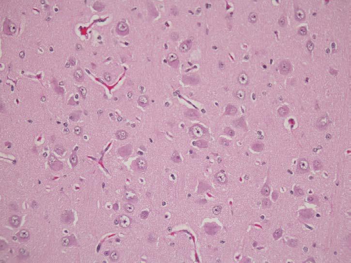
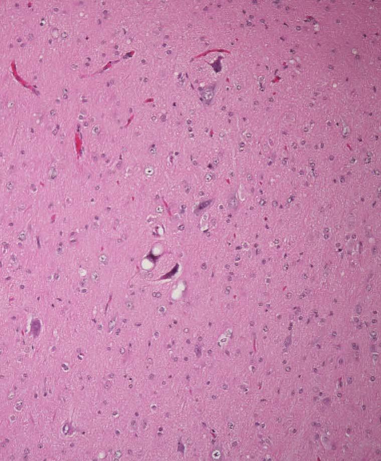
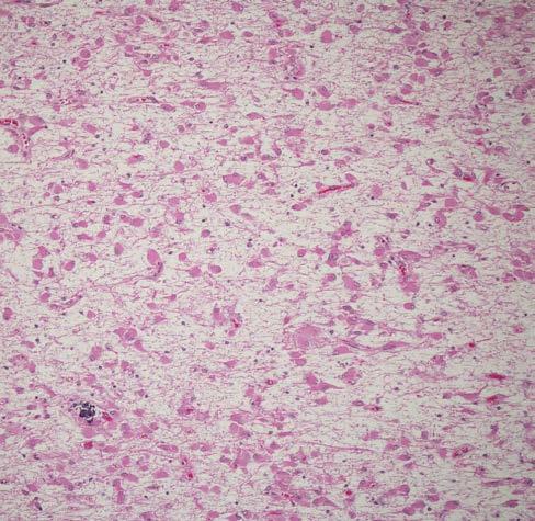
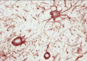
43 Cortical Dysplasia with Cytomegaly Unilateral dysplasia hemimegalencephaly Focal cortical dysplasia Tuberous Sclerosis


44 Normal
45


46 Hemimegalencephaly Focal cortical dysplasia

47


48 gfap cd34
49 Three-tiered ILAE classification system for FCDs FCD Type I (isolated) Focal Cortical Dysplasia with abnormal radial cortical lamination (FCD Ia) Focal Cortical Dysplasia with abnormal tangential cortical lamination (FCD Ib) Focal Cortical Dysplasia with abnormal radial and tangential cortical lamination (FCD Ic) FCD Type II (isolated) FCD Type III (associated with principal lesion) Focal Cortical Dysplasia with dysmorphic neurons (FCD IIa) Cortical lamination abnormalities in the temporal lobe associated with hippocampal sclerosis (FCD IIIa) Cortical lamination abnormalities adjacent to a glial or glio-neuronal tumor (FCD IIIb) Focal Cortical Dysplasia with dysmorphic neurons and balloon cells (FCD IIb) Cortical lamination abnormalities adjacent to vascular malformation (FCD IIIc) Cortical lamination abnormalities adjacent to any other lesion acquired during early life, e.g., trauma, ischemic injury, encephalitis (FCD IIId) The clinico-pathological spectrum of Focal Cortical Dysplasias: a consensus classification proposed by an ad hoc Task Force of the ILAE Diagnostic Methods Commission Blumcke I, et al Epilepsia 2011 Jan;52(1):


50 Tuberous Sclerosis


51 Tuberous Sclerosis NF GFAP

52 Tuberous Sclerosis: etiology Locus heterogeneity with disease-determining genes mapped to chromosome 9q34 (TSC1 gene; product hamartin) and 16p13.3 (TSC2 gene; product tuberin). Allelic loss (i.e. loss of heterozygosity) for 16p13.3 has been demonstrated in hamartomas, a cortical tuber, and a giant cell astrocytoma from tuberous sclerosis patients. This is consistent with the hypothesis that TSC2 acts as a tumor suppressor gene. TSC1 and 2 gene products are strategically important in cell growth and turnover Crino PB, Nathanson KL, Henske EP. N Engl J Med 2006; 355(13): )
53 Hydrocephalus Enlargement of cerebral ventricles due to increased CSF Distinguish from hydrocephalus due to decreased brain volume Etiologically heterogeneous


54 CSF overproduction Hydrocephalus Compromized absorption

55 Ventricular System
56 Hydrocephalus-Aqueduct obstruction The Aqueduct is the narrowest part of ventricular system, irregular curved tube with 2 constrictions either side of central ampulla Narrowest part in children 0.15mm 2, (mean 0.5mm 2) Obstruction can result from Stenosis: sporadic, rarely recessive, X-linked form is the most common type of genetic hydrocephalus, Atresia Gliosis/ septum Vascular malformation (Galen)


57 atresia gliosis


58 Septum


59 Stenosis


60 X-linked hydrocephalus: absent medullary pyramids
61 Hydrocephalus and L1CAM L1CAM (cell-adhesion molecule) Xq28 Mutations in this gene may cause up to 25% of congenital hydrocephalus in males Loss-of-function (»failed interaction with cytoskeletal protein, ankyrin) Not all cases of aqueductal stenosis/hydrocephalus are due to L1CAM mutations
62 L1CAM Diseases X-linked hydrocephalus Stenosis of aqueduct of Sylvius MASA syndrome (MR, aphasia, shuffling gait, adducted thumbs) Complicated spastic paraparesis 1 Corpus callosum hypoplasia, retardation, adducted thumbs, spastic paraplegia (lower limbs), hydrocephalus All can occur in same family: CRASH syn
63 Holoprosencephaly (HPE) Developmental defect of the forebrain (prosencephalon) Incomplete separation of the cerebral hemispheres into distinct right and left halves Other brain findings that can be seen in association with HPE do not necessarily indicate HPE (ACC, absent septum pellucidum) Prevalence: 1:16,000 live births 1:250 conceptuses


64 Alobar HPE


65 Semilobar HPE


66 Lobar HPE
67 HPE Clinical Features Cleft lip/palate Eye anomalies Pituitary dysfunction (including SIADH) Congenital nasal pyriform aperture stenosis Seizures Hypotonia

68
69 HPE Microforms Anosmia Single central maxillary incisor Cleft lip/palate Congenital nasal pyriform aperture stenosis (presents like choanal atresia) Ocular hypotelorism Microcephaly Developmental delay / mental retardation Seizures
70 HPE Etiology Genetic and etiologic heterogeneity Teratogens Diabetes, Ethanol, retinoic acid, cholesterol synthesis inhibitors Genetic factors Cytogenetic abnormalities - Trisomy 13 Genetic defects Syndromes - Smith-Lemli-Opitz Incomplete penetrance Variable expressivity
71 HPE Genes 7q36 SHH 13q32 ZIC2 2p21 SIX3 18p11 TGIF 9q22 PTCH 3p23 TDGF1 2q14 GLI2 Other loci have been described
72 HPE Genes Many PHE patients do not have a defined genetic cause Patients with detected mutations in the HPE genes to date have alterations in one allele HPE may be due to the interactions of multiple genetic and environmental influences
73 Agenesis of the Corpus Callosum Isolated (silent clinically, or subtle) Syndromic, Aicardi, Andermann, Meckel, Inborn errors of metabolism, chromosomal defects Possible pathogenetic mechanisms Probst bundle of misdirected fibers Midline glial sling: exptal manipulation, acallosal mice Mechanical defect suggested by hamartoma/ lipoma


74 Agenesis of the Corpus Callosum Normal Probst bundles


75
Cerebral Malformation gene panel
 Cerebral Malformation gene panel Dr John Livingston Consultant Paediatric Neurologist Leeds Teaching Hospitals NHS Trust on behalf of Yorkshire Regional Genetics Service Leeds UK Cerebral Malformation
Genetic test for Bilateral frontoparietal polymicrogyria
 Genetic test for Bilateral frontoparietal polymicrogyria Daniela Pilz, Cardiff UKGTN Genetic testing for neurological conditions; London February 26 th 2013 Region-specific Polymicrogyria (PMG) bilateral
Malformations of cortical development. Clinical and imaging findings.
 Malformations of cortical development. Clinical and imaging findings. Poster No.: C-2086 Congress: ECR 2012 Type: Educational Exhibit Authors: I. Alba de Caceres, B. García-Castaño, L. Ibañez, E. Roa,
Malformations of cortical development. Clinical and imaging findings.
 Malformations of cortical development. Clinical and imaging findings. Poster No.: C-2086 Congress: ECR 2012 Type: Educational Exhibit Authors: I. Alba de Caceres, B. García-Castaño, L. Ibañez, E. Roa 1
Test Information Sheet
 Prenatal Lissencephaly Panel Sequence Analysis and Exon-Level Deletion/Duplication Testing* of 24 Genes Panel Gene List: ACTB, ACTG1, X, ATP6V0A2, B3GALNT2*, B4GAT1*, DCX, FKRP*, FKTN, GMPPB*, ISPD, LAMB1,
MALFORMATIONS OF CORTICAL DEVELOPMENT: A PICTORIAL REVIEW
 MALFORMATIONS OF CORTICAL DEVELOPMENT: A PICTORIAL REVIEW Padmaja K. Naidu, M.D. Usha D. Nagaraj, M.D. William T. O Brien, Sr., D.O. AOCR Annual Conference 2018 Scottsdale, Arizona @CincyKidsRad facebook.com/cincykidsrad
intracranial anomalies
 Chapter 5: Fetal Central Nervous System 84 intracranial anomalies Hydrocephaly Dilatation of ventricular system secondary to an increase in the amount of CSF. Effects of hydrocephalus include flattening
Epilepsy and malformations of the cerebral cortex
 Abnormalities of cortical development and epilepsy Epileptic Disord 2003; 5 (Suppl 2): S 9 S 26 Epilepsy and malformations of the cerebral cortex Renzo Guerrini, Federico Sicca, Lucio Parmeggiani Institute
Cortical malformations and epilepsy: role of MR imaging
 Cortical malformations and epilepsy: role of MR imaging Poster No.: C-0921 Congress: ECR 2013 Type: Scientific Exhibit Authors: S. Jerbi, O. Bradai, S. Haj Slimene, Y. Abdelhafidh, H. HAMZA; Mahdia/TN
Malformations of the Nervous System November 10, Dr. Peter Ostrow
 Malformations of the Nervous System November 10, 2016 Dr. Peter Ostrow Malformations of the Nervous System 1. Abnormal closure of the neural tube 1. Disorders of forebrain formation 1. Cortical anomalies
Neuro-Anatomical Study of a Rare Brain Malformation: Lissencephaly, A Report of Eleven Patients
 Original Research Article Neuro-Anatomical Study of a Rare Brain Malformation: Lissencephaly, A Report of Eleven Patients Kataria Sushma K 1, Gurjar Anoop S 2,*, Parakh Manish 3, Gurjar Manisha 4, Parakh
Symposium: OB/GY US (Room B) CNS Anomalies
 CNS Anomalies") 82 Symposium: OB/GY US (Room B) 11 : 50 1 2 : 10 CNS Anomalies Brain area Midline structure S u p r a t e n t o r i a l ventricular system Cerebral hemisphere Posterior fossa Head size and shape Image
82 Symposium: OB/GY US (Room B) 11 : 50 1 2 : 10 CNS Anomalies Brain area Midline structure S u p r a t e n t o r i a l ventricular system Cerebral hemisphere Posterior fossa Head size and shape Image
Developmental Posterior Fossa Abnormalities with Associated Supratentorial Findings
 Developmental Posterior Fossa Abnormalities with Associated Supratentorial Findings Seattle Children s Hospital Christopher J Hurt, MD Gisele E Ishak, MD Dennis W Shaw, MD Introduction Barkovich has classified
Advanced multimodal imaging in malformations of cortical development
 Advanced multimodal imaging in malformations of cortical development Seok Jun Hong (sjhong@bic.mni.mcgill.ca) NOEL Neuroimaging of Epilepsy Lab MICA Multimodal Imaging and Connectome Analysis Lab w4 w5
Supplementary Online Content
 Supplementary Online Content Honein MA, Dawson AL, Petersen E, et al; US Zika Pregnancy Registry Collaboration. Birth Defects Among Fetuses and Infants of US Women With Laboratory Evidence of Possible
Central nervous system. Obstetrics Content Outline Obstetrics - Fetal Abnormalities
 Obstetrics Content Outline Obstetrics - Fetal Abnormalities Many congenital malformations of the CNS result from incomplete closure of the neural tube Effective February 2007 10 16% the most common neural
Imaging in Epilepsy. Nucharin Supakul, MD Ramathibodi Hospital, Mahidol University August 22, 2015
 Imaging in Epilepsy Nucharin Supakul, MD Ramathibodi Hospital, Mahidol University August 22, 2015 Nothing to disclose Outline Role of Imaging and pitfalls Imaging protocol Case scenarios Clinical & Electrophysiologic
New Classification of Focal Cortical Dysplasia: Application to Practical Diagnosis
 New Classification of Focal Cortical Dysplasia: Application to Practical Diagnosis Original Article Journal of Epilepsy Research pissn 2233-6249 / eissn 2233-6257 Yoon-Sung Bae, MD 1, Hoon-Chul Kang, MD,
syndromes associated with brain malformation - dysmorphology Renske Oegema, MD, PhD clinical geneticist UMC Utrecht, NL Milano, 2018
 syndromes associated with brain malformation - dysmorphology Renske Oegema, MD, PhD clinical geneticist UMC Utrecht, NL Milano, 2018 Miller Dieker syndrome, del 17p13.3 incl LIS1 en YWHAE Postnatal mild
Cerebral corticogenesis includes 3 major steps: 1) cell. Developmental Differences of the Major Forebrain Commissures in Lissencephalies
 cell. Developmental Differences of the Major Forebrain Commissures in Lissencephalies") ORIGINAL RESEARCH S. Kara P. Jissendi-Tchofo A.J. Barkovich Developmental Differences of the Major Forebrain Commissures in Lissencephalies BACKGROUND AND PURPOSE: Changes of the major forebrain commissures
ORIGINAL RESEARCH S. Kara P. Jissendi-Tchofo A.J. Barkovich Developmental Differences of the Major Forebrain Commissures in Lissencephalies BACKGROUND AND PURPOSE: Changes of the major forebrain commissures
The University of Chicago Genetic Services Laboratories LaboLaboratories. Molecular Testing for Lissencephaly
 The University of Chicago Genetic Services Laboratories LaboLaboratories 5841 S. Maryland Ave., Rm. G701, MC 0077, Chicago, Illinois 60637-9130 ucgslabs@genetics.uchicago.edu dnatesting.uchicago.edu CLIA
Fetal Medicine. Case Presentations. Dr Ermos Nicolaou Fetal Medicine Unit Chris Hani Baragwanath Hospital. October 2003
 Case Presentations Dr Ermos Nicolaou Fetal Medicine Unit Chris Hani Baragwanath Hospital October 2003 Case 1 Ms A M 22year old P0 G1 Referred from Sebokeng Hospital at 36w for polyhydramnios On Ultrasound:
Prenatal Prediction of The Neurologically Impaired Neonate By Ultrasound
 Prenatal Prediction of The Neurologically Impaired Neonate By Ultrasound Robert H. Debbs, D.O.,F.A.C.O.O.G. Professor of OB-GYN Perelman School of Medicine, University of Pennsylvania Director, Pennsylvania
Developmental Neuropathology
 Developmental Neuropathology Pathology, Radiology, and Clinical Correlations Reid Heffner MD Distinguished Teaching Professor Department of Pathology and Anatomy I HAVE NO CONFLICTS OF INTEREST OR DISCLOSURES
Evaluation and treatment of epilepsy in multiply handicapped individuals
 Epilepsy & Behavior 3 (2002) S2 S6 Epilepsy & Behavior www.academicpress.com Evaluation and treatment of epilepsy in multiply handicapped individuals Andrew J. Cole * MGH Epilepsy Service, Massachusetts
CNS Embryology 5th Menstrual Week (Dorsal View)
") Imaging of the Fetal Brain; Normal & Abnormal Alfred Abuhamad, M.D. Eastern Virginia Medical School CNS Embryology 5th Menstrual Week (Dorsal View) Day 20 from fertilization Neural plate formed in ectoderm
Imaging of the Fetal Brain; Normal & Abnormal Alfred Abuhamad, M.D. Eastern Virginia Medical School CNS Embryology 5th Menstrual Week (Dorsal View) Day 20 from fertilization Neural plate formed in ectoderm
Malformations of cortical development
 Abnormalities of cortical development and epilepsy Epileptic Disord 2003; 5 (Suppl 2): S 59 S 66 Morphological neuroimaging of malformations of cortical development Ludovico D Incerti Department of Neuroradiology,
Elliott Sherr, MD University of California, San Francisco
 University of California, San Francisco ACC AND A SSOCIATED F EATURES MRI features associated with ACC Clinical diagnoses found in individuals with ACC Clinical Syndromes in which ACC is a component or
Developmental Neuropathology
 Developmental Neuropathology EARLY Anterior closure E26 Posterior closure E28 Anencephaly E16-E26 Spina Bifida Holoprosencephaly (anterior midline closure) MID-GESTATION Neuronal migration Gyral formation
Molecular Testing for Lissencephaly
 Molecular Testing for Lissencephaly Clinical Features: Classic Lissencephaly (LIS) or Lissencephaly Type 1 is a smooth or nearly smooth cerebral surface caused by deficient neuronal migration. The spectrum
Long-term follow-up of type 1 lissencephaly: survival is related to neuroimaging abnormalities
 DEVELOPMENTAL MEDICINE & CHILD NEUROLOGY ORIGINAL ARTICLE Long-term follow-up of type 1 lissencephaly: survival is related to neuroimaging abnormalities MARIE-CLAIRE Y DE WIT 1 JOJANNEKE DE RIJK-VAN ANDEL
High Resolution MRI in the evaluation of cerebral focal cortical dysplasias
 High Resolution MRI in the evaluation of cerebral focal cortical dysplasias Poster No.: C-0966 Congress: ECR 2012 Type: Scientific Exhibit Authors: M. Stefanetti, E. Antichi, T. Tartaglione, F. Lanza,
Neonatal Hypotonia Guideline Prepared by Dan Birnbaum MD August 27, 2012
 Neonatal Hypotonia Guideline Prepared by Dan Birnbaum MD August 27, 2012 Hypotonia: reduced tension or resistance to range of motion Localization can be central (brain), peripheral (spinal cord, nerve,
Epilepsy in children: Review of the main causes detectable by MRI
 Epilepsy in children: Review of the main causes detectable by MRI Poster No.: C-2182 Congress: ECR 2014 Type: Educational Exhibit Authors: A. A. S. M. D. Santos, T. C. R. S. SANTOS, A. Monteiro ; 1 1 1
SWISS SOCIETY OF NEONATOLOGY. Raine syndrome: clinical and radiological features of a case from the United Arab Emirates
 SWISS SOCIETY OF NEONATOLOGY Raine syndrome: clinical and radiological features of a case from the United Arab Emirates December 2014 2 Abu Asbeh J, Bystricka A, Qadir M, Nikolay M, Khan J, Neonatal Intensive
Neuropathology Specialty Conference
 Neuropathology Specialty Conference March 22, 2010 Case 2 Rebecca Folkerth, MD Brigham and Women s Hospital Children s Hospital Harvard Medical School Clinical History 18-gestational-week fetus found on
EGI Clinical Data Collection Form Cover Page
 EGI Clinical Data Collection Form Cover Page Please find enclosed the EGI Clinical Data Form for my patient. This form was completed by: On (date): _ Page 1 of 14 EGI Clinical Data Form Patient Name: Date
Development of the Nervous System. Leah Militello, class of 2018
 Development of the Nervous System Leah Militello, class of 2018 Learning Objectives 1. Describe the formation and fate of the neural tube and neural crest including timing and germ layer involved. 2. Describe
Miller-Dieker syndrome. William B Dobyns, MD
 Miller-Dieker syndrome William B Dobyns, MD MDS phenotype Early clinical course Prenatal Prenatal growth DEF 11/26 Polyhydramnios 13/24 Neonatal Neonatal resuscitation 06/20 Neonatal jaundice 08/21 Growth
Complex Hydrocephalus
 2012 Hydrocephalus Association Conference Washington, DC - June 27-July1, 2012 Complex Hydrocephalus Marion L. Walker, MD Professor of Neurosurgery & Pediatrics Primary Children s Medical Center University
Department of Cognitive Science UCSD
 Department of Cognitive Science UCSD Verse 1: Neocortex, frontal lobe, Brain stem, brain stem, Hippocampus, neural node, Right hemisphere, Pons and cortex visual, Brain stem, brain stem, Sylvian fissure,
BALANCE 13 DISORDERS OF WATER DISORDERS CHARACTERISED BY POLYDIPSIA AND POLYURIA. (vasopressin deficiency) 1 [primary] [secondary 6C] insipidus
![BALANCE 13 DISORDERS OF WATER DISORDERS CHARACTERISED BY POLYDIPSIA AND POLYURIA. (vasopressin deficiency) 1 [primary] [secondary 6C] insipidus](/thumbs/80/80940031.jpg "BALANCE 13 DISORDERS OF WATER DISORDERS CHARACTERISED BY POLYDIPSIA AND POLYURIA. (vasopressin deficiency) 1 [primary] [secondary 6C] insipidus") Wit JM, Ranke MB, Kelnar CJH (eds): ESPE classification of paediatric endocrine diagnosis. 13. Disorders of water balance. Horm Res 2007;68(suppl 2):96 97 ESPE Code Diagnosis OMIM ICD10 13 DISORDERS OF
Wit JM, Ranke MB, Kelnar CJH (eds): ESPE classification of paediatric endocrine diagnosis. 13. Disorders of water balance. Horm Res 2007;68(suppl 2):96 97 ESPE Code Diagnosis OMIM ICD10 13 DISORDERS OF
Appendix 3.5 Case Inclusion Guidance for Potentially Zika-related Birth Defects
 Appendix 3.5 Case Inclusion Guidance for Potentially Zika-related Birth Defects Appendix 3.5 A3.5-1 Case Definition Appendix 3.5 Case Inclusion Guidance for Potentially Zika-related Birth Defects Contents
Imaging of Pediatric Epilepsy MRI. Epilepsy: Nonacute Situation
 Imaging of Pediatric Epilepsy Epilepsy: Nonacute Situation MR is the study of choice Tailor MR study to suspected epileptogenic zone Temporal lobe Extratemporal A. James Barkovich, MD University of California
Characteristic features of CNS pathology. By: Shifaa AlQa qa
 Characteristic features of CNS pathology By: Shifaa AlQa qa Normal brain: - The neocortex (gray matter): six layers: outer plexiform, outer granular, outer pyramidal, inner granular, inner pyramidal, polymorphous
NYEIS Version 4.3 (ICD) ICD - 10 Codes Available in NYEIS at time of version launch (9/23/2015)
 ICD - 10 Codes Available in NYEIS at time of version launch (9/23/2015)") D82.1 Di George's syndrome E63.9 Nutritional deficiency, unspecified E70.21 Tyrosinemia E70.29 Other disorders of tyrosine metabolism E70.30 Albinism, unspecified E70.5 Disorders of tryptophan metabolism
D82.1 Di George's syndrome E63.9 Nutritional deficiency, unspecified E70.21 Tyrosinemia E70.29 Other disorders of tyrosine metabolism E70.30 Albinism, unspecified E70.5 Disorders of tryptophan metabolism
Heterotopia represent malformations of cortical development
 ORIGINAL RESEARCH PEDIATRICS Location of Periventricular Nodular Heterotopia Is Related to the Malformation Phenotype on MRI G. González, L. Vedolin, B. Barry, A. Poduri, C. Walsh, and A.J. Barkovich ABSTRACT
Vascular Malformations of the Brain. William A. Cox, M.D. Forensic Pathologist/Neuropathologist. September 8, 2014
 Vascular Malformations of the Brain William A. Cox, M.D. Forensic Pathologist/Neuropathologist September 8, 2014 Vascular malformations of the brain are classified into four principal groups: arteriovenous
RESEARCH ARTICLE RELATIVE FREQUENCY OF HYDROCEPHALUS IN RASHT PEDIATRIC PATIENTS
 RESEARCH ARTICLE RELATIVE FREQUENCY OF HYDROCEPHALUS IN RASHT PEDIATRIC PATIENTS Elham BIDABADI MD Assistant Professor of Pediatric Neurology, Guilan University of Medical Sciences,Guilan,Iran Corresponding
Chapter 11 Development and Diseases
 Chapter 11 Development and Diseases Fertilization Development of the Nervous System Cleavage (Blastula, Gastrula) Neuronal Induction- Neuroblast Formation Cell Migration Axon Growth/Target innervation
Prenatal Diagnosis of Central Nervous System (CNS) Pathologies: does Fetal MRI help in their management?
 Pathologies: does Fetal MRI help in their management?") Prenatal Diagnosis of Central Nervous System (CNS) Pathologies: does Fetal MRI help in their management? Daniela Prayer, Division of Neuroradiology and Musculoskeletal Radiology Medical University Vienna/Austria
Prenatal Diagnosis of Central Nervous System (CNS) Pathologies: does Fetal MRI help in their management? Daniela Prayer, Division of Neuroradiology and Musculoskeletal Radiology Medical University Vienna/Austria
Neurocutaneous Syndromes. Phakomatoses
 Neurocutaneous Syndromes Phakomatoses Financial Disclosures I have NO SIGNIFICANT FINANCIAL, GENERAL, OR OBLIGATION INTERESTS TO REPORT Neurocutaneous Syndomes Definition Entities Diagnosis/ Presentation
AAP ZIKA ECHO (EXTENSION FOR COMMUNITY HEALTHCARE OUTCOMES)
") AAP ZIKA ECHO (EXTENSION FOR COMMUNITY HEALTHCARE OUTCOMES) HOUSEKEEPING ITEMS For educational and quality improvement purposes, this ECHO session will be recorded Project ECHO collects participation data
AAP ZIKA ECHO (EXTENSION FOR COMMUNITY HEALTHCARE OUTCOMES) HOUSEKEEPING ITEMS For educational and quality improvement purposes, this ECHO session will be recorded Project ECHO collects participation data
Epilepsy & Behavior Case Reports
 Epilepsy & Behavior Case Reports 1 (2013) 45 49 Contents lists available at ScienceDirect Epilepsy & Behavior Case Reports journal homepage: www.elsevier.com/locate/ebcr Case Report Partial disconnection
Central nervous system
 Chapter 2 Central nervous system NORMAL SONOGRAPHIC ANATOMY The fetal brain undergoes major developmental changes throughout pregnancy. At 7 weeks of gestation, a sonolucent area is seen in the cephalic
Syndrome of the month
 JMed Genet 1996;33:319-323 Syndrome of the month 319 Institute of Medical Genetics, University Hospital of Wales, Heath Park, Cardiff CF4 4XN, UK D T Pilz Centre for Human Genetics, 117 Manchester Road,
KYAMC Journal Vol. 8, No.-1, July Two Cases of Holoprosencephalies
 Case Report Two Cases of Holoprosencephalies Sharif M M 1, Parvin K 2, Rahman M T 3, Ullah N 4, Islam S 5 Abstract Two pregnant women with around 33-34 weeks of gestation were reported to Gynaecology and
Pediatric Neuroimaging in Epilepsy. Bhagwan Moorjani, MD, FAAP, FAAN Hope Neurologic Center La Quinta, CA
 Pediatric Neuroimaging in Epilepsy Bhagwan Moorjani, MD, FAAP, FAAN Hope Neurologic Center La Quinta, CA Neuroimaging in Childhood Neuroimaging issues are distinct from adults Sedation/anesthesia Motion
Genetics of Hereditary Spastic Paraplegia Dr. Arianna Tucci
 Genetics of Hereditary Spastic Paraplegia 1 Clinical Research Fellow Institute of Neurology University College London Hereditary spastic paraplegia: definition Clinical designation for neurologic syndromes
2. Subarachnoid Hemorrhage
 Causes: 2. Subarachnoid Hemorrhage A. Saccular (berry) aneurysm - Is the most frequent cause of clinically significant subarachnoid hemorrhage is rupture of a saccular (berry) aneurysm. B. Vascular malformation
Hemimegalencephaly without seizures: report of a case and review of literature
 Romanian Neurosurgery Volume XXXI Number 3 2017 July-September Article Hemimegalencephaly without seizures: report of a case and review of literature Agrawal Atul, Dutta Gautam, Singh Daljit, Sachdeva
Anatomy Lab (1) Theoretical Part. Page (2 A) Page (2B)
 Theoretical Part. Page (2 A) Page (2B)") Anatomy Lab (1) This sheet only includes the extra notes for the lab handout regarding the theoretical part, as for the practical part it includes everything the doctor mentioned. Theoretical Part Page
Anatomy Lab (1) This sheet only includes the extra notes for the lab handout regarding the theoretical part, as for the practical part it includes everything the doctor mentioned. Theoretical Part Page
SWISS SOCIETY OF NEONATOLOGY. A rare cause of neonatal seizures
 SWISS SOCIETY OF NEONATOLOGY A rare cause of neonatal seizures October 2006 2 Hagmann C, Robertson NJ, Centre for Perinatal Brain Research, Institute for Women s Health, University College London, London
Supplemental Information
 ARTICLE Supplemental Information SUPPLEMENTAL TABLE 6 Mosaic and Partial Trisomies Thirty-eight VLBW infants were identified with T13, of whom 2 had mosaic T13. T18 was reported for 128 infants, of whom
PATIENTS AND METHODS:
 ORIGINAL ARTICLE Pattern of congenital brain malformations at a referral hospital in Saudi Arabia: An MRI study Ibrahim A. Alorainy BACKGROUND: More than 2000 different congenital cerebral malformations
EGI Clinical Data Collection Form Cover Page
 EGI Clinical Data Collection Form Cover Page Please find enclosed the EGI Clinical Data Form for my patient. This form was completed by: On (date): Page 1 of 15 Patient Name: Date of Birth: MM/DD/YYYY
Fetal CNS MRI. Daniela Prayer. Division of Neuroradiology And Musculoskeletal Radiology. Medical University of Vienna, AUSTRIA
 Fetal CNS MRI Daniela Prayer Division of Neuroradiology And Musculoskeletal Radiology Medical University of Vienna, AUSTRIA Methods Normal development Malformations Acquired pathology MR- methods for assessment
Radiologic-Pathologic Correlation
 Radiologic-Pathologic Correlation Alobar Holoprosencephaly Mauricio Castillo, 1 Thomas W. Bouldin, James H. Scat/iff, and Kinuko Suzuki Case Report After 37 weeks of gestation, a girl was born at another
Imaging of tuberous sclerosis complex
 Imaging of tuberous sclerosis complex Poster No.: C-0388 Congress: ECR 2015 Type: Educational Exhibit Authors: M. V. Vu#kovi#, N. Menkovic, A. Petkovic, S. Ognjanovic, 1 2 1 1 1 1 1 1 J. Markov, M. Ilic,
COPY NUMBER VARIANT ANALYSIS OF PATIENTS WITH MALFORMATIONS OF CORTICAL DEVELOPMENT
 Yale University EliScholar A Digital Platform for Scholarly Publishing at Yale Yale Medicine Thesis Digital Library School of Medicine 11-9-2009 COPY NUMBER VARIANT ANALYSIS OF PATIENTS WITH MALFORMATIONS
Magnetic resonance imaging findings and Spectrum of Etiologies in children epilepsy
 Magnetic resonance imaging findings and Spectrum of Etiologies in children epilepsy Poster No.: C-2262 Congress: ECR 2015 Type: Educational Exhibit Authors: B. Alami, L. Tazi, Z. Traoré, M. Y. Alaoui Lamrani,
Joana Ramalho, MD C. Ryan Miller, MD, PhD
 Joana Ramalho, MD C. Ryan Miller, MD, PhD Case 1 3 month old baby girl Presented with new onset of seizures Newborn. Questionable blurring of the gray-white junction within the right occipital lobe. Findings
Case 1B. 46,XY,-14,+t(14;21)
") Case 1B 46,XY,-14,+t(14;21) G-banded Chromosome telomere centromere G-dark bands AT-rich few genes G-pale bands GC-rich many genes telomere ideograms ideograms Conventional (light microscopy) p = short
Case 1B 46,XY,-14,+t(14;21) G-banded Chromosome telomere centromere G-dark bands AT-rich few genes G-pale bands GC-rich many genes telomere ideograms ideograms Conventional (light microscopy) p = short
Fetal obstructive hydrocephalus. Charles Raybaud Hospital for Sick Children, University of Toronto
 Fetal obstructive hydrocephalus Charles Raybaud Hospital for Sick Children, University of Toronto charles.raybaud@sickkids.ca Extensive definition No mention of aqueductal stenosis Purpose Working definition
Announcement. Danny to schedule a time if you are interested.
 Announcement If you need more experiments to participate in, contact Danny Sanchez (dsanchez@ucsd.edu) make sure to tell him that you are from LIGN171, so he will let me know about your credit (1 point).
CEREBRUM. Dr. Jamila EL Medany
 CEREBRUM Dr. Jamila EL Medany Objectives At the end of the lecture, the student should be able to: List the parts of the cerebral hemisphere (cortex, medulla, basal nuclei, lateral ventricle). Describe
Chromosome 13. Introduction
 Chromosome 13 Chromosome Disorder Outreach Inc. (CDO) Technical genetic content provided by Dr. Iosif Lurie, M.D. Ph.D Medical Geneticist and CDO Medical Consultant/Advisor. Ideogram courtesy of the University
EEG IN FOCAL ENCEPHALOPATHIES: CEREBROVASCULAR DISEASE, NEOPLASMS, AND INFECTIONS
 246 Figure 8.7: FIRDA. The patient has a history of nonspecific cognitive decline and multiple small WM changes on imaging. oligodendrocytic tumors of the cerebral hemispheres (11,12). Electroencephalogram
A novel POMT2 mutation causes mild congenital muscular dystrophy with normal brain MRI
 Brain & Development 31 (2009) 465 468 Case report A novel POMT2 mutation causes mild congenital muscular dystrophy with normal brain MRI Terumi Murakami a,b, Yukiko K. Hayashi a, *, Megumu Ogawa a, Satoru
The congenital muscular dystrophies in 2004: a century of exciting progress
 Neuromuscular Disorders 14 (2004) 635 649 Review The congenital muscular dystrophies in 2004: a century of exciting progress Francesco Muntoni a, *, Thomas Voit b a Department of Paediatrics and Neonatal,
Supratentorial Brain Malformations. Edward Yang, MD PhD Department of Radiology Boston Children s Hospital 1 May 2015/ SPR 2015
 Supratentorial Brain Malformations Edward Yang, MD PhD Department of Radiology Boston Children s Hospital 1 May 2015/ SPR 2015 Disclosures: Consultant, Corticometrics LLC Objectives 1) Review major steps
P. Hitchcock, Ph.D. Department of Cell and Developmental Biology Kellogg Eye Center. Wednesday, 16 March 2009, 1:00p.m. 2:00p.m.
 Normal CNS, Special Senses, Head and Neck TOPIC: CEREBRAL HEMISPHERES FACULTY: LECTURE: READING: P. Hitchcock, Ph.D. Department of Cell and Developmental Biology Kellogg Eye Center Wednesday, 16 March
Dr H. Gharebaghian MD Neurologist Department of Neurology Kermanshah Faculty of Medicine
 Dr H. Gharebaghian MD Neurologist Department of Neurology Kermanshah Faculty of Medicine Definitions Seizures are transient events that include symptoms and/or signs of abnormal excessive hypersynchronous
Neurosonography: State of the art
 Neurosonography: State of the art Lisa H Lowe, MD, FAAP Professor and Academic Chair, University MO-Kansas City Pediatric Radiologist, Children s Mercy Hospitals and Clinics Learning objectives After this
High spatial resolution reveals excellent detail in pediatric neuro imaging
 Publication for the Philips MRI Community Issue 46 2012/2 High spatial resolution reveals excellent detail in pediatric neuro imaging Achieva 3.0T with 32-channel SENSE Head coil has become the system
Medical Conditions Resulting in High Probability of Developmental Delay and DSCC Screening Information
 Jame5. L.Jma5, ~reuiry Medical Conditions Medical Conditions Resulting in High Probability of Developmental Delay and DSCC Screening Information I Not Listed later Children with medical conditions which
Differential localisation of hamartin and tuberin and increased S6 phosphorylation in a tuber
 Differential localisation of hamartin and tuberin in a tuber 10 Differential localisation of hamartin and tuberin and increased S6 phosphorylation in a tuber Floor Jansen*, Robbert Notenboom*, Mark Nellist*,
Development of Brain Stem, Cerebellum and Cerebrum
 Development of Brain Stem, Cerebellum and Cerebrum The neural tube cranial to the 4th pair of somites develop into the brain. 3 dilatations and 2 flexures form at the cephalic end of the neural tube during
Medical Genetics Branch National Human Genome Research Institute National Institutes of Health
 What I Know Best: Holoprosencephaly Max Muenke Medical Genetics Branch National Human Genome Research Institute National Institutes of Health Bethesda, Maryland, USA mamuenke@mail.nih.gov Second European
Meet Libby. Corneal Dysgenesis, Degeneration, and Dystrophies Definitions. Dr. Victor Malinovsky
 Meet Libby Corneal Dysgenesis, Degeneration, and Dystrophies 2006 Dr. Victor Malinovsky Definitions Dysgenesis: (congenital anomalies) A development disorder that results in a congenital malformation of
Adult-Onset Neurologic Dysfunction Associated with Cortical Malformations
 AJNR Am J Neuroradiol 20:1037 1043, June/July 1999 Adult-Onset Neurologic Dysfunction Associated with Cortical Malformations Woo Ho Cho, David Seidenwurm, and A. James Barkovich BACKGROUND AND PURPOSE:
Approach to the Genetic Diagnosis of Neurological Disorders
 Approach to the Genetic Diagnosis of Neurological Disorders Dr Wendy Jones MBBS MRCP Great Ormond Street Hospital for Children National Hospital for Neurology and Neurosurgery What is a genetic diagnosis?
Molecular alterations in epilepsy-associated malformations of cortical development Boer, K.
 UvA-DARE (Digital Academic Repository) Boer, K. Link to publication Citation for published version (APA): Boer, K. (2009).. General rights It is not permitted to download or to forward/distribute the text
Epilepsy surgery is an increasingly recognized therapeutic
 J Neurosurg Pediatrics 14:68 80, 2014 AANS, 2014 Magnetic resonance imaging abnormalities in the resection region correlate with histopathological type, gliosis extent, and postoperative outcome in pediatric
Approach to Intellectual Disability
 Approach to Intellectual Disability Dr Prajnya Ranganath Definition Significant sub-average intellectual function existing concurrently with deficits in adaptive behaviour and manifested during the developmental
Tumors of the Nervous System
 Tumors of the Nervous System Peter Canoll MD. PhD. What I want to cover What are the most common types of brain tumors? Who gets them? How do they present? What do they look like? How do they behave? 1
Neuroanatomy. Assistant Professor of Anatomy Faculty of Medicine The University of Jordan Dr Maha ELBeltagy
 Neuroanatomy Dr. Maha ELBeltagy Assistant Professor of Anatomy Faculty of Medicine The University of Jordan 2018 Development of the Central Nervous System Development of the nervous system Development
Herczegfalvi Ágnes. Tuberous sclerosis. Case history. SE. II. sz. Gyermekklinika. Budapest, Febr
 Herczegfalvi Ágnes Tuberous sclerosis Case history SE. II. sz. Gyermekklinika Budapest, Febr. 2016. Case history 1. GD. DOB: 05-03-1989 Pre and perinatal history: normal His development was normal till
The Brain: Prenatal and Postnatal Effects of Congenital Heart Disease. Dianna M. E. Bardo, M D Swedish Cherry Hill Radia, Inc.
 The Brain: Prenatal and Postnatal Effects of Congenital Heart Disease Dianna M. E. Bardo, M D Swedish Cherry Hill Radia, Inc. Seattle, WA embryology We recognize the VACTERL association and frequency of
List the conditions known as neurophakomatosis and demonstrate their clinical findings:
 Neurophakomatosis: List the conditions known as neurophakomatosis and demonstrate their clinical findings: Phacos (Greek): mole or freckle. Neurologic abnormalities combined with skin or retinal pigmented