The Influence of Static Stretching of Knee Flexors on Knee Biomechanics
|
|
|
- Leslie Quinn
- 5 years ago
- Views:
Transcription

1 Wright State University CORE Scholar Browse all Theses and Dissertations Theses and Dissertations 2018 The Influence of Static Stretching of Knee Flexors on Knee Biomechanics Joshua David Perrin Wright State University Follow this and additional works at: Part of the Anatomy Commons Repository Citation Perrin, Joshua David, "The Influence of Static Stretching of Knee Flexors on Knee Biomechanics" (2018). Browse all Theses and Dissertations This Thesis is brought to you for free and open access by the Theses and Dissertations at CORE Scholar. It has been accepted for inclusion in Browse all Theses and Dissertations by an authorized administrator of CORE Scholar. For more information, please contact
2 THE INFLUENCE OF STATIC STRETCHING OF KNEE FLEXORS ON KNEE BIOMECHANICS A thesis submitted in partial fulfillment of the requirements for the degree of Master of Science by JOSHUA DAVID PERRIN B.S., Wright State University, Wright State University
3 WRIGHT STATE UNIVERSITY GRADUATE SCHOOL June 27, 2018 I HEREBY RECOMMEND THA THE THESIS PREPARED UNDER MY SUPERVISION BY Joshua David Perrin ENTITLED: The Influence of Static Stretching of Knee Flexors on Knee Biomechanics BE ACCEPTED IN PARTIAL FULFILLMENT OF THE REQUIREMENTS FOR THE DEGREE OF Master of Science. Andrew Froehle, Ph.D. Thesis Advisor Eric Bennett, Ph.D. Department Chair Committee on Final Examination David Ladle, Ph.D. Drew Pringle, Ed.D., FACSM Barry Milligan, Ph.D. Interim Dean of the Graduate School
4 ABSTRACT Perrin, Joshua David. M.S. Department of Neuroscience, Cell Biology and Physiology, Wright State University, The Influence of Static Stretching of Knee Flexors on Knee Biomechanics. There is a greater incidence of non-contact anterior cruciate ligament (ACL) injury in female athletes compared to males. The higher rate of ACL injury in female athletes is most likely multifactorial and is influenced by intrinsic and extrinsic risk factors. This study examines the effects of an extrinsic factor static stretching of the hamstrings on knee biomechanics. Twelve female athletes performed drop vertical jump (DVJ) tasks before and after a hamstrings static stretching protocol. Knee kinematics and kinetics were recorded during the contact phase of the DVJ. The results of this study revealed stretching had no significant effect on knee kinetics or kinematics. Our initial hypothesis that static stretching of the knee flexors would increase the incident of biomechanical ACL injury risk factors was not supported. iii
5 TABLE OF CONTENTS I. INTRODUCTION...1 Knee Joint Anatomy....3 Ligaments.4 Muscles..6 Kinematic and Kinetic Factors Behind ACL Injury..7 Intrinsic ACL Injury Risk Factors.9 Anatomical...9 Hormonal...11 Menarche & Age of 12 Neuromuscular...13 Asymmetry...15 Extrinsic ACL Injury Risk Factors 17 Fatigue...17 Stretching...18 Hypothesis..21 II. MATERIALS AND METHODS...22 Participants.22 Data Collection and Instrumentation.22 Experimental Procedure.24 iv
6 Drop Vertical Jump 24 Stretching Protocol 25 Statistical Analysis III. IV. RESULTS..33 DISCUSSION 45 Effects of Static Stretching on Kinetics and Kinematics of the Knee...45 Influence of Participant Bias..51 Variation in Static Stretching Protocols and Analysis...52 Practicality of the Research...53 Study Limitations...54 Conclusions 56 V. REFERENCES..57 v
7 LIST OF FIGURES Figure 1. Images of the drop vertical jump task Images of the static stretching protocol Protocol Timeline Pearson correlation values of knee abduction angle pre- and poststretching at various time points of the DVJ contact phase Pearson correlation values of knee flexion angle pre- and post-stretching at various time points of the DVJ contact phase Pearson correlation values of knee abduction moment pre- and poststretching at various time points of the DVJ contact phase...43 vi
8 LIST OF TABLES Table 1. The effects of static stretching on mean (SD) knee kinematic and kinetic variables at various time points of the DVJ contact phase Effect sized and the probability of a higher value post-stretch than pre-stretch at various time points of the DVJ contact phase vii
9 I. INTRODUCTION The knee is the largest joint in the body [1, 2]. It is a modified hinge type of synovial joint located between the two longest lever arms: the femur and the tibia [3]. Though the knee is designed to sustain large loads, it is extremely vulnerable to injuries [1, 2]. Injury to the knee can damage one of the primary knee stabilizers the anterior cruciate ligament or ACL [3]. The ACL is one of the most commonly injured knee structures [4]. ACL injury can occur from contact or noncontact [4]. Contact injuries can be defined as forces from another person being applied to the athlete s knee or person [4]. Non-contact injuries can be defined as an injury due to indirect forces like higher ground reaction forces and internal muscle forces these types of injuries are more common and are the focus of this study [4]. The majority of non-contact injuries occur during cutting, landing, or running and jumping maneuvers because these activities occur in multiples of body mass that can be greater than two to three times normal bodyweight [5, 6]. Both contact and non-contact injuries can be debilitating [7]. Generally, there are two methods of treatment after injuring an ACL: rehabilitation without surgical intervention or reconstructive surgery [8, 7]. A study published in The Journal of Bone and Joint Surgery determined ACL reconstruction surgery to be more cost-effective than rehabilitation alone when taking into account 1
10 societal and economic factors, however, the procedure is still costly and comes with a significant recovery period [8]. Several studies have taken a look at the impacts of ACL reconstruction surgery [8, 7, 4]. One study evaluated 229,446 outpatient arthroscopic ACL reconstructions over a 9- year period [7]. The median immediate procedure cost (defined as a 3-day window centered around the surgery) was $9, Total health care utilization (defined as 3 months prior to surgery and 6 months after) was 13, Over the course of the study ( ) the immediate procedure costs increased by 41% and the total cost of health care utilization increased by 34%. Along with the cost of reconstructive surgery, patients faced a course of rehabilitation that typically lasted 6 months [7]. Often overlooked are the life-long effects of ACL injury and reconstruction. When taking into account societal and economic factors, the mean lifetime cost to society for a typical patient undergoing ACL reconstruction can be as high as $38,121 [8]. This value illustrates the immediate and long-term economic impact of an ACL injury. Though any athlete can incur this economic burden, a quick review of the literature reveals a greater incidence of non-contact ACL injuries in female athletes compared to males [9, 10, 11]. Title IX is a federal law that was passed in 1972 [12, 11]. The law prohibits discrimination on the basis of sex in any federally funded educational program or activity [12, 11]. As a result, there was an increase in female athletic participation [12, 11]. Along with the increasing participation was an increased rate of female sport-related injuries when compared to males [9, 11, 10]. Many researchers noticed this trend and wanted to 2
11 know why. Two researchers, in particular, Elizabeth Arendt and Randall Dick, wanted to determine if females were more susceptible to ACL injury than males. In 1995, Arendt and Dick wanted to test their hypothesis. To do so, they evaluated the incidence of ACL injuries in men and women s sports across a broad sample over the course of several years. What they found was that females were much more likely to sustain an ACL injury in soccer (0.31 vs 0.13) and basketball (0.29 vs 0.07). These injury rate values represent the number of injuries per 1000 athlete-exposures [11, 10]. Many others continued to evaluate the ACL injury gender disparity and found similar results [13, 14, 15, 16, 17]. The higher rate of ACL injury in females is most likely multifactorial and includes both extrinsic and intrinsic factors. Extrinsic factors are external to the athlete and intrinsic factors are inherent [18, 19]. In order to have an understanding of the factors that affect ACL injury rates, one must have a basic understanding of knee anatomy. Knee Joint Anatomy The largest joint in the human body is the knee [2, 1]. It is a complex modified hinge type of synovial joint that allows for flexion and extension in the sagittal plane as well as a small amount of varus and valgus rotation about the frontal plane [2, 1, 3]. The joint also permits a small amount of rotation around a vertical axis during flexion and extension [2, 1, 3]. At its core, the knee is a three joint structure composed of the medal and lateral femorotibial joints and the patellofemoral joint [2, 1, 3]. The femorotibial joint bears 3
12 most of the body s weight and is formed by the articulation of the medial and lateral condyles of the femur and tibia [2, 3]. The second joint, patellofemoral, is formed by an articulation between the patella and the patellar surface of the femur [2, 3]. This articulation increases the mechanical advantage of the quadriceps muscles [2, 3]. Unlike many other joints, the bony articulations that form the knee are relatively incongruent [2]. As a result, the knee joint is stabilized by associated structures. Primarily stabilizing the knee joint are ligaments [3]. Ligaments Ligaments are fibrous bands of tissue that connect bone to bone and are thought to play a role in joint proprioception through cutaneous receptors [3]. There are several ligaments that surround the knee, each of which provides stability in a specific direction. The ligaments of the knee can be divided into extracapsular and intracapsular [2]. There are five extracapsular ligaments: patellar, medial collateral (MCL), lateral collateral (LCL), oblique popliteal, and arcuate popliteal [2]. The latter two ligaments strengthen the joint capsule posterolaterally [2]. The remaining three ligaments play a larger role in knee stabilization [2]. The patellar ligament is a continuation of the quadriceps femoris tendon and the anterior ligament of the knee joint [3]. It originates from the apex of the patella and inserts into the tibial tuberosity of the tibia [3]. Inserting into the patellar ligament are the medial and lateral patellar retinacula [2]. These aponeurotic expansions of the vastus 4
13 medialis and vastus lateralis play an important role in maintaining alignment of the patella relative to the patellar surface of the femur [2]. Reinforcing the medial and lateral aspects of the knee are the collateral ligaments [2, 3]. The MCL extends from the medial epicondyle of the femur to the medial condyle and the superior part of the medial surface of the tibia [2]. It provides stability to the medial aspect of the knee and prevents excessive valgus movement during external rotation [2, 3]. It s counterpart, the LCL, originates from the lateral epicondyle of the femur and extends inferiorly to the lateral surface of the fibular head [2]. This ligament provides stability to the lateral aspect of the knee and prevents excessive varus movement and external rotation at all positions of knee flexion [2, 3]. Within the knee joint capsule are two strong intracapsular ligaments: PCL and ACL [2]. Also known as the cruciate ligaments, they obliquely cross each other within the joint capsule, like the letter X [2]. Because of this crossing pattern, they limit medial rotation of the tibia in relation to the femur to roughly 10 and permit 60 of lateral rotation [2]. The oblique orientation of the cruciate ligaments also ensures one of them is tense at any angle of knee flexion [2]. These ligaments are responsible for maintaining contact between the tibia and femur during flexion of the knee [2]. This makes these ligaments very important knee stabilizers. The PCL specifically protects against posterior displacement of the tibia in relation to the femur [3]. It arises from the posterior intercondylar area of the tibia and attaches to the anterior part of the lateral surface of the medial condyle of the femur [2]. Preventing excessive anterior and rotational displacement of the tibia in relation to the femur is the ACL [3]. This ligament arises from the anterior intercondylar area of the 5
14 tibia [2]. It then extends superiorly, posteriorly, and laterally to attach to the posterior part of the medial side of the lateral condyle of the femur [2]. The purpose of ligaments surrounding the knee is to prevent excessive movement. This is achieved passively, as each ligament prevents movement in a specific direction [2, 3]. During movement of the knee, ligaments can become at risk for injury. To combat the increased risk of injury, ligaments work in conjunction with muscles. Muscles, with their attaching tendons, dynamically reinforce the knee through movement [2, 3]. Muscles Most movement of the knee exists in the sagittal plane as flexion and extension [3]. To a smaller degree, the knee experiences rotation about the transverse plane as well as varus and valgus stress about the frontal plane [3]. There are many muscles that act on the knee to perform these movements, however, there are arguably two groups of muscles that are more important than the others: quadriceps and hamstrings. On the anterior aspect of the knee joint is the insertion site of four large muscles found on the anterior thigh: the quadriceps [2]. The rectus femoris, vastus medialis, vastus lateralis, and vastus intermedius work in the sagittal plane [2]. Contraction of these muscles produces extension [2]. Differential contraction of these muscles produces varus/valgus movements. Contraction of the vastus lateralis tends to have a valgus moment arm and contraction of the vastus medialis tends to have a varus moment arm. On the posterior aspect of the knee are a group of muscles that act in opposition to the quadriceps: the hamstrings [2]. Flexion is produced when the biceps femoris, 6
15 semitendinosus, and semimembranosus contract [2]. Just like the quadriceps, differential contraction of these muscles produces varus and valgus movements. Contraction of the lateral hamstring muscles, the biceps femoris, tends to produce a valgus moment arm and contraction of the medial hamstring muscles, the semitendinosus and semimembranosus, tends to produce a varus moment arm. Kinematic and Kinetic factors behind ACL injury In a prospective study by Hewett et al., adolescent female athletes who actively participated in high-risk sports such as soccer, basketball, and volleyball were measured for neuromuscular control using joint angles and joint moments during a jump-landing task. Out of 205 participants, 9 went on to rupture their ACL during follow-up. The 9 injured participants had significantly different knee posture and loading compared to the participants who did not suffer injury. At landing, the injured athletes had knee abduction angles that were 8 greater when compared to their uninjured counterparts. Injured athletes also had a 2.5 times greater knee abduction moment and 20% higher ground reaction force (GRF). The stance time was 16% shorter. In short, increased motion, force, and moments occurred more quickly. Findings from this study and others are important because they identify kinematic and kinetic mechanisms for ACL injury [17]. Kinematics can be described as motion without referencing the forces causing the motion [1, 6]. Several studies, just like the one above, found dynamic valgus to be a kinematic mechanism for ACL injury. In Understanding and Preventing Noncontact 7
16 Anterior Cruciate Ligament Injuries, dynamic knee valgus was once again identified as a risk factor for ACL injury. Dynamic valgus is a combination of movements involving the hip, knee, and ankle joints [17]. An individual experiencing dynamic valgus will have femoral adduction, knee abduction, and eversion of the ankle [17]. Dynamic valgus can also be described as the position or motion of the distal femur (at the knee joint) toward the midline of the body and the distal tibia away from the midline of the body [17]. Dynamic valgus has also been correlated with increased anterior tibial translation when combined with low knee flexion angles, which puts the ACL under tension [6]. Kinetics is a study based on kinematics [1, 6]. It incorporates the forces that cause motion [1, 6]. One common force associated with ACL injury is ground reaction force (GRF). GRFs are the opposing linear forces applied from the ground while weight bearing [6]. As we saw in the study above, higher GRFs are correlated with increased ACL injury [17]. The mechanism by which GRFs are applied also plays a role in ACL injury risk. Non-contact ACL injuries typically occur when landing from a jump, cutting, pivoting, or with an unplanned change of direction [6]. A joint moment is another force to consider when discussing ACL injuries. Moments are the net effect of the eccentric forces of all structures acting on a joint to produce rotational motion [6]. Since the ACL prevents anterior tibial translation, many studies have focused on flexion and extension moments produced by the hamstrings and quadriceps. Reviewing the literature reveals more anterior tibial translation occurs with smaller knee flexor moments [6]. As we will discuss in the following sections, there are extrinsic and intrinsic factors that affect the rate at which an individual experiences these risky kinematic and 8
17 kinetic mechanisms [18, 19]. Extrinsic factors can include the weather, type of playing surface, or the type of shoe the athlete is wearing [19, 20]. Recent research suggests fatiguing of the muscles surrounding the knee joint through exercise and stretching may also be a factor that increases the likelihood of ACL injury [21]. Intrinsic factors can be divided into anatomical, hormonal, age at menarche, neuromuscular, genetic, cognitive, and previous injury [18, 19]. Each factor, extrinsic or intrinsic, contributes to ACL injury risk in its own way. Intrinsic ACL Injury Risk Factors Anatomical It is thought that there exists a difference in ACL injury rates between men and women because of natural anatomic variations between sexes [18]. Due to the location of the ACL, many researchers speculated that one of the anatomic variations that exist would be appreciated in the intercondylar notch of the femur [18]. It had been suggested that a smaller intercondylar notch would impinge the ACL, limiting its range of motion, and putting it at a higher risk for injury at specific knee positions [18]. Several studies aimed at measuring intercondylar notch fell to the same conclusion: individuals with a smaller intercondylar notch were at a higher risk for ACL injury and females, on average, had a smaller intercondylar notch than their male counterparts [18, 22]. Another anatomical factor that has been shown to affect ACL injury rates is general joint laxity [18, 9]. An increase in knee laxity has been associated with increased ACL injury risk 9
18 [18, 9]. As with the previous risk factor, this too comes with a gender disparity. Females have greater knee and generalized joint laxity than males [9, 3]. Having increased knee joint laxity puts them at a greater risk for ACL injury. A third anatomic variant that was found between sexes is the effect of BMI on ACL injury risk [22]. In a study by Uhorchak et al., 895 United States Military Academy cadets (739 men, 120 women) were evaluated over a 4-year period for non-contact ACL injury risk factors. Over the course of the study, 29 complete ACL tears were found (21 in men, 8 in women). Smaller notch width and increased generalized joint laxity were recognized as factors for increased ACL injury and a significant main effect for sex was found. A higher than normal body mass index (BMI) also indicated an increased risk for ACL injury. Interestingly, a higher than normal BMI only affected female cadets. Throughout the study a general trend of increased ACL injury risk in females when compared to males can be found, however, the gender disparity was at a climax when risk factors were combined and when evaluating relative risk. In this study, 100% of females who displayed notch width, BMI, and generalized laxity risk factors experienced complete ACL tears. None of the males with the combination of these risk factors were injured [22]. Though this was one study, taking these data at face value reveals an even greater risk for ACL injuries in females when compared to males than previously thought. Continuing to evaluate individual risk factors is a must and more research into how stacked individual risk factors affect ACL injury risk need to continue. 10
19 Hormonal One basic difference between the sexes is the presence of a cyclic hormonal cycle in females [23]. The menstrual cycle consists of several phases, each of which has characteristic rises and falls in different hormone levels [23]. At the beginning of the menstrual cycle (day 1), estrogen and progesterone are at their lowest [23, 24]. During the follicular phase of the menstrual cycle estrogen is at it s highest and during the luteal phase is when the highest levels of progesterone can be appreciated [23, 24]. High levels of these hormones have been correlated with increased ACL injury, which is thought to be because of increased ACL laxity [24]. At peak estrogen and progesterone levels, an increase in ACL laxity can be appreciated [24]. High levels of estrogen have also been shown to affect muscle strength, tendon and ligament length, aerobic and anaerobic capacity, endurance, and isokinetic strength [14]. These data suggest that estrogen affects systems outside of the ACL that may increase injury risk rates. Despite the amount of data, the point at which females are at their highest risk for ACL injury is still up for debate. A consensus statement from the Hunt Valley II meeting stated that there was evidence to support the hypothesis that the highest incidence of ACL injury occurred during the early and late follicular phases. Contradicting this statement was a case-control study published after the conference. Befuddling, as it may seem, the findings just echo the need for more research. 11
20 Menarche & Age of It would be impossible to discuss the effects hormones play on ACL injury risk without mentioning the effects of puberty. In Hewett et al. s article Anterior Cruciate Ligament Injuries in Female Athletes, the effect hormones have on biomechanics and neuromuscular characteristics were explored. During puberty, the tibia and femur grow at rapid rates. Increasing the length of these bones, as well as others, will increase the height of the individual. A taller individual will have a higher center of mass and, typically, a greater overall mass. Increasing the two longest lever arms in the body, raising the center of mass, and increasing overall mass will increase the amount of force on the knee. In order to compensate for the increased force, increased dynamic lower extremity control is needed. The increased demand for lower extremity control is the reason ACL injury rates increase for both genders, however, girls have a higher rate of ACL injury immediately after the growth spurt. The appreciable difference in ACL injury rates between males and females can be attributed to a divergence of neuromuscular patterns that manifests in the form of girls having decreased adaptation after puberty. After puberty, males demonstrate increased power, strength, and coordination with age. Females, on the other hand, show little change throughout maturation [14]. There is an abundance of research that agrees with the conclusion that females undergo a change at puberty that increases their risk for ACL injury [9, 25, 15, 16, 26]. Wanting to understand this relationship more, Hewett et al tested middle school and high school-aged girls and boys for dynamic knee movements and lower limb length. The results revealed increased shank and thigh length for males and females as well as 12
21 increased height and mass. Despite the increase in anthropometric measures, postpubertal female peak muscular torque about the knee remained steady with increasing age. Males had significantly greater quadriceps torque with increasing age. The dynamic movements at the knee also favored the males and prepubertal females. Postpubertal females had significantly more medial rotation and increased valgus moments at the knee. These data support the hypothesis that postpubertal females have poor neuromuscular control of the knee joint when compared to males and prepubertal females [26]. Recent research from our lab suggests earlier age at menarche negatively affects biomechanical and neuromuscular development [27, 28]. The narrowing of the gait base of support is a normal part of pubertal development [29], and affects dynamic knee alignment [27, 28]. Menarche is associated with an increase in estrogen levels, which induces the cessation of skeletal muscle growth [30, 31, 32]. If estrogen levels increase too soon, as in the case with early menarche, then the narrowing of the gait base of support is interrupted. Interrupting normal gait narrowing will result in retention of a more immature and wider base of support [27]. The wider base of support translates into more valgus knee angles and moments in the frontal plane [27]. Increased valgus has been identified to increase the risk for ACL injury [26]. These findings are suggestive of earlier age at menarche increasing the risk for ACL injury. Neuromuscular Many studies document a post-pubertal increase in neuromuscular strength and coordination for males; however, this is not always the case for females [14]. Because of 13
22 this gender disparity, much of the research into ACL injuries concerns neuromuscular risk factors. One of the most commonly researched neuromuscular risk factors concerns the hamstrings to quadriceps (H:Q) ratio. The hamstring muscles, producing flexion of the knee, share an agonistic relationship with the ACL [2, 3, 14, 6]. Research supports the hypothesis that increasing the strength of the hamstrings will decrease the stress placed on the ACL [33, 34]. Inversely, the quadriceps muscles, producing extension of the knee, have an antagonistic relationship with the ACL [2, 3, 14]. Increased quadriceps activation has been associated with increased anterior tibial translation and increased ACL elongation [12]. Despite the danger to the ACL, the forces produced by the quadriceps are immensely important for locomotion. Without them, extension at the knee joint would be impossible. To allow for extension at the knee and protection of the ACL, a proper H:Q ratio must be maintained. This supports the theory that when the ratio of hamstrings strength to quadriceps strength is unbalanced, injuries can occur. Some research has suggested age at menarche in females has an effect on their H:Q ratio by affecting the rate at which their musculature develops, which in turn influences neuromuscular control of the knee [25, 26]. After menarche, females have a lower H:Q ratio than males because the strength of their quadriceps increases more than the strength of their hamstrings [25, 26]. This can make them more susceptible to ACL injury due to an increase in valgus alignment and loading of the knee [26]. Moreover, recent work in our lab suggests that earlier menarche is associated with greater valgus alignment and loading [27, 28], pointing to the possibility that the timing of menarche is related to the H:Q ratio. 14
23 Asymmetry In 1970, S. R. Chibber and Inderbir Singh studied the weight of right and left lower limbs of 10 cadavers. The results showed one limb being heavier than the other, suggesting limb dominance. Interestingly, in this study, the heavier lower limb was often on the contralateral side of the dominant upper limb. From this, it was concluded that there was lower limb asymmetry, however, there was no correlation between dominance in the upper and lower limbs. Findings such as these made it clear that there was a difference between right and left limbs, however, dominant and non-dominant may not be the correct way to distinguish them [35]. In a study by Fort-Vanmeerhaeghe, Gabriel Gual, Daniel Romero-Rodriguez, and Viswanath Unnitha, subjective lower limb dominance and objective lower limb strength were compared. Males and females were used for this study. The results of the study showed that there was little correlation between subjective dominance (leg dominance as reported by the participant) and objective leg strength. Out of all participants, only 40% described the dominant leg as being the one to score higher in strength tests. These findings once again supported the conclusion that there was lower limb asymmetry, but they also showed subjective lower limb dominance might not always be the strongest leg. Another important finding was that females had higher rates of asymmetry than males [36]. Other studies comparing males to females have come to similar conclusions. For example, in a study by Ford et al, females experienced more valgus at the dominant leg than the non-dominant leg. They postulated the dominant leg might be able to experience 15
24 higher forces and torques while the non-dominant leg may be able to manage only average forces [37]. Pappas and Carpes found similar results in their study. The reasons for increased lower limb asymmetry in females are most likely multifactorial. As previously discussed, after puberty, females have significantly lower neuromuscular development compared to their male counterparts [14]. Cowley et al. studied these neuromuscular differences and found females to have increased GRF and valgus of the dominant leg during a cutting maneuver. Interestingly, during a drop vertical jump, the non-dominant leg was the one experiencing higher GRF. The change in kinetics may indicate a change in lower limb dominance depending on the type of activity [38]. Understanding asymmetry is important because recent research into lower limb asymmetry has shown a side-to-side strength disparity may increase injury risk (Hewett, Stroupe, Nance, & Noyes, 1996; Hewett, et al., Biomechanical Measures of Neuromuscular Control and Valgus Loading of the Knee Predict Anterior Cruciate Ligament Injury Risk in Female Athletes, 2005; Hewett, Myer, & Ford, Decrease in Neuromuscular Control About the Knee with Maturation in Female Athletes, 2004). More research into this topic needs to be done. In order to stratify participant populations, a standardized method of determining limb dominance needs to be established. This may include subjective dominance through questioning or measuring the strength of the limb. However, each of these methods must be taken with trepidation because the possibility of lower limb dominance reversal exists. Perhaps the best method of dealing with limb asymmetry is through preventative training programs in lieu of retrospective research. 16
25 A study has even shown that a proper plyometric training program can be effective at correcting lower limb asymmetry [39]. Extrinsic ACL Injury Risk Factors Fatigue Fatigue is an extrinsic factor that changes the biomechanics of the knee by affecting mechanisms centrally and peripherally [40]. These changes manifest in the form of decreased force production, impaired neuromuscular coordination and precision, slower muscle reaction times, increased joint laxity, and weakened proprioceptive capabilities [21, 41, 42, 43, 44, 45, 46, 47]. Given what is known, the effects of fatigue on ACL injury rates have been voraciously studied. In a study by Chappell et al., recreational athletes were fatigued and then assessed for knee joint angles, resultant forces, and moments. The results showed males and females had significantly increased peak proximal tibial anterior shear forces, valgus moments, and decreased knee flexion angles during 3 stop-jump tasks when fatigued. These fatigued athletes demonstrated altered motor strategies, possibly resulting in the increased anterior tibial shear forces. The increased anterior tibial shear forces correlated with increased strain on the ACL. The results suggest subjects had experienced fatigueinduced increased ACL injury risk because of decreased knee flexion angle. Interestingly, the data also suggest females experienced knee valgus moments in conjunction with decreased knee flexion angles. The increased knee valgus moments in females puts them 17
26 at increased ACL injury risk when compared to males [21]. Stretching Stretching of skeletal muscle prior to engaging in physical activity is a common practice. Two common types are dynamic and static stretching [48, 49]. Evidence suggests both types have a positive impact on overall joint mobility/range of motion (ROM) [48, 49]. Data suggest both types of stretches also affect the peak torque (PT) of a muscle, however, they do so in different ways [49]. Affecting the PT of a muscle may or may not affect overall athletic performance by changing neuromuscular control [48]. In regards to the knee, changing PT may indirectly affect the ACL through changes in the H:Q ratio [50]. As previously discussed, the H:Q ratio is the ratio of hamstrings strength to quadriceps strength. The hamstring muscles are agonists to the ACL and the quadriceps are antagonists. When the ratio of the strength in these muscle groups becomes unbalanced, particularly when the quadriceps is exceedingly stronger than the hamstrings, more stress is placed on the ACL [12, 34, 3, 2, 6]. In the following paragraphs, I will discuss the different types of stretches and, using current literature, speculate answers to the questions about whether or not a specific preexercise stretch will affect PT, affect the H:Q ratio, affect overall athletic performance, or increase the risk of ACL injury. Two dynamic stretching methods are commonly recognized: active and ballistic [49]. Active stretching is the process of gently moving a limb through its entire range of motion several times [49]. This method of stretching, when performed properly, has been 18
27 shown to increase ROM [49, 48, 51, 52]. The effect it has on PT of a muscle and athletic performance is somewhat controversial. Some studies report positive effects [49, 53, 54, 55, 56, 57]. One study reports no effect at all [58]. Another study even reports possible negative effects as a result of dynamic stretching [59]. Despite the existence of one negative study, most researchers can agree that active dynamic stretching does not appear to have a detrimental effect on PT or athletic performance. The effects of dynamic stretching have also been applied to the H:Q ratio. One article directly tackled the topic and found dynamic stretching had no effect on the H:Q ratio [50]. There are data that support the theory of dynamic stretching having a positive influence. Because of these findings, it can be speculated that dynamic stretching does not negatively affect ACL injury rates. Contrary to active dynamic stretching, ballistic stretching has been correlated with increased risk for injury [49]. It involves rapid, not gentle, movements and is typically not recommended anymore [49]. Static stretching is one of the most common types of stretching. It involves holding a specific position that applies tension to a muscle or muscle group for a short period of time [49]. The process is repeated several times to achieve the desired result [49]. The stretch can be performed actively (without a partner) or passively (with a partner) [49]. Static stretching has been correlated with increased range of motion, just like dynamic stretching, however, that is where the similarities end [49, 48]. The effects of static stretching become confusing because the majority of research suggests the activity has negative effects [60, 61, 62, 56, 54, 63, 64, 53, 57, 65], while some studies report no effect at all [66, 67]. The lack of agreeable data can be attributed to the many variables that exist between research studies. Some of the prominent 19
28 variables include participant population, type of stretch, duration of stretch, and other activities performed in conjunction with the static stretch exercise. Despite some confusion about static stretching s effects, it has been shown to affect the H:Q ratio [60]. Several trends can be identified when looking at studies that report no negative effects from static stretching. The first, and perhaps easiest to identify, is the population of participants. Two studies, in particular, used elite athletes [66, 68]. The data from these athletes revealed no changes at all from static stretching. This was evidence suggesting a trained athlete was unaffected by the activity. The second and perhaps more important category is the duration of the stretch. Research has shown static stretching for less than 45 seconds does not produce negative effects [69, 70]. On the other hand, stretching for 60 seconds or more, with increased frequency, can produce negative effects [70]. These negative effects can manifest in the form of increased ACL injury. It is well supported that static stretching can decrease the PT of a muscle [71]. Further research has determined that static stretching of the hamstrings can adversely affect the H:Q ratio by decreasing the maximum torque of the hamstrings [50]. The decreased maximum torque of the hamstrings increases the chances for an ACL injury because they normally act to stabilize the knee, decrease valgus moment, and dampen anterior tibial translation [3, 2]. Interestingly, the effects of static stretching are not permanent or irreversible. Some studies report negative effects on performance lasting as long as 24 hours poststretch [56]. One theory that is supported with data is that static stretching with sport specific activity or warm-up can reverse or mitigate negative effects [72, 73, 74]. Athletes 20
29 that perform static stretching with a sport specific activity almost always mitigate or completely reverse the effects of static stretching [72, 73]. Hypothesis The higher rate of ACL injury in females is most likely multifactorial and includes both extrinsic and intrinsic factors. Extrinsic factors are external to the athlete and can include weather, type of playing surface, or the type of shoe the athlete is wearing. Other external factors include fatiguing and stretching of muscles and surrounding structures. Intrinsic factors can be divided into anatomical, hormonal, age at menarche, neuromuscular, genetic, cognitive, and previous injury. This study was interested in the effects of static stretching due to its effects of altering neuromuscular control. It is well supported that static stretching can decrease the PT of a muscle. Further research has determined that static stretching of the hamstrings can adversely affect the H:Q ratio by decreasing the maximum torque of the hamstrings. The decreased maximum torque of the hamstrings increases the chances for an ACL injury because they normally act to stabilize the knee, decrease valgus moment, and dampen anterior tibial translation. In our study, a static stretching protocol was completed to assess for changes in kinematic and kinetic data. Due to the known effects of static stretching, we have postulated that static stretching of the knee flexors will increase biomechanical ACL injury risk factors. 21
30 II. MATERIALS AND METHODS Participants Twelve active females (aged = ± 3.8 years, mass = ± kg, height = ± 6.96 cm, body mass index = ± 3.83) volunteered for this study. All participants were Wright State University students, aged 18 years or older, who were healthy with no history of anterior cruciate ligament injury or any other injuries that might affect lower limb biomechanics. Inclusion criteria for the study included recreational activity, participating in organized sports and/or moderate exercise at least two times per week. Informed consent was obtained, and Wright State University s Institutional Review Board approved the study. Data Collection and Instrumentation After acquiring informed consent, anthropometric measurements were obtained using standard methods [75]. The measurements included height (cm), sitting height (cm), weight (kg), bi-iliac breadth (cm), right foot length (cm), and left foot length (cm). Biomechanical data were obtained using the LHRC s three-dimensional motional capture laboratory. The laboratory is equipped with six Osprey cameras (Motion Analysis Corp., Santa Rosa, CA) that are synchronized with three force plates embedded in a 15m 22
31 walkway (two AMTI OR , Advanced Medical Technology Inc., Watertown, MA; and one Kistler Type 9281B11, Kistler Instruments, Winterthur, Switzerland). Cameras captured subject motion at a rate of 120 frames per second, recording the movement of reflective markers placed on precise anatomical landmarks. Cameras captured the motion of 25 reflective markers placed on each subject. The Helen Hayes marker set was used, including bilateral markers at the head of the second metatarsal, heel, medial and lateral malleoli, shank, medial and lateral femoral epicondyles, thigh, anterior superior iliac spine, acromion process, lateral humeral epicondyle, mid-wrist, and a unilateral marker on the sacrum. After static trials, the medial femoral epicondyle and medial malleoli markers were removed. Participants were required to perform six trials pre-stretching routine (three per leg) and six trials post-stretching routine (three per leg). Any trials with poor marker recognition were removed from the dataset. For each variable, participant averages across viable trials for each leg were used in the analysis. Biomechanical data were collected using Cortex 6.0 software and was processed using MacGait 1.0 software (Motion Analysis Corp., Santa Rosa, CA). Reported moments are external moments, which were calculated using inverse dynamics and normalized for body mass. Ground reaction forces are expressed as multiples of body weight. This study uses the following conventions for angle and moment signs: positive values refer to adduction/varus and flexion; negative values refer to abduction/valgus and extension. 23
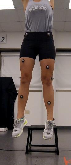
32 Experimental Procedure Data were collected during a single visit to Wright State University s Lifespan Health Research Center. Prior to marker placement, participants completed a questionnaire regarding menstrual history, sports participation history, and ACL injury history. After completing the questionnaire, participants were taken to an exam room and anthropometric measurements were obtained. Once anthropometric measurements were obtained, makers were placed on the subject and they performed a drop vertical jump (DVJ) task. Participants immediately transitioned into the stretching protocol and then completed a second DVJ task, completing the protocol. A visual representation of the procedure is represented in Figure 3. Drop Vertical Jump To perform the DVJ task, participants were instructed to jump down from a height of 31 cm onto a force plate and then jump as high as they could with arms stretched vertically after landing with both feet at the same time. DVJ is demonstrated in Figure 1. To prevent bias, participants were told to jump onto the gray square on the floor and were unaware of the presence of a force plate. The participant landed on the ground with both feet hitting the ground at the same time with each trial, however, only one foot landed on the force plate. The participant landed on the force plate with her dominant foot three times and then landed on the force plate with her non-dominant foot three times for a total of six trials. Prior to data collection, each subject performed practice 24
33 jumps to ensure they understood the procedure and were able to land properly on the force plate. Stretching Protocol Stretching of the hamstrings was induced by having subjects perform a routine consisting of five unassisted stretches. The five unassisted stretches included (1) a standing unilateral toe-touch to the dominant leg; (2) a standing unilateral toe-touch to the non-dominant leg; (3) a bilateral toe touch; (4) a seated hurdler stretch to the dominant leg; (5) a seated hurdler stretch to the non-dominant leg. Each stretch is demonstrated in Figure 2. Each stretch was held for 30 seconds, four times, with 15 seconds of rest between. The stretches were held with some discomfort but not pain, as described by the participant. The hamstring stretches and duration that they would be held was chosen based on previous static stretching research. Several studies evaluating the effects of static stretching on the hamstrings revealed a decrease in maximum muscle torque after holding each stretch for 30 seconds, four times, with a short rest period in between [54, 50]. A comprehensive review of static stretching times reveals no effect of static stretching less than 45 seconds with some effects being noticeable after 60 seconds of total stretching [70]. Our participants performed each stretch for a total of 2 minutes. This is much greater than 60 seconds. After completing the first DVJ task, participants immediately start the static stretching protocol. The instructor demonstrated the stretching exercises for the 25
34 participant. Each participant verbalized understanding of the stretching exercise. After completing the stretching protocol, participants once again completed a DVJ task. Statistical Analysis Statistical analysis of the data began with the identification of the contact phase of the DVJ task. Initial contact (IC) was determined as the first frame in which ground reaction forces (GRF) were measured. Toe-off (TO) was determined as the final frame in which GRF was measured. DVJ trials were grouped according to leg dominance. Leg dominance was determined by which leg the participant would use to kick a soccer ball. Data for the knee and the hip were analyzed. At the knee, frontal and sagittal angles, as well as frontal knee moments, were analyzed at IC, TO, maximum, and minimum for the duration of the contact phase, and at peak vgrf using paired t-tests. The peak vgrf was also analyzed via a paired t-test, with an additional sensitivity analysis to test for the effects of removing an outlier. Statistical analysis was conducted using the JMP 12.0 and SAS 9.3 integrated suite of statistical software. 26
 (B)")
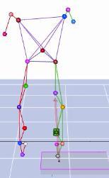
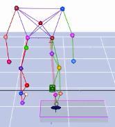
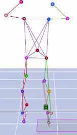
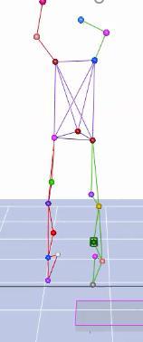
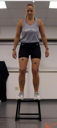
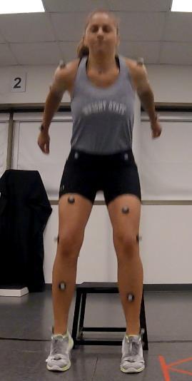
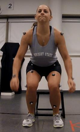
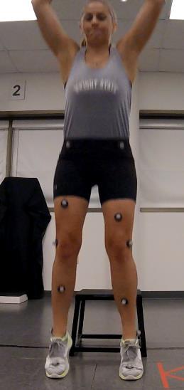
35 Figure 1. (A) (B) 27
36 Figure 1. Drop vertical jump (DVJ) task performed three times per leg by participants. (A) displays a series of images from Cortex 6.0 software showing movement of reflective markers placed on precise anatomical landmarks and showing force vectors during the contact phase of the DVJ task. (B) shows the corresponding video images of the DVJ task. Images are in chronological order from left to right. [76] 28

37 Figure 2. 29
38 Figure 2. The five unassisted stretches included (A) a standing unilateral toe-touch to the dominant leg; (B) a standing unilateral toe-touch to the non-dominant leg; (C) a bilateral toe touch; (D) a seated hurdler stretch to the dominant leg; (E) a seated hurdler stretch to the nondominant leg. 30
39 Figure 3. 31
40 Figure 3. Timeline of events that occurred during a participant s visit to the LHRC. Numbers on the horizontal axis represent time in minutes. 32
41 III. RESULTS During the stretching protocol, participants completed 5 unassisted stretches. Each of the stretches was held for 30 seconds, four times, with 15-second rest periods inbetween. The stretches were held to a point of discomfort but not pain, as described by the participant. Knee kinematics and kinetics were measured for all 12 subjects. Knee kinematics and kinetic measurements are summarized in Table 1. The results of our data revealed stretching had no significant effect. Knee abduction angle showed no significant change at IC (P= 0.50), TO (P= 0.81), Max (P= 0.39), and Min (P= 0.38). Knee flexion angles in the sagittal plane also showed no change at IC (P= 0.80), TO (P= 0.43), Max (P= 0.98), and Min (P= 0.95). Knee abduction moment was not significantly affected by stretching at IC (P= 0.98), TO (P= 0.43), Max (P=0.84), or Min (P= 0.43). Effect size for repeated measures were calculated. Hedges gav values for knee abduction angle at IC (g= 0.07), TO (g= 0.03), Max (g= 0.09), and Min (g= 0.17) showed a very small effect. Hedges gav values for knee flexion angle at IC (g= 0.06), TO (g= 0.09), Max (g= <0.01), and Min (g= 0.01) also showed a very small effect. Finally, hedges gav values for knee abduction moment at IC (g= 0.01), TO (g= 0.20), Max (g= 0.03), and Min (g= 0.16) showed a very small effect. Hedges gav values are summarized in Table 2. 33
42 The probabilities that any person would have a higher value post-stretching than they would pre-stretch routine were also calculated. The probability that someone would have a higher value post-stretch than pre-stretch for knee abduction angle at IC (0.56), TO (0.52), Max (0.57), and Min (0.58) were essentially equal to 50%. The probability that someone would have a higher value post-stretch than pre-stretch for knee flexion angle at IC (0.52), TO (0.57), Max (0.50), and Min (0.51) were approximately equal to 50%. Finally, the probability that someone would have a higher value post-stretch than pre-stretch for knee abduction moment at IC (0.50), TO (0.57), Max (0.52), and Min (0.57) were also approximately equal to 50%. The probability of a higher value poststretch than pre-stretch is summarized in Table 2. Pearson correlation values for knee abduction angle pre- and post- stretching routine at IC (r= ), TO (r= ), Max (r= ), and Min (r= ) are summarized in Figure 3. Pearson correlation values for knee flexion angle pre- and poststretching routine at IC (r= ), TO (r= ), Max (r= ), and Min (r= ) are summarized in Figure 4. Pearson correlation values for knee abduction moment pre- and post-stretching routine at IC (r= ), TO (r= ), Max (r= ), and Min (r= ) are summarized in Figure 5. 34
5/13/2016. ACL I Risk Factors AAP Position Statement. Anterior Cruciate Ligament Injuries: Diagnosis, Treatment and Prevention.
 ACL I Risk Factors AAP Position Statement Timothy E. Hewett, PhD 2016 Chicago Sports Medicine Symposium Chicago, Illinois August 5-7, 2016 2015 MFMER slide-1 Anterior Cruciate Ligament Injuries: Diagnosis,
Sports Medicine 15. Unit I: Anatomy. The knee, Thigh, Hip and Groin. Part 4 Anatomies of the Lower Limbs
 Sports Medicine 15 Unit I: Anatomy Part 4 Anatomies of the Lower Limbs The knee, Thigh, Hip and Groin Anatomy of the lower limbs In Part 3 of this section we focused upon 11 of the 12 extrinsic muscles
Knee valgus in self-initiated vertical jump landings: developmental and gender comparisons
 Bridgewater State University Virtual Commons - Bridgewater State University Movement Arts, Health Promotion and Leisure Studies Faculty Publications Movement Arts, Health Promotion and Leisure Studies
RN(EC) ENC(C) GNC(C) MN ACNP *** MECHANISM OF INJURY.. MOST IMPORTANT *** - Useful in determining mechanism of injury / overuse
 ENC(C) GNC(C) MN ACNP *** MECHANISM OF INJURY.. MOST IMPORTANT *** - Useful in determining mechanism of injury / overuse") HISTORY *** MECHANISM OF INJURY.. MOST IMPORTANT *** Age of patient Sport / Occupation - Certain conditions are more prevalent in particular age groups (Osgood Schlaters in youth / Degenerative Joint Disease
HISTORY *** MECHANISM OF INJURY.. MOST IMPORTANT *** Age of patient Sport / Occupation - Certain conditions are more prevalent in particular age groups (Osgood Schlaters in youth / Degenerative Joint Disease
Anatomy and Biomechanics
 Introduction Increased participation= increased injury rates Females were found to be 5.4 times more likely to sustain injury than males. And females injured their ACL ad a rate of 7.8 times more than
The Lower Limb II. Anatomy RHS 241 Lecture 3 Dr. Einas Al-Eisa
 The Lower Limb II Anatomy RHS 241 Lecture 3 Dr. Einas Al-Eisa Tibia The larger & medial bone of the leg Functions: Attachment of muscles Transfer of weight from femur to skeleton of the foot Articulations
CHAPTER 8: THE BIOMECHANICS OF THE HUMAN LOWER EXTREMITY
 CHAPTER 8: THE BIOMECHANICS OF THE HUMAN LOWER EXTREMITY _ 1. The hip joint is the articulation between the and the. A. femur, acetabulum B. femur, spine C. femur, tibia _ 2. Which of the following is
The Knee. Prof. Oluwadiya Kehinde
 The Knee Prof. Oluwadiya Kehinde www.oluwadiya.sitesled.com The Knee: Introduction 3 bones: femur, tibia and patella 2 separate joints: tibiofemoral and patellofemoral. Function: i. Primarily a hinge joint,
ACL and Knee Injury Prevention. Presented by: Zach Kirkpatrick, PT, MPT, SCS
 ACL and Knee Injury Prevention Presented by: Zach Kirkpatrick, PT, MPT, SCS ACL Anatomy ACL Mechanism of Injury Contact ACL Tear Noncontact ACL Tear ACL MOI and Pathology Common in young individual who
In the name of god. Knee. By: Tofigh Bahraminia Graduate Student of the Pathology Sports and corrective actions. Heat: Dr. Babakhani. Nov.
 In the name of god Knee By: Tofigh Bahraminia Graduate Student of the Pathology Sports and corrective actions Heat: Dr. Babakhani Nov. 2014 1 Anatomy-Bones Bones Femur Medial/lateral femoral condyles articulate
Knee Joint Anatomy 101
 Knee Joint Anatomy 101 Bone Basics There are three bones at the knee joint femur, tibia and patella commonly referred to as the thighbone, shinbone and kneecap. The fibula is not typically associated with
Myology of the Knee. PTA 105 Kinesiology
 Myology of the Knee PTA 105 Kinesiology Objectives Describe the planes of motion and axes of rotation of the knee joint Visualize the origins and insertions of the muscles about the knee List the innervations
Biomechanics of the Knee. Valerie Nuñez SpR Frimley Park Hospital
 Biomechanics of the Knee Valerie Nuñez SpR Frimley Park Hospital Knee Biomechanics Kinematics Range of Motion Joint Motion Kinetics Knee Stabilisers Joint Forces Axes The Mechanical Stresses to which
Balanced Body Movement Principles
 Balanced Body Movement Principles How the Body Works and How to Train it. Module 3: Lower Body Strength and Power Developing Strength, Endurance and Power The lower body is our primary source of strength,
Ligamentous and Meniscal Injuries: Diagnosis and Management
 Ligamentous and Meniscal Injuries: Diagnosis and Management Daniel K Williams, MD Franciscan Physician Network Orthopedic Specialists September 29, 2017 No Financial Disclosures INTRODUCTION Overview of
BIOMECHANICAL INFLUENCES ON THE SOCCER PLAYER. Planes of Lumbar Pelvic Femoral (Back, Pelvic, Hip) Muscle Function
 Muscle Function") BIOMECHANICAL INFLUENCES ON THE SOCCER PLAYER Functional performance of the soccer player reflects functional capability of certain specific muscle and muscle groups of the back, pelvis and hip to work
BIOMECHANICAL INFLUENCES ON THE SOCCER PLAYER Functional performance of the soccer player reflects functional capability of certain specific muscle and muscle groups of the back, pelvis and hip to work
Recognizing common injuries to the lower extremity
 Recognizing common injuries to the lower extremity Bones Femur Patella Tibia Tibial Tuberosity Medial Malleolus Fibula Lateral Malleolus Bones Tarsals Talus Calcaneus Metatarsals Phalanges Joints - Knee
Joints of the Lower Limb II
 Joints of the Lower Limb II Lecture Objectives Describe the components of the knee and ankle joint. List the ligaments associated with these joints and their attachments. List the muscles acting on these
The Female Athlete: Train Like a Girl. Sarah DoBroka Wilson, PT, SCS Ron Weathers, PT, DPT, ATC, LAT
 The Female Athlete: Train Like a Girl Sarah DoBroka Wilson, PT, SCS Ron Weathers, PT, DPT, ATC, LAT Page 1 of 6 The Female Athlete: Train Like a Girl Sarah DoBroka Wilson PT, SCS Ron Weathers PT, DPT,
DIFFERENCES IN THE MECHANICS BETWEEN THE DOMINANT AND NON-DOMINANT PLANT LIMB DURING INSTEP SOCCER KICKING. Cassidy M. Berlin.
 DIFFERENCES IN THE MECHANICS BETWEEN THE DOMINANT AND NON-DOMINANT PLANT LIMB DURING INSTEP SOCCER KICKING by Cassidy M. Berlin A thesis submitted in partial fulfillment of the requirements for the degree
Practical 1 Worksheet
 Practical 1 Worksheet ANATOMICAL TERMS 1. Use the word bank to fill in the missing words. reference side stand body arms palms anatomical forward All anatomical terms have a(n) point which is called the
ACL Rehabilitation and Return To Play
 ACL Rehabilitation and Return To Play Seth Gasser, MD Director of Sports Medicine Florida Orthopaedic Institute Introduction Return to Play: the point in recovery from an injury when a person is safely
Prevention and Treatment of Injuries. Anatomy. Anatomy. Chapter 20 The Knee Westfield High School Houston, Texas
 Prevention and Treatment of Injuries Chapter 20 The Knee Westfield High School Houston, Texas Anatomy MCL, Medial Collateral Ligament LCL, Lateral Collateral Ligament PCL, Posterior Cruciate Ligament ACL,
To describe he knee joint, ligaments, structure & To list the main features of other lower limb joints
 To describe he knee joint, ligaments, structure & neurovascular supply To demonstrate the ankle joint anatomy To list the main features of other lower limb joints To list main groups of lymph nodes in
Rehabilitation of an ACL injury in a 29 year old male with closed kinetic chain exercises: A case study
 Abstract Objective: This paper will examine a rehabilitation program for a healthy 29 year old male who sustained an incomplete tear of the left ACL. Results: Following a 9 week treatment plan focusing
Re training Movement Behavior for ACL Injury Prevention and Rehabilitation: A Matter of Strength or Motor Control?
 Re training Movement Behavior for ACL Injury Prevention and Rehabilitation: A Matter of Strength or Motor Control? Christopher M. Powers, PT, PhD, FACSM, FAPTA Beth Fisher, PT, PhD, FAPTA Division of Biokinesiology
Knee Ligament Function 3 properties allow ligaments mechanical functions to limit motion: Attachment Location
 29 muscles attach Lumbar Spine Muscles Transversospinalis group Rotatores Interspinales Intertransversarii Semispinalis Multifidus Erector spinae Iliocostalis Longissimus Spinalis Quadratus lumborum Latissimus
What is Kinesiology? Basic Biomechanics. Mechanics
 What is Kinesiology? The study of movement, but this definition is too broad Brings together anatomy, physiology, physics, geometry and relates them to human movement Lippert pg 3 Basic Biomechanics the
CHANGES IN LOWER-LIMB MUSCLE FORCES WITH PROPHYLACTIC KNEE BRACING DURING LANDING AND STOP-JUMP TASKS
 CHANGES IN LOWER-LIMB MUSCLE FORCES WITH PROPHYLACTIC KNEE BRACING DURING LANDING AND STOP-JUMP TASKS Katie Ewing 1, Rezaul Begg 2, Peter Lee 1 Department of Mechanical Engineering, University of Melbourne,
Female Athlete Injury Prevention
 Female Athlete Injury Prevention Startling Facts Huge rise in knee ligament injuries among young females engaging in sport and exercise Females athletes participating in jumping and pivoting sports are
ACL Injury Prevention: Considerations for Children and Adolescents
 ACL Injury Prevention: Considerations for Children and Adolescents Susan Sigward PhD, PT, ATC Human Performance Laboratory University of Southern California 1 ACL Injury Season ending/ Career ending 82%
Musculoskeletal Examination Benchmarks
 Musculoskeletal Examination Benchmarks _ The approach to examining the musculoskeletal system is the same no matter what joint or limb is being examined. The affected and contralateral region should both
Rehabilitation Guidelines for Anterior Cruciate Ligament (ACL) Reconstruction
 Reconstruction") Rehabilitation Guidelines for Anterior Cruciate Ligament (ACL) Reconstruction The knee is the body's largest joint, and the place where the femur, tibia, and patella meet to form a hinge-like joint. These
Rehabilitation Guidelines for Anterior Cruciate Ligament (ACL) Reconstruction The knee is the body's largest joint, and the place where the femur, tibia, and patella meet to form a hinge-like joint. These
Discrepancies in Knee Joint Moments Using Common Anatomical Frames Defined by Different Palpable Landmarks
 Journal of Applied Biomechanics, 2008, 24, 185-190 2008 Human Kinetics, Inc. Discrepancies in Knee Joint Moments Using Common Anatomical Frames Defined by Different Palpable Landmarks Dominic Thewlis,
When are athletes ready for return to sports??? Functional Testing for Return to Sports. Important Factors Involved in Return to Sport
 Functional Testing for Return to Sports Meg Jacobs PT Momentum Physical Therapy and Sports Rehab Mjacobs@wegetyouhealthy.com When are athletes ready for return to sports??? Post ACL reconstruction, average
During the initial repair and inflammatory phase, focus should be on placing the lower limbs in a position to ensure that:
 The Anatomy Dimensions series of tutorials and workbooks is aimed at improving anatomical and pathological understanding for body movement professionals. It is ideal for teachers in disciplines such as
Commonality of ACL Injuries and Prevention Methods in Women. Anterior cruciate ligament or ACL injuries have become more common in
 4/6/12 Period 2 Commonality of ACL Injuries and Prevention Methods in Women Anterior cruciate ligament or ACL injuries have become more common in the past decade than at any other time in our history.
UNIT 7 JOINTS. Knee and Ankle Joints DR. ABDEL-MONEM A. HEGAZY
 UNIT 7 JOINTS Knee and Ankle Joints BY DR. ABDEL-MONEM A. HEGAZY (Degree in Bachelor of Medicine and Surgery with honor 1983, Dipl."Gynaecology and Obstetrics "1989, Master "Anatomy and Embryology "1994,
TREATMENT GUIDELINES FOR GRADE 3 PCL TEAR
 GENERAL CONSIDERATIONS Posterior cruciate ligament (PCL) injuries occur less frequently than anterior cruciate ligament (ACL) injuries, but are much more common than previously thought. The PCL is usually
CHAPTER 1: 1.1 Muscular skeletal system. Question - text book page 16. Question - text book page 20 QUESTIONS AND ANSWERS. Answers
 QUESTIONS AND ANSWERS CHAPTER 1: 1.1 Muscular skeletal system Question - text book page 16 Using the information on pages 12 to 14 above, complete the table below. joint joint type articulating bones associated
Range Of Motion And Plantar Foot Pressures In Those With And Without A Lateral Hip Shift During An Overhead Squat
 Illinois State University ISU ReD: Research and edata Theses and Dissertations 5-25-2016 Range Of Motion And Plantar Foot Pressures In Those With And Without A Lateral Hip Shift During An Overhead Squat
Mechanisms of ACL Injury: Implications for Rehabilitation, Injury Prevention & Return to Sport Decisions. Overarching research theme:
 Mechanisms of ACL Injury: Implications for Rehabilitation, Injury Prevention & Return to Sport Decisions Associate Professor Co Director, Musculoskeletal Biomechanics Research Laboratory University of
Anterior Cruciate Ligament Surgery
 Anatomy Anterior Cruciate Ligament Surgery Roger Ostrander, MD Andrews Institute Anatomy Anatomy Function Primary restraint to anterior tibial translation Secondary restraint to internal tibial rotation
Current trends in ACL Rehab. James Kelley, MDS, PT
 Current trends in ACL Rehab James Kelley, MDS, PT Objectives Provide etiological information Discuss the criteria for having an ACL reconstruction Review the basic rehabilitation principles behind ACL
ACL RECONSTRUCTION HAMSTRING METHOD. Presents ACL RECONSTRUCTION HAMSTRING METHOD. Multimedia Health Education
 HAMSTRING METHOD Presents HAMSTRING METHOD Multimedia Health Education Disclaimer Stephen J. Incavo MD This movie is an educational resource only and should not be used to make a decision on Anterior Cruciate
BIOMECHANICS AND CONTEXT OF ACUTE KNEE INJURIES. Uwe Kersting MiniModule Idræt Biomekanik 2. Objectives
 BIOMECHANICS AND CONTEXT OF ACUTE KNEE INJURIES Uwe Kersting MiniModule 06 2011 Idræt Biomekanik 2 1 Objectives Know about the static and dynamic organisation of the knee joint (anatomy & function) Be
KNEE INJURIES IN FEMALE SOCCER PLAYERS: A FOCUS ON THE ACL
 KNEE INJURIES IN FEMALE SOCCER PLAYERS: A FOCUS ON THE ACL Item Type text; Electronic Thesis Authors PEÑA, VANESSA NICOLE Publisher The University of Arizona. Rights Copyright is held by the author. Digital
Literature Review of Female Anterior Cruciate Ligament Injuries
 Eastern Michigan University DigitalCommons@EMU Senior Honors Theses Honors College 2004 Literature Review of Female Anterior Cruciate Ligament Injuries Allison M. Nichol Follow this and additional works
Strengthening the ACL and Knee Health Warm-up Program
 Strengthening the ACL and Knee Health Warm-up Program Researched and compiled by: Jordan Holtz B.S., NCSF- PT Workouts and Exercises recommended and proven by: Dr. Laura Inverarity, P.T., D.O. Elizabeth
ACL AND PCL INJURIES OF THE KNEE JOINT
 ACL AND PCL INJURIES OF THE KNEE JOINT Dr.KN Subramanian M.Ch Orth., FRCS (Tr & Orth), CCT Orth(UK) Consultant Orthopaedic Surgeon, Special interest: Orthopaedic Sports Injury, Shoulder and Knee Surgery,
and K n e e J o i n t Is the most complicated joint in the body!!!!
 K n e e J o i n t K n e e J o i n t Is the most complicated joint in the body!!!! 1-Consists of two condylar joints between: A-The medial and lateral condyles of the femur and The condyles of the tibia
Hip joint Type: Articulating bones:
 Ana (242 ) Hip joint Type: Synovial, ball & socket Articulating bones: Formed between head of femur and lunate surface of acetabulum of hip bone. Capsule: it is a strong fibrous sleeve connecting the articulating
Copyright 2012 by The McGraw-Hill Companies, Inc. All rights reserved. McGraw-Hill/Irwin
 CHAPTER 8: THE LOWER EXTREMITY: KNEE, ANKLE, AND FOOT KINESIOLOGY Scientific Basis of Human Motion, 12 th edition Hamilton, Weimar & Luttgens Presentation Created by TK Koesterer, Ph.D., ATC Humboldt State
Knee Joint Assessment and General View
 Knee Joint Assessment and General View Done by; Mshari S. Alghadier BSc Physical Therapy RHPT 366 m.alghadier@sau.edu.sa http://faculty.sau.edu.sa/m.alghadier/ Functional anatomy The knee is the largest
Knee Multiligament Rehabilitation
 Knee Multiligament Rehabilitation Orlando Valle, PT, MSPT, SCS, CSCS Director Ironman Sports Medicine Institute TMC Orlando.Valle@memorialhermann.org 4 Major Ligaments ACL PCL MCL LCL (PLC) Anatomy Function
ANTICIPATORY EFFECTS ON LOWER EXTREMITY KINETICS AND KINEMATICS DURING A LAND AND CROSS STEP MANEUVER IN FEMALE VOLLEYBALL PLAYERS
 ANTICIPATORY EFFECTS ON LOWER EXTREMITY KINETICS AND KINEMATICS DURING A LAND AND CROSS STEP MANEUVER IN FEMALE VOLLEYBALL PLAYERS A THESIS SUBMITTED TO THE GRADUATE SCHOOL IN PARTIAL FULFILLMENT OF THE
Muscle Testing of Knee Extensors. Yasser Moh. Aneis, PhD, MSc., PT. Lecturer of Physical Therapy Basic Sciences Department
 Muscle Testing of Knee Extensors Yasser Moh. Aneis, PhD, MSc., PT. Lecturer of Physical Therapy Basic Sciences Department Muscle Testing of Knee Extensors othe Primary muscle Quadriceps Femoris -Rectus
The Effects of Augmented Feedback on Knee Valgus Angles and Muscle Activity During a Jump Landing Task. Michelle M. Bensman BS, LAT, ATC
 The Effects of Augmented Feedback on Knee Valgus Angles and Muscle Activity During a Jump Landing Task Michelle M. Bensman BS, LAT, ATC A thesis submitted to the faculty of The University of North Carolina
HAMSTRING NEUROMECHANICAL PROPERTIES AND BIOMECHANICS IN AN ACL INJURED POPULATION. David Robert Bell
 HAMSTRING NEUROMECHANICAL PROPERTIES AND BIOMECHANICS IN AN ACL INJURED POPULATION David Robert Bell A dissertation submitted to the faculty of the University of North Carolina at Chapel Hill in partial
ChiroCredit.com Presents Biomechanics: Focus on
 ChiroCredit.com Presents Biomechanics: Focus on the Knee Presented by: Ivo Waerlop, DC Shawn Allen, DC 1 Focus on The Knee 2 Pertinent Anatomy Femur Tibia Fibula Patella Prepatellar bursa Infrapatellar
The Knee Joint By Prof. Dr. Muhammad Imran Qureshi
 The Knee Joint By Prof. Dr. Muhammad Imran Qureshi Structurally, it is the Largest and the most complex joint in the body because of the functions that it performs: Allows mobility (flexion/extension)
Knee Injuries. PSK 4U Mr. S. Kelly North Grenville DHS. Medial Collateral Ligament Sprain
 Knee Injuries PSK 4U Mr. S. Kelly North Grenville DHS Medial Collateral Ligament Sprain Result from either a direct blow from the lateral side in a medial direction or a severe outward twist Greater injury
Girls and Sports. Title IX. You Go Girl! : Female Athlete Hot Topics. Women in Sports
 You Go Girl! : Female Athlete Hot Topics Rebecca A. Demorest, MD Associate Director, Pediatric and Young Adult Sports Medicine Children s Hospital & Research Center Oakland rdemorest@mail.cho.org Girls
Effects of Altered Surface Inclinations on Knee Kinematics During Drop Landing
 Pursuit - The Journal of Undergraduate Research at the University of Tennessee Volume 7 Issue 1 Article 14 April 2016 Effects of Altered Surface Inclinations on Knee Kinematics During Drop Landing Cicily
Muscles of the Thigh. 6.1 Identify, describe the attachments of and deduce the actions of the muscles of the thigh: Anterior group
 Muscles of the Thigh 6.1 Identify, describe the attachments of and deduce the actions of the muscles of the thigh: Anterior group Sartorius: This is a long strap like muscle with flattened tendons at each
Military Movement Training Program Improves Jump-Landing Mechanics Associated With Anterior Cruciate Ligament Injury Risk
 Military Movement Training Program Improves Jump-Landing Mechanics Associated With Anterior Cruciate Ligament Injury Risk Brett D. Owens, MD; Kenneth L. Cameron, PhD, ATC; Michele L. Duffey, MS; Donna
educate OBJECTIVES AND TAKE-HOME ACL INJURY BY THE NUMBERS 12/4/2011 LIVE ON-SCREEN DEMONSTRATION
 LIVE ON-SCREEN DEMONSTRATION Derek Hirai, MS, ATC Gina Biviano, MA, ATC PRE-EMPTIVE STRIKE: ACL Injury Prevention Techniques OBJECTIVES AND TAKE-HOME Identify risk factors In-office testing/evaluation
The Knee. Clarification of Terms. Osteology of the Knee 7/28/2013. The knee consists of: The tibiofemoral joint Patellofemoral joint
 The Knee Clarification of Terms The knee consists of: The tibiofemoral joint Patellofemoral joint Mansfield, p273 Osteology of the Knee Distal Femur Proximal tibia and fibula Patella 1 Osteology of the
Uranie P. Browne. Chapel Hill 2007
 A Comparison of Risk Factors Between a Cutting Task and a Stop-Jump as it Relates to the Non Contact Anterior Cruciate Ligament Injury Uranie P. Browne A thesis submitted to the faculty of the University
Objectives. The BIG Joint. Case 1. Boney Architecture. Presenter Disclosure Information. Common Knee Problems
 3:30 4:15 pm Common Knee Problems SPEAKER Christopher J. Visco, MD Presenter Disclosure Information The following relationships exist related to this presentation: Christopher J. Visco, MD: Speaker s Bureau
Lateral knee injuries
 Created as a free resource by Clinical Edge Based on Physio Edge podcast episode 051 with Matt Konopinski Get your free trial of online Physio education at Orthopaedic timeframes Traditionally Orthopaedic
TOTAL KNEE ARTHROPLASTY (TKA)
") TOTAL KNEE ARTHROPLASTY (TKA) 1 Anatomy, Biomechanics, and Design 2 Femur Medial and lateral condyles Convex, asymmetric Medial larger than lateral 3 Tibia Tibial plateau Medial tibial condyle: concave
TOTAL KNEE ARTHROPLASTY (TKA) 1 Anatomy, Biomechanics, and Design 2 Femur Medial and lateral condyles Convex, asymmetric Medial larger than lateral 3 Tibia Tibial plateau Medial tibial condyle: concave
The Knee. Two Joints: Tibiofemoral. Patellofemoral
 Evaluating the Knee The Knee Two Joints: Tibiofemoral Patellofemoral HISTORY Remember the questions from lecture #2? Girth OBSERVATION TibioFemoral Alignment What are the consequences of faulty alignment?
The Knee. Tibio-Femoral
 The Knee Tibio-Femoral Osteology Distal Femur with Proximal Tibia Largest Joint Cavity in the Body A modified hinge joint with significant passive rotation Technically, one degree of freedom (Flexion/Extension)
Do Persons with PFP. PFJ Loading? Biomechanical Factors Contributing to Patellomoral Pain: The Dynamic Q Angle. Patellofemoral Pain: A Critical Review
 Biomechanical Factors Contributing to Patellomoral Pain: The Dynamic Q Angle Division Biokinesiology & Physical Therapy Co Director, oratory University of Southern California Movement Performance Institute
Upper Limb Biomechanics SCHOOL OF HUMAN MOVEMENT STUDIES
 Upper Limb Biomechanics Phases of throwing motion 1. Wind up Starts: initiate first movement Ends: lead leg is lifted & throwing hand removed from glove COG raised 2. Early Cocking Start: lead leg is lifted
Knee Movement Coordination Deficits. ICD-9-CM: Sprain of cruciate ligament of knee
 1 Knee Movement Coordination Deficits Anterior Cruciate Ligament ACL Tear ICD-9-CM: 844.2 Sprain of cruciate ligament of knee ACL Insufficiency ICD-9-CM: 717.83 Old disruption of anterior cruciate ligament
Functional Movement Test. Deep Squat
 Functional Movement Test Put simply, the FMS is a ranking and grading system that documents movement patterns that are key to normal function. By screening these patterns, the FMS readily identifies functional
CONTROL OF THE BOUNDARY CONDITIONS OF A DYNAMIC KNEE SIMULATOR
 CONTROL OF THE BOUNDARY CONDITIONS OF A DYNAMIC KNEE SIMULATOR J. Tiré 1, J. Victor 2, P. De Baets 3 and M.A. Verstraete 2 1 Ghent University, Belgium 2 Ghent University, Department of Physical Medicine
Hamstring Strain. 43 Thames Street, St Albans, Christchurch 8013 Phone: (03) Website: philip-bayliss.com.
 Website: philip-bayliss.com.") 43 Thames Street, St Albans, Christchurch 8013 Phone: (03) 356 1353. Website: philip-bayliss.com Hamstring Strain The hamstring muscles are very susceptible to tears, strains and other common sporting
43 Thames Street, St Albans, Christchurch 8013 Phone: (03) 356 1353. Website: philip-bayliss.com Hamstring Strain The hamstring muscles are very susceptible to tears, strains and other common sporting
Anterior knee pain.
 Anterior knee pain What are the symptoms? Anterior knee pain is very common amongst active adolescents and athletes participating in contact sports. It is one of the most common problems/injuries seen
Anatomy. Anatomy deals with the structure of the human body, and includes a precise language on body positions and relationships between body parts.
 Anatomy deals with the structure of the human body, and includes a precise language on body positions and relationships between body parts. Proper instruction on safe and efficient exercise technique requires
Knee Injury Assessment
 Knee Injury Assessment Clinical Anatomy p. 186 Femur Medial condyle Lateral condyle Femoral trochlea Tibia Intercondylar notch Tibial tuberosity Tibial plateau Fibula Fibular head Patella Clinical Anatomy
Comparative Effectiveness Of Injury Prevention Programs In Female Adolescent Athletes
 University of Connecticut DigitalCommons@UConn Doctoral Dissertations University of Connecticut Graduate School 9-14-2015 Comparative Effectiveness Of Injury Prevention Programs In Female Adolescent Athletes
Learning Objectives. Epidemiology 7/22/2016. What are the Medical Concerns for the Adolescent Female Athlete? Krystle Farmer, MD July 21, 2016
 What are the Medical Concerns for the Adolescent Female Athlete? Krystle Farmer, MD July 21, 2016 Learning Objectives Discuss why females are different than males in sports- the historical perspective.
The Dance Hall by Vincent van Gogh,1888
 The Dance Hall by Vincent van Gogh,1888 Articulations of the pelvic girdle Lumbosacral joints, sacroiliac joints & pubic symphysis The remaining joints of the lower limb Hip joint Knee joint Tibiofibular
Fitness Theory Exam Review
 - 1 - Fitness Theory Exam Review 1. Identify the risk factors for cardio-vascular disease Controllable Uncontrollable 2. What health improvements could be made by being physically active? 3. What behaviours
EMG Analysis of Lower Extremity Muscles in Three Different Squat Exercises
 REPORT EMG Analysis of Lower Extremity Muscles in Three Different Squat Exercises Gaston Ariel NISHIWAKI 1, Yukio URABE 1 and Kosuke TANAKA 1 1 Department of Sport Rehabilitation, Graduate School of Health
Human Anatomy Biology 351
 Human Anatomy Biology 351 Lower Limb Please place your name on the back of the last page of this exam. You must answer all questions on this exam. Because statistics demonstrate that, on average, between
Copyright 2003 Pearson Education, Inc. publishing as Benjamin Cummings. Dr. Nabil Khouri MD, MSc, Ph.D
 Dr. Nabil Khouri MD, MSc, Ph.D Pelvic Girdle (Hip) Organization of the Lower Limb It is divided into: The Gluteal region The thigh The knee The leg The ankle The foot The thigh and the leg have compartments
ANTERIOR CRUCIATE LIGAMENT PREVENTION: EFFECT OF NEUROMUSCULAR TRAINING COMPLIANCE ON MUSCULAR STRENGTH DEVELOPMENT
 University of Kentucky UKnowledge Theses and Dissertations--Rehabilitation Sciences Rehabilitation Sciences 2013 ANTERIOR CRUCIATE LIGAMENT PREVENTION: EFFECT OF NEUROMUSCULAR TRAINING COMPLIANCE ON MUSCULAR
ACL Athletic Career. ACL Rupture - Warning Features Intensive pain Immediate swelling Locking Feel a Pop Dead leg Cannot continue to play
 FIMS Ambassador Tour to Eastern Europe, 2004 Belgrade, Serbia Montenegro Acute Knee Injuries - Controversies and Challenges Professor KM Chan OBE, JP President of FIMS Belgrade ACL Athletic Career ACL
APTA Intro to Identity. The Movement System The Kinesiopathologic Model Movement System Impairment Syndromes of the Knee THE HUMAN MOVEMENT SYSTEM
 The Movement System The Kinesiopathologic Model Movement System Impairment Syndromes of the Knee Shirley Sahrmann, PT, PhD, FAPTA Professor Emerita Statement of Privacy To protect the privacy of the subjects
University of Kentucky Doctoral Dissertations
 University of Kentucky UKnowledge University of Kentucky Doctoral Dissertations Graduate School 2009 CHANGES IN LONGITUDINALLY ASSESSED BIOMECHANICAL PARAMETERS RELATED TO INCREASED RISK OF ANTERIOR CRUCIATE
Diane Vives, MS, CSCS, *D Owner, Fit4Austin, Austin, TX Member of the Board of Directors, NSCA
 Diane Vives, MS, CSCS, *D Owner, Fit4Austin, Austin, TX Member of the Board of Directors, NSCA 1. Introduction a. Through my experience as a strength coach over the last 11 years, I have often been faced
ANTICIPATORY EFFECTS ON LOWER EXTREMITY KINEMATICS AND KINETICS DURING CUTTING MOVEMENTS
 ANTICIPATORY EFFECTS ON LOWER EXTREMITY KINEMATICS AND KINETICS DURING CUTTING MOVEMENTS By RYAN ASHLEY MIZELL A THESIS PRESENTED TO THE GRADUATE SCHOOL OF THE UNIVERSITY OF FLORIDA IN PARTIAL FULFILLMENT
Anterior Cruciate Ligament (ACL) Injuries
 Injuries") Anterior Cruciate Ligament (ACL) Injuries Mark L. Wood, MD The anterior cruciate ligament (ACL) is one of the most commonly injured ligaments of the knee. The incidence of ACL injuries is currently estimated
Anterior Cruciate Ligament (ACL) Injuries Mark L. Wood, MD The anterior cruciate ligament (ACL) is one of the most commonly injured ligaments of the knee. The incidence of ACL injuries is currently estimated
CHAPTER 4: The musculo-skeletal system. Practice questions - text book pages QUESTIONS AND ANSWERS. Answers
 CHAPTER 4: The musculo-skeletal system Practice questions - text book pages 64-66 1) A prime mover of hip flexion is the: a. rectus femoris. b. Iliopsoas. c. vastus muscles. d. gluteus maximus. b. Key
Primary Movements. Which one? Rational - OHS. Assessment. Rational - OHS 1/1/2013. Two Primary Movement Assessment: Dynamic Assessment (other)
") Primary Movements Practical Application for Athletic Trainers Two Primary Movement Assessment: NASM-CES Overhead Squat Single-leg Squat Dynamic Assessment (other) Single-leg Step Off Functional Movement
Primary Movements Practical Application for Athletic Trainers Two Primary Movement Assessment: NASM-CES Overhead Squat Single-leg Squat Dynamic Assessment (other) Single-leg Step Off Functional Movement