Neuroradiology Case of the Day
|
|
|
- Arlene Miles
- 5 years ago
- Views:
Transcription


1 Neuroradiology Case of the Day 76 th CAR Annual Meeting, Montreal, Quebec April 27, 2013 Eugene Yu, MD Assistant Professor of Radiology and Otolaryngology-Head and Neck Surgery Head and Neck Imaging Princess Margaret Hospital University of Toronto
2 Disclosure of Commercial Interests Neither I nor my immediate family members have a financial relationship with a commercial organization that may have a direct or indirect interest in the content.
3 The Case History: 62 year old female presents with 6 month history of left facial fullness, trismus and left V3 distribution numbness


4
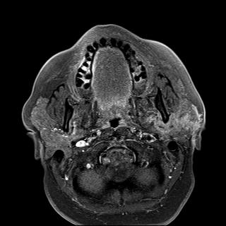
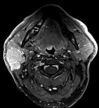
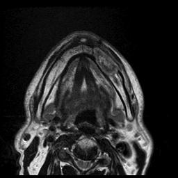
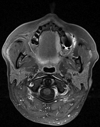
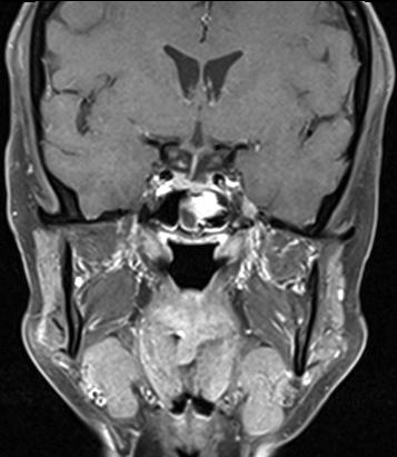
5 Left parotid mass
6
7
8

9
10 T2WI

11
12
13
14
15 Enhanced T1WI
16
17

18
19
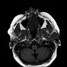
20
21
22
23
24
25
26

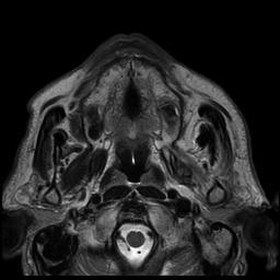
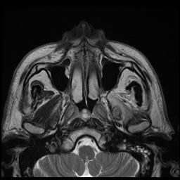
27 Foramen ovale

28

29

30
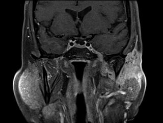
31
32
33 Case of the Day Considerations: Parotid mass - nonspecific Curvilinear structure - to cavernous sinus Clinical features are concerning Trismus, numbness Dealing with a neoplasm - malignant


34 Parotid Malignancy

35 Parotid Malignancy Perineural disease Facial nerve

36


37 Pierces parotid fascia Into the gland Communicates with CN 7 via two rami
38 Perineural Tumor Spread Adenoid cystic carcinoma parotid mass Perineural tumor spread Auriculotemporal nerve Cavernous sinus
39 Highlight Perineural Disease
40 Perineural Disease Spread of tumor along nerves Neurotropic carcinomatous spread First described in mid-1800 s

41 Nerve Sheath Anatomy
42 Perineural Invasion Liebig et al, 2009 Cancer Tumor cells within any of the three layers of the nerve sheath Tumor foci outside of the nerve with involvement > 33% of the nerve s circumference

43 Perineural Invasion Physiological basis Due to reciprocal nerve signaling interactions between tumor and nerves

44 Perineural Invasion Tumor cells express proteinases Help migrate through extracellular matrix and nerve sheath

45 Perineural Invasion What it does not involve Not tracking along lymphatics Lymphatics do not penetrate into the nerve sheath Not due to spread along path of least resistance Collagen, basement membrane
46 Perineural Invasion Pathologist: Perineural invasion is a contiguous process
47 Perineural Invasion Significance of PNI 3x increased rate of local recurrence Decreased survival 30% 5yr Increased LN recurrence Influence therapy Can make disease unresectable Influence surgical approach XRT, chemotherapy
48 Perineural Tumor Spread Perineural tumor spread: PTS Different from perineural invasion Macroscopic tracking of tumor along nerves Tumor uses nerve as a scaffold Visible on imaging
49 Clinical Symptoms Clinical features: 40% asymptomatic - imaging may be the first to pick it up Pain Paresthesia Muscle weakness Diplopia
50 Disease Entities Squamous cell carcinoma Melanoma Adenoid cystic carcinoma Lymphoma
51 Imaging Features Computed Tomography and MRI: Increased size of nerve, enhancement Widened neuroforamen Erosion of adjacent bony margins Loss of normal fat planes Muscle atrophy
52 How to Image MRI scan > CT scan Contrast enhanced scanning Narrow FOV 16-18cm High resolution matrix Slice thickness 3mm


53 Imaging Features Magnetic resonance imaging: T1 pre gadolinium scan T1 post gad ± fat saturation
54 Imaging Features Image all the way back along the course of the nerve Antegrade as well as retrograde spread
55
56
57 Nerve enlargement
58

59

60

61

62
63

64

65 Denervation atrophy
66

67
68
69
70
71
72 Loss of fluid signal in Meckel s cave

73

74

75

76

77

78

79

80

81

82

83
84 Conclusion ATN Perineural tumor spread Significance of PNTS Anatomy - knowing what to look for Clinical history

85 Pterygopalatine Fossa Pterygopalatine Fossa Space between posterior margin of maxillary sinus and the pterygoid process V2 as it exits foramen rotundum travels through here - predominantly fat density
86 Pterygopalatine Fossa The relay station : Nasal cavity - sphenopalatine foramen Masticator space - pterygomaxillary fissure Orbit - Superior/inferior orbital fissure Intracranial - Foramen Rotundum, Vidian canal

87

88

89
90 ACC

91

92 Enhancement and widening Greater palatine foramen

93

94

95

96 Pterygopalatine foramen

97 V2 along foramen rotundum
98

99
100

101

102

103 Greater palatine foramen

104 Pterygopalatine fossa

105

106

107
108

109
110 Right sided ACC palate
111
112
113
114
115
116
117
118
119
120
121
122

123
124
125
126
127 Any palate tumor Check the palatine foramen and PPF
128 Conclusion ATN Perineural tumor spread Significance of PNTS Imaging technique Knowing what to look for Clinical history
129 Perineural Tumor Spread Adenoid cystic carcinoma Can we diagnose its full extent?


130
131 Perineural Tumor Spread Retrospective studies Various histology of HN cancer Except Hanna, et al - ACC Gandhi, et al - cutaneous HN cancer Pre-operative MRI ± CT Surgical resection and pathology evaluation
132 Perineural Tumor Spread Nemzek et al, pts 19 patients - picked only those with (+) histo PNS and deemed resectable MRI sensitivity for detection 95% Sensitivity to map full extent 63% Hanna et al, consecutive patients - all ACC underwent resection CT sens 88% spec 89% MRI sens 100% spec 85% Gandhi et al, patients - cutaneous malignancy, clinically suspected PND (+) MRI MRI sens 100% Sensitivity to map full extent 83.3%
133
134
Perineural Tumor Spread. In Head & Neck Cancer
 Head and Neck Imaging Conference University of Perineural Tumor Spread In Head & Neck Cancer Philip Chapman MD University of Alabama, Birmingham OBJECTIVES: 1. Define (PNTS) 2. Distinguish from pathologic
Perineural Tumor Spread (PNS) Perineural Tumor Spread (PNS) PNS Anatomic Considerations. Perineural Tumor Spread-Imaging
 Perineural Tumor Spread (PNS) PNS Anatomic Considerations. Perineural Tumor Spread-Imaging") Imaging of Perineural Tumor Spread in Head and Neck Cancer Lawrence E. Ginsberg, MD Departments of Diagnostic Radiology and Head and Neck Surgery University of Texas M.D. Anderson Cancer Center Houston,
Imaging of Perineural Tumor Spread in Head and Neck Cancer Lawrence E. Ginsberg, MD Departments of Diagnostic Radiology and Head and Neck Surgery University of Texas M.D. Anderson Cancer Center Houston,
Patterns of perineural spread of head and neck malignancies.
 Patterns of perineural spread of head and neck malignancies. Poster No.: C-1234 Congress: ECR 2014 Type: Educational Exhibit Authors: C. Martins Jarnalo, G. Lycklama à Nijeholt, E. Sanchez-Aliaga, 1 1
ARTICLE. Imaging the cranial nerves in cancer
 Cancer Imaging (2004) 4, S1 S5 DOI: 10.1102/1470-7330.2004.0006 CI ARTICLE Vincent Chong Department of Diagnostic Radiology, Singapore General Hospital, Outram Road, Singapore 169608, Singapore Corresponding
Imaging Perineural Spread in the Head &
 Imaging Perineural Spread in the Head & Neck Tumours Vincent Chong, MD MBA FRCR Professor Department of Diagnostic Imaging National University Health System Singapore Overview: Perineural Spread Review
The Pterygopalatine Fossa: Postoperative MR Imaging Appearance
 AJNR Am J Neuroradiol 21:1315 1319, August 2000 The Pterygopalatine Fossa: Postoperative MR Imaging Appearance Ling-Ling Chan, June Chong, Ann M. Gillenwater, and Lawrence E. Ginsberg BACKGROUND AND PURPOSE:
NEXT STOP : Central Station "Pterygopalatine fossa"
 NEXT STOP : Central Station "Pterygopalatine fossa" Poster No.: C-1359 Congress: ECR 2015 Type: Educational Exhibit Authors: I. Alba de Caceres, A. Paniagua, L. Ibañez, J. A. Blanco ; 1 1 1 1 2 2 Madrid/ES,
No IN THE SUPREME COURT OF ALABAMA
 E-Filed 09/22/2017 @ 03:05:41 PM Honorable Julia Jordan Weller Clerk Of The Court No. 1881555 IN THE SUPREME COURT OF ALABAMA Ex parte Doyle Lee Hamm, * * In re. State of Alabama * Petitioner, * Fourth
Temporal region. temporal & infratemporal fossae. Zhou Hong Ying Dept. of Anatomy
 Temporal region temporal & infratemporal fossae Zhou Hong Ying Dept. of Anatomy Temporal region is divided by zygomatic arch into temporal & infratemporal fossae. Temporal Fossa Infratemporal fossa Temporal
Temporal fossa Infratemporal fossa Pterygopalatine fossa Terminal branches of external carotid artery Pterygoid venous plexus
 Outline of content Temporal fossa Infratemporal fossa Pterygopalatine fossa Terminal branches of external carotid artery Pterygoid venous plexus Boundary Content Communication Mandibular division of trigeminal
Infratemporal fossa: Tikrit University college of Dentistry Dr.Ban I.S. head & neck Anatomy 2 nd y.
 Infratemporal fossa: This is a space lying beneath the base of the skull between the lateral wall of the pharynx and the ramus of the mandible. It is also referred to as the parapharyngeal or lateral pharyngeal
PTERYGOPALATINE FOSSA
 PTERYGOPALATINE FOSSA Outline Anatomical Structure and Boundaries Foramina and Communications with other spaces and cavities Contents Pterygopalatine Ganglion Especial emphasis on certain arteries and
Imaging: When to get MRI, CT or PET-CT?
 Imaging: When to get MRI, CT or PET-CT? Alina Uzelac, D.O. Assistant Clinical Professor Neuroradiology UCSF Department of Radiology and Biomedical Imaging San Francisco General Hospital Overview CT MRI
MR and CT anatomy and the pathology of skull base focusing on pterygopalatine fossa
 MR and CT anatomy and the pathology of skull base focusing on pterygopalatine fossa Poster No.: C-1688 Congress: ECR 2010 Type: Educational Exhibit Topic: Head and Neck Authors: S. Kandatsu, R. Kishimoto,
Major Anatomic Components of the Orbit
 Major Anatomic Components of the Orbit 1. Osseous Framework 2. Globe 3. Optic nerve and sheath 4. Extraocular muscles Bony Orbit Seven Bones Frontal bone Zygomatic bone Maxillary bone Ethmoid bone Sphenoid
Juvenile Angiofibroma
 Juvenile Angiofibroma Disclaimer The pictures used in this presentation have been obtained from a number of sources. Their use is purely for academic and teaching purposes. The contents of this presentation
Omran Saeed. Luma Taweel. Mohammad Almohtaseb. 1 P a g e
 2 Omran Saeed Luma Taweel Mohammad Almohtaseb 1 P a g e I didn t include all the photos in this sheet in order to keep it as small as possible so if you need more clarification please refer to slides In
ORIGINAL ARTICLE. head and neck cancer frequently necessitates combined extracranial and intracranial approaches
 ORIGINAL ARTICLE The Sensitivity and Specificity of High-Resolution Imaging in Evaluating Perineural Spread of Adenoid Cystic Carcinoma to the Skull Base Ehab Hanna, MD; Emre Vural, MD; Emmanuel Prokopakis,
Dr.Ban I.S. head & neck anatomy 2 nd y جامعة تكريت كلية طب االسنان مادة التشريح املرحلة الثانية أ.م.د. بان امساعيل صديق 6102/6102
 جامعة تكريت كلية طب االسنان مادة التشريح املرحلة الثانية أ.م.د. بان امساعيل صديق 6102/6102 Pterygopalatine fossa: The pterygopalatine fossa is a cone-shaped depression, It is located between the maxilla,
Parotid Gland. Parotid Gland. Largest of 3 paired salivary glands (submandibular; sublingual) Ramus of Mandible. Medial pterygoid.
 Ramus of Mandible. Medial pterygoid.") Parotid region Parotid Gland Largest of 3 paired salivary glands (submandibular; sublingual) Ramus of Mandible Medial pterygoid Cross section of mandible Masseter D S SCM Parotid Gland Mastoid Process
Parotid region Parotid Gland Largest of 3 paired salivary glands (submandibular; sublingual) Ramus of Mandible Medial pterygoid Cross section of mandible Masseter D S SCM Parotid Gland Mastoid Process
Malignant growth Maxilla management an analysis
 ISSN: 2250-0359 Volume 3 Issue 2 2013 Malignant growth Maxilla management an analysis *Balasubramanian Thiagarajan *Geetha Ramamoorthy *Stanley Medical College Abstract: Malignant tumors involving maxilla
RADIOLOGY TEACHING CONFERENCE
 RADIOLOGY TEACHING CONFERENCE John Athas, MD Monica Tadros, MD Columbia University, College of Physicians & Surgeons Department of Otolaryngology- Head & Neck Surgery September 27, 2007 CT SCAN IMAGING
Trigeminal Nerve Worksheets, Distributions Page 1
 Trigeminal Nerve Worksheet #1 Distribution by Nerve Dr. Darren Hoffmann Dental Gross Anatomy, Spring 2013 We have drawn out each of the branches of CN V in lecture and you have an idea now for their basic
The International Federation of Head and Neck Oncologic Societies. Current Concepts in Head and Neck Surgery and Oncology
 The International Federation of Head and Neck Oncologic Societies Current Concepts in Head and Neck Surgery and Oncology www.ifhnos.net The International Federation of Head and Neck Oncologic Societies
Mohammad Hisham Al-Mohtaseb. Lina Mansour. Reyad Jabiri. 0 P a g e
 2 Mohammad Hisham Al-Mohtaseb Lina Mansour Reyad Jabiri 0 P a g e This is only correction for the last year sheet according to our record. If you already studied this sheet just read the yellow notes which
Paranasal Sinuses: Neoplastic Lesions
 Pravin Mundada Department of Radiology, Geneva University Hospital, Switzerland Paranasal Sinuses: Neoplastic Lesions ESHNR 2017 Lisbon, Portugal Layout of the presentation Clinical & imaging features
Easily detected signs of perineural tumour spread in head and neck cancer
 Insights into Imaging (2018) 9:1089 1095 https://doi.org/10.1007/s13244-018-0672-8 PICTORIAL REVIEW Easily detected signs of perineural tumour spread in head and neck cancer Jan Willem Dankbaar 1 & Frank
Anatomic Relations Summary. Done by: Sohayyla Yasin Dababseh
 Anatomic Relations Summary Done by: Sohayyla Yasin Dababseh Anatomic Relations Lecture 1 Part-1 - The medial wall of the nose is the septum. - The vestibule lies directly inside the nostrils (Nares). -
From GTV to CTV: A Critical Step Towards Cure. Kenneth Hu, MD Associate Professor New York University Langone Medical Center June 21, 2017
 From GTV to CTV: A Critical Step Towards Cure Kenneth Hu, MD Associate Professor New York University Langone Medical Center June 21, 2017 Head and Neck Cancer Model for Understanding CTV Expansion Radiation
Management of Salivary Gland Malignancies. No Disclosures or Conflicts of Interest. Anatomy 10/4/2013
 Management of Salivary Gland Malignancies Daniel G. Deschler, MD Director: Division of Head and Neck Surgery Massachusetts Eye & Ear Infirmary Massachusetts General Hospital Professor Harvard Medical School
Introduction to Local Anesthesia and Review of Anatomy
 5-Sep Introduction and Anatomy Review 12-Sep Neurophysiology and Pain 19-Sep Physiology and Pharmacology part 1 26-Sep Physiology and Pharmacology part 2 Introduction to Local Anesthesia and Review of
Maxilla, ORBIT and infratemporal fossa. Neophytos C Demetriades MD, DDS, MSc Associate professor European University of Cyprus School of Medicine
 Maxilla, ORBIT and infratemporal fossa Neophytos C Demetriades MD, DDS, MSc Associate professor European University of Cyprus School of Medicine MAXILLA Superior, middle, and inferior meatus Frontal sinus
Extramedullary Multiple Myeloma in the Head and Neck: A Pictorial Essay
 Canadian Association of Radiologists Journal 64 (2013) 363e369 Neuroradiology / Neuroradiologie Extramedullary Multiple Myeloma in the Head and Neck: A Pictorial Essay Michael Chan, BHSc a, Eric Bartlett,
Sinonasal Tumors. Objectives. Objectives. Incidence of Paranasal Sinus Tumors. Demographics of Paranasal Sinus Tumors. Paranasal Sinus Tumors
 Sinonasal Tumors Objectives Incidence and demographics of sinonasal tumors Separating tumors from inflammatory changes Common and notable histologic types of sinonasal tumors Staging of sinonasal tumors
Unknown Cases from the Participants
 Unknown Cases from the Participants Case 1: 1 Case 1: Case 1: DDX? Answer on next slide Case 1: MS V5 Neuropathy Case 2: Case 2: 76 year old woman Ultrasound for multinodular goiter finds suspicious nodule
Nasopharyngeal Carcinoma: Recognizing the Radiographic Features in Children
 AJNR Am J Neuroradiol 26:1575 1579, June/July 2005 Nasopharyngeal Carcinoma: Recognizing the Radiographic Features in Children Hilda E. Stambuk, Snehal G. Patel, Kristine M. Mosier, Suzanne L. Wolden,
Nasopharyngeal Carcinoma. Rusty Stevens, MD Christopher Rassekh, MD
 Nasopharyngeal Carcinoma Rusty Stevens, MD Christopher Rassekh, MD Introduction Rare in the US, more common in Asia High index of suspicion required for early diagnosis Nasopharyngeal malignancies SCCA
Research Article Length and Geometric Patterns of the Greater Palatine Canal Observed in Cone Beam Computed Tomography
 International Dentistry Volume 2010, Article ID 292753, 6 pages doi:10.1155/2010/292753 Research Article Length and Geometric Patterns of the Greater Palatine Canal Observed in Cone Beam Computed Tomography
C. Douglas Phillips, MD FACR Director of Head and Neck Imaging Weill Cornell Medical Center NewYork Presbyterian Hospital
 C. Douglas Phillips, MD FACR Director of Head and Neck Imaging Weill Cornell Medical Center NewYork Presbyterian Hospital Objectives Review basics of head and neck imaging Discuss our spatial approach
Salivary Gland Imaging. Mary Scanlon MD FACR October 2016
 Salivary Gland Imaging Mary Scanlon MD FACR October 2016 Objectives Recognize normal and abnormal anatomy Discuss work up, management and differential diagnosis of commonly referred clinical scenarios
Head&Neck Imaging. ssregypt.com. Parapharyngeal Spaces. Mamdouh mahfouz MD
 Head&Neck Imaging Parapharyngeal Spaces ssregypt.com Mamdouh mahfouz MD mamdouh.m5@gmail.com Definitio n Fat filled triangular space lateral the pharynx Extends from the skull base to the oropharynx Parapharyngeal
Skull-2. Norma Basalis Interna Norma Basalis Externa. Dr. Heba Kalbouneh Associate Professor of Anatomy and Histology
 Skull-2 Norma Basalis Interna Norma Basalis Externa Dr. Heba Kalbouneh Associate Professor of Anatomy and Histology Norma basalis interna Base of the skull- superior view The interior of the base of the
Trigeminal Nerve Anatomy. Dr. Mohamed Rahil Ali
 Trigeminal Nerve Anatomy Dr. Mohamed Rahil Ali Trigeminal nerve Largest cranial nerve Mixed nerve Small motor root and large sensory root Motor root Nucleus of motor root present in the pons and medulla
APRIL
 APRIL - 2003 OCTOBER - 2003 February 2009 [KU 652] Sub. Code : 4131 FIRST B.D.S DEGREE EXAMINATION (Modified Regulations III) Paper I HUMAN ANATOMY, HISTOLOGY AND EMBRYOLOGY Time : Three hours
The International Federation of Head and Neck Oncologic Societies. Current Concepts in Head and Neck Surgery and Oncology
 The International Federation of Head and Neck Oncologic Societies Current Concepts in Head and Neck Surgery and Oncology www.ifhnos.net The International Federation of Head and Neck Oncologic Societies
MAXILLA, ORBIT & PTERYGOPALATINE FOSSA. Neophytos C Demetriades MD, DDS, MSc Associate professor European University of Cyprus School of Medicine
 MAXILLA, ORBIT & PTERYGOPALATINE FOSSA Neophytos C Demetriades MD, DDS, MSc Associate professor European University of Cyprus School of Medicine Maxilla MAXILLA Superior, middle, and inferior meatus Frontal
NASOPHARYNX MALIGNANT NEOPLASM MOHAMMED ALESSA MBBS, FRCSC ASSISTANT PROFESSOR, CONSULTANT OTOLARYNGOLOGY, HEAD & NECK SURGRY KING SAUD UNIVERSITY
 NASOPHARYNX MALIGNANT NEOPLASM MOHAMMED ALESSA MBBS, FRCSC ASSISTANT PROFESSOR, CONSULTANT OTOLARYNGOLOGY, HEAD & NECK SURGRY KING SAUD UNIVERSITY Epidemiology Anatomy Histopathology Clinical presentation
Parotid Gland, Temporomandibular Joint and Infratemporal Fossa
 M1 - Anatomy Parotid Gland, Temporomandibular Joint and Infratemporal Fossa Jeff Dupree Sanger 9-057 jldupree@vcu.edu Parotid gland: wraps around the mandible positioned between the mandible and the sphenoid
EXTRACRANIAL MENINGIOMA PRESENTING AS INFRATEMPORAL FOSSA MASS: A CASE SERIES
 Case Series EXTRACRANIAL MENINGIOMA PRESENTING AS INFRATEMPORAL FOSSA MASS: A CASE SERIES Sunil Mathew * 1, Reddy Ravikanth 2, Vijaykishan B 3. ABSTRACT Extradural meningioma occurs as extracranial extension
NASAL SEPTUM ADENOID CYSTIC CARCINOMA: A CASE REPORT
 NASAL SEPTUM ADENOID CYSTIC CARCINOMA: A CASE REPORT Shu-Yu Tai, 1 Chen-Yu Chien, 2 Chih-Feng Tai, 2,4 Wen-Rei Kuo, 2,4 Wan-Ting Huang, 3 and Ling-Feng Wang 2,4 Departments of 1 Family Medicine, 2 Otolaryngology
FOR CMS (MEDICARE) MEMBERS ONLY NATIONAL COVERAGE DETERMINATION (NCD) FOR MAGNETIC RESONANCE IMAGING:
 MEMBERS ONLY NATIONAL COVERAGE DETERMINATION (NCD) FOR MAGNETIC RESONANCE IMAGING:") National Imaging Associates, Inc. Clinical guidelines SINUS MRI Original Date: November 2007 Page 1 of 5 CPT Codes: 70540, 70542, 70543 Last Review Date: July 2014 NCD 220.2 MRI Last Effective Date: July
National Imaging Associates, Inc. Clinical guidelines SINUS MRI Original Date: November 2007 Page 1 of 5 CPT Codes: 70540, 70542, 70543 Last Review Date: July 2014 NCD 220.2 MRI Last Effective Date: July
Refresher Course EAR TUMOR. Sasikarn Chamchod, MD Chulabhorn Hospital
 Refresher Course EAR TUMOR Sasikarn Chamchod, MD Chulabhorn Hospital Reference: Perez and Brady s Principles and Practice of radiation oncology sixth edition Outlines Anatomy Epidemiology Clinical presentations
AJCC Staging of Head & Neck Cancer (7 th edition, 2010) -LIP & ORAL CAVITY-
 -LIP & ORAL CAVITY-") TX: primary tumor cannot be assessed T0: no evidence of primary tumor Tis: carcinoma in situ. T1: tumor is 2 cm or smaller AJCC Staging of Head & Neck Cancer (7 th edition, 2010) -LIP & ORAL CAVITY- T2:
TX: primary tumor cannot be assessed T0: no evidence of primary tumor Tis: carcinoma in situ. T1: tumor is 2 cm or smaller AJCC Staging of Head & Neck Cancer (7 th edition, 2010) -LIP & ORAL CAVITY- T2:
Case Report Squamous Cell Carcinoma of the External Auditory Canal: ACaseReport
 Case Reports in Otolaryngology Volume 2011, Article ID 615210, 4 pages doi:10.1155/2011/615210 Case Report Squamous Cell Carcinoma of the External Auditory Canal: ACaseReport Harry Boamah, 1 Glenn Knight,
Head & Neck Clinical Sub Group. Network Agreed Imaging Guidelines for UAT and Thyroid Cancer. Measure Nos: 11-1C-105i & 11-1C-106i
 Greater Manchester, Lancashire & South Cumbria Strategic Clinical Network & Senate Head & Neck Clinical Sub Group Network Agreed Imaging Guidelines for UAT and Thyroid Cancer Measure Nos: 11-1C-105i &
(CYLINDROMA) ATLAS OF HEAD AND NECK PATHOLOGY ADENOID CYSTIC CARCINOMA
 ATLAS OF HEAD AND NECK PATHOLOGY ADENOID CYSTIC CARCINOMA") (CYLINDROMA) This malignant tumor is poorly encapsulated and while seemingly well defined within the affected gland, there is usually infiltration of surrounding tissue on closer examination. The cut surface
(CYLINDROMA) This malignant tumor is poorly encapsulated and while seemingly well defined within the affected gland, there is usually infiltration of surrounding tissue on closer examination. The cut surface
Perineural Invasion of Head and Neck Skin Cancer: Diagnostic and Therapeutic Implications
 Curr Oncol Rep (2013) 15:128 133 DOI 10.1007/s11912-012-0288-y HEAD AND NECK CANCERS (EY HANNA, SECTION EDITOR) Perineural Invasion of Head and Neck Skin Cancer: Diagnostic and Therapeutic Implications
Case Studies in the Skull Base
 Case Studies in the Skull Base Amy C Tsai, MD Neuroradiology Fellow Department of Radiology and Imaging Sciences University of Utah Health Sciences Center Salt Lake City, Utah, USA No disclosures related
Trigeminal Nerve (V)
") Trigeminal Nerve (V) Lecture Objectives Discuss briefly how the face is developed. Follow up the course of trigeminal nerve from its point of central connections, exit and down to its target areas. Describe
Trigeminal Nerve (V) Lecture Objectives Discuss briefly how the face is developed. Follow up the course of trigeminal nerve from its point of central connections, exit and down to its target areas. Describe
Chapter 7: Head & Neck
 Chapter 7: Head & Neck Osteology I. Overview A. Skull The cranium is composed of irregularly shaped bones that are fused together at unique joints called sutures The skull provides durable protection from
PLEOMORPHIC ADENOMA OF LATERAL WALL OF NOSE A RARE PRESENTATION
 ISSN: 2250-0359 Volume 4 Issue 1 2014 PLEOMORPHIC ADENOMA OF LATERAL WALL OF NOSE A RARE PRESENTATION *USHA KUMAR MAHESH *RATNAKAR MADHAVARAO POTEKAR * B.L.D.E UNIVERSITY ABSTRACT: The aim of the article
EVERYTHING YOU WANTED TO KNOW ABOUT. Robin Billet, MA, CTR, Head & Neck CTAP Member May 9, 2013
 EVERYTHING YOU WANTED TO KNOW ABOUT. Robin Billet, MA, CTR, Head & Neck CTAP Member May 9, 2013 Head and Neck Coding and Staging Head and Neck Coding and Staging Anatomy & Primary Site Sequencing and MPH
Neuroradiology MR Protocols
 Neuroradiology MR Protocols Brain protocols N 1: Brain MRI without contrast N 2: Pre- and post-contrast brain MRI N 3 is deleted N 4: Brain MRI without or pre-/post-contrast (seizure protocol) N 5: Pre-
Radiation Technology, Hyogo Ion Beam Medical Center, Tatsuno, Hyogo, JAPAN
 Analysis of Visual Loss Due to Radiation- Induced Optic Neuropathy After Particle Therapy for Head and Neck and Skull Base Tumors Adjacent to Optic Nerves Y. Demizu 1, M. Murakami 1, D. Miyawaki 1, Y.
Bisection of Head & Nasal Cavity 頭部對切以及鼻腔. 解剖學科馮琮涵副教授 分機
 Bisection of Head & Nasal Cavity 頭部對切以及鼻腔 解剖學科馮琮涵副教授 分機 3250 E-mail: thfong@tmu.edu.tw Outline: The structure of nose The concha and meatus in nasal cavity The openings of paranasal sinuses Canals, foramens
OPEN ACCESS ATLAS OF OTOLARYNGOLOGY, HEAD & NECK OPERATIVE SURGERY
 OPEN ACCESS ATLAS OF OTOLARYNGOLOGY, HEAD & NECK OPERATIVE SURGERY INFERIOR MAXILLECTOMY Tumours of the hard palate and superior alveolus may be resected by inferior maxillectomy (Figure 1). A Le Fort
The orbit-2. Dr. Heba Kalbouneh Assistant Professor of Anatomy and Histology
 The orbit-2 Dr. Heba Kalbouneh Assistant Professor of Anatomy and Histology Eyelids The eyelids (act like the curtains) protect the eye from injury and excessive light by their closure The upper eyelid
Research Article Expanded Endoscopic Endonasal Treatment of Primary Intracranial Tumors within the Paranasal Sinuses
 ISRN Minimally Invasive Surgery Volume 2013, Article ID 129780, 5 pages http://dx.doi.org/10.1155/2013/129780 Research Article Expanded Endoscopic Endonasal Treatment of Primary Intracranial Tumors within
THE PALATOVAGINAL CANAL. Done by: Sultan alanazy. 30/3/2016
 THE PALATOVAGINAL CANAL Done by: Sultan alanazy. 30/3/2016 Why? It is rarely mentioned in the medical literature and is even omitted in articles and textbook chapters describing the anatomy of the pterygopalatine
What is ACC? (Adenoid Cystic Carcinoma)
") What is ACC? (Adenoid Cystic Carcinoma) 10-9-10 Where ACC Occurs ACC (Adenoid Cystic Carcinoma) is a rare and unique form of cancer that is known to be unpredictable in nature, with a typical growth pattern
What is ACC? (Adenoid Cystic Carcinoma) 10-9-10 Where ACC Occurs ACC (Adenoid Cystic Carcinoma) is a rare and unique form of cancer that is known to be unpredictable in nature, with a typical growth pattern
Dr.Ban I.S. head & neck anatomy 2 nd y. جامعة تكريت كلية طب االسنان املرحلة الثانية أ.م.د. بان امساعيل صديق 6102/6102
 جامعة تكريت كلية طب االسنان التشريح مادة املرحلة الثانية أ.م.د. بان امساعيل صديق 6102/6102 Parotid region The part of the face in front of the ear and below the zygomatic arch is the parotid region. The
Cranial nerves.
 Cranial nerves eaglezhyxzy@163.com Key Points of Learning Name Components Passing through Peripheral distribution Central connection Function Cranial nerves Ⅰ olfactory Ⅱ optic Ⅲ occulomotor Ⅳ trochlear
USCAP Neuropathology night panel CASE 2
 USCAP Neuropathology night panel CASE 2 B.K. Kleinschmidt-DeMasters MD University of Colorado at Denver and Health Sciences Center Denver, Colorado The Chinese Wall, Flat Tops Wilderness, Colorado Clinical
Imaging Work-Up of a Neck Mass - Adults & Children
 Disclosures Imaging Work-Up of a Neck Mass - Adults & Children I have nothing to disclose Christine M Glastonbury MBBS Professor of Radiology & Biomedical Imaging Otolaryngology-Head & Neck Surgery and
Anatomy and Physiology. Bones, Sutures, Teeth, Processes and Foramina of the Human Skull
 Anatomy and Physiology Chapter 6 DRO Bones, Sutures, Teeth, Processes and Foramina of the Human Skull Name: Period: Bones of the Human Skull Bones of the Cranium: Frontal bone: forms the forehead and the
*in general the blood supply of the nose comes from branches of the internal and external carotid arteries.
 In the previous lecture we talked about the anatomy of the nasal cavity, today we will talk about its blood supply, venous drainage, innervations, and finally about the paranasal sinuses. When we describe
Bony orbit Roof The orbital plate of the frontal bone Lateral wall: the zygomatic bone and the greater wing of the sphenoid
 Bony orbit Roof: Formed by: The orbital plate of the frontal bone, which separates the orbital cavity from the anterior cranial fossa and the frontal lobe of the cerebral hemisphere Lateral wall: Formed
Protocol of Radiotherapy for Head and Neck Cancer
 106 年 12 月修訂 Protocol of Radiotherapy for Head and Neck Cancer Indication of radiotherapy Indication of definitive radiotherapy with or without chemotherapy (1) Resectable, but medically unfit, or high
Salivary ultrasound. Dr T J Beale Royal National Throat Nose & Ear and UCLH Hospitals London UK
 Salivary ultrasound Dr T J Beale Royal National Throat Nose & Ear and UCLH Hospitals London UK Two main groups of patients with presenting symptoms of: Obstructive or chronic inflammatory symptoms (salivary
ENDOSCOPIC SURGERY has. Endoscopic Transnasal Approach to the Pterygopalatine Fossa ORIGINAL ARTICLE. John M. DelGaudio, MD
 Endoscopic Transnasal Approach to the Pterygopalatine Fossa John. DelGaudio, D ORIGINAL ARTICLE Objective: To describe an endoscopic transnasal approach to the pterygopalatine fossa (PPF). Design: Case
Dr. Sami Zaqout, IUG Medical School
 The skull The skull is composed of several separate bones united at immobile joints called sutures. Exceptions? Frontal bone Occipital bone Vault Cranium Sphenoid bone Zygomatic bones Base Ethmoid bone
Boundaries Septum Turbinates & Meati Lamellae Drainage Pathways Variants
 The Fastest 20 Minutes in Michelle A. Michel, MD Professor of Radiology and Otolaryngology Medical College of Wisconsin, Milwaukee Overview Nasal cavity Anterior skull base Ostiomeatal complex Frontal
Polymorphous Low-Grade. December 5 th, 2008
 Polymorphous Low-Grade Adenocarcinoma December 5 th, 2008 Epidemiology Represents 2 nd or 3 rd most common minor salivary gland malignancy (17-26%) 1 st mucoepidermoid carcinoma Rare in reported Asian
CT of Nasopharyngeal Carcinoma: Significance of Widening of the Preoccipital Soft Tissue on Axial Scans
 839 CT of Nasopharyngeal Carcinoma: Significance of Widening of the Preoccipital Soft Tissue on Axial Scans John Hoe 1 Axial CT scans of 60 patients with biopsy-proved nasopharyngeal carcinoma were reviewed
The Palatovaginal Canal: Can It Be Identified on Routine CT and MR Imaging?
 Zoran Rumboldt 1,2 Mauricio Castillo 1 Jeffrey K. Smith 1 Received ugust 28, 2001; accepted after revision January 24, 2002. 1 Department of Radiology, C #7510, University of North Carolina at Chapel Hill,
Lec [8]: Mandibular nerve:
![Lec [8]: Mandibular nerve:](/thumbs/94/121295776.jpg "Lec [8]: Mandibular nerve:") Lec [8]: Mandibular nerve: The mandibular branch from the trigeminal ganglion lies in the middle cranial fossa lateral to the cavernous sinus. With the motor root of the trigeminal nerve [motor roots lies
Lec [8]: Mandibular nerve: The mandibular branch from the trigeminal ganglion lies in the middle cranial fossa lateral to the cavernous sinus. With the motor root of the trigeminal nerve [motor roots lies
NCCN GUIDELINES ON PROTON THERAPY (AS OF 4/23/18) BONE (Version , 03/28/18)
 BONE (Version , 03/28/18)") BONE (Version 2.2018, 03/28/18) NCCN GUIDELINES ON PROTON THERAPY (AS OF 4/23/18) Radiation Therapy Specialized techniques such as intensity-modulated RT (IMRT); particle beam RT with protons, carbon ions,
BONE (Version 2.2018, 03/28/18) NCCN GUIDELINES ON PROTON THERAPY (AS OF 4/23/18) Radiation Therapy Specialized techniques such as intensity-modulated RT (IMRT); particle beam RT with protons, carbon ions,
Trigeminal nerve. Slide in bold and please go back to see the pictures, if I skipped any part of record that because it wasn t clear to me
 Trigeminal nerve Slide in bold and please go back to see the pictures, if I skipped any part of record that because it wasn t clear to me Hala nsour 2/26/2018 P a g e 1 this lecture contain two topics
Q&A. Fabulous Prizes. Collecting Cancer Data: Pharynx 12/6/12. NAACCR Webinar Series Collecting Cancer Data Pharynx
 Collecting Cancer Data Pharynx NAACCR 2012 2013 Webinar Series Q&A Please submit all questions concerning webinar content through the Q&A panel. Reminder: If you have participants watching this webinar
Skull-2. Norma Basalis Interna. Dr. Heba Kalbouneh Assistant Professor of Anatomy and Histology
 Skull-2 Norma Basalis Interna Dr. Heba Kalbouneh Assistant Professor of Anatomy and Histology Norma basalis interna Base of the skull- superior view The interior of the base of the skull is divided into
Congenital Neck Masses C. Stefan Kénel-Pierre, MD
 Congenital Neck Masses C. Stefan Kénel-Pierre, MD SUNY-LICH Medical Center Department of Surgery Case Presentation xx year old male presents with sudden onset left lower neck swelling x 1 week Denies pain,
DISCLOSURES LEARNING OBJECTIVES WE WILL NOT DISCUSS. CSB: Birdseye View MESSAGE NAVIGATING THE SELLA AND CENTRAL SKULL BASE
 NAVIGATING THE SELLA AND CENTRAL SKULL BASE Christopher P. Hess, M.D., Ph.D. DISCLOSURES Research Support, General Electric SLIDES: http://www.radiology.ucsf.edu/research/meetings/rsna LEARNING OBJECTIVES
Mick Spillane. Medical. Intensity-Modulated Radiotherapy for Sinonasal Tumors
 Mick Spillane Medical Formatted: Left Intensity-Modulated Radiotherapy for Sinonasal Tumors F Division of Radiotherapy, Department of Oncology (I. M., L. V., W. D. N.), and Division of Head and Neck Surgery,
Introduction to Head and Neck Anatomy
 Introduction to Head and Neck Anatomy Nervous Tissue Controls and integrates all body activities within limits that maintain life Three basic functions 1. sensing changes with sensory receptors 2. interpreting
Catholic University of Louvain, St - Luc University Hospital Head and Neck Oncology Programme. Anatomopathology. Pathology 1 Sept.
 Anatomopathology Pathology 1 Anatomopathology Biopsies Frozen section Surgical specimen Peculiarities for various tumor site References Pathology 2 Biopsies Minimum data, which should be given by the pathologist
Abstract. Samuel Hahn, M.D. 1 James N. Palmer, M.D. 1 Nithin D. Adappa, M.D. 1
 19 A Catecholamine-Secreting Skull Base Sinonasal Paraganglioma Presenting with Labile Hypertension in a Patient with Previously Undiagnosed Genetic Mutation Samuel Hahn, M.D. 1 James N. Palmer, M.D. 1
Evaluation of Neck Mass. Disclosure. Learning Objectives 3/24/2014. Karen T. Pitman MD, FACS Banner MDACC, Gilbert AZ. Nothing to disclose
 Evaluation of Neck Mass Karen T. Pitman MD, FACS Banner MDACC, Gilbert AZ Nothing to disclose Disclosure Learning Objectives 1. Describe a systematic method to evaluate a patient with a neck mass 2. Select