Iliofemoral outflow obstruction. - Acute and chronic DVT - Michael K.W. Lichtenberg, MD. Venous Center Arnsberg
|
|
|
- George Hoover
- 5 years ago
- Views:
Transcription


1 Iliofemoral outflow obstruction - Acute and chronic DVT - Awareness, Diagnostic approach, Treatment strategies Michael K.W. Lichtenberg, MD Venous Center Arnsberg


2 EVIDENCE / GUIDELINES General Antithrombotic Therapy for VTE Disease Antithrombotic Therapy and Prevention of Thrombosis, 9th ed: American College of Chest Physicians Evidence-Based Clinical Practice Guidelines Clive Kearon, MD, PhD ; Elie A. Akl, MD, MPH, PhD ; Anthony J. Comerota, MD ; Paolo Prandoni, MD, PhD ; Henri Bounameaux, MD ; Samuel Z. Goldhaber, MD, FCCP ; Michael E. Nelson, MD, FCCP ; Philip S. Wells, MD ; Michael K. Gould, MD, FCCP ; Francesco Dentali, MD ; Mark Crowther, MD ; and Susan R. Kahn, MD
3 Dedicated treatment recommendations Lichtenberg et al: Standards for Recanalization of Chronic Venous Outflow Obstructions. VASA accepted
4 Agenda Epidemiology Venous Anatomy Venous Disease Patient Selection Stent Placement
5 Venous thromboembolism (VTE) in Europe. The number of VTE events and associated morbidity and mortality Thromb Haemost Oct;98(4): Outpatient During hospital stay Total VTE Deep vein thrombosis Pulmonary embolism VTE associated death Patient on anticoagulation Patient not on anticoag. Sudden death Chronic complications Postthrombotic Syndrome b Pulm. Hypertension Ereignisse pro Jahr in 6 europäischen Ländern
6 VTE Incidence Framingham cohort : 104,091 person-years 297 incident VTE (PE+DVT) IR: 20.3/10,000
7 Mortality rates
8 Conservative Therapy
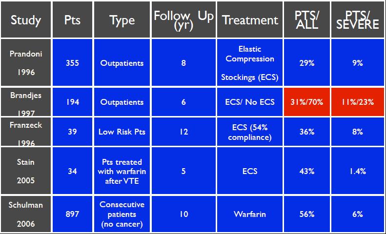
9 Study Population Mean follow-up PTS, % (n) PTS ulcer, % Overall Mild Moderate Severe (n) Reverse Galanaud (2012) Ten Cate-Hoek (2010) SOX trial Kahn (2014) 367 1st unprovoked proximal DVT 6 months 31.6 (116) 79.3 (92) 15.5 (18) 5.2 (6) 1.7 (2) 125 Proximal DVT 2 years 29.6 (37) 7.5 (3) ~ 0.8 (1) 806 First proximal DVT 2 years 51.3 (185) 67.6 (119) 17.0 (30) 15.3 (27) 4.4 (17) ECS arm 51.4 (178) 66.1 (111) 22.0 (37) 11.9 (20) 4.1 (16) ELATE Kahn (2005) 145 unprovoked proximal DVT 2.2 years 37 (55) 11 (4) ~ 1.4 (n = 2) CANANO 1st proximal DVT 3 years 35.6 (47) 6.4 (3) Prandoni (2004) First proximal DVT 2 years up to 5 years 25.7 (23) 49.1 (44) 13.0 (3) 22.7 (10) 2.2 (2) 6.7 (6) EINSTEIN trial Proximal DVT 5 years 29 (45) 89 (40) 11 (5) 2 (1) Cheung (2016) 40 (66) 91 (60) 9 (6) 6 (4)
10 Kahn S. et al. Determinants of health-related quality of life during the 2 years following deep vein thrombosis. Journal of Thrombosis and Haemostasis 2009, 6:

11 Anually costs PTS: $ No PTS: $
12
13 Venous Anatomy


14 Venous Versus Arterial Anatomy These are not arteries. Peter Neglen, MD Arteries Low volume High pressure Pulsatile flow Stiffer vessel walls Thick muscle layer No Valves Venous Valve Veins High volume Low pressure Phasic flow High compliance Thin muscle layer Valves Images courtesy P. Neglen MD

15 Venous Anatomy

16 Pelvic Landmarks Iliofemoral veins extend from confluence of iliac veins at IVC (L4-L5) to lesser trochanter L4 L5 Superior Iliac Spine Bony landmarks are useful for access, wire guidance and stent placement Pubic Tubercle Lesser Trochanter Inguinal Ligament
17 Venous Disease Pathophysiology Etiology

18
19 Iliofemoral Venous Disease Central Peripheral Collateral Obstruction Deep/superficial/perforator Segmental, axial Incompetent collaterals Reflux Venous hypertension Microvascular pathology Leg pain/swelling/lds/ulceration Stiff joints (ulcer patients) Vein wall stiffness Vein lumen geometry Calf venous volume
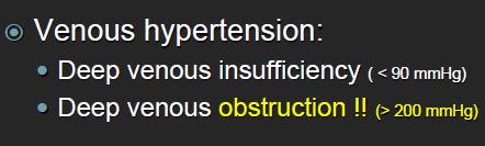
20 Post Thrombotic Syndrome Ambulatory Venous Pressures & Symptoms 28 mmhg Asymptomatic 36 mmhg Varicosities 41 mmhg Edema 47 mmhg Hyperpigmentation 60 mmhg Ulceration Greater pressure associated with worse PTS symptoms Linder et al: J Vasc Surg 1986
21 Ulcer Healing Diskussion
22 NIVL treatment better than reflux treatment Diskussion
23 Diskussion
24 Venous Disease Pathophysiology Etiology


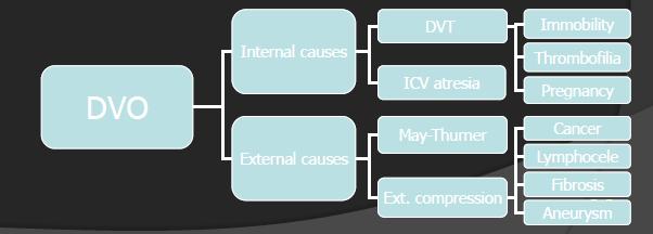
25 Three Primary Etiologies Etiology NIVL Non-thrombotic iliac vein lesions Acute DVT Postthrombotic iliac vein lesions Other Etiologies Benign or malignant tumors Retroperitoneal fibrosis Atresia of the IVC Miscellaneous iatrogenic injury, irradiation, cysts and aneurysms


26 Non-Thrombotic Iliac Vein Lesion (NIVL) Etiology NIVL is where veins are impinged, compressed, or damaged by a neighboring artery or structure NIVL s may precipitate iliofemoral DVT 24% of NIVL s thought to be clinically significant 1 NIVL s are highly under appreciated due to lack of accurate diagnosis by standard venography imaging. Venography was only 66% sensitive, with 34% of venograms appearing normal. IVUS had a diagnostic sensitivity of > 90% 2 Focal Anterior/Posterior Pinch Force 1. Marston W, Fish D, Incidence of and risk factors for iliocaval venous obstruction in patients with active or healed venous leg ulcers. J Vasc Surg 2011;54: Raju S, Neglén P. High prevalence of nonthrombotic iliac vein lesions in chronic venous disease: A permissive role in pathogenicity, J Vasc Surg 2006;44:136-44
27 Non-Thrombotic Iliac Vein Lesion (NIVL) Etiology NIVL s present as: Left-right ratio = 3:1 Female-male ratio = 4:1 Proximal (iliac artery crossing) and distal lesion (hypogastric artery crossing) Median age 54 years (range: 18-90) NIVL: underlies May-Thurner or Cockett s Syndrome A syndrome is a set of signs and symptoms that appear together and characterize a medical condition. Focal Anterior/Posterior Pinch Force NIVL clinical impact without previous DVT May be permissive of future development of chronic venous disease CVD May lead to venous valve reflux Raju S, Neglén P. High prevalence of nonthrombotic iliac vein lesions in chronic venous disease: A permissive role in pathogenicity, J Vasc Surg 2006;44:
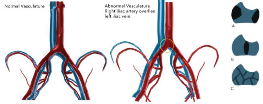
28 May Thurner Syndrom Ung BJ et al. May-Thurner Syndrome Complicated by Acute Iliofemoral Vein Thrombosis: Helical CT Venography for Evaluation of Long-Term Stent Patency and Changes in the Iliac Vein, AJR 2010; 195: )
29
30 Non-Thrombotic Iliac Vein Lesion (NIVL) Etiology The impact of non-thrombotic iliac vein lesion (NIVL) on acute DVT and postthrombotic obstruction Often underlying NIVLs found (left 84%, right 66%) Chung JW, Yoon CJ. Acute iliofemoral deep vein thrombosis: evaluation of underlying anatomic abnormalities by spiral CT venography. J Vasc Interv Radiol. 2004;15: Stenting of the stenosis after early clot removal improves patency from 27-44% to 86-93% Juhan CM, Alimi YS. Late results of iliofemoral venous thrombectomy. J Vasc Surg 1997;25: Mickley V, Schwagierek R. Left iliac venous thrombosis caused by venous spur: treatment with thrombectomy and stent implantation. J Vasc Surg 1998;28: Wohlgemuth,WA, Weber H. PTA and stenting of benign venous stenoses in the pelvis: long-term results. Cardiovasc Intervent Radiol. 2000; 23: Poor recanalization with external compression of the iliac vein (70-80% remains obstructed) Fraser D, Moody A. Iliac compression syndrome and recanalization of femoropopliteal and iliac venous thrombosis: A prospective study with magnetic resonance venography. J Vasc Surg. 2004;40:
31 Acute DVT Etiology Treatment Goal is to Reduce DVT Recurrence and Postthrombotic Syndrome Treatment window = two weeks Patients with iliofemoral DVT (IFDVT) have a twofold increased risk of developing PTS Venous stenting in conjunction with thrombus removal is safe and effective and has low incidence of PTS Images courtesy P. Neglen MD Foegh P, Jensen LP. Factors associated with long-term outcome in 191 patients with ilio-femoral DVT treated with catheter-directed thrombolysis. Eur J Vasc Endovasc Surg. 2017;53(3): Engelberger RP, Fahrni J, Willenberg T, et al. Fixed low-dose ultrasound-assisted catheter-directed thrombolysis followed by routine stenting of residual stenosis for acute iliofemoral deep-vein thrombosis. Thromb Haemost. 2014;111(6): ten Cate-Hoek AJ, Henke PK. The post thrombotic syndrome: Ignore it and it will come back to bite you. Blood Rev. 2016;30(2):131-7.


32 Recurrent DVT Rate Etiology Clinical course of DVT after the first episode of symptomatic deep venous thrombosis following traditional systemic anticoagulant therapy. Study Design: Prospective Study of 355 Patients with First Episode of DVT Follow-up Period Recurrent DVT Rate 2 years 17.5% 5 years 24.6% 8 years 30.3% Image courtesy P. Neglen MD Prandoni P, Lensing AW, Cogo A, et al. The long-term clinical course of acute deep venous thrombosis. Ann Intern Med. 1996;125(1):1-7.

33
34 American Venous Forum Promoting venous and lymphatic health Society for Clinical Vascular Surgery 2. Indications for early thrombus removal 2.1. We suggest a strategy of early thrombus removal in selected patients meeting the following criteria: (a) a first episode of acute iliofemoral deep venous thrombosis (b) symptoms <14 days in duration (c) a low risk of bleeding (d) ambulatory with good functional capacity and an acceptable life expectancy (Grade 2C)

 Follow up 5")
35 Venous Claudication in Iliofemoral Thrombosis Long-term Effects on Venous Hemodynamics, Clinical Status, and Quality of Life (Ann Surg 2004;239: ) 39 patients with prior iliofemoral DVT (22-86 years) Follow up 5 years * * * *
36 Patient Selection
37 Lichtenberg et al: Standards for Recanalization of Chronic Venous Outflow Obstructions. VASA accepted

38 Clinical assessment pdf
39 Patient Selection for Successful Venous Stenting Clinical severity of the disease Don t treat the lesion, treat the patient Findings on Investigations Treatment Considerations Can the patient be stented? Assess landing zones Sufficient inflow to the CFV? Need for endophlebectomy?


40 Clinical Severity of the Disease CEAP Classification Clinical Severity Visual Indications for Treatment Accessed July 23, 2017
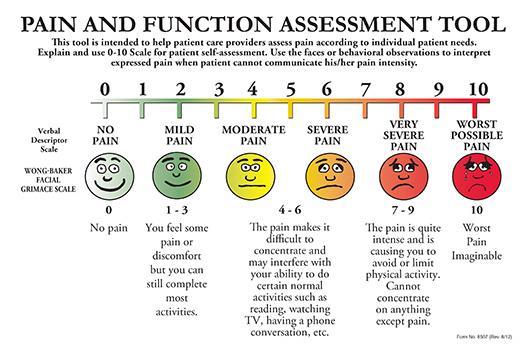
41 VAS Pain Scale Visual Analog Scale / Accessed August 2, 2017
42 Clinical Severity of the Disease Clinical Severity Specific clinical signs and symptoms Severe C 3, C 4-6 (CEAP) and/or pain >5 (VAS) Additional indications for treatment abdominal collaterals, atypical varicose veins, early varicose vein recurrence venous claudication postthrombotic disease pain out of proportion to lesion no detectable lesion explaining symptoms


43 Initial Investigation and Imaging Modalities Investigations Initial Patient Investigations Duplex Doppler scanning (incl. pelvic outflow) Transfemoral antegrade and descending venogram MRV, CT-V or IVUS Ascending venography DUPLEX IVUS CIV A CT Ascending Venography Venography Images courtesy P. Neglen MD
44 Positive Indicators of Obstruction on Tests Investigations Stenosis/occlusion on DUS, venogram, MR-V, CT-V or IVUS Presence of collaterals Positive pressure test Absence of respiratory variations in the groin...but the absence of collaterals, no pressure gradient and phasic variations in the groin does not exclude significant obstruction
45 Evaluating Findings to Determine if Clinically Significant Easy to measure a hemodynamically significant arterial obstruction, while impossible in the venous system Unknown at what degree an obstruction is hemodynamically significant No test to assess hemodynamically significant stenosis is available Morphological area/diameter stenosis >50% is considered significant Neglén P, Hollis KC, Olivier J, and Raju S. Stenting of the venous outflow in chronic venous disease: long-term stent-related outcome, clinical, and hemodynamic result. J Vasc Surg 2007;46: Hartung, Otero A, Boufi M et al. Mid-term result of endovascular treatment for symptomatic chronic nonmalignant iliocaval venous occlusive disease. J Vasc Surg 2005;42: Neglén P, Raju S. Proximal lower extremity chronic venous outflow obstruction: Recognition and treatment. Seminars in Vascular Surgery 2002; 15:57-64.
46 Can the patient be stented? Attempt to assess the central and peripheral extent of the disease before the intervention is scheduled using DUS, venogram, CTV, MRV etc. 1. Central landing zone single lumen a. Is the IVC patent? b. Does the disease involve the IVC? c. Is the potential outflow of the stent system appropriate? d. Is the contralateral venous outflow compromised? 2. Peripheral landing zone single lumen a. Is the CFV involved? b. Is there a potential landing zone in the CFV above the profundafemoral vein confluence? c. Is there a sufficient inflow from the periphery to sustain patency of a stent placed in the pelvic outflow?


47 Identifying the Landing Zones Outflow of the stent system is usually not an issue, but inflow to the CFV segment is. A one-lumen segment of the CFV vein is preferable with a reasonable inflow from the profunda and/or the femoral veins. Images courtesy of Prof. A. Comerota


48 Assessment of the Inflow Inflow is Vital for Patency FV Profunda vein inflow Images courtesy of Prof. A. Comerota
 NIVL s can be challenging")


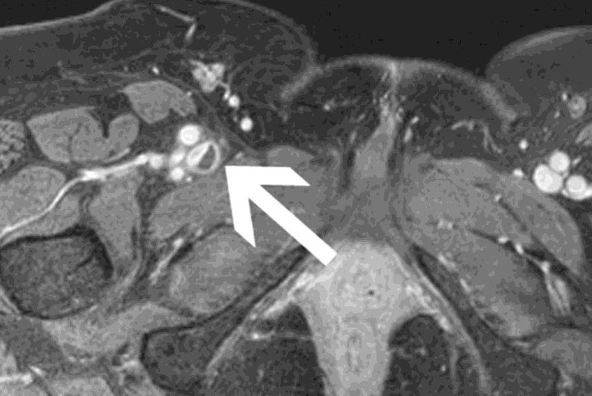
49 Non-Thrombotic Iliac Vein Lesion (NIVL) NIVL s can be challenging to visualize using venography alone. IVUS is often used to characterize the lesion. Venography IVUS Images courtesy P. Neglen MD

50 Thrombus age

51 Thrombus age
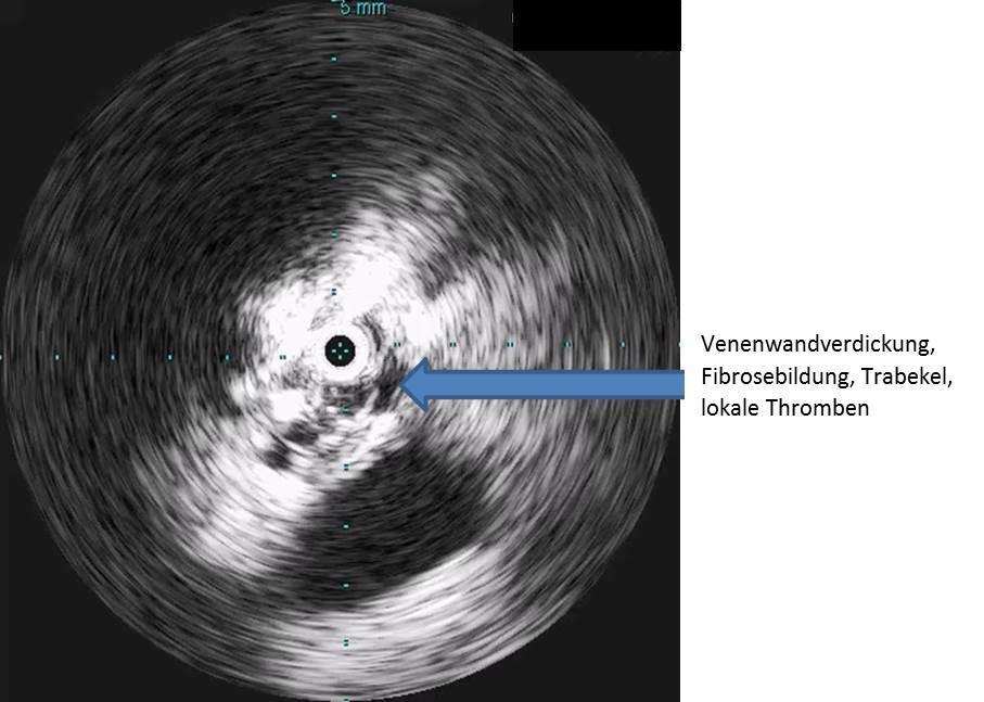
52 Lichtenberg et al: Standards for Recanalization of Chronic Venous Outflow Obstructions. VASA accepted Advantages of IVUS Dynamic measurement of area and the degree of stenosis Analysis of morphological changes in the vein (the formation of fibrosis, scars, thrombi) Dynamic evaluation of compression, such as in the presence of the May-Thurner syndrome No need for contrast medium in patients with kidney failure Exact determination of the diameter and length of the required vein stent Exact placement of the vein stent Stent analysis after implantation
53 Stents decrease flow resistance with a circular shape Shape defined by Aspect Ratio Aspect Ratio = Maximum Diameter to Minimum Diameter Perfect Circle Diameter = 14 Max Diameter = 14 Min Diameter = 7 Max Diameter = 14 Min Diameter = 3.5 Aspect Ratio Smaller Aspect Ratio = Better Lumen Quality
54 Summary Intervention should be considered after thorough patient diagnosis and investigation Combine conservative treatment (anticoagulation therapy) with invasive procedures Stenting should be considered if patient has a lesion >50%, is symptomatic, has good inflow and good landing zones, and a guidewire can cross the lesion
55 Endovascular intervention Optimal Interventional Setting Access Identifying Obstructive Lesion Stent Sizing Planning Stent Stack
56 Lichtenberg et al: Standards for Recanalization of Chronic Venous Outflow Obstructions. VASA accepted Optimal Interventional Setting Appropriate operating table with C-arm: Power injector Subtraction Image magnifier External ultrasound for cannulation guidance Consider general anesthesia in all, but especially, cases of with occlusion, bilateral disease, and IVC involvement Consider positioning of arms, IV lines, cables, etc. to limit interference with the C-arm With venography, multi-planar (45, 60, 90 ) views are, generally, required Availability of intravascular ultrasound (IVUS)
57 Access Options Ipsilateral versus contralateral access Femoral vein Facilitates recanalization of occlusions from below ( pushability ) Evaluation of the inflow to the stent Placement of the stent in relationship to distal tributaries Popliteal vein Cases of catheter-directed thrombectomy Access for inflow to femoral/common femoral vein Jugular vein Ensure a sterile back table is provided to support stent deployment Profunda vein When a large profunda vein is the main inflow to iliofemoral veins

 Mid-thigh access Entire CFV visualized Too high a")
 to be visualized Assessment of the flow into the")
58 Ipsilateral Mid-femoral Access The tip of the inserted sheath needs to be below the confluence of the profunda and femoral veins (anatomical landmark - trochanter minor) Mid-thigh access Entire CFV visualized Too high a stick This is to allow: The entire common femoral vein (CFV) to be visualized Assessment of the flow into the stent system

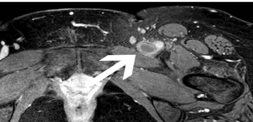
 At the mid-thigh, the")

59 Identifying the Femoral Vein Slide the U/S probe distally from the CFV and identify the profunda-femoral vein confluence FA FV Acute angle (Transverse image shown) At the mid-thigh, the femoral vein will stay separated from the artery Note: Acute, rather than obtuse, angle of the needle track Images courtesy P. Neglen MD

60 Patient in frog leg position on table Use ultrasound to identify femoral vein U/S gel facilitates imaging Transverse (shown above) or longitudinal approaches Initial venipuncture with 20- to 22-ga needle on a 10-mL syringe Introduce guide wire Replace needle with introducer sheath Return patient s legs to supine position


61 Watch out for foreshortening- This is a definite potential disadvantage of this site of access Brite Tip Sheath is key
62 Stent Placement Optimal Interventional Setting Access Identifying Obstructive Lesion Stent Sizing Planning Stent Stack
63 Lichtenberg et al: Standards for Recanalization of Chronic Venous Outflow Obstructions. VASA accepted Identifying the Obstructive Lesion Venography Ideally, power injector with subtraction Multi-planar views (45, 60, 90 ) to identify location of stenosis and extent Shows collaterals and inflow/outflow However: Underestimates stenosis by 30% Inaccurate location or extent on venogram in 41% Normal venogram findings in 17-25% Hingorani A, Alhabouni S, Ascher E, et al. Role of IVUS versus venograms in assessment of iliac-femoral vein stenosis. J Vasc Surg. 2010;52:804. Raju S, Neglen P. High prevalence of nonthrombotic iliac vein lesions in chronic venous disease: a permissive role in pathogenicity. J Vasc Surg. 2006;44: Neglen P, Raju S. Intravascular ultrasound scan evaluation of the obstructed vein. J Vasc Surg. 2002;35: Forauer AR, Gemmete JJ Intravascular ultrasound in the diagnosis and treatment of iliac vein compression (May-Thurner) syndrome. J Vasc Interv Radiol. 2002;13(5):523-7.
64 Reference Vein Diameter (RVD) Each Target Vein Segment (TVS) has an RVD: Will be measured in normal healthy vein Must accurately assess lesion and landing zones Appropriate stent diameter estimated (in order of preference): The venous segment immediately peripheral to the TVS. The venous segment immediately central to the TVS. The contralateral venous segment at the same level as the TVS. The literature-reported vein diameter of the CIV (16 mm), EIV (14 mm), and CFV (12 mm). Gasparis A. Labropuolous N. (2011, February) Venous Outflow Obstruction, Managing Intervention After Iliofemoral Thrombolysis. Endovascular Today..

65 Identifying Lesion and RVD - NIVL Use the tightest stenosis in any projection RVD for EIV is the peripheral EIV above the inguinal ligament RVD for CIV is peripheral CIV above the IIV/EIV confluence CIV stenosis EIV stenosis CIV RVD CIV RVD CIV RVD EIV RVD EIV RVD EIV RVD Images courtesy P. Neglen MD


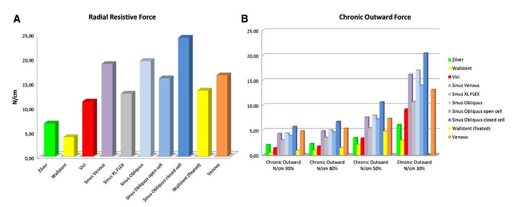
66 Identifying Lesion and RVD - PTS Occlusion Stenosis RVD contralateral vein RVD CFV The last option being the literature-reported vein diameter Images courtesy P. Neglen MD
67 Stent Placement Optimal Interventional Setting Access Identifying Obstructive Lesion Stent Sizing Planning Stent Stack
68 Graaf et al., Cardiovasc Intervent Radiol. 2015;38: (30).


69 C6 Stadium, 58 Jahre, Z.n. peripartaler Thrombose vor 30 Jahren
 4 x")

70 Sinus XL Stent (22 x 80 mm) 4 x Veniti Stent (16 x 120 mm + 14 x 60 mm)


71 Special Considerations: Confluence of Internal and External Iliac Veins AP view De novo stenosis: Stents landing at confluence of two veins, in different planes, with change in inflow rate De novo stenosis Straightening and tenting of vein Images courtesy P. Neglen MD


72 Going around the curve 120 mm stent Images courtesy P. Neglen MD


73 Special Considerations: Inguinal Ligament A concern? Lichtenberg et al: Standards for Recanalization of Chronic Venous Outflow Obstructions. VASA accepted Stenting across the inguinal ligament should be avoided. Guidelines present risk/benefits: Risks No data on venous stent fracture Increased risk of early in-stent stenosis Benefits Stenting down to a normal flow segment is more important than avoiding crossing the inguinal ligament. Stents should not overlap at the inguinal ligament. Mahnken A, Thompson K, de Haan M, O Sullivan G. CIRSE Practice Guidelines on Iliocaval Stenting. Cardiovasc Intervent Radiol 2014;37:
74 Successful Venous Stenting Understand the venous disease and the obstructive lesion Careful selection of patients Use optimal setting and techniques Adequate anticoagulation therapy and surveillance
75 Lichtenberg et al: Standards for Recanalization of Chronic Venous Outflow Obstructions. VASA accepted 6 months anticoagulation
 Boston Wallstent")
 Trial")


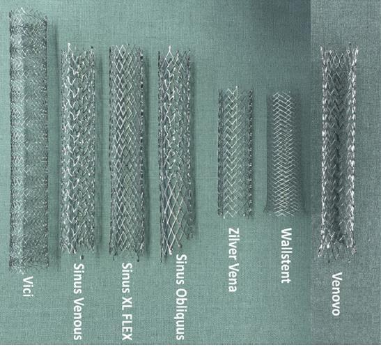
76 Venous Stent Options (CE) Boston Wallstent Optimed Cook Zilver Vena VIVO (EU) Trial presented Veniti Vici VIRTUS Trial ongoing Optimed Sinus obliquus Sinus Obliquus-01-NIS ongoing Bard Venovo VERNACULAR Trial ongoing Medtronic ABRE ABRE Clinical Study ongoing


77 Stent patency Stent Choice Force/ Flexibility Placement Technical Flow Inflow Outflow Clotting Anti-coagulation


78 Venous Stent Attributes Self-expandable Crush resistant across length of stent Sufficient chronic outward force Sufficient wall coverage Flexibility sufficient to resist kink at physiological angles Durability allowing repeated shortening, twisting, and bending at the groin Minimal foreshortening on deployment and balloon dilation Predictable, consistent deployment Strength Flexibility Lumen quality


79 there is not a perfect venous stent for the whole system..


80 Different venous stents for different locations High radial force Radial force plus flexibility flexibility, kink resistance, low fracture rate

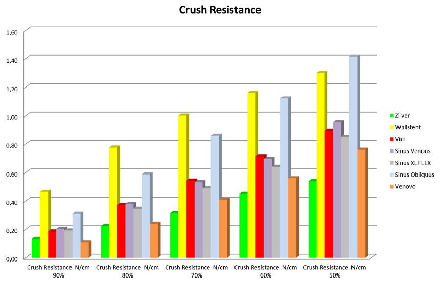
81 Stents decrease flow resistance with a circular shape Shape defined by Aspect Ratio Aspect Ratio = Maximum Diameter to Minimum Diameter Perfect Circle Diameter = 14 Max Diameter = 14 Min Diameter = 7 Max Diameter = 14 Min Diameter = 3.5 Aspect Ratio Smaller Aspect Ratio = Better Lumen Quality

 Cho H, Kim JK. Stent Compression in IVCS Associated with Acute Ilio-Femoral DVT. Korean J Radiol.")
82 48 patients with iliac compression and acute DVT followed for average of 20 months Follow-up was performed with CT venography Stent compression considered significant if lumen compression was greater that 50% (Aspect Ratio 1:2, or 2) Significant stent compression was inversely correlated with stent patency (p < 0.001) Cho H, Kim JK. Stent Compression in IVCS Associated with Acute Ilio-Femoral DVT. Korean J Radiol. 2015;16(4):
83
84

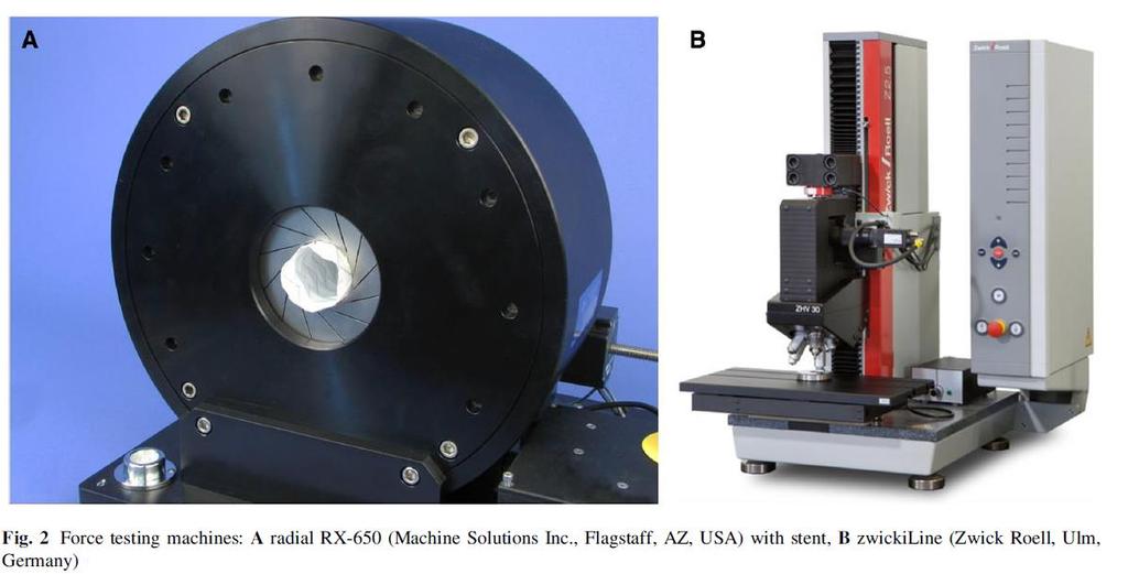
85 Radial resistive force: Force during loading Chronic outward force: Force during unloading Tests start here fully deployed, then loaded to an outer diameter to 4 mm (2)
86
87
88


89 Venous Stenting is Safe and Efficacious 37 Studies, 2,869 Patients, technical success ranged from 94%-96% AT = Acute Thrombotic, CPT=Chronic Post Thrombotic NT=Nonthrombotic 4
90 Primary patency 64 % Hybrid intervention Primary patency 90 % Primary patency 37 %
91

; Non Thrombotic (25%) Core Labs Venography: Syntactx IVUS: St.")
92 VIRTUS Feasibility Trial Design Objective Safety Assess safety & effectiveness in achieving patency of target venous lesion through 12-M post stent placement 30 days Post-thrombotic Effectiveness Primary 12-M Principal Investigators Study Design Patient Population Dr. William Marston Dr. Mahmood Razavi Prospective, multicenter, single arm nonrandomized, up to 45 sites worldwide 200 subjects with clinically significant chronic nonmalignant obstruction of the iliofemoral venous segment first 30 were feasibility. Image Courtesy of Mr. Stephen Black Non-thrombotic Etiologies: Post Thrombotic (75%); Non Thrombotic (25%) Core Labs Venography: Syntactx IVUS: St. Lukes DUS: VasCore/MGH X-Ray: Syntactx Image Courtesy of Mr. Mahmood Razavi

93 12-month Patency Data Secondary 100% Assisted-primary 96% Primary 93% Razavi M, et al. J Vasc Surg Venous Lymphat Disord Dec 28. pii: S X(17)
94 Patient Outcome Measures 63% of patients had 50% VCSS score reduction 81% of patients with pain reduction at 12 months 78% of patients considered QOL improved Baseline N=30 6 months N=26* P value 12 months N=27 P value VCSS 1 10 (2-25) 5 (0-30) < (0-23) <.001 VAS 2 60 (6-98) 23 (0-84) (0-94).001 CIVIQ (24-97) 28 (20-91) (20-89) <.001 * At 6 months, 27 patients had VCSS scores. The 1 patient with 6-month VCSS data (and no VAS or CIVIQ- 20 data) at 6 months only had completed form responses for 3 of 10 VCSS domains (all 0 s). 1. VCSS venous clinical severity score 2. VAS visual analogue scale 3. CIVIQ-20 chronic venous insufficiency quality of life questionnaire Razavi M, et al. J Vasc Surg Venous Lymphat Disord Dec 28. pii: S X(17)
95 Arnsberg Venous Registry > 300 patients included since 2013 Objective Assess safety & effectiveness in achieving patency of target venous lesion through 36 months post venous stent placement in patients with non thrombotic iliac vein lesions and post thrombotic iliac vein lesions. Effectiveness Primary 12-M // Clincal 12 -M Principle Investigators Dr. Michael Lichtenberg Dr. Rick de Graaf Study Design Ongoing prospective, single arm, single center non-randomized registry FU 1 (4 weeks), FU 2 (6 months), FU 3 (12 months), FU 4 (24 months), FU 5 (36 months) Patient Population Subjects with clinically significant chronic non-malignant obstruction of the iliofemoral venous segment Study is sponsored by German Venous Center Arnsberg
96 Arnsberg Venous Registry Venovo Venous Stent Objective Assess safety & effectiveness in achieving patency of target venous lesion through 36 months post venous stent placement in patients with non thrombotic iliac vein lesions and post thrombotic iliac vein lesions. Effectiveness Primary 12-M // Clincal 12 -M Principle Investigators Dr. Michael Lichtenberg Dr. Rick de Graaf Study Design Ongoing prospective, single arm, single center non-randomized registry FU 1 (4 weeks), FU 2 (6 months), FU 3 (12 months), FU 4 (24 months), FU 5 (36 months) Patient Population Subjects with clinically significant chronic non-malignant obstruction of the iliofemoral venous segment Study is sponsored by German Venous Center Arnsberg
97 Demographics/Medical History Demographics/ Comorbidity No. (%) Age 57 (19-89) Male 35 (44%) Female 45 (56%) Post-thrombotic 50 (63%) Non-thrombotic 30 (37%) Prev. PE 8 (10%) Prev. DVT 43 (48%) High Blood Pressure 40 (50%) Renal Disease 6 (8%) Stroke 3 (3%) Cancer 9 (11%) Diabetes 11 (14%) Smoker 13 (16%) CEAP Score, prior stent No. (%) 1 0 (0%) 2 1 (1%) 3 41 (51%) 4 28 (36%) 5 8 (10%) 6 2 (2%) Signs/Symptoms prior stent No. (%) Pain (incl. venous claudication) 78 (98%) Varicose Veins 63 (79%) Edema 62 (78%) Pigment Changes 41 (51%) Ulcers 10 (8%) Use Compression Stockings 68 (85%) 48% > CEAP C4
 @ 12 -M Etiology Primary")
98 Patency analysis NIVL vs. PTS --- NIVL --- PTS Patency Results by duplex 12 -M Etiology Primary Patency % Secondary Patency % NIVL 96.6% 100% PTS 95.7% 95%
99 9.6±5.41 Mean rvcss score (±SD) 5.6± ± ±2.4 N=79 N=77 N=59 N=52 51% had substantial clinical improvement (rvcss 2 Venous claudication and persistent swelling improved 8/10 venous ulceration were 12 - M
100 Arnsberg Venous Registry VENITI VICI VENOUS STENT System Objective Assess safety & effectiveness in achieving patency of target venous lesion through 36 months post stent placement (VENITI VICI Stent) Effectiveness Primary 12-M // Clincal 12 -M Principle Investigators Dr. Michael Lichtenberg Dr. Rick de Graaf Study Design Ongoing prospective, single arm, single center non-randomized registry FU 1 (4 weeks), FU 2 (6 months), FU 3 (12 months), FU 4 (24 months), FU 5 (36 months) Patient Population Subjects with clinically significant chronic non-malignant obstruction of the iliofemoral venous segment
101 Demographic / Clinical data 90 patients Demographic/comorbidity No. (%) Age 57.4±16.4 Male 43 (48%) Female 47 (52%) Post-thrombotic Syndrome 49 (54%) Non-thrombotic 41 (46%) History of venous 81 (90%) thromboembolic disease Pulmonary embolism 22 (24%) Deep vein thrombosis 43 (48%) Coronary Artery Disease 6 (7%) Myocardial Infarction 1 (1%) Congestive Heart Failure 7 (8%) High Blood Pressure 48 (55%) Renal Disease 6 (7%) Stroke 3 (3%) Cancer 13 (14%) Diabetes 13 (14%) Smoker (current or previous) a 15 (17%) CEAP score, prior to stenting 1 0 (0%) 2 1 (1%) 3 56 (62%) 4 20 (22%) 5 8 (9%) 6 4 (4%) Signs and symptoms, prior to stenting b Pain (inc. venous claudication) 89 (99%) Varicose veins 83 (92%) Edema 89 (99%) Pigment Changes 41 (46%) Ulcers 10 (11%) Use of compression stockings 88 (98%)

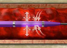
102 Patency rates non-thrombotic vs. post-thrombotic 12 months 12 months
103 Clinical efficacy: rvcss analysis Baseline N=90 1 month N=56 P value 6 months N=29 P value 12 months N=13 P value All Patients 8 (4, 27) 4 (1, 15) < (0, 12) < (0, 15).008
104 5 4, Mean CEAP score (±SD) , ,5 2 1,5 1 0,5 0 N=90 N=82 N=50 N=21 Baseline FU1 FU2 FU3


105 Physical Characteristics Bending Test 12 x 60 mm Stents tested 10mm Vessel diameter Position Stent end 45mm away from peak Centerline Radius blueflow Venous stent Minimum open diameter Boston Scientific Wallstent Minimum open diameter 10mm 6,0mm 5,5mm Blueflow Venouse Stent Position Stent end 45mm away from Peak Centerline Radius 10mm Boston Scientific Wallstent Position Stent end 45mm away from Peak Centerline Radius 10mm
106 Take home message Use dedicated venous stents! Choose wisely - based on lesion morphology Choose wisely based on stent technology
107 Venous Thrombectomy Benefits of an Endovascular Approach for Rapid Flow Restoration in DVT
108 DVT / VT - what do we need to know? Who ist the patient? (KI lysis, KI post.int.med., Preg., Tumor, MTS, coag.dis., Age, etc.) Who is the enemy? (acute, chronic, acute on chronic) What are our arms? (recanalisation devices, IVC-Filter, IVUS, Stent) What are our opportunities? (time, ICU/IMC, capacity for reintervention/control, post interv. surveillance) Reimbursement

109 Venous Thrombus Treatment Options: Traditional Therapy


110 Traditional Therapy Initial therapy of LMW heparin or unfractionated heparin Long term oral anticoagulants (3-6 months) Compression stockings to reduce swelling Comerota et al; Iliofemoral Venous Thrombosis ; J Vasc Surg 2007; 46:
111 Traditional Therapy Prevents clot propagation Reduces risk of pulmonary embolism May provide moderate symptomatic relief Advantages Easily administered without specialized skills Low cost of medications / appliances Accepted as standard of care Anticoagulation does NOT: Resolve clot Reduce risk of venous valvular damage Prevent venous hypertension Prevent or reduce severity of Post Thrombotic Syndrome (PTS) Rapidly resolve symptoms
 Design: Multicenter, randomized, placebo-controlled trial of active (N=410) vs placebo (N=396) ECS Key Inclusion Criteria: First indicative, proximal DVT (with or without coexisting")
112 SOX Trial Elastic Compression Stockings vs Placebo Control Objective: To evaluate the effectiveness of elastic compression stockings (ECS), compared with placebo stockings to prevent post-thrombotic syndrome (PTS) Design: Multicenter, randomized, placebo-controlled trial of active (N=410) vs placebo (N=396) ECS Key Inclusion Criteria: First indicative, proximal DVT (with or without coexisting pulmonary embolism or distal DVT) Primary Endpoint: PTS diagnosed at 6 months or later using Ginsberg s criteria (ie, leg pain and swelling of 1 month) Kahn SR, Shapiro S, Wells PS, et al SOX trial investigators. Lancet Mar 8;383(9920):880-8
113 SOX Trial Results Elastic Compression Stockings vs Placebo Control ECS did not prevent PTS after a first proximal DVT, hence our findings do not support routine wearing of ECS after DVT PTS incidence rate at 2 years: 14.2% for active ECS vs. 12.7% for placebo (p=0.58) a a HR adjusted for center 1.13, 95% CI ; p=0.58 PTS, post-thrombotic syndrome Kahn SR, Shapiro S, Wells PS, et al SOX trial investigators. Lancet Mar 8;383(9920):880-8
 Chronic 6 months (organized thrombus, fibrin-rich stable and difficult to remove) Mixed")
 Pharmacomechanical")
114 Considerations for Endovascular Intervention Thrombus age/extent Acute 14 days (fresh thrombus, easier to remove) Sub-acute 6 months (fibrinbound, more stable clot increases difficulty) Chronic 6 months (organized thrombus, fibrin-rich stable and difficult to remove) Mixed morphology Symptoms Pain, leg swelling, difficulty walking Life-style limiting Anatomy Common femoral or higher Iliofemoral DVT are typically most symptomatic Thrombus Acute Sub-acute Chronic PPS, Power Pulse TM Spray Mechanical Thrombectomy (eg, AngioJet) Pharmacomechanical Thrombectomy (eg, AngioJet + PPS)

115 Pharmacomechanical Thrombectomy (PMT) Combination of drug and mechanical thrombectomy to remove thrombus Allows medication to soften the clot, followed by mechanical action to remove the clot Advantages Minimally invasive Removes thrombus Can reduce procedure time/length of ICU stay May provide rapid symptomatic relief Potential for reduced lytic dosage Postthrombotic Syndrome; Patricia E. Thorpe, MD, FSIR; October 2007; Endovascular Today Limitations Specialized skills required Higher cost of disposables Effectiveness may be reduced in long-standing chronic thrombus
 Controlled, randomized trial of ultrasound-assisted CDT (N=24) vs conventional CDT (N=24) Thrombolysis regimen for both groups: 20 mg r-tpa over")
116 BERNUTIFUL Trial: Ultrasound-Assisted CDT vs Conventional CDT Objective: Assess whether the addition of intravascular highfrequency, low-power ultrasound energy facilitates the resolution of thrombosis during catheter-directed thrombolysis (CDT) Controlled, randomized trial of ultrasound-assisted CDT (N=24) vs conventional CDT (N=24) Thrombolysis regimen for both groups: 20 mg r-tpa over 15 hours Patients with acute (<2 weeks) iliofemoral DVT Primary efficacy endpoint was the percentage of thrombus load reduction from baseline to 15 hours Engelberger R, et al. Circ Cardiovasc Interv Jan;8(1). pii: e


117 pharmacomechanical thrombus fragmentation ultrasound accelerated thrombolysis: EKOS Fr catheter and 135 cm working length - 6, 12, 18, 24, 30, 40 and 50 cm treatment zones


118 pharmacomechanical thrombus fragmentation ultrasound accelerated thrombolysis: EKOS Fibrin Separation Non-cavitational ultrasound separates fibrin without fragmentation of emboli Active Drug Delivery Drug is actively driven into clot by Acoustic Streaming Fibrin without Ultrasound Fibrin With Ultrasound Acoustic streaming drives lytic into clot Braatan et al. Thrmob Haemost 1997;78: Francis et al. Ultrasound in Medicine and Biology, 1995;21(5): Soltani et al. Physics in Medicine and Biology, 2008; 53: EKOS Acoustic Pulse Thrombolysis is a minimally invasive system for dissolving thrombus.
119 BERNUTIFUL Trial Results: Ultrasound-Assisted CDT vs Conventional CDT The addition of intravascular ultrasound did not facilitate thrombus resolution CDT (N=24) CDT + Ultrasound (N=24) P Post-CDT 3 Months Thrombus Load Reduction 54%±27% 55%±27% 0.91 Bleeding Complication Rate 8.3% 12.5% >0.99 Primary Venous Patency 96% 100% 0.33 PTS Severity (Villalta score) 3.0± ± CDT regimen: 20 mg r-tpa over 15 hours Length-Adjusted Thrombus score was based on venographic filling defects in segments along the indwelling CDT catheter Thrombus load and complication rates were similar after CDT or ultrasound-assisted CDT 3-month outcomes did not differ significantly between groups Rates of adjunctive therapy use were similar between groups (angioplasty and stenting 83% vs 80%, P>.99; adjunctive thrombus removal 46% vs 29%, P=.37) Engelberger R, et al. Circ Cardiovasc Interv Jan;8(1). pii: e
120 Mechanical Thrombectomy Minimally invasive thrombectomy method Allows for rapid thrombus removal Supports less dose and duration of lytic agents Decreased bleeding Potential for vessel (endothelial) trauma Can be used for both arterial and venous clots
121 Therapeutic options in the treatment of DVT Conservative medical treatment (eg. LMWH, OAC, DOAC) OP / Fogarty Systemic thrombolysis Endovascular approaches local thrombolysis thrombus fragmentation and removal by Ballon-PTA, Basket, Aspiration thrombus fragmentation Tretorola (Teleflex) Cleaner 15 / XT (Argon) Mantis (Invamed) pharmacomechanical thrombolysis AngioJet (Boston Sc.) EkoSonic (BTG) mechanical thrombectomy devices Aspirex (Straub) Indigo (Penumbra) Angiovac (Argon) ClearLumen (Walkvascular)
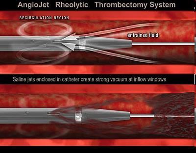
122 pharmacomechanical thrombus fragmentation AngioJet

")


123 Bench Simulations Power Pulse TM Delivery ZelanteDVT TM Simulated Clot Model of Thrombus Removal ~4x more thrombus removal with ZelanteDVT Solent TM Omni Clot tube model. Catheter advanced at 1mm/sec with Power Pulse (foot pedal) delivery of fluid Fluid delivered with ZelanteDVT disperses within the clot 1 pass ZelanteDVT TM 4 passes Solent TM Omni BSC fiber clot 100 in a 22 mm tube BSC data on file. Bench test results may not necessarily be indicative of clinical performance.


124 PEARL and PEARL II Clinical Registries PEripheral Use of AngioJet TM Rheolytic Thrombectomy With Mid Length Catheters Universitätsmedizin Rostock
125 PEARL Registry Objectives Determine efficacy of thrombus removal from baseline to final angiogram Evaluate clinical outcomes of treated patients at defined intervals of 3, 6, and 12 months Characterize clinical events Characterize treatment options used with the AngioJet System Estimate rate of AngioJet Thrombectomy-related adverse events Universitätsmedizin Rostock
 AngioJet PMT + CDT 172 (52%) 96% of patients had Grade II/III (50%-100%) clot reduction Clot reduction grade distribution not affected by symptom duration or")
126 PEARL Registry: Venous Cohort 329 patients 73% of cases completed in <24 hours Treatment AngioJet Thrombectomy alone (Rheolytic) AngioJet + Lytic by AngioJet (PMT) Frequency 13 (4%) 115 (35%) AngioJet Rheolytic + CDT 29 (9%) AngioJet PMT + CDT 172 (52%) 96% of patients had Grade II/III (50%-100%) clot reduction Clot reduction grade distribution not affected by symptom duration or treatment group Significant improvements over baseline in both physical & mental component scores of the SF-12 (P<.0001) 83% freedom from rethrombosis at 12 months 78% with continued clinical benefit (no recurrent thrombosis or worsened condition in the treated limb) at 12 months Garcia,MJ, et al. J Vasc Interv Radiol 2015; 26: Universitätsmedizin Rostock
127 PEARL Comparison Treatment of Lower Extremity DVT PEARL * Venous CaVenT Registry CDT Standard # of Patients # of Sites Prior DVT 40% 31% 10% 9% Primary Treatment AngioJet Thrombectomy With or Without PMT CDT CDT LMWH Stent Placement 35% 33% 17% NA Primary access Popliteal Popliteal Popliteal NA Male 57% 48% 64% 62% Age (mean) 52.2 yrs 47.5 yrs 53.3 yrs 50.0 yrs Treatment Location Limbs Involved Iliofemoral femoral pop Left=62%; Right=38% *Garcia,MJ, et al. J Vasc Interv Radiol 2015; 26: Mewissen MW, Seabrook GR. Radiology 1999:211:39-49 Enden, Haig Y. Lancet 2012:379:31-38 Patient Characteristics Iliofemoral femoral pop Left=61%; Right=39% Left=60%; Right=40% CFV or iliofemoral Left=62%; Right=38% CDT, catheter-directed thrombolysis; CFV, common femoral vein; LMWH, low molecular weight heparin; PMT, pharmacomechanical thrombolysis Results from different clinical investigations are not directly comparable. Information provided Universitätsmedizin for educational purposes Rostockonly
128 PEARL Comparison Treatment of Lower Extremity DVT Onset of DVT Symptom s PEARL * Venous CaVenT Registry CDT Standard Acute 67% ( 14 days) 66% ( 10 Days ) 100% 21 days Chronic 33% (>14 days) 16% (>10 Days ) NA Acute & Chronic NA 19% NA Primary Lytic TPA Urokinase TPA NA CDT Drip Times (mean) Procedur e Times Bleeding Complications CDT (N=29) CDT+PPS/RL (N=172) PPS/RL (N=115) 17 hrs 48 hrs 57.6 hrs (2.4 days) 40.9 hrs NA NA NA 22.0 hrs NA NA NA 2.0 hrs NA NA NA 4.5% (major & minor combined) *Garcia,MJ, et al. J Vasc Interv Radiol 2015; 26: Mewissen MW, Seabrook GR. Radiology 1999:211:39-49 Enden, Haig Y. Lancet 2012:379:31-38 Treatment Characteristics 11% (major); 16% (minor) 22% (major & minor combined) NA 0% CDT, catheter-directed thrombolysis; PMT, pharmacomechanical thrombolysis; PPS, power-pulse spray; RL, rheolytic; TPA, tissue plasminogen activator Results from different clinical investigations are not directly comparable. Information provided Universitätsmedizin for educational purposes Rostockonly
129 PEARL Comparison Treatment of Lower Extremity DVT Treatment Effectiveness Overall % Thrombus Removal By Lytic Groups: % thrombus removal CDT (N=28) CDT+PP S/RL (N=167) PPS/RL (N=113) Acute: % Thrombus Removal Chronic: % Thrombus Removal Primary Patency Freedom from Rethrombosis PEARL* Venous CaVenT Registry CDT Standard 96% 83% 89% NA 93% NA NA 97% NA NA 95% NA NA 97% 86% 89% 95% 68% NA NA 6 Mon= 87%; 12 Mon=83% *Garcia,MJ, et al. J Vasc Interv Radiol 2015; 26: Mewissen MW, Seabrook GR. Radiology 1999:211:39-49 Enden, Haig Y. Lancet 2012:379: Mon=65%; 12 Mon=60% 6 Mon = 65.9% 6 Mon = 47.4% NA NA NA CDT, catheter-directed thrombolysis; PPS, power-pulse spray; RL, rheolytic Results from different clinical investigations are not directly comparable. Information provided Universitätsmedizin for educational purposes Rostockonly


130 Mechanical thrombectomy devices AngioVac Indigo Aspirex Argon Penumbra Straub no GW no GW 0,018 GW Separator 0,025 GW OTW 22F 3,4F, 5F, 6F, 8F 6F, 8F, 10F 75, 120 cm 85,115, 135, 150 cm 85, 95, 110 cm Aspiration Aspiration Aspiration


131 mechanical thrombectomy: Indigo CAT5 CAT6 1.6 more aspiration* CAT more aspiration* Size 3,4F 5F 6F 8F MAC 42 ml/min 168 ml/min 270 ml/min 480 ml/min


132 mechanical thrombectomy: Angiovac


133 mechanical thrombectomy: Aspirex Size Length cm GW OD mm rvd mm Rotation rpm MAC ml/min Head 6 F 110 0,018 2, L-shape 135 0,018 2,0 8 F 85 0,018 2, L-shape 110 0,018 2,6 10 F 110 0,025 3, shape GW-Guidewire, OD-outer diameter, rvd-recommended Vessel Diameter, MAC-maximum aspiration capacity
134 PI AB ATTRACT Trial Proximal DVT Meets eligibility criteria & Provided informed consent Pre-Randomization Initiate anticoagulants and complete baseline assessments Control Arm 5 days heparin and immediately bridge to warfarin Randomization (1:1) Treatment Arm 5 days heparin concurrent with PCDT procedure then warfarin 692 patients at 56 U.S. centers Determining if the use of PCDT and /or CDT in acute DVT reduces occurrence of post thrombotic syndrome (PTS) over 24 months Anticoagulant therapy Elastic compression stockings Trellis -8 AngioJet System Catheter-Directed Thrombolysis Long-Term Treatment >3 months warfarin and daily use of compression stockings Follow-Up Early (10 days & 30 days) Late (6,12,18 & 24 months) ATTRACT data source: Dr. Suresh Vedantham, Washington University Hospital, St. Louis, MO 2016 Boston Scientific Corporation or its affiliates. All rights reserved. PI AA
, or Trellis -8 (Isolated Thrombolysis) Catheter-Directed Thrombolysis Additional rt-pa (total max 35 mg) Balloon maceration Aspiration/mechanical")
135 PCDT Treatment If good inflow to popliteal a Treatment Arm 5 days heparin concurrent with PCDT procedure then warfarin Trellis -8 AngioJet System Catheter-Directed Thrombolysis If poor inflow to popliteal AngioJet System (PowerPulse Thrombolysis), or Trellis -8 (Isolated Thrombolysis) Catheter-Directed Thrombolysis Additional rt-pa (total max 35 mg) Balloon maceration Aspiration/mechanical thrombectomy 90% thrombus removed and flow restored, or Maximum rt-pa dose or infusion time reached, or Overt bleeding or complication requiring cessation of therapy a Lower half of the popliteal vein and 1 major calf vein tributary are free of occlusive thrombus. rt-pa, recombinant tissue plasminogen activator; PCDT, pharmacomechanical catheter-directed thrombolysis Vedantham S, et al. Rationale and design of the ATTRACT Study: a multicenter randomized trial to evaluate pharmacomechanical catheter-directed thrombolysis for the prevention of postthrombotic syndrome in patients with proximal deep vein thrombosis. Am Heart J. 2013;165(4): e3
136 ATTRACT Outcomes Primary Outcome : cumulative occurrence of PTS between 6-24 months using the Villalta Scale Villalta Score> 5 or presence of an ulcer The question the study was designed to answer PTS Severity: Villalta, VCSS, CEAP Class QOL: SF-36, VEINES-QOL/Sym measures Symptoms: Likert pain scale, calf circumference Costs: Bleeds, VTE, deaths, US/economic
137 ATTRACT Cohort Characteristics 692 patients randomized: 337 PCDT, 355 No-PCDT 62% mean, median age 53 years, 25% previous VTE 57% had IFDVT, median 6 days from DVT diagnosis Baseline medical factors & use of anticoagulation, compression, anti-platelet therapy did not differ PCDT performance = consistent with past studies Median dose 21mg TPA; median 17 hours treatment Venography: mean thrombus removal 74% (p < 0.001) 94% of patients had 50% of their thrombus removed
138 Performance of PCDT INITIAL PCDT METHOD Trellis (Technique A) 50 Patients (15%) Angiojet (Technique B) 75 Patients (23%) ADJUNCTIVE PROCEDURE Balloon maceration (56%) Balloon angioplasty (56%) Angiojet (55%) Aspiration (19%) Trellis (14%) Stent placement (30%) Infuse-First (Technique C) 194 Patients (59%)
139 ATTRACT trial Outcome (24 mo) PCDT (n=336) no PCDT (n=335) P value Any PTS 46,7 % 48,2% 0.56 Recurrent VTE 12,5% 8,5% 0.09 Generic QOL (SF-36 PCS) 11,8 10, VENOUS QOL (VEINES) 27,7 23, Moderate or Severe PTS 17,9% 23,7% MS-PTS IFDVT 18,4% 28,2% MS-PTS FPDVT 17,1% 18,1% Major bleed 1,7% 0,3% Any bleed 4,5% 1,7% PTCD less effective in patients 65 years (p = 0.038) SIR, Washington
140 Study Outcomes Short-Term Effects of PCDT Outcome PCDT N=336 No-PCDT N=355 P Value Major Bleeding (10 days) 1.7% 0.3% Any Bleeding (10 days) 4.5% 1.7% Leg Pain (10 days) Leg Pain (30 days) Leg Swelling (10 days) Leg Swelling (30 days) No fatal or intracranial bleeds in either arm (10 day) PCDT Arm: ¾ transfusions & 2 embolization's
141 Study Outcomes Long-Term Effects of PCDT Outcome (24 Months) PCDT N=336 No-PCDT N=355 P Value Any PTS 46.7% 48.2% 0.56 Recurrent VTE 12.5% 8.5% 0.09 Generic QOL (SF-36 PCS) Venous QOL (VEINES) Moderate to Severe PTS 17.9% 23.7% MS PTS: IFDVT 18.4% 28.2% MS PTS: FPDVT 17.1% 18.1% PCDT less effective in patients 65 years old (P = 0.038)
142 Conclusion PCDT does not prevent PTS, does increase bleeding Most DVT patients can avoid unhelpful procedure Need better understanding of pathogenesis of PTS PCDT reduces early DVT symptoms and PTS severity Open vein hypothesis likely relevant to PTS progression Suggest targeting to IFDVT based on higher risk of PTS
143 CaVenT ATTRACT Arnsberg N Control group without treatment YES YES No Age, years Symptom duration, days <21 <14 < - 4 weeks Ascending femoropopliteal DVT 52 % 43% 25% Descending iliofemoral DVT 48% 57% 75% Mean tpa dose, mg 55 (variable) 21 (max 35 mg) 0 Major bleeding 9.0% 1.7% 0 Definition of criteria for stenting NO NO Yes ( stenosis > 50%) IVUS % Dedicated venous stents NO NO YES Stenting rate 17% 30% 100% Overall PTS mts 41% 47% 36% Patency rate 75% (2y) Not evaluated 92%

144 One word on ATTRACT Reality In 59 % of cases no dedicated venous stent was used Vedantham S, et al. Pharmacomechanical Catheter-Directed Thrombolysis for Deep-Vein Thrombosis. N Engl J Med Dec 7;377(23):
145 The ATTRACT failures Inclusion of femoropopliteal DVT (43%) No definition of criteria for stenting No IVUS No dedicated venous stents (59%) Outcome 24 months PCDT (n=336) No-PCDT (n=355) P Value Recurrent DVT 12.5% 8.5% Placebo around 10% recurrent DVT ASA 6% recurrent DVT NOACS 1% recurrent DVT


146 Venous Thrombus Treatment Options: Proactive Endovascular Treatment
147 Clinical follow-up study with the ASPIREX S Endovascular System to investigate safety and effectiveness in treatment of iliofemoral DVT patients - ARNSBERG ASPIREX REGISTRY - Responsible principal investigator (PI): M. Lichtenberg, R. de Graaf Study sponsored by Vascular Clinical Research Department, Arnsberg
![Patient Demographics Total N (%) 56 (100 %) Age Mean (Median [Range]) in years 52 (51 [17-89]) Female N (%) 37 (66 %) Male N (%) 19 (34 %) General Medical History N (%) Smoking status (valid](/docs-images/90/101637249/images/148-0.jpg "observations) 55 (100 %) Current 9 (16 %) Former 4 (7 %) Hypertension 56 (100%) Yes 28 (50 %) Immobilisation (valid observations) 55 (100 %) Yes 4 (7 %) Malignancy 56 (100 %) Current active 4 (7 %)")
148 Patient Demographics Total N (%) 56 (100 %) Age Mean (Median [Range]) in years 52 (51 [17-89]) Female N (%) 37 (66 %) Male N (%) 19 (34 %) General Medical History N (%) Smoking status (valid observations) 55 (100 %) Current 9 (16 %) Former 4 (7 %) Hypertension 56 (100%) Yes 28 (50 %) Immobilisation (valid observations) 55 (100 %) Yes 4 (7 %) Malignancy 56 (100 %) Current active 4 (7 %) Condition post 5 (5 %) Oral contraceptive 56 (100 %) Yes 21 (38 %) No 35 (62%) Study is sponsored by Klinikum Arnsberg
149 Diagnostic details (contd.) N (%) Type of occlusion 56 (100 %) Acute (< 14 days) 40 (71 %) Subacute (> 14 days) 13 (23 %) Chronic 2 (4 %) Acute / Chronic 1 (2 %) N (%) Location of occlusion (vessel) 56 (100 %) Left complete pelvic veins including com. femoral vein, left sup. femoral vein (may also include profunda femoral vein and 42 (75 %) distal part of IVC) Left common iliac vein only 7 (13 %) Left common iliac vein / Left external iliac vein without com. femoral vein 3 (5 %) Right complete pelvic veins 4 (7 %) Length of occlusion [mm] N=56 (100 %) Statistics Mean (SD) (72.0) Median (Range) (60 410) Study is sponsored by Klinikum Arnsberg
150 Aspirex treatment (contd.) N (%) Heparin [IU] 56 (100) 5, (89 %) 10,000 3 (5 %) 7,000 OR 7,500 OR 9,000 (1 patient each) 3 (5 %) Thrombolysis 56 (100 %) No 52 (93 %) Yes (bolus) 4 (7%) Technical success Yes 56 (100 %) Stent rate 100 % Mean (SD) 1.9 (1.2) Median (Range) 2 (0 6) Treatment duration [min] Mean (SD) 94.2 (44.8) Median (Range) 81.5 ( ) Study is sponsored by Klinikum Arnsberg
151 Patency analysis: DUS with restenosis < 50% N (%) Patency on FU month 1 53/56 (95%) Patency on FU month 6 (valid observations) 51/56 (91%) Patency on FU month 12 (valid observations) 47/56 (84%) Risk for re-thrombosis: - Symptoms > 10 days - CFV and PV involved - > 1 DVT in past Study is sponsored by Klinikum Arnsberg
152 Outcome: Post thrombotic syndrome after 12 months N (%) CEAP Score < 3, rvcss Score < 3) 43 (77 %) CEAP Score > 3, rvcss Score > 3) 13 (23 %) Study is sponsored by Klinikum Arnsberg
Clinical results of venous stents. Michael K. W. Lichtenberg MD, FESC
 Clinical results of venous stents Michael K. W. Lichtenberg MD, FESC Conflict of Interest - Disclosure Within the past 12 months, I or my spouse/partner have had a financial interest/arrangement or affiliation
Venous stent experience in Arnsberg Michael K. W. Lichtenberg MD, FESC
 Venous stent experience in Arnsberg Michael K. W. Lichtenberg MD, FESC IMPORTANT INFORMATION: These materials are intended to describe common clinical considerations and procedural steps for the on-label
- Our patients with iliofemoral DVT - Effective thrombus removal with purely mechanical thrombectomy can lead to better outcomes
 - Our patients with iliofemoral DVT - Effective thrombus removal with purely mechanical thrombectomy can lead to better outcomes Michael K. W. Lichtenberg, FESC Conflict of Interest - Disclosure Within
Patency rates and clinical results of the Veniti VICI Stent for treatment of iliac vein lesion Data from the Arnsberg Venous Registry
 Patency rates and clinical results of the Veniti VICI Stent for treatment of iliac vein lesion Data from the Arnsberg Venous Registry Michael K. W. Lichtenberg MD, FESC Conflict of Interest - Disclosure
REKANALISATION CHRONISCH VENÖSER VERSCHLÜSSE. Michael K. W. Lichtenberg, FESC
 REKANALISATION CHRONISCH VENÖSER VERSCHLÜSSE Michael K. W. Lichtenberg, FESC Conflict of Interest - Disclosure Within the past 12 months, I or my spouse/partner have had a financial interest/arrangement
VIRTUS: Trial Design and Primary Endpoint Results
 VIRTUS: Trial Design and Primary Endpoint Results Mahmood K. Razavi, MD St. Joseph Cardiac and Vascular Center Orange, CA, USA IMPORTANT INFORMATION: These materials are intended to describe common clinical
Should We Be More Aggressive in the Treatment of Acute DVT?
 DISCLOSURES Consultant Penumbra, Inc. UCSF Vascular Surgery Symposium April 6, 2017 K. Pallav Kolli, MD Assistant Professor of Clinical Radiology University of California, San Francisco 17 yo male, DVT
Techniques for thrombus removal in acute DVT Benefits of an Endovascular Approach for Rapid Flow Restoration in DVT
 Techniques for thrombus removal in acute DVT Benefits of an Endovascular Approach for Rapid Flow Restoration in DVT Michael K. W. Lichtenberg, MD, FESC Vascular Centre Arnsberg, Germany Disclosure Speaker
Aggressive endovascular management of ilio-femoral DVT. thrombotic syndrome. is the key in preventing post
 CACVS 2017 Aggressive endovascular management of ilio-femoral DVT is the key in preventing post thrombotic syndrome ALI AMIN MD, FACS,FACC, RVT CHIEF OF ENDOVASCULAR INTERVENTIONS READING HEALTH SYSTEM
Improved clinical outcomes Evidence on venous mechanical thrombectomy followed by stenting
 Improved clinical outcomes Evidence on venous mechanical thrombectomy followed by stenting Michael K. W. Lichtenberg, MD, FESC Vascular Centre Arnsberg, Germany German Venous Centre Arnsberg, Germany Disclosure
Improved clinical outcomes Evidence on venous thrombectomy followed by stenting
 Improved clinical outcomes Evidence on venous thrombectomy followed by stenting Michael K. W. Lichtenberg, MD, FESC Vascular Centre Arnsberg, Germany Venous Centre Arnsberg, Germany Disclosure Speaker
Iliofemoral DVT: Miminizing Post-Thrombotic Syndrome
 Iliofemoral DVT: Miminizing Post-Thrombotic Syndrome Catherine K. Chang, MD FACS Vascular Surgery San Diego Southern California Permanente Medical Group Acute Deep Venous Thrombosis Incidence & Outcomes
Pharmaco-mechanical techniques stand alone procedures? Peter Neglén, MD, PhD SP Vascular Center Limassol Cyprus
 Pharmaco-mechanical techniques stand alone procedures? Peter Neglén, MD, PhD SP Vascular Center Limassol Cyprus Faculty Disclosure Peter Neglén, M.D., Ph.D Stockholder/Founder of Veniti, Inc. Member, Medical
The evidence for venous interventions is evolving- many patients do actually benefit. Nils Kucher University Hospital Bern Switzerland
 The evidence for venous interventions is evolving- many patients do actually benefit Nils Kucher University Hospital Bern Switzerland Disclosure Speaker name: Nils Kucher X X I have the following potential
Venous interventions in DVT
 Venous interventions in DVT Sriram Narayanan Chief of Vascular and Endovascular Surgery, Tan Tock Seng Hospital A/Prof of Surgery, National University of Singapore ANTI-COAGULATION LMWH Warfarin x 6m Acute
Percutaneous Mechanical Thrombectomy for Acute Iliofemoral DVT with the Aspirex Catheter: The Dijon Experience
 JFICV 2018, Beaune Percutaneous Mechanical Thrombectomy for Acute Iliofemoral DVT with the Aspirex Catheter: The Dijon Experience Prof. Romaric LOFFROY, MD, PhD, FCIRSE Chief, Department of Vascular and
THERE IS NO ROLE FOR SURGICAL THERAPY FOR DVT
 THERE IS NO ROLE FOR SURGICAL THERAPY FOR DVT Tara D. Balint, MD FACS Sentara RMH Thursday, June 14, 2018 1 Objectives of treatment for DVT Prevent death from PE Prevent recurrent VTE Prevent post-thrombotic
Aspirex for Upper and Lower Extremity DVT
 Aspirex for Upper and Lower Extremity DVT Steven Kum MD Vascular & Endovascular Surgeon Director of Vascular Service Changi General Hospital Singapore Disclosure Speaker name:... I have the following potential
Treatment of Chronic DVT with EKOS: Reproducing ACCESS PTS Data in Every Day Clinical Practice
 Treatment of Chronic DVT with EKOS: Reproducing ACCESS PTS Data in Every Day Clinical Practice Mert Dumantepe, MD Acibadem Altunizade Hospital, Istanbul, Turkey Department of Cardiovascular Surgery Disclosure
Interventional Treatment VTE: Radiologic Approach
 Interventional Treatment VTE: Radiologic Approach Hae Giu Lee, MD Professor, Dept of Radiology Seoul St. Mary s Hospital The Catholic University of Korea Introduction Incidence High incidence: 250,000-1,000,000/year
The Ideal Venous Stent and Early Results from Venous Stent Trials PNEC, Seattle
 The Ideal Venous Stent and Early Results from Venous Stent Trials 2017 PNEC, Seattle Bill Marston MD Professor, Div of Vascular Surgery University of N. Carolina DISCLOSURES William Marston, MD Consultant/Advisory
Not all Leg DVT s are the Same: Which Patients Benefit from Interventional Therapy? Case 1:
 12/16/2015 Not all Leg DVT s are the Same: Which Patients Benefit from Interventional Therapy? Constantino S.Peña, FSIR, FSCCT, FAHA Interventional Radiologist Medical Director, Vascular Imaging Miami
Technique de recanalisation: mon expérience avec Aspirex
 JFICV 2017, Deauville Thrombose veineuse profonde aiguë en 2017 Technique de recanalisation: mon expérience avec Aspirex Romaric LOFFROY Département de Radiologie Diagnostique et Thérapeutique CHU Hôpital
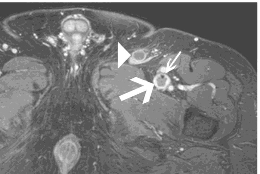
Case Study of Implantation of a VICI VENOUS STENT - Combined NIVL and PTS Stenting
 Case Study of Implantation of a VICI VENOUS STENT - Combined NIVL and PTS Stenting Courtesy of Mr. Stephen Black United kingdom Patient History 25 y/o female 2011: Conservatively treated ilio-femoral DVT
Chronic Iliocaval Venous Occlusive Disease
 none Chronic Iliocaval Venous Occlusive Disease David Rigberg, M.D. Clinical Professor of Surgery Division of Vascular Surgery University of California Los Angeles Chronic Venous Occlusive Disease Chronic
On Which Criteria Do You Select Your Stent for Ilio-femoral Venous Obstruction? North American Point of View
 On Which Criteria Do You Select Your Stent for Ilio-femoral Venous Obstruction? North American Point of View Peter Gloviczki, MD Ying Huang, MD, PhD Division of Vascular and Endovascular Surgery, Mayo
Future Devices of Venous Interventions
 Future Devices of Venous Interventions Director of Peripheral Vascular Medicine Department of Shin Kong Wu Ho-Su Memorial Hospital, Taiwan Interventional Cardiologist Tien-Yu Wu MD Disclosure Speaker name:...
What Really Matters to Patient is QOL: Veniti Virtus Venous Feasibility Trial
 DISCLOSURES Speaker name: Lowell S. Kabnick, MD, FACS... I have the following potential conflicts of interest: Consultant and shareholder, VENITI, Inc. Consultant to BARD What Really Matters to Patient
Chronic deep venous occlusions: Case planning, recanalization and stent technique
 Chronic deep venous occlusions: Case planning, recanalization and stent technique Michael K. W. Lichtenberg, FESC German Venous Center Arnsberg, Germany Conflict of Interest - Disclosure Within the past
Venous stenting in Marseille
 Venous stenting in Marseille Olivier HARTUNG, MD, MSc CHU Nord, Marseille, FRANCE Disclosure Speaker name: Olivier HARTUNG I have the following potential conflicts of interest to report: x Consulting :
Complex Iliocaval Reconstruction PNEC. Seattle WA. Bill Marston MD Professor, Div of Vascular Surgery University of N.
 Complex Iliocaval Reconstruction 2017 PNEC. Seattle WA Bill Marston MD Professor, Div of Vascular Surgery University of N. Carolina DISCLOSURES William Marston, MD Consultant/Advisory Board: Veniti, Cardinal
Optimal Utilization of Thrombolytics
 April 8-9, 2011 New York LaGuardia Marriott COMPLETE MANAGEMENT OF VENOUS DISEASE Optimal Utilization of Thrombolytics Anthony J. Comerota, MD, FACS, FACC Director, Jobst Vascular Institute Adjunct Professor
A Dedicated Venous Self-expanding Oblique Hybrid Nitinol Stent (Sinus-Obliquus Stent)
") A Dedicated Venous Self-expanding Oblique Hybrid Nitinol Stent (Sinus-Obliquus Stent) Anna Stuck, Rolf P. Engelberger, Nils Kucher Division of Angiology Cantonal Hospital Fribourg & Bern University Hospital
A Dedicated Venous Self-expanding Oblique Hybrid Nitinol Stent (Sinus-Obliquus Stent) Anna Stuck, Rolf P. Engelberger, Nils Kucher Division of Angiology Cantonal Hospital Fribourg & Bern University Hospital
Innovative Endovascular Approach to Pulmonary Embolism by Ultrasound Enhanced Thrombolysis. Prof. Ralf R.Kolvenbach MD,PhD,FEBVS
 Innovative Endovascular Approach to Pulmonary Embolism by Ultrasound Enhanced Thrombolysis Prof. Ralf R.Kolvenbach MD,PhD,FEBVS Catheter-based thrombolysis Local administration of lytic agent Higher local
Venous Stents Placed Below the Inguinal Ligament: No Worries
 Venous Stents Placed Below the Inguinal Ligament: No Worries Disclosure Speaker name: Lowell S. Kabnick, MD, FACS, FACPh, RPhS... I have the following potential conflicts of interest to report: Consulting:
Successful recanalisation of venous thrombotic occlusions with Aspirex mechanical thrombectomy. Michael K. W. Lichtenberg
 Successful recanalisation of venous thrombotic occlusions with Aspirex mechanical thrombectomy Michael K. W. Lichtenberg Disclosure Speaker name: Michael Lichtenberg... I have the following potential conflicts
The Evidence Base for Treating Acute DVT
 The Evidence Base for Treating Acute DVT Mr Chung Sim Lim Consultant Vascular Surgeon and Honorary Lecturer Royal Free London NHS Foundation Trust and University College London NIHR UCLH Biomedical Research
VIVO-EU Results: Prospective European Study of the Zilver Vena TM Venous Stent in the Treatment of Symptomatic Iliofemoral Venous Outflow Obstruction
 VIVO-EU Results: Prospective European Study of the Zilver Vena TM Venous Stent in the Treatment of Symptomatic Iliofemoral Venous Outflow Obstruction Gerard J O Sullivan, M.D. and Jennifer McCann-Brown,
Ileo Femoral DVT Review and Update
 Ileo Femoral DVT Review and Update Ammar Safar, MD, FSCAI, FACC, FACP, RPVI Interventional Cardiology & Endovascular Medicine Deep Vein Thrombosis Venous thromboembolism is a major national health problem,
Intervention for Deep Venous Thrombosis and Pulmonary Embolus
 Intervention for Deep Venous Thrombosis and Pulmonary Embolus Michael R. Jaff, DO Paul and Phyllis Fireman Endowed Chair in Vascular Medicine Massachusetts General Hospital Professor of Medicine Harvard
Copy Here. The Easy One.. What is the Role of Thrombus Removal in Acute Proximal DVT after ATTRACT? Deep Venous Thrombosis Spectrum
 What is the Role of Thrombus Removal in Acute Proximal DVT after ATTRACT? Mitchell J. Silver DO FACC FSVM RPVI Director, Center for Critical Limb Care Riverside Methodist Hospital Ohio Health Heart and
PEARL Registry Update Overview Venous Arterial AV Access
 PEARL Registry Update Overview Venous Arterial AV Access PEARL Registry Overview (as of 10 Sep12*) Overview Venous Arterial AV Access HOME Topic Data Support Comments Study Design Prospective, non-randomized,
VERNACULAR Trial & Clinical Experience with the VENOVO Venous Stent
 Stephen Black, MD VERNACULAR Trial & Clinical Experience with the VENOVO Venous Stent 1 Speaker Disclaimers The speakers presentation today is on behalf of Bard Peripheral Vascular, Inc. Any discussion
Acute Venous Thrombosis: Thrombus Removal with Adjunctive Catheter-Directed Thrombolysis (ATTRACT Trial)
") Acute Venous Thrombosis: Thrombus Removal with Adjunctive Catheter-Directed Thrombolysis (ATTRACT Trial) N Engl J Med. Volume 377(23):2240-2252. December 7, 2017 Wednesday, July 11, 2018, 1:00pm ET Guest
Acute Venous Thrombosis: Thrombus Removal with Adjunctive Catheter-Directed Thrombolysis (ATTRACT Trial) N Engl J Med. Volume 377(23):2240-2252. December 7, 2017 Wednesday, July 11, 2018, 1:00pm ET Guest
How to best approach chronic venous occlusions?
 How to best approach chronic venous occlusions? Prof. Nils Kucher Director Venous Thromboembolism Reseach Group University Hospital Bern nilskucher.com Disclosure Speaker name: Nils Kucher X X I have the
Understanding of the importance of venous
 The Critical Need for an Iliofemoral Venous Obstruction Classification System An overview of a potential classification system to better identify and treat iliofemoral venous outflow obstruction. BY WILLIAM
Ultrasound-assisted catheter-directed thrombolysis: Does it really work? The BERNUTIFUL trial
 Ultrasound-assisted catheter-directed thrombolysis: Does it really work? The BERNUTIFUL trial Rolf P. Engelberger Division of Angiology CHUV, Lausanne & Inselspital, Bern Switzerland Disclosure Speaker
Surgical approach for DVT. Division of Vascular Surgery Department of Surgery Seoul National University College of Medicine
 Surgical approach for DVT Seung-Kee Min Division of Vascular Surgery Department of Surgery Seoul National University College of Medicine Treatment Options for Venous Thrombosis Unfractionated heparin &
BC Vascular Day. Contents. November 3, Abdominal Aortic Aneurysm 2 3. Peripheral Arterial Disease 4 6. Deep Venous Thrombosis 7 8
 BC Vascular Day Contents Abdominal Aortic Aneurysm 2 3 November 3, 2018 Peripheral Arterial Disease 4 6 Deep Venous Thrombosis 7 8 Abdominal Aortic Aneurysm Conservative Management Risk factor modification
Acoustic Pulse Thrombolysis Treatment
 Acoustic Pulse Thrombolysis Treatment BTGVascular.com SETTING THE STANDARD FOR VASCULAR THERAPIES Quickly & safely dissolve thrombus with the EKOS System. The Acoustic Pulse Difference Acoustic Pulse Thrombolysis
Straub Endovascular System &
 Straub Endovascular System & S t r a u b E n d o v a s c u l a r To o l s Straub Endovascular System Effective debulking in occluded arteries and veins Effective debulking in many indications Rotarex
Canadian Society of Internal Medicine Annual Meeting 2017 Toronto, ON
 Canadian Society of Internal Medicine Annual Meeting 2017 Toronto, ON How to Prevent and Manage the Post-Thrombotic Syndrome? Jean-Philippe Galanaud Clinical Thromboembolism & Division of GIM Sunnybrook,
The hallmark of percutaneous thrombus management
 Treating Venous Thromboembolism Without Lytic Medications What the present and the near future will bring in terms of techniques and devices to remove venous thrombus. BY CONSTANTINO S. PEÑA, MD; RIPAL
The hallmark of percutaneous thrombus management
 Treating Venous Thromboembolism Without Lytic Medications What the present and the near future will bring in terms of techniques and devices to remove venous thrombus. BY CONSTANTINO S. PEÑA, MD; RIPAL
Michael Meuse, M.D. Vascular and Interventional Radiology
 Michael Meuse, M.D. Vascular and Interventional Radiology Iliac Vein Compression Syndrome Left CIV compressed by right CIA Virchow 1851: DVT L>R May and Thurner 1954: venous spurs Cockett and Thomas 1965:
The Conservative and Active Management of Post Thrombotic Syndrome
 The Conservative and Active Management of Post Thrombotic Syndrome Stephen Black Consultant Vascular Surgeon Clinical Lead for Venous and Lymphoedema Surgery Guys and St Thomas Hospital London How important
Imaging, it s central role in planning and guiding intervention. Prof. Luis Izquierdo. MD, PhD, FEBVS
 Imaging, it s central role in planning and guiding intervention Prof. Luis Izquierdo. MD, PhD, FEBVS IMPORTANT INFORMATION: These materials are intended to describe common clinical considerations and procedural
Duplex ultrasound is first-line imaging for all
 Our Protocol for Transabdominal Pelvic Vein Duplex Ultrasound A summary of s protocol for pelvic vein duplex ultrasonography, including equipment, patient positioning, ultrasound settings, and technique.
PEARL REGISTRY Post Market Registry
 PEARL REGISTRY Post Market Registry Real World Use of the AngioJet Thrombectomy System in the Peripheral Vascular System PI-315422-AA JUN2015 Objectives AngioJet Action Enrollment General Indications &
Starting with deep venous treatment
 Starting with deep venous treatment Carsten Arnoldussen, MD Interventional Radiologist Maastricht University Medical Centre, Maastricht VieCuri Medical Centre, Venlo The Netherlands Background Maastricht
VIRTUS Trial: Pivotal Cohort 12-Month Primary Safety and Efficacy Results of the VICI Venous Stent System
 VIRTUS Trial: Pivotal Cohort 12-Month Primary Safety and Efficacy Results of the VICI Venous Stent System Mahmood K. Razavi, MD St. Joseph Cardiac and Vascular Center Orange, CA, USA Disclosure Speaker
Venogram Versus Intravascular Ultrasound for Diagnosing and Treating Iliofemoral Vein Obstruction (VIDIO)
") Venogram Versus Intravascular Ultrasound for Diagnosing and Treating Iliofemoral Vein Obstruction (VIDIO) Report From a Multicenter, Prospective Study of Iliofemoral Vein Interventions Paul J. Gagne, MD,
Venogram Versus Intravascular Ultrasound for Diagnosing and Treating Iliofemoral Vein Obstruction (VIDIO) Report From a Multicenter, Prospective Study of Iliofemoral Vein Interventions Paul J. Gagne, MD,
Use of EKOS Catheter in the management of Venous Mr. Manoj Niverthi, Mr. Sarang Pujari, and Ms. Nupur Dandavate, The GTF Group
 Use of EKOS Catheter in the management of Venous Thromboembolism @ Mr. Manoj Niverthi, Mr. Sarang Pujari, and Ms. Nupur Dandavate, The GTF Group Introduction Georgia Thrombosis Forum (GTF, www.gtfonline.net)
Venous Thrombosis. Magnitude of the Problem. DVT 2 Million PE 600,000. Death 60,000. Estimated Cost of VTE Care $1.5 Billion/year.
 Venous Thrombosis Magnitude of the Problem DVT 2 Million Postthrombotic Syndrome 800,000 PE 600,000 Death 60,000 Silent PE 1 Million Pulmonary Hypertension 30,000 Estimated Cost of VTE Care $1.5 Billion/year
NATIONAL INSTITUTE FOR HEALTH AND CARE EXCELLENCE
 NATIONAL INSTITUTE FOR HEALTH AND CARE EXCELLENCE INTERVENTIONAL PROCEDURES PROGRAMME Interventional procedure overview of Interventional procedure overview of ultrasound-enhanced, catheterdirected thrombolysis
Anticoagulation therapy following endovascular treatment of iliofemoral deep vein thrombosis
 Anticoagulation therapy following endovascular treatment of iliofemoral deep vein thrombosis Tim Sebastian, M.D. University Hospital Zurich Clinic for Angiology Disclosure Speaker name: Tim Sebastian I
Implications from the ACCP 2012 Consensus Guidelines for the Management of Thrombosis: a case based approach
 Implications from the ACCP 2012 Consensus Guidelines for the Management of Thrombosis: a case based approach Prof. I. Baumgartner Head Clinical and Interventional Angiology About the ACCP guidelines Widely
Acute Versus Chronic DVT Imaging in the Vascular Lab Heather Gornik, MD, RVT, RPVI
 Acute Versus Chronic DVT Imaging in the Vascular Lab Heather Gornik, MD, RVT, RPVI Cleveland Clinic Heart and Vascular Institute Heather L. Gornik, MD has the following relationships to disclose: CVR Global
2017 Florida Vascular Society
 Current Management of Venous Leg Ulcers: How to Identify Patients with Correctable Venous Disease and Interventional Procedures to Heal and Prevent Recurrence 2017 Florida Vascular Society Bill Marston
Emerging Tools for Lytic-Free, Single-Session Treatment of Venous Thromboembolic Disease
 FEATURED TECHNOLOGY THE CLOTTRIEVER AND FLOWTRIEVER SYSTEMS Emerging Tools for Lytic-Free, Single-Session Treatment of Venous Thromboembolic Disease The ClotTriever Outcomes (CLOUT) registry principal
Deep Venous Pathology. Eberhard Rabe Department of Dermatology University of Bonn Germany
 Deep Venous Pathology Eberhard Rabe Department of Dermatology University of Bonn Germany Disclosures None for this presentation Consultant: Sigvaris, EUROCOM Speakers bureau: Bayer Vital, Aspen, Boehringer,
EKOS. Interventional Vascular 3 February, Imagine where we can go.
 EKOS Interventional Vascular 3 February, 2015 Imagine where we can go. Forward-looking statement This presentation and information communicated verbally to you may contain certain projections and other
Percutaneously Inserted AngioVac Suction Thrombectomy for the Treatment of Filter-Related. Iliocaval Thrombosis
 Percutaneously Inserted AngioVac Suction Thrombectomy for the Treatment of Filter-Related Iliocaval Thrombosis Faiz D. Francis, DO; Gianvito Salerno, MD; Sabbah D. Butty, MD Abstract In the setting of
Early clinical outcomes of a novel rheolytic directional thrombectomy technique for patients with iliofemoral deep vein thrombosis
 56 Original communication Early clinical outcomes of a novel rheolytic directional thrombectomy technique for patients with iliofemoral deep vein thrombosis Jörn F. Dopheide 1, Tim Sebastian 1, Rolf P.
Complex ilio-caval revascularization in chronic venous obstruction with the Venovo Stent. Michael K. W. Lichtenberg MD, FESC
 Complex ilio-caval revascularization in chronic venous obstruction with the Venovo Stent Michael K. W. Lichtenberg MD, FESC German Venous Center Arnsberg, Germany Not available for sale or distribution
4/30/2018 CLOT+ In patients with an acute proximal deep vein thrombosis, pharmacomechanical catheter-directed thrombolysis does not reduce t
 In patients with an acute proximal deep vein thrombosis, pharmacomechanical catheter-directed thrombolysis does not reduce the rate of post-thrombotic syndrome Question In patients who have symptomatic
Innovative Endovascular Approach to Pulmonary Embolism by Ultrasound Enhanced Thrombolysis. Prof. Ralf R.Kolvenbach MD,PhD,FEBVS
 Innovative Endovascular Approach to Pulmonary Embolism by Ultrasound Enhanced Thrombolysis Prof. Ralf R.Kolvenbach MD,PhD,FEBVS Conflict of Interest BTG Standard PE therapy ANTICOAGULATION (AC) HEPARIN
Image-Guided Approach to Treatment of Patients with Nonthrombotic
 Image-Guided Approach to Treatment of Patients with Nonthrombotic May Thurner Syndrome Brian DeRubertis, MD, FACS Associate Professor of Surgery Division of Vascular Surgery UCLA School of Medicine Los
Post-thrombotic syndrome (PTS), often the
, often the") Revascularization of Chronic Venous Occlusion in the Setting of Post-Thrombotic Syndrome Jon George, MD; Deepakraj Gajanana; Sean Janzer; Vincent Figueredo; Dennis Morris From the Einstein Heart and Vascular
Revascularization of Chronic Venous Occlusion in the Setting of Post-Thrombotic Syndrome Jon George, MD; Deepakraj Gajanana; Sean Janzer; Vincent Figueredo; Dennis Morris From the Einstein Heart and Vascular
Expanding Horizons: AngioVac Suction Thrombectomy at UTHealth
 Expanding Horizons: AngioVac Suction Thrombectomy at UTHealth Naveed Saqib, MD Assistant Professor Department of Cardiothoracic and Vascular Surgery McGovern Medical School The University of Texas Science
Complete Evaluation of the Chronic Venous Patient: Recognizing deep venous obstruction. Erin H. Murphy, MD Rane Center
 Complete Evaluation of the Chronic Venous Patient: Recognizing deep venous obstruction Erin H. Murphy, MD Rane Center Disclosure Speaker name: Erin H. Murphy... I have the following potential conflicts
NATIONAL INSTITUTE FOR HEALTH AND CARE EXCELLENCE
 NATIONAL INSTITUTE FOR HEALTH AND CARE EXCELLENCE INTERVENTIONAL PROCEDURES PROGRAMME Interventional procedure overview of Interventional procedure overview of ultrasound-enhanced, catheterdirected thrombolysis
Selection and work up for the right patients suspected of deep venous disease
 Selection and work up for the right patients suspected of deep venous disease R A G H U K O L L U R I, M S, M D, R V T S Y S T E M M E D I C A L D I R E C T O R V A S C U L A R M E D I C I N E / V A S
Patency and Clinical Outcomes of a Dedicated, Self-Expanding, Hybrid Oblique Stent Used in the Treatment of Common Iliac Vein Compression
 676803JETXXX10.1177/1526602816676803Journal of Endovascular TherapyStuck et al research-article2016 Clinical Investigation Patency and Clinical Outcomes of a Dedicated, Self-Expanding, Hybrid Oblique Stent
Mechanical treatment for acute DVT. Laurent Casbas, MD Toulouse, France
 Laurent Casbas, MD Toulouse, France Disclosure of Interest Speaker name: Laurent Casbas I have the following potential conflicts of interest to report: Consulting Medtronic General Electric Acute Venous
Methods of Thrombus Fragmentation & Extraction. Methods of Thrombus Extraction
 Thrombus Fragmentation and Extraction: Clinical Evidence and Practical Application No Relevant Disclosures Venita Chandra, MD Clinical Assistant Professor of Surgery Division of Stanford Medical School,
BY DAVID GILLESPIE, MD; MARCIA JOHANSSON, RN, MS; AND CAROLYN GLASS, MD
 Stent Placement fter DVT Thrombolysis/ Mechanical Thrombectomy Which patients should be stented and which should not? Y DVID GILLESPIE, MD; MRCI JOHNSSON, RN, MS; ND CROLYN GLSS, MD It is an exciting time
Evidence for endovascular therapy of iliofemoral DVT: CAVENT, ATTRACT, CAVA and more to come
 Evidence for endovascular therapy of iliofemoral DVT: CAVENT, ATTRACT, CAVA and more to come Stephen Black Consultant Vascular Surgeon Reader in Venous Surgery Or rephrased To lyse or not to lyse: that
DEEP VENOUS THROMBOSIS A PRACTICAL APPROACH TO IMPROVING CLINICAL OUTCOMES
 DEEP VENOUS THROMBOSIS A PRACTICAL APPROACH TO IMPROVING CLINICAL OUTCOMES Jose M. Borromeo M.D. Vascular Surgeon Iowa Heart Center Disclosures: AstraZeneca Pharmaceuticals Cook CVRx LeMaitre Vascular,
What is the optimal time window for treating deep venous thrombosis? Acute vs subacute vs chronic
 What is the optimal time window for treating deep venous thrombosis? Acute vs subacute vs chronic Peter A. Schneider, MD Chief of Vascular Therapy Kaiser Foundation Hospital Honolulu, Hawaii Disclosure
Management of Acute DVT Extending From the Tibial Veins to the Common Iliac Vein Using the AngioJet Thrombectomy System
 FEATURED TECHNOLOGY: ANGIOJET THROMBECTOMY SYSTEM Sponsored by Boston Scientific Corporation Management of Acute DVT Extending From the Tibial Veins to the Common Iliac Vein Using the AngioJet Thrombectomy
ENHANCING YOUR OPTIONS
 ANGIOJET ULTRA Thrombectomy System ENHANCING YOUR OPTIONS FOR RESTORING FLOW When you need the versatility and power to restore flow Refined from experience in over 700,000 cases worldwide, today s AngioJet
5 year quality of life data after EKOS treatment in acute DVT
 5 year quality of life data after EKOS treatment in acute DVT Jochen Grommes MD European Vascular Center Aachen-Maastricht GxUS-EKO-2016-0006 LINC 2016 Disclosure of speaker s interests conflict of interest
J Jpn Coll Angiol, 2009, 49:
 Online publication August 27, 2009 1 2 J Jpn Coll Angiol, 2009, 49: 247 254 deep vein thrombosis, thrombolytic therapy, catheter-directed thrombolysis, inferior vena cava filter, pulmonary thromboembolism
Introduction What Causes Peripheral Vascular Disease? How Do Doctors Treat Peripheral Vascular Disease?... 9
 Patient Information Table of Contents Introduction... 3 What is Peripheral Vascular Disease?... 5 What Are Some of the Symptoms of Peripheral Vascular Disease?... 7 What Causes Peripheral Vascular Disease?...
Prevention of VTE Sequelae: Post-thrombotic Syndrome and Chronic Thromboembolic Pulmonary Hypertension
 Prevention of VTE Sequelae: Post-thrombotic Syndrome and Chronic Thromboembolic Pulmonary Hypertension Susan R. Kahn MD MSc Professor of Medicine, McGill University Director, Jewish General Hospital Centre
Management of Post-Thrombotic Syndrome
 Management of Post-Thrombotic Syndrome Thanainit Chotanaphuti Phramongkutklao College of Medicine Bangkok, Thailand President of CAOS Asia President of Thai Hip & Knee Society President of ASEAN Arthroplasty
What is New in Acute Pulmonary Embolism? Interventional Treatment. Prof. Nils Kucher University Hospital Bern Switzerland
 What is New in Acute Pulmonary Embolism? Interventional Treatment Prof. Nils Kucher University Hospital Bern Switzerland nils.kucher@insel.ch Disclosure of Interest Dr. Kucher received research grants
DEEP VEIN THROMBOSIS (DVT): TREATMENT
: TREATMENT") DEEP VEIN THROMBOSIS (DVT): TREATMENT OBJECTIVE: To provide an evidence-based approach to treatment of patients presenting with deep vein thrombosis (DVT). BACKGROUND: An estimated 45,000 patients in Canada
DEEP VEIN THROMBOSIS (DVT): TREATMENT OBJECTIVE: To provide an evidence-based approach to treatment of patients presenting with deep vein thrombosis (DVT). BACKGROUND: An estimated 45,000 patients in Canada
Re-intervention for occluded iliac vein stents
 Review Article Re-intervention for occluded iliac vein stents Stacey Black 1, Amy Janicek 2, M. Grace Knuttinen 3 1 University of Arizona, Tucson, Arizona, USA; 2 Arizona State Radiology, Tucson, Arizona,