Radiology Afterhours: ATAI and Interesting Cases
|
|
|
- Basil Dalton
- 5 years ago
- Views:
Transcription
1 Radiology Afterhours: ATAI and Interesting Cases Jeff Dunkle, MD February 21, 2011
2 Disclosures: I have nothing to disclose.


3 Assistant Professor of Clinical Radiology, Department of Radiology and Imaging Sciences, Indiana University School of Medicine. Section Chief, Emergency Radiology. President-elect, Indiana Radiological Society. Interests: Aortic Trauma, Staffing Operations.
4 Acute Traumatic Aortic Injury Learning objectives: Understand epidemiology and pathogenesis of ATAI. Describe spectrum of CT and CXR findings of ATAI. Understand treatment options and related anatomic considerations.
5 ATAI: Acute Traumatic Aortic Injury Spectrum of ATAI ranges from isolated intimal injury to full thickness tears. The most common type of (survivable) ATAI is laceration of the intima and media with pseudoaneurysm formation. Patients with full thickness tears (intima, media, and adventia) usually exsanguinate rapidly.
6 ATAI: Spectrum of injury: Circumferential tear: 45% Non-circumferential tear: 55% Full thickness: 35% Partial thickness: 65%
7 ATAI: Key Points Death is immediate in 80-90% of cases. Ascending aorta Full thickness tears Of those who survive long enough to be imaged: Descending aorta 50% die within 24 hours. >90% mortality if untreated. Factors affecting survivability: Co-injuries Time-to-treatment Age (>80% mortality in those > 55) ATAI is the 2 nd most common cause of death from blunt trauma.
8 ATAI: Key Points Mechanism: Blunt force deceleration and crush injuries: MVC (80%) Other (20%) Fall from height Pedestrian versus car Crush injuries
9 ATAI: MVC 10-20% of all high speed MVA fatalities due to ATAI. Incidence of ATAI associated with MVA unchanged despite increased seatbelt usage. Factors associated with increased risk of ATAI in MVC: High speed (>30mph/ 50kph) Sudden deceleration > 20 mph Head on collision versus side impact: Side impact probably has higher association with ATAI Impact of side / curtain airbags unclear Cabin intrusion
10 ATAI: Pathophysiology: Complex, likely multifactorial: Torsional stress Osseous pinch Water hammer effect
11 ATAI: Key Points Sites of injury seen at imaging: 80-90% involve the descending aorta Aortic Isthmus 5-10% involve the ascending aorta 5% involve the aorta at the diaphragm Blunt force injuries to the abdominal aorta are rare.
12 ATAI: Key Points Epidemiology: ATAI is a disease of adults. ATAI in children is rare. ATAI shows no sex, race, or geographic predilection
13 ATAI: Clinical presentation: Clinical signs and symptoms of ATAI are insensitive and nonspecific. ATAI cannot be excluded on clinical grounds. Imaging is the mainstay of diagnosis.
14 ATAI: Clinical presentation: Signs: Symptoms: Heart murmur Paraplegia Pseudocoarctation syndrome Other chest injuries High volume chest tube output Chest pain Cough Hoarseness Interscapular pain
15 Imaging of ATAI MDCT is the diagnostic test of choice for ATAI. Sensitivity >98% Specificity near 100%
16 CT and ATAI: Pros Fast Noninvasive Reliable NPV close to 100% Readily available Excellent anatomic detail Volumetric imaging Multi-planar reformats Comprehensive injury assessment One stop shopping Cons IV contrast Ionizing radiation Cost
17 Imaging of ATAI: Primary imaging modalities: CT: Mainstay of trauma imaging. CXR: Variable utility. Angiography: Useful in problem solving or complex injuries Secondary imaging modalities: Trans-esophageal Echocardiography (TEE) Excellent visualization of ascending Ao MRI: Useful in rare cases.
18 MDCT and ATAI: Technique: no consensus on specific protocol. C/A/P? Neck angio? CTA only? Delays? General considerations: Optimize technique with timeliness.
19 MDCT and ATAI: Our protocol: C/A/P with IV contrast. 130 cc standard dose, 3-4 cc/sec. 25sec/75sec standard delays for chest and abdomen/pelvis respectfully. Dual phase of upper abdomen. Isotropic data set. Review in axial, sagittal, and coronal planes. Thin sections available on workstation for problem solving.

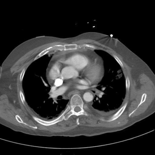
20 Example: Typical ATAI on MDCT
21 MDCT and ATAI: Direct findings: Indirect findings: Ao luminal irregularity Mediastinal hematoma Intimal flap Concomitant injuries Pseudoaneurysm Ao contrast extravasation
22 MDCT and ATAI: Findings of ATAI: Direct: Ao luminal irregularity Intimal flap Pseudoaneurysm Contrast extravasation Indirect: Mediastinal hematoma Other injuries


23 Other typical ATAI cases:

24 Other typical ATAI cases:


25 Other typical ATAI cases:


26 Other typical ATAI cases:

27 Other typical ATAI cases:

28 Example: Not-so-typical ATAI

29 Example: Not-so-typical ATAI

30 Example: Not-so-typical ATAI


31 Example: Not-so-typical ATAI


32 ATAI: Injury at Diaphragm

33 ATAI: Ascending Ao injury:

34 ATAI: Ascending Ao injury:
35 MDCT and ATAI: Pitfalls: Ductus bump Atherosclerotic plaque, penetrating ulcers Streak artifact Cardiac motion

36 CT pitfalls: Streak artifact Motion artifact Aortic injury

37 CT pitfalls: Streak artifact Motion artifact Normal
38 CT Pitfalls: Ductus diverticulum: Remnant of closed ductus arteriosus. Convex bulge along the anterior undersurface of aortic isthmus. Smooth margins and obtuse angles with the aortic lumen typifies a normal ductus bump

39 Ductus Bump
40 Ductus Bump Smooth margins. Obtuse angles. No concerning associated findings. No MS hematoma No intimal flap


41 What about this?

42 Dx: PDA in an adult.


43 What about this?


44 What about this? 24 hr. follow-up with ECG-gating

45 additional imaging
46 What about this? Radiology consensus: ATAI with small pseudoaneurysm.

47 What about this? Radiology consensus: ATAI with small pseudoaneurysm. Surgical findings: No injury. Chronic atheromatous plaque with penetrating ulcer.
48 MDCT and ATAI: Pitfalls: Reassuring factors: Absence of MS hematoma Obtuse angles Smooth margins Lack of intimal flap Calcification Consider CT follow-up (+/- angio) for equivocal cases
49 CXR and ATAI:
50 CXR and ATAI: Goal: Identify evidence of MS hematoma. If CXR normal, presume no ATAI. CXR valuable for immediately life-threatening conditions, and to assess line/tube positions.
51 CXR and ATAI: Goal: Identify evidence of MS hematoma. If CXR normal, presume no ATAI. Best case scenario: 7% of ATAI have normal or near-normal CXR. Regardless of CXR findings, if mechanism suspicious, CT evaluation is warranted.
52 CXR and ATAI: Then

53 CXR and ATAI: Now
54 CXR and ATAI: Most aortic injuries are associated with mediastinal hematoma. NPV near 100% PPV only ~20% The presence of a ATAI may or may not result in mediastinal hematoma. An abnormal CXR correlates poorly with the presence of ATAI.
55 CXR findings of ATAI: Indistinct or abnormal Ao contour Obscuration of the AP window Widened L (or R) paraspinal stripe Deviation of support tubes and trachea to R Superior mediastinal widening (>8cm) L sided pleural fluid collections/apical capping 1st or 2nd rib fractures
56 ATAI?
57 ATAI: Findings of ATAI: Direct: Ao luminal irregularity Intimal flap Pseudoaneurysm Contrast extravasation Indirect: Mediastinal hematoma Other injuries
58 ATAI: Minimal Aortic Injury What does minimal mean? Low grade ATAI. Injury Isolated to intima. No indirect findings (except those attributable to concomitant injury).
59 Minimal Aortic Injury: Where did it come from?
60 ATAI: Minimal Aortic Injury Compromise 10% of ATAI % may be unapparent on angiography. Conservative therapy becoming more common.
61 ATAI: Minimal Aortic Injury Natural history varies: Resolve How quickly? Stabilize Worsen As many as 50% may progress to pseudoaneurysm. Recommend: CT follow-up.

62 ATAI: Minimal Aortic Injury
63 Treatment of ATAI: Open surgical repair Endovascular therapy
64 Treatment of ATAI Morbidity and Mortality: Surgery; Morality: % Paraplegia: % Complications: % Endovascular: Mortality: % Paraplegia: 0-0.8% Complications: %
65 Treatment of ATAI: Complications Surgery Death Paraplegia Stroke Upper extremity ischemia Other perioperative complications. Endograft Endoleak Infection Stent failure Stent migration Death Stroke Upper extremity ischemia Paraplegia Access site complications
66 Treatment of ATAI: Surgery Pros: Established technique Morbidity stats improving Definitive therapy Long term data available Cons: Big, complicated surgery Concomitant injuries complicate pre-surgical planning. Historically non-trivial paraplegia rates
67 Treatment of ATAI: Endograft Pros: Much simpler procedure than open repair. Quick. Can be more easily coordinated with treatment of other injuries. Cons: Long term data unavailable. Equipment limitations.
68 Treatment of ATAI Pre-endograft placement: What to include in CT report? Aortic arch anatomy and variation if present Vertebral artery dominance Pre-existent stenosis, atherosclerotic disease Post-operative changes (CABG) Measure Ao below and above injury Proximity to LSA origin (1.5-2cm preferable) Injury length

69 Acute Traumatic Aortic Injury Learning objectives: Understand epidemiology and pathogenesis of ATAI. Describe spectrum of CT and CXR findings of ATAI. Understand treatment options and related anatomic considerations.
70 ATAI in the 21 st century.

71 ATAI in the 23 rd Century?
72 Thank you.
73 References: 1.Creasy JD, Chiles C, Routh WD, Dyer RB. Overview of Traumatic Injury of the Thoracic Aorta. RadioGraphics 1997; Jan-Feb; 17(1): Fishman JE, Nuñez D Jr, Kane A, Rivas LA, Jacobs WE. Direct versus Indirect Signs of Traumatic Aortic Injury Revealed by Helical CT: Performance Characteristics and Interobserver Agreement. AJR Am J Roentgenol 1999; 172(4): Malhotra AK, Fabian TC, Croce MA, Weiman DS, Gavant ML, Pate JW. Minimal Aortic Injury: A Lesion associated with Advancing Diagnostic Techniques. J Trauma 2001; 51: Steenburg SD, Ravenel JG, Ikonomidis JS, Schonholz C, Reeves S. Acute Traumatic Aortic Injury: Imaging Evaluation and Management. Radiology 2008; 248: Alonso RC, Nacenta SB, et al. Kidney in Danger: CT Findings in Blunt and Penetrating Renal Trauma. RadioGraphics 2009; 29: Linsenmaier U, Wirth S, et al. Diagnosis and Classification of Pancreatic and Duodenal Injuries in Emergency Radiology. RadioGraphics 2008; 28: Hamilton, JD, Kumaraval M, Censullo ML, et al. Multidetector CT Evaluation of Active Extravasation of Blunt Abdominal and Pelvic Trauma Patients. RadioGraphics 2208; 28: Vu M, Anderson SW, et al. CT of blunt abdominal and pelvic vascular injury. Emergency Radiology 2010; 17: Kaewlai R, Avery LL, et al. Multidetector CT of Blunt Thoracic Trauma. RadioGraphics 2008; 28: Daly KP, Ho CP, et al. Traumatic Retroperitoneal Injuries; Review of Multidetector CT Findings. RadioGraphics 2008; 28: Prasad KR, Kumar A, et al. CT in post-traumatic hypoperfusion complex--a pictorial review. Emergency Radiology, online Publication: 23 December Yoon W, Yong YJ, et al. CT in Blunt Liver Trauma. RadioGraphics 2005; 25: Mirvis SE, Whitley NO, Gens DR, Blunt Splenic Trauma in Adults: CT-based classification and correlation with prognosis and treatment. Radiology 1989; 171: Shanmuganathan K, et al. Nonsurgical Manaement of Blunt Splenic Injury: Use of CT Criteria to Select Patients for Splenic Arteriography and Potential Endovascular Therapy. Radiology 2000; 217: Morgan TA, Steenburg SD, et al. Acute Traumatic Aortic Injuries: Posttherapy Multidetector CT Findings. RadioGraphics 2010; 30: Neschis DG, Scalea TM, et al. Blunt Aortic Injury. NEJM 2008;
CT of Acute Thoracic Aortic Syndromes Stuart S. Sagel, M.D.
 CT of Acute Thoracic Aortic Syndromes Stuart S. Sagel, M.D. Thoracic Aortic Aneurysms Atherosclerotic Dissection Penetrating ulcer Mycotic Inflammatory (vasculitis) Traumatic Aortic Imaging Options Catheter
CT Imaging of Blunt and Penetrating Vascular Trauma DENNIS FOLEY MEDICAL COLLEGE WISCONSIN
 CT Imaging of Blunt and Penetrating Vascular Trauma DENNIS FOLEY MEDICAL COLLEGE WISCONSIN THORACO ABDOMINAL TRAUMA 0 10 20 30 40 50 60 5 cc/sec 30 secs 1.25 mm/ 55 mm Z1.375 2.5 mm/ 55 mm Z 1.375 Grade
account for 10% to 15% of all traffic fatalities majority fatal at the scene 50% who survive the initial injury die in the first 24 hours 90% die
 account for 10% to 15% of all traffic fatalities majority fatal at the scene 50% who survive the initial injury die in the first 24 hours 90% die within the first month if aorta not repaired 30-90% overall
Acute Aortic Syndromes
 Acute Aortic Syndromes Carole J. Dennie, MD Acute Thoracic Aortic Syndromes Background Non-Traumatic Acute Thoracic Aortic Syndromes Carole Dennie MD FRCPC Associate Professor of Radiology and Cardiology
Imaging of Thoracic Trauma: Tips and Traps. Arun C. Nachiappan, MD Associate Professor of Clinical Radiology University of Pennsylvania
 Imaging of Thoracic Trauma: Tips and Traps Arun C. Nachiappan, MD Associate Professor of Clinical Radiology University of Pennsylvania None Disclosures Objectives Describe blunt and penetrating traumatic
Traumatic aortic injury: CT findings, mimics, and therapeutic options
 Review Article Traumatic aortic injury: CT findings, mimics, and therapeutic options Ethany L. Cullen, Eric J. Lantz, C. Michael Johnson, Philip M. Young Department of Radiology, Mayo Clinic, Rochester,
Update on Acute Aortic Syndrome
 SUNDAY Update on Acute Aortic Syndrome Diana Litmanovich, MD Learning objectives To be familiar with the definition, natural history, and imaging findings of acute aortic syndrome, including: I. Aortic
TEVAR FOR! THORACIC AORTIC TRAUMA"
 10th HKL Vascular Surgery Conference and Workshop" TEVAR FOR! THORACIC AORTIC TRAUMA" Dr Hanif Hussein" Vascular and General Surgeon" Department of Surgery" Hospital Kuala Lumpur" Source: MIROS! Thoracic
Advances in MDCT of Thoracic Trauma
 Baltic Congress of Radiology, Riga 2010 Advances in MDCT of Thoracic Trauma Robert A. Novelline, MD Professor of Radiology, Harvard Medical School Director of Emergency Radiology, Massachusetts General
Haemodynamically unstable patient with chest trauma
 HR J Clinical Case - Test Yourself Interventional Haemodynamically unstable patient with chest trauma Dimitrios Tomais, Theodoros Kratimenos, Dimosthenis Farsaris Interventional Radiology Unit, Radiology
Blunt Thoracic Aortic Injury
 September 2004 Blunt Thoracic Aortic Injury Richelle Williams, Harvard Medical School, Year III Blunt Aortic Injury ~8000 deaths/year in the U.S. Most common cause of sudden death following: - high-speed
Aortic CT: Intramural Hematoma. Leslie E. Quint, M.D.
 Aortic CT: Intramural Hematoma Leslie E. Quint, M.D. 43 M Mid back pain X several months What type of aortic disease? A. Aneurysm with intraluminal thrombus B. Chronic dissection with thrombosed false
Thoracic aortic trauma A.T.O.ABDOOL-CARRIM ACADEMIC HEAD VASCULAR SURGERY DEPARTMENT OF SURGERY UNIVERSITY OF WITWATERSRAND
 Thoracic aortic trauma A.T.O.ABDOOL-CARRIM ACADEMIC HEAD VASCULAR SURGERY DEPARTMENT OF SURGERY UNIVERSITY OF WITWATERSRAND Thoracic Aortic Trauma In USA and CANADA 7500-8000 die of blunt thoracic aortic
Katarzyna J. Macura 1, Frank M. Corl, Elliot K. Fishman, David A. Bluemke
 Pictorial Essay Pathogenesis in cute ortic Syndromes: ortic neurysm Leak and Rupture and Traumatic ortic Transection Katarzyna J. Macura 1, Frank M. Corl, Elliot K. Fishman, David. luemke T his pictorial
ACUTE AORTIC SYNDROMES
 ACUTE AORTIC SYNDROMES AGNETA FLINCK MD, PhD Dept. of Thoracic Radiology Sahlgrenska University Hospital ACUTE AORTIC SYNDROMES Aortic dissection Intramural hematoma (IMH) 5-20% Penetrating atherosclerotic
Development of a Branched LSA Endograft & Ascending Aorta Endograft
 Development of a Branched LSA Endograft & Ascending Aorta Endograft Frank R. Arko III, MD Sanger Heart & Vascular Institute Carolinas Medical Center Charlotte, North Carolina, USA Disclosures Proximal
Interventional Radiology in Trauma. Vikash Prasad, MD, FRCPC Vascular and Interventional Radiology The Moncton Hospital
 Interventional Radiology in Trauma Vikash Prasad, MD, FRCPC Vascular and Interventional Radiology The Moncton Hospital Disclosures None relevant to this presentation Shareholder Johnson and Johnson Goal
Thoracic and Great Vessel Imaging and Intervention
 Thoracic and Great Vessel Imaging and Intervention Shelley R. Marder, M.D. The Kaiser Permanente Medical Group Clinical Faculty, SFGH, UCSF Traumatic aortic injury Shearing / shoveling forces at junction
IMAGING the AORTA. Mirvat Alasnag FACP, FSCAI, FSCCT, FASE June 1 st, 2011
 IMAGING the AORTA Mirvat Alasnag FACP, FSCAI, FSCCT, FASE June 1 st, 2011 September 11, 2003 Family is asking $67 million in damages from two doctors Is it an aneurysm? Is it a dissection? What type of
Role of the Radiologist
 Diagnosis and Treatment of Blunt Cerebrovascular Injuries NORDTER Consensus Conference October 22-24, 2007 Clint W. Sliker, M.D. University of Maryland Medical Center R Adams Cowley Shock Trauma Center
Computed tomography angiography (CTA) of
 of") Multidetector CT in the Evaluation of Thoracic Aortic Disease The pre-eminent platform for the planning and surveillance of TEVAR patients. BY PETER S. FAIL, MD, FACC, FACP, AND VINOD NAIR, MD, FACC Computed
Multidetector CT in the Evaluation of Thoracic Aortic Disease The pre-eminent platform for the planning and surveillance of TEVAR patients. BY PETER S. FAIL, MD, FACC, FACP, AND VINOD NAIR, MD, FACC Computed
Case 8036 Multiple penetrating atherosclerotic ulcers
 Case 8036 Multiple penetrating atherosclerotic ulcers Santiago I, Seco M, Curvo-Semedo L Section: Cardiovascular Published: 2010, Feb. 22 Patient: 78 year(s), male Clinical History A 78-year-old hypertensive
Diseases of the Aorta
 Diseases of the Aorta ASE Review 2018 Susan E Wiegers, MD, FASE, FACC Professor of Medicine My great friend Dr. Roberto Lang Disclosure None related to this presentation 1 Objectives Aneurysm Dissection
Echocardiographic Evaluation of the Aorta
 Echocardiographic Evaluation of the Aorta William F. Armstrong M.D. Director Echocardiography Laboratory Professor of Medicine University of Michigan The Aorta: What to Evaluate Dimensions / shape Atherosclerotic
Acute Aortic Syndromes
 Acute Aortic Syndromes None Disclosures Smita Patel, M.B.B.S., M.R.C.P., F.R.C.R. Associate Professor, University of Michigan Ann Arbor, MI Objectives To review common CTA findings of acute aortic syndromes
CT angiography techniques. Boot camp
 CT angiography techniques Boot camp Overview Basic concepts Contrast administration arterial opacification Time scan acquisition during the arterial phase Protocol examples Helical non-gated CTA Pulmonary
Pediatric Abdomen Trauma
 Pediatric Abdomen Trauma Susan D. John, MD, FACR Pediatric Trauma Trauma is leading cause of death and disability in children and adolescents Causes and effects vary between age groups Blunt trauma predominates
MISSED FINDINGS IN EMERGENCY RADIOLOGY: CASE BASE SESSION 5 th Nordic Trauma Radiology Course Oslo, Norway
 MISSED FINDINGS IN EMERGENCY RADIOLOGY: CASE BASE SESSION 5 th Nordic Trauma Radiology Course Oslo, Norway K.SHANMUGANATHAN M.D. EASILY MISSED FINDINGS IN EMERGENCY RADIOLOGY OBJECTIVES Commonly missed
Traumatic aortic rupture was first described in 1557 by Vesalius (1). However, acute traumatic aortic injuries (ATAIs) remained rare until the advent
. However, acute traumatic aortic injuries (ATAIs) remained rare until the advent") Note: This copy is for your personal non-commercial use only. To order presentation-ready copies for distribution to your colleagues or clients, contact us at www.rsna.org/rsnarights. REVIEWS AND COMMENTARY
Note: This copy is for your personal non-commercial use only. To order presentation-ready copies for distribution to your colleagues or clients, contact us at www.rsna.org/rsnarights. REVIEWS AND COMMENTARY
Aortic Coarctation: Evaluation with Computed Tomography Angiography in Pediatric Patients
 Med. J. Cairo Univ., Vol. 83, No. 2, June: 63-70, 2015 www.medicaljournalofcairouniversity.net Aortic Coarctation: Evaluation with Computed Tomography Angiography in Pediatric Patients MOHAMED ZAKI, M.D.
Fundamentals, Techniques, Pitfalls, and Limitations of MDCT Interpretation and Measurement
 Fundamentals, Techniques, Pitfalls, and Limitations of MDCT Interpretation and Measurement 3 rd Annual Imaging & Physiology Summit November 20-21, 21, 2009 Seoul, Korea Wm. Guy Weigold, MD, FACC Cardiovascular
Critical Evaluation of Chest Computed Tomography Scans for Blunt Descending Thoracic Aortic Injury
 Critical Evaluation of Chest Computed Tomography Scans for Blunt Descending Thoracic Aortic Injury Brian A. Bruckner, MD, Daniel J. DiBardino, MD, Todd C. Cumbie, BS, Charles Trinh, MD, Shanda H. Blackmon,
TEVAR for trauma is here to stay: Advances in the Treatment of Blunt Thoracic Aortic Injury
 TEVAR for trauma is here to stay: Advances in the Treatment of Blunt Thoracic Aortic Injury Megan Brenner MD MS RPVI FACS Associate Professor of Surgery Division of Trauma/Surgical Critical Care, RA Cowley
Disclosures: Acute Aortic Syndrome. A. Michael Borkon, M.D. Director of CV Surgery Mid America Heart Institute Saint Luke s Hospital Kansas City, MO
 Acute Aortic Syndrome Disclosures: A. Michael Borkon, M.D. Director of CV Surgery Mid America Heart Institute Saint Luke s Hospital Kansas City, MO No financial relationships to disclose 1 Acute Aortic
Χρόνιος διαχωρισμός. υπερηχοκαρδιογραφική. αορτής. παρακολούθηση ή άλλη; Α. Παπασπυρόπουλος ΕΠΙΜΕΛΗΤΗΣ ΓΝ.ΝΙΚΑΙΑΣ ΠΕΜΠΤΗ
 Χρόνιος διαχωρισμός αορτής υπερηχοκαρδιογραφική παρακολούθηση ή άλλη; Α. Παπασπυρόπουλος ΕΠΙΜΕΛΗΤΗΣ ΓΝ.ΝΙΚΑΙΑΣ ΠΕΜΠΤΗ 8-2-2018 The Normal Aorta (conduit function + control ) *Aortic expansion is about
Case 9799 Stanford type A aortic dissection: US and CT findings
 Case 9799 Stanford type A aortic dissection: US and CT findings Accogli S, Aringhieri G, Scalise P, Angelini G, Pancrazi F, Bemi P, Bartolozzi C Department of Diagnostic and Interventional Radiology, University
Role of imaging in evaluation of genitourinary i trauma Spectrum of GU injuries Relevance of imaging findings in determining management Focus on MDCT
 Genitourinary Tract Injuries 6 th Nordic Course Scott D. Steenburg, MD Assistant Professor University of Maryland Department of Radiology Division of Trauma and Emergency Radiology R Adams Cowley Shock
Multimodality Imaging of the Thoracic Aorta
 Multimodality Imaging of the Thoracic Aorta Steven Goldstein MD, FACC Director Noninvasive Cardiology MedStar Heart and Vascular Institute Washington Hospital Center Saturday, October 8, 2016 DISCLOSURE
Computed tomography (CT) and magnetic resonance
 and magnetic resonance") Trends in Emergency ortic Imaging Determining optimal imaging techniques for each patient. Y PERRY CHOI, MD, ND HMID MOJIIN, MD Computed tomography (CT) and magnetic resonance imaging (MRI) play vital
Trends in Emergency ortic Imaging Determining optimal imaging techniques for each patient. Y PERRY CHOI, MD, ND HMID MOJIIN, MD Computed tomography (CT) and magnetic resonance imaging (MRI) play vital
Abdominal Aortic Aneurysms. A Surgeons Perspective Dr. Derek D. Muehrcke
 Abdominal Aortic Aneurysms A Surgeons Perspective Dr. Derek D. Muehrcke Aneurysm Definition The abnormal enlargement or bulging of an artery caused by an injury or weakness in the blood vessel wall A localized
MODERN METHODS FOR TREATING ABDOMINAL ANEURYSMS AND THORACIC AORTIC DISEASE
 MODERN METHODS FOR TREATING ABDOMINAL ANEURYSMS AND THORACIC AORTIC DISEASE AAA FACTS 200,000 New Cases Each Year Ruptured AAA = 15,000 Deaths per Year in U.S. 13th Leading Cause of Death 80% Chance of
Free Esophageal Perforation Following Hybrid Visceral Debranching and Distal Endograft Extension to Repair a Ruptured Thoracoabdominal Aortic
 Free Esophageal Perforation Following Hybrid Visceral Debranching and Distal Endograft Extension to Repair a Ruptured Thoracoabdominal Aortic Aneurysm History A 56-year-old gentleman, who had been referred
I have the following financial relationships to disclose:
 Novel Approaches to Endovascular Management of Aortic Aneurysms Rodney A White, MD Medical Director, Vascular Services MemorialCare Heart & Vascular Institute Long Beach Memorial Hospital Long Beach, California
Multidetector CTA for Diagnosing Blunt Cerebrovascular Injuries
 Multidetector CTA for Diagnosing Blunt Cerebrovascular Injuries 4 th Nordic Trauma Course 2006 Stuart E. Mirvis, M.D., FACR Department of Diagnostic Radiology and Nuclear Medicine, University of Maryland
Disclosure Information
 Coronary CTA Pearls and Pitfalls Ricardo C. Cury, MD, FSCCT, FAHA, FACC Chairman of Radiology Radiology Associates of South Florida Director of Cardiac Imaging Miami Cardiac and Vascular Institute Past-President
Case Report 1. CTA head. (c) Tele3D Advantage, LLC
 Tele3D Advantage, LLC") Case Report 1 CTA head 1 History 82 YEAR OLD woman with signs and symptoms of increased intra cranial pressure in setting of SAH. CT Brain was performed followed by CT Angiography of head. 2 CT brain Extensive
Case Report 1 CTA head 1 History 82 YEAR OLD woman with signs and symptoms of increased intra cranial pressure in setting of SAH. CT Brain was performed followed by CT Angiography of head. 2 CT brain Extensive
Animesh Rathore, MD 4/21/17. Penetrating atherosclerotic ulcers of aorta
 Animesh Rathore, MD 4/21/17 Penetrating atherosclerotic ulcers of aorta Disclosures No financial disclosures Thank You Dr. Panneton for giving this lecture for me. I am stuck at Norfolk with an emergency
EVAR and TEVAR: Extending Their Use for Rupture and Traumatic Injury. Conflict of Interest. Hypotensive shock 5/5/2014. none
 EVAR and TEVAR: Extending Their Use for Rupture and Traumatic Injury Bruce H. Gray, DO MSVM FSCAI Professor of Surgery/Vascular Medicine USC SOM-Greenville Greenville, South Carolina none Conflict of Interest
Post-Op Aorta: Differentiating Normal Post-Op vs. Complications. Linda C. Chu, MD Assistant Professor of Radiology Johns Hopkins University
 Post-Op Aorta: Differentiating Normal Post-Op vs. Complications Linda C. Chu, MD Assistant Professor of Radiology Johns Hopkins University No disclosures Disclosures Goals and Objectives To review CT technique
Case 47 Clinical Presentation
 93 Case 47 C Clinical Presentation 45-year-old man presents with chest pain and new onset of a murmur. Echocardiography shows severe aortic insufficiency. 94 RadCases Cardiac Imaging Imaging Findings C
SUPPLEMENTAL MATERIAL
 SUPPLEMENTL MTERIL Marie erna, Martin Kocher, Rohit Philip Thomas. cute aorta, overview of acute T findings and endovascular treatment options (doi: 10.5507/bp.2016.060) Fig. 1. : Non-enhanced T, hemopericardium
CT IMAGING OF BLUNT SPLENIC INJURY: A PICTORIAL ESSAY
 CT IMAGING OF BLUNT SPLENIC INJURY: A PICTORIAL ESSAY Radhiana H, Azian AA, Ahmad Razali MR, Amran AR, Azlin S, S Kamariah CM Department of Radiology International Islamic University Malaysia Kuantan,
Four-year Surgical Results for Traumatic Aortic Injury in China Medical University Hospital, Mid-Taiwan
 Four-year Surgical Results for Traumatic Aortic Injury in China Medical University Hospital, Mid-Taiwan Yi-Chun Lin 林怡均 (5 th grade medical student), MingLi Li 李明禮, Chih-Hsiang Hsu, Ching-Feng Wu, Hui-Han
Neurological Complications of TEVAR. Frank J Criado, MD. Union Memorial-MedStar Health Baltimore, MD USA
 ISES Online Neurological Complications of Frank J Criado, MD TEVAR Union Memorial-MedStar Health Baltimore, MD USA frank.criado@medstar.net Paraplegia Incidence is 0-4% after surgical Rx of TAAs confined
TRAUMATIC CAROTID &VERTEBRAL ARTERY INJURIES
 TRAUMATIC CAROTID &VERTEBRAL ARTERY INJURIES ALBERTO MAUD, MD ASSOCIATE PROFESSOR TEXAS TECH UNIVERSITY HEALTH SCIENCES CENTER EL PASO PAUL L. FOSTER SCHOOL OF MEDICINE 18TH ANNUAL RIO GRANDE TRAUMA 2017
Advances in Treatment of Traumatic Aortic Transection
 Advances in Treatment of Traumatic Aortic Transection Himanshu J. Patel MD University of Michigan Medical Center Author Disclosures Consulting fees from WL Gore Inc. There is no disease more conducive
Typical and atypical imaging of thoracic and abdominal aortic rupture
 Typical and atypical imaging of thoracic and abdominal aortic rupture Poster No.: C-0453 Congress: ECR 2014 Type: Educational Exhibit Authors: J. Isogai, T. Ichihara, T. Inoue, T. Kanamori ; Asahi/JP,
Performance of the conformable GORE TAG device in Type B aortic dissection from the GORE GREAT real world registry
 University of Milan Thoracic Aortic Research Center Performance of the conformable GORE TAG device in Type B aortic dissection from the GORE GREAT real world registry Santi Trimarchi, MD, PhD Associate
IMH/Penetrating Aortic Ulcers/ Saccular Aneurysms: How to manage and when to intervene
 IMH/Penetrating Aortic Ulcers/ Saccular Aneurysms: How to manage and when to intervene UCSF Vascular Surgery Symposium 2018 Sukgu M Han, MD, MS Assistant Professor of Clinical Surgery Co-director, Comprehensive
Effective Utilization of Imaging. John V. Roberts, M.D. Premier Radiology Abdominal Imaging
 Effective Utilization of Imaging John V. Roberts, M.D. Premier Radiology Abdominal Imaging Safety Contrast and Radiation What to order Abdomen/Pelvis Brain/Spine Chest Musculoskeletal Ob/Gyn Head and Neck
Multidetector CT of Blunt Traumatic Venous Injuries in the Chest, Abdomen, and Pelvis 1
 Note: This copy is for your personal non-commercial use only. To order presentation-ready copies for distribution to your colleagues or clients, contact us at www.rsna.org/rsnarights. TRAUMA/EMERGENCY
Abdominal Ultrasonography
 Abdominal Ultrasonography David A. Masneri, DO, FACEP, FAAEM Assistant Professor of Emergency Medicine Assistant Director, Emergency Medicine Residency Medical Director, Operational Medicine Division Center
Screening and Management of Blunt Cereberovascular Injuries (BCVI)
") Grady Memorial Hospital Trauma Service Guidelines Screening and Management of Blunt Cereberovascular Injuries (BCVI) BACKGROUND Blunt injury to the carotid or vertebral vessels (blunt cerebrovascular injury
Grady Memorial Hospital Trauma Service Guidelines Screening and Management of Blunt Cereberovascular Injuries (BCVI) BACKGROUND Blunt injury to the carotid or vertebral vessels (blunt cerebrovascular injury
Vascular CT Protocols
 Vascular CT Protocols V 1D: Chest and abdominal CT angiogram (aortic dissection protocol) V 1T: Chest CT angiogram (aortic trauma protocol) V 2: Abdominal and pelvis CT angiogram (aortic aneurysm protocol)
Is computed tomography angiography really useful in. of coronary artery disease?
 Is computed tomography angiography really useful in screening patients with high risk of coronary artery disease? Myeong-Ki Hong, M.D. Ph D Professor of Medicine Division of Cardiology, Severance Cardiovascular
General Imaging. Imaging modalities. Incremental CT. Multislice CT Multislice CT [ MDCT ]
![General Imaging. Imaging modalities. Incremental CT. Multislice CT Multislice CT [ MDCT ]](/thumbs/76/74079340.jpg "General Imaging. Imaging modalities. Incremental CT. Multislice CT Multislice CT [ MDCT ]") General Imaging Imaging modalities Conventional X-rays Ultrasonography [ US ] Computed tomography [ CT ] Radionuclide imaging Magnetic resonance imaging [ MRI ] Angiography conventional, CT,MRI Interventional
General Imaging Imaging modalities Conventional X-rays Ultrasonography [ US ] Computed tomography [ CT ] Radionuclide imaging Magnetic resonance imaging [ MRI ] Angiography conventional, CT,MRI Interventional
Coronary Artery Imaging. Suvipaporn Siripornpitak, MD Inter-hospital Conference : Rajavithi Hospital
 Coronary Artery Imaging Suvipaporn Siripornpitak, MD Inter-hospital Conference : Rajavithi Hospital Larger array : cover scan area Detector size : spatial resolution Rotation speed : scan time Retrospective
New Cardiovascular Devices and Interventions: Non-Contrast MRI for TAVR Abhishek Chaturvedi Assistant Professor. Cardiothoracic Radiology
 New Cardiovascular Devices and Interventions: Non-Contrast MRI for TAVR Abhishek Chaturvedi Assistant Professor Cardiothoracic Radiology Disclosure I have no disclosure pertinent to this presentation.
CT Chest. Verification of an opacity seen on the straight chest X ray
 CT Chest Indications: To assess equivocal plain x-ray findings Staging of lung neoplasm Merastatic workup of extra thoraces malignancies Diagnosis of diffuse lung diseases with HRCT Assessment of bronchietasis
Case 1. Aortic Disasters. Case 2. Case 3. Diagnosis, Imaging Techniques and Management
 Aortic Disasters Diagnosis, Imaging Techniques and Management Eric R. Snoey, MD Alameda County Medical Center Oakland, CA 1 Case 1 51 yo female presents with sharp anterior chest pain while at rest. She
Intravascular Ultrasound in the Treatment of Complex Aortic Pathologies. Naixin Kang, M.D. Vascular Surgery Fellow April 26 th, 2018
 Intravascular Ultrasound in the Treatment of Complex Aortic Pathologies Naixin Kang, M.D. Vascular Surgery Fellow April 26 th, 2018 DISCLOSURES Nothing To Disclose 2 ENDOVASCULAR AORTIC INTERVENTION Improved
Follow-up of Aortic Dissection: How, How Often, Which Consequences Euro Echo 2011
 Follow-up of Aortic Dissection: How, How Often, Which Consequences Euro Echo 2011 Susan E. Wiegers, MD, FASE Director of Clinical Echocardiography Hospital of the University of Pennsylvania Disclosure
Radiological Investigations of Abdominal Trauma
 76 77 Investigations of Abdominal Trauma Introduction: Trauma to abdominal organs is a common cause of patient morbidity and mortality among trauma patients. Causes of abdominal trauma include blunt injuries,
Enter modality here Enter modality here, enter none if none. Principal Modality (2): Case Report # [] Date accepted: April 2015
![Enter modality here Enter modality here, enter none if none. Principal Modality (2): Case Report # [] Date accepted: April 2015](/thumbs/83/87309292.jpg "Enter modality here Enter modality here, enter none if none. Principal Modality (2): Case Report # [] Date accepted: April 2015") Radiological Category: Enter category here Principal Modality (1): Principal Modality (2): Enter modality here Enter modality here, enter none if none Case Report # [] Submitted by: Varun Rachakonda, M.D.
Radiological Category: Enter category here Principal Modality (1): Principal Modality (2): Enter modality here Enter modality here, enter none if none Case Report # [] Submitted by: Varun Rachakonda, M.D.
Richard L. Hallett, MD
 SCCT 2015 LAS VEGAS, NV 18 JULY 2015 Richard L. Hallett, MD Chief, Cardiovascular Imaging Northwest Radiology Network Indianapolis St. Vincent Heart Center of Indiana Adjunct Assistant Professor of Radiology
Case report of traumatic aortic disruption: a lethal injury requiring rapid and accurate diagnosis to lower mortality
 Hong Kong Journal of Emergency Medicine Case report of traumatic aortic disruption: a lethal injury requiring rapid and accurate diagnosis to lower mortality EYL Mak and CW Kam Traumatic aortic disruption
Ultrasound. Computed tomography. Case studies. Utility of IQon Spectral CT in. cardiac imaging
 Ultrasound Computed tomography Case studies Utility of IQon Spectral CT in cardiac imaging Cardiac imaging is a challenging procedure where it is necessary to image a motion-free heart. This requires a
Pediatric CT Protocols (18 years old or less)
") Pediatric CT Protocols (18 years old or less) Ped1: Head CT Ped2: Cervical spine CT Ped3: Sinus CT Ped4: Neck CT Ped5: Chest CT Ped6: Abdomen and pelvis CT Ped7: Thoracic or lumbar spine CT Ped8: Extremity
Pediatric CT Protocols (18 years old or less) Ped1: Head CT Ped2: Cervical spine CT Ped3: Sinus CT Ped4: Neck CT Ped5: Chest CT Ped6: Abdomen and pelvis CT Ped7: Thoracic or lumbar spine CT Ped8: Extremity
Endovascular therapy for Ischemic versus Nonischemic complicated acute type B aortic dissection (catbad).
.") Endovascular therapy for Ischemic versus Nonischemic complicated acute type B aortic dissection (catbad). AS. Eleshra, MD 1, T. Kölbel, MD, PhD 1, F. Rohlffs, MD 1, N. Tsilimparis, MD, PhD 1,2 Ahmed Eleshra
Endovascular therapy for Ischemic versus Nonischemic complicated acute type B aortic dissection (catbad). AS. Eleshra, MD 1, T. Kölbel, MD, PhD 1, F. Rohlffs, MD 1, N. Tsilimparis, MD, PhD 1,2 Ahmed Eleshra
CT Imaging of Atherosclerotic Plaque. William Stanford MD Professor-Emeritus Radiology University of Iowa College of Medicine Iowa City, IA
 CT Imaging of Atherosclerotic Plaque William Stanford MD Professor-Emeritus Radiology University of Iowa College of Medicine Iowa City, IA PREVALENCE OF CARDIOVASCULAR DISEASE In 2006 there were 80 million
CT angiography in type I acute aortic dissection complicated with malperfusion - a visual review of obstruciton patterns
 CT angiography in type I acute aortic dissection complicated with malperfusion - a visual review of obstruciton patterns Eneva M. St. Ekaterna University Hospital Report objectives 1. Review malperfusion
CT Versus MR for the Runoff
 CT Versus MR for the Runoff Robert R. Edelman, M.D. Dept. of Radiology NorthShore University HealthSystem Feinberg School of Medicine, Northwestern University Magnetic Resonance Computed Tomography Radio
Pediatric Isolated Trachea Rupture Treated with a Conservative Approach İ Akdulum 1, M Öztürk 2, N Dağ 1, A Sığırcı 1 ABSTRACT
 Pediatric Isolated Trachea Rupture Treated with a Conservative Approach İ Akdulum 1, M Öztürk 2, N Dağ 1, A Sığırcı 1 ABSTRACT Tracheobronchial rupture as a result of blunt thoracic trauma is extremely
Radiation Exposure in Pregnancy. John R. Mayo UNIVERSITY OF BRITISH COLUMBIA
 Radiation Exposure in Pregnancy John R. Mayo UNIVERSITY OF BRITISH COLUMBIA Illustrative Clinical Scenario 32 year old female 34 weeks pregnant with recent onset shortness of breath and central chest pain
Case Acute ascending thoracic aortic rupture due to penetrating atherosclerotic ulcer
 Case 12305 Acute ascending thoracic aortic rupture due to penetrating atherosclerotic ulcer Lopes Dias J, Costa NV, Leal C, Alves P, Bilhim T Section: Chest Imaging Published: 2014, Dec. 19 Patient: 68
Complex Thoracic and Abdominal Aortic Repair Using Hybrid Techniques
 Complex Thoracic and Abdominal Aortic Repair Using Hybrid Techniques Tariq Almerey MD, January Moore BA, Houssam Farres MD, Richard Agnew MD, W. Andrew Oldenburg MD, Albert Hakaim MD Department of Vascular
MAKING THE GRADE FOR PEDIATRIC TRAUMA THE REVIEW AND IMPLEMENTATION OF COMPUTED TOMOGRAPHIC (CT) GRADING FOR SOLID ABDOMINAL ORGAN INJURY
 GRADING FOR SOLID ABDOMINAL ORGAN INJURY") MAKING THE GRADE FOR PEDIATRIC TRAUMA THE REVIEW AND IMPLEMENTATION OF COMPUTED TOMOGRAPHIC (CT) GRADING FOR SOLID ABDOMINAL ORGAN INJURY AUTHORS & DISCLOSURE OF COMMERCIAL INTEREST: Jennifer Thomas 1
MAKING THE GRADE FOR PEDIATRIC TRAUMA THE REVIEW AND IMPLEMENTATION OF COMPUTED TOMOGRAPHIC (CT) GRADING FOR SOLID ABDOMINAL ORGAN INJURY AUTHORS & DISCLOSURE OF COMMERCIAL INTEREST: Jennifer Thomas 1
Intraabdominal Active Bleeding: Helical CT, MDCT (64slice), DSA and Homeostatic Embolization Findings
, DSA and Homeostatic Embolization Findings") Intraabdominal Active Bleeding: Helical CT, MDCT (64slice), DSA and Homeostatic Embolization Findings Poster No.: C-2495 Congress: ECR 2012 Type: Educational Exhibit Authors: B. ALPARSLAN, N. YILDIRIM,
Intraabdominal Active Bleeding: Helical CT, MDCT (64slice), DSA and Homeostatic Embolization Findings Poster No.: C-2495 Congress: ECR 2012 Type: Educational Exhibit Authors: B. ALPARSLAN, N. YILDIRIM,
Abdominal Solid Organ Injury
 Abdominal Solid Organ Injury 9th Nordic Trauma Radiology Course Aarhus, Denmark May 23-26, 2016 K.SHANMUGANATHAN M.D. ABDOMINAL TRAUMA OBJECTIVES Splenic injury Late arterial / early p-v phase imaging
The utility of intravascular ultrasound compared to angiography in the diagnosis of blunt traumatic aortic injury
 From the Society for Vascular Surgery The utility of intravascular ultrasound compared to angiography in the diagnosis of blunt traumatic aortic injury Ali Azizzadeh, MD, a Jaime Valdes, MD, a Charles
Early Clinical Results with the Valiant Mona LSA Branch Stent-Graft
 Early Clinical Results with the Valiant Mona LSA Branch Stent-Graft Frank R. Arko III, MD Professor of Cardiovascular Surgery Director, Endovascular Surgery Co-Director, Aortic Institute Carolinas Medical
Bilateral blunt carotid artery injury: A case report and review of the literature
 CASE REPORT Bilateral blunt carotid artery injury: A case report and review of the literature S Cheddie, 1 MMed (Surg), FCS (SA); B Pillay, 2 FCS (SA), Cert Vascular Surgery; R Goga, 2 FCS (SA) 1 Department
Penetrating Neck Injuries. Jason Levine MD Lutheran Medical Center July 22, 2010
 Penetrating Neck Injuries Jason Levine MD Lutheran Medical Center July 22, 2010 CASE PRESENTATION 19 YO M 3 Stab Wounds Right zone I neck SW 2 SW anterior abdomen Left epigastrium anterior axillary line
Advances in Emergency Imaging
 Hampton Symposium,, October 16 th, 2010 Advances in Emergency Imaging Robert A. Novelline, MD Professor of Radiology, Harvard Medical School Director of Emergency Radiology, Massachusetts General Hospital
Abdominal Solid Organ Injury
 Abdominal Solid Organ Injury 8 th Nordic Course Stockholm, Sweden May 19-22, 2014 K.SHANMUGANATHAN M.D. ABDOMINAL TRAUMA OBJECTIVES Splenic injury Late arterial / early p-v phase imaging Liver injury Blunt
Acute Type B dissection. Closure of the infra diaphragmatic tear: how and when?
 Acute Type B dissection. Closure of the infra diaphragmatic tear: how and when? Prof. Olgierd Rowiński II Department of Clinical Radiology Medical University of Warsaw Disclosure Speaker name: Olgierd
Abdominal Vascular Emergencies in MDCT Imaging
 Abdominal Vascular Emergencies in MDCT Imaging Poster No.: C-0913 Congress: ECR 2016 Type: Educational Exhibit Authors: K. SHIRODKAR, D. N. Dasappa, S. L. DEVARU, D. S. 1 2 2 2 2 2 1 Nandikoor, A. R. Patil,
Blunt Traumatic Rupture of the Aorta
 ORIGINAL ARTICLE Blunt traumatic aortic rupture Blunt Traumatic Rupture of the Aorta Shen-Feng Chao, Bee-Song Chang Department of Thoracic and Cardiovascular Surgery, Buddhist Tzu Chi General Hospital
The role of multidetector computed tomography versus digital subtraction angiography in triaging care and management in abdominopelvic trauma
 Singapore Med J 2016; 57(9): 497-502 doi: 10.11622/smedj.2015179 The role of multidetector computed tomography versus digital subtraction angiography in triaging care and management in abdominopelvic trauma