14 Endoleak: Definition, Diagnosis, and Management
|
|
|
- Bernadette Floyd
- 5 years ago
- Views:
Transcription
1 Endoleak: Definition, Diagnosis, and Management Endoleak: Definition, Diagnosis, and Management David Valenti and Jafar Golzarian CONTENTS 14.1 Introduction Classification and Significance of Endoleaks Type I Endoleak Type II Endoleak Type III Endoleak Type IV Endoleak Type V Endoleak or Endotension Diagnosis of Endoleaks Computed Tomography Color Doppler Ultrasound Magnetic Resonance Imaging (MRI) Angiography Treatment of Endoleak Type I Endoleak Type IA Endoleak Type IB Endoleak Type IC Endoleak Type II Endoleak Transarterial Approach Translumbar Approach Other Embolic Materials Type III Endoleak Type V Endoleak Endotension Conclusion 249 References Introduction Endoleak is defined as the persistent perfusion of the aneurysmal sac after endovascular aortic aneurysm repair (EVAR). A leak can appear during the first 30 days after implantation. This type of leak is called primary endoleak. Secondary endoleak is one that occurs after 30 days. Leaks may also be classified as graft-related or non graft-related. The incidence of endoleak varies from 10% to 50% [1, 2]. In a report from EUROSTAR registry, the incidence of early endoleak was 18% [1]. A total of 69% of these leaks were graft related; 70% sealed spontaneously during the first 6 months without difference between graft-related and non graft-related endoleaks. There is not always a rational explanation of the cause of spontaneous resolution of some endoleaks and persistence or late occurrence of some others. The presence of outflow vessels (mainly lumbar arteries and inferior mesenteric artery) partially explains this phenomenon [3]. A leak communicating with these outflow vessels seldom disappears spontaneously [4]. Thus, these vessels should be identified. Whatever the cause of a persistent leak, it should be identified, monitored and treated. The details of EVAR will not be discussed here, except in relation to endoleaks. This chapter will review classification and significance, diagnosis and treatment options for different types of endoleak. D. Valenti, MD Royal Victoria Hospital, McGill University Health Centre, McGill University, 687 Pine Avenue West, Suite A451, Montreal, H3A 1A1, Canada J. Golzarian, MD Professor of Radiology, Director, Vascular and Interventional Radiology, University of Iowa, Department of Radiology, 200 Hawkins Drive, 3957 JPP, Iowa City, IA 52242, USA 14.2 Classification and Significance of Endoleaks A generally accepted anatomic classification for endoleak has been developed over the years [5]. In this system, leaks are defined by their inflow source, regardless of the number and type of other vessels involved in the outflow (Table 14.1).
2 236 D. Valenti and J. Golzarian Table Endoleak classification Types I A B C II A B III A B IV V Mechanism Flow originates from ineffective endograft seal at fixation zones Proximal Distal Iliac occluder Branch vessel retrograde flow Single vessel (simple) Two or more vessels creating a circuit (complex) Flow results from structural endograft failure Junctional separation (modular devices) Endograft fracture or holes Minor (<2 mm) Major ( 2 mm) Endograft fabric porosity (<30 days after endograft implantation) Endotension Type I Endoleak Type I endoleak is caused by failure to achieve a circumferential seal at either the proximal (type IA) or distal end (type IB) of the stentgraft. Type IC endoleak is due to non-occluded iliac artery in patients with aorto-mono-iliac stent and femoral femoral bypass. With type I endoleak, the aneurysm is perfused directly from the aorta or the iliac arteries (inflows). The leak usually communicates through a channel (sometimes multiple channels) with the aneurysmal sac. There are several outflow vessels, mainly lumbar arteries and inferior mesenteric artery (IMA) that communicate with the channel and or the sac (Figs. 14.1, 14.2). The pressure within a type I leak is systemic. The tension on the aortic wall remains high. Causes of primary type I endoleak include inappropriate anatomy, with a significantly angulated neck, significant calcification/plaque at the proximal or distal landing zone, a non-circular landing zone, malpositioning of the stentgraft, type of endograft and under-dilation of the stentgraft. Secondary type I endoleak can be due to aneurysm re-modeling, resulting in stentgraft migration, progressive dilatation of the proximal neck, design and dimensions of stentgrafts or unfavourable infrarenal necks including the conically shaped neck and neck shorter than 15 mm. Grafts whose fixation relies on radial force are more prone to caudal migration and type I endoleak than grafts with hooks [6]. Endothelialization of bare stents at the landing zones may contribute to a certain fixation, but endothelialization of the fabric itself does not occur. Proximal bare stent separation, as seen with the Vanguard device, and hook fractures, as seen with the EVT device, are also causes of delayed type I endoleak. Oversizing the graft by 20% is recommended to prevent a delayed endoleak. At the iliac level, type IB endoleak occurs when the limb of the graft is too short or migrates upward due to the sac s retraction pressure. Although a type I endoleak can seal spontaneously, risk of rupture is high and intervention is indicated [4, 7, 8] Type II Endoleak A type II endoleak corresponds to the retrograde filling of the aneurysm mainly from lumbar arteries and/or IMA but also in rare situations from sacral, gonadal or accessory renal artery (Figs. 14.3, 14.4). Type II endoleaks can be associated with aneurysmal expansion and rupture; however, this risk is much less than with the type I and III endoleaks (0.5 versus 3.4 %) [9, 10]. A leak in the setting of a shrinking aneurysm can generally be followed, without immediate intervention. It is well established that up to 40 % of type II endoleaks will seal spontaneously. Some have advocated intervening in all endoleaks persisting beyond 3 6 months, while other groups recommended observing leaks in the absence of aneurysm expansion. We favor the last approach. In our experience with biphasic helical CT follow-up of more than 300 patients treated by EVAR from 1994 to 1998, only three patients needed intervention for type II endoleak Type III Endoleak Type III endoleaks are caused by a structural failure of the implanted device, including junctional separation of modular components, due to migration or changes in vessel morphology with aneurysm shrinkage, holes in the fabric, and fabric tears due to graft strut fracture or erosion (Figs. 14.5, 14.6). Graft disconnections were not infrequent with the first stentgraft generation due to a short overlap between the main body and the limb [11]. Type III leaks allow direct communication between the aorta and aneurysm sac. They have systemic arterial pressure. Similar to type I leak, type
3 Endoleak: Definition, Diagnosis, and Management 237 III endoleak needs to be treated aggressively [10]. Type III endoleaks are considered to be the most dangerous, since there is an acute re-pressurization of the sac Type IV Endoleak Type IV leaks are caused by porosity of the graft fabric. They are seen at the time of device implantation, as a faint blush on the post-implantation angiogram, when patients are fully anti-coagulated. It is important to rule out other types of endoleak before labeling a leak as type IV. They are rarely seen with current devices and will seal spontaneously. If a leak persists, other types should be excluded Type V Endoleak or Endotension Endotension (or type V endoleak) corresponds to continued aneurysm expansion in the absence of a confirmed endoleak [12, 13]. The expansion of the aneurysm in a type V endoleak may be due to an undiagnosed endoleak, presumably with very slow flow and suboptimal imaging (e.g. no delayed helical CT acquisition). Endotension has been reported up to 18% in [14] a study evaluating the significance of endotension in 658 patients. The authors demonstrated that endotension is rare and concluded that it may represent missed endoleak rather than true aneurysm expansion in the absence of perigraft flow [15]. However, in most situations, endotension corresponds to an accumulation of yellowish fluid (seroma) [16]. Endotension is more common with eptfe grafts due to ultra-filtration through graft pores Diagnosis of Endoleaks Computed Tomography Contrast enhanced helical computed tomography or CT angiography (CTA) is considered the imaging technique of choice for the detection of endoleak. CTA is reported to be superior to aortography for the demonstration of small leak [17]. The technique is also able to demonstrate the patency of lumbar arteries and IMA. However, selective aneurysmal angiography is superior to CTA for the detection of outflow vessels [4, 18]. The value of biphasic or triphasic CT scanning has been established for follow up of EVAR [19, 20]. Some authors favor obtaining an unenhanced helical CT series. Rozenblit at al. have demonstrated that the unenhanced series were helpful to diagnose an indeterminate endoleak in one patient [20]. Important mimickers of endoleak include calcification, contrast within the folds of unsupported portions of the graft and residual endosac contrast from the initial procedure when early CT follow-up is obtained at 1 3 days. This pseudo-endoleak was seen in up to 57% of patients [21]. It has been demonstrated that delayed acquisition uncovered up to 11% of endoleaks that were missed by arterial phase alone [19, 20]. An optimal CT protocol for the monitoring of the aorta after endoluminal therapy should include a delayed acquisition (Fig. 14.7) Color Doppler Ultrasound Color Doppler ultrasound (CDUS) is a noninvasive and cost-effective imaging modality. It is highly dependent on the operator and has limitations in obese patients and those with excessive bowel gas. Patients should be evaluated after 5 6 h fasting in supine and lateral position. The aorta is evaluated both transversally and longitudinally. Leak is suspected when a reproducible color and Doppler signal inside the aneurysm is visualized. Variable success is reported for the detection and localization of the source of endoleaks with ultrasound, depending on technical factors, the imaging protocol, and the image quality. Reported sensitivities for overall endoleak detection range between 12% and 100%, with specificities of 74% 99% [22 26]. In a series of 55 patient with CDUS compared to biphasic CTA, CT was superior in detection of small leak. Discrepancies between helical CT and CDUS were observed in eight patients (14.5%). In five cases, a small perigraft leak that was clearly demonstrated by helical CT was not found on CDUS. All these leaks were small and disappeared during the follow-up. For the diagnosis of endoleak, the sensitivity, the specificity, the positive and negative pre-


.")
.")

4 238 D. Valenti and J. Golzarian a b c d e Fig. 14.1a e. Type I endoleak. a CT scan, arrow shows large type IA endoleak from proximal end of an abdominal aortic stent graft (arrow). b Aortogram confirms the type IA endoleak (arrows). c Palmaz stent placement at the proximal end to achieve a circumferential seal. Palmaz 5014, 47 mm long at 14-mm diameter. d Deployment of a balloon expandable Palmaz stent. e Follow-up CT showing no leak

.")
.")

.")
, the sac (white arrow) and lumbar")

5 Endoleak: Definition, Diagnosis, and Management 239 a b c d f e g Fig. 14.2a f. A high-risk (ASA IV) patient with an abdominal aortic aneurysm (AAA). a An aortogram shows an AAA, a long irregular neck with occlusion of left renal artery (arrow). b Follow-up angiogram 1 year after aorto-mono-iliac stentgraft implantation. There is a type I endoleak (large arrow) due to a significant angulation of the proximal aortic neck (small arrow). c Selective aneurysmal sac catheterization from brachial approach shows the channel (black arrow), the sac (white arrow) and lumbar arteries acting as outflow vessels (small black arrows). d,e Embolization of the channel with multiple coils. Angiogram shows no more endoleak. f CT scan obtained before embolization demonstrates the endoleak. g CT scan at the same level as image (f), obtained 1 year after embolization shows no endoleak with significant shrinkage of the aneurysm


6 240 D. Valenti and J. Golzarian b a c d e f
.")


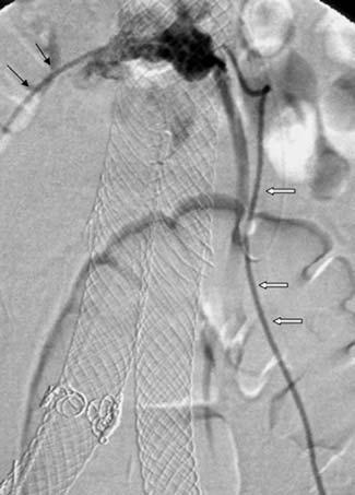
7 Endoleak: Definition, Diagnosis, and Management 241 g h i j Fig. 14.3a j. Type II endoleak. a Contrast enhanced CT shows an important peri-prosthetic leak (arrow). b Left internal iliac artery angiogram demonstrates a type II leak (black arrow) from iliolumbar artery (white arrow). c,d Translumbar approach has been used for the treatment of this endoleak. CT and volume rendering reconstruction shows the position of the needle and the aneurysmal sac. The pressure in the sac was then measured showing a systolic pressure of 180 mmhg. e Injection of the sac shows the involvement of the IMA (black arrow). f Embolization of the origin of the IMA. g After IMA embolization, the origin of the lumbar artery is embolized. Arrow shows the lumbar artery as the outflow vessel. h The aneurysmal sac is then embolized with coils. At this point, the systolic pressure in the sac drops to 100 mmhg. At the end of the procedure, the sac pressure was close to zero. i The tract (arrow) was then embolized with Gelfoam. j CT obtained 8 months after embolization shows no more endoleak


8 242 D. Valenti and J. Golzarian a b c d e f
. b Right lateral (curved arrow) and anterior position of the endoleak (black arrow).")
 and spermatic artery (white arrows) acting as outflow.")
. The IMA was then embolized. f Angiogram shows no more endoleak with transient spasm of the arc of Riolan (arrows).")
 a b Fig. 14.5a,b. Type III endoleak due to a hole in the fabric.")
9 Endoleak: Definition, Diagnosis, and Management 243 g h Fig. 14.4a h. Type II endoleak. a,b CT scan demonstrates an endoleak involving both IMA and lumbar arteries. a Posterior endoleak (white arrow). b Right lateral (curved arrow) and anterior position of the endoleak (black arrow). c Superior mesenteric angiogram demonstrates the opacification of IMA (curved arrow) through arc of Riolan (large black arrow) and the endoleak cavity (small black arrow). d Selective microcatheter placement in the sac. Angiogram shows the aneurysm, one lumbar artery (black arrows) and spermatic artery (white arrows) acting as outflow. e Coil embolization at the origin of the lumbar artery (white arrow) was initiated however, the catheter was pushed back and the distal end of the coil released at the origin of the IMA (black arrow). The IMA was then embolized. f Angiogram shows no more endoleak with transient spasm of the arc of Riolan (arrows). g Left common iliac angiogram shows no endoleak from iliolumbar artery. h Enhanced CT obtained after embolization demonstrates a small endoleak from lumbar artery (arrow) a b Fig. 14.5a,b. Type III endoleak due to a hole in the fabric. a Aortogram demonstrates the endoleak (large black arrow) with IMA (small black arrow) and a lumbar artery (white arrow) acting as an outflow vessel. b The wire is passed through the hole in the aortic aneurysm (arrow)


10 244 D. Valenti and J. Golzarian a b c Fig. 14.6a c. Type III endoleak due to incomplete seal at the junction between components. a Angiogram from left groin demonstrates a type III endoleak (white arrow) and a lumbar artery (black arrow). b Palmaz stent placement inflated to 12 mm. c Control angiogram shows no more endoleak a b Fig. 14.7a,b. Biphasic helical CT. a Arterial phase demonstrates no endoleak. b Delayed phase showed a type II endoleak (white arrow)
11 Endoleak: Definition, Diagnosis, and Management 245 dictive values CDUS as compared to helical CT were respectively 77%, 90%, 85%, and 85% [26]. Administration of an ultrasound contrast agent can increase the sensitivity for detecting endoleaks with color and power Doppler by 33% 300%; however, the specificity may decrease by 17% to 30% [27 31]. Utilizing an ultrasound contrast agent may also enable detection of endoleaks that are not seen by CT angiography [30, 31] Magnetic Resonance Imaging (MRI) MRI and MR angiography can provide all the information during EVAR follow-up for Nitinol based stentgrafts. As to detection of endoleaks, results are comparable to CT angiography for detecting type I and type III endoleaks. Depending on the CT section thickness and imaging protocol, MR angiography may yield a greater sensitivity to detect slow flow type II endoleaks [32 35]. Blood pool magnetic resonance angiography has been found useful in detecting small endoleaks. A study of six patients after EVAR using Ferumoxytol, a blood pool agent, showed four low flow endoleaks that were not detected by CT. Most importantly these patients also demonstrated no reduction in endograft size after EVAR [36]. Contrast enhanced MRA (CEMRA) with time-resolved (TR) technique provides dynamic angiographic information similar to conventional angiography. TR-CEMRA affords a more comprehensive evaluation than standard MR angiography. The source and flow direction of endoleaks can be depicted, improving the characterization of the inflow and potential outflow of endoleaks. As this information impacts decision making for appropriate management, with advances in parallel imaging to reduce MR scan time, TR-CEMRA may become the routine method for post-evar MR angiography [37]. Phase contrast imaging can be applied to demonstrate endoleak direction and quantify flow and velocity [38]. Although all these non-invasive techniques are reliable to demonstrate an endoleak, the characterization and the type of endoleak can still be difficult. Angiographic examination should include a global pigtail injection of the aorta at the level of renal arteries and inside the stentgraft. A flush catheter is placed just above the proximal attachment site. A power injector is used to achieve an adequate flow rate, (10 15 ml/s for 2 s). Next, the catheter is withdrawn within the graft (to a level just above the flow divider in a bifurcated device). Frontal and/or bilateral oblique views are obtained, to search for distal type I and type III leaks. For these images the flow injection rate is decreased to 5 10 ml/s, to avoid reflux up to the level of the proximal attachment site, which could confuse the interpretation. Finally, selective arteriogram of the superior mesenteric artery (SMA) and both internal iliac arteries should be obtained, to hunt for type II leaks. On all the acquisitions it is important to carry the imaging out into the venous phase (i.e s) to search for slowly filling type II leaks. Images are acquired at 2 3 frames/s for the first 10 s, after this the frame rate can be lowered to frames/s. In case of type I endoleak, the origin of the sac is catheterized by placing the catheter between the stentgraft and aortic wall and intra-aneurysmal injection is performed for optimal evaluation of the outflow vessels Treatment of Endoleak There is a consensus that type I and III leaks should be treated on a relatively urgent basis. There is still debate regarding the treatment of stable type II leaks. Multiple algorithms are proposed for the treatment of the endoleaks (Table 14.2). In this chapter, we will discuss the treatment options for each type of endoleak separately. Table Treatment algorithm Angiography Digital subtraction angiography (DSA) remains the gold standard for characterization of the endoleaks and their endovascular treatment.
12 246 D. Valenti and J. Golzarian Type I Endoleak Multiple modalities are available for the treatment of type I endoleak (Table 14.3). The choice of the optimal treatment is based on the source of the leak. Our policy in this matter is to use the least invasive yet the most durable treatment Type IA Endoleak Placement of a proximal cuff or extension endograft is the most commonly used treatment in case of proximal endoleak associated with malpositioning, angulated neck or migration. This technique needs a new cut down and is not always feasible due to different anatomical and technical challenges. In case of proximal endoleak associated with an irregular neck with no migration, simple balloon angioplasty with large balloons (25 30 mm) or large Palmaz stent placement could be sufficient to apply the stentgraft to the aortic wall. This procedure can be performed under local anesthesia using a long 12- F sheath that can allow the passage of a large Palmaz stent (Fig. 14.1). Embolization and coiling of the aneurysmal sac and the outflow vessels has been proposed as an alternative treatment for type I endoleak in selected patients [4, 39 42]. Historically, this technique was used when proximal extension cuffs were not available. With current devices, proper size cuffs are generally always available. The majority of patients treated with this technique had extensive medical co-morbidities and short or highly angulated proximal neck. Although there have been concerns about the long-term efficacy of this technique, the results seem to be encouraging. Gorich et al. [40] have successfully treated 13 patients with embolization (mean follow-up: 6.8 months). Sheehan et al. [41] have reported a high clinical success rate in nine patients with type I endoleak treated just by coil embolization with a mean follow-up of 24 months. We have treated 32 patients with type I endoleak from 1996 to The majority of the patients received a Corvita stentgraft (n, 28), two Talent endografts and two AneuRx. All patients were considered high risk for surgery. Embolization was successful in 29 patients with the occlusion of the outflow vessels and the aortic channel and/or sac. Three patients with large neck had persistent endoleak after several procedures. Six patients were lost to follow-up. Among Table Treatment of persistent type I endoleak Extension stentgraft or cuff Balloon angioplasty Bare stent Embolization Surgical conversion the remaining 26 patients, four died of cardiac disease between 7 to 90 days after the procedure. In all, 22 patients could be followed with a mean followup of 38.6 months. The aneurysm shrank in 15 patients and remained stable in five and increased in two patients with persistent endoleak. None of the patients with successful embolization has developed a new endoleak or an aneurysmal expansion (Fig. 14.2). This study confirms that upon achievement of thrombosis, embolization of the outflow vessels and the sac can be associated with long-term clinical success and freedom from endoleak Technique of Embolization The key to success for type I endoleak is to disrupt the communications between the inflow and outflow vessels involved in the leak. Careful review of contrast enhanced CT scans will be helpful prior to the procedure, to select the best vascular access site (femoral or brachial). An aortogram is performed to more precisely define the entry site of the endoleak. Thereafter, the aneurysmal sac is selectively catheterised using either a 5-F multipurpose catheter or a 5-F cobra catheter by brachial or femoral access. In some situations a Side-winder II reverse curve catheter can be used. An intra-sac angiogram is then performed to better evaluate the outflow vessels. A selective occlusion of the outflow vessels is then performed (Fig. 14.2). If catheterization of the outflow vessels is difficult to achieve, coils can be placed in front of their origin. After outflow vessel embolization, the aneurysmal pouch and/or the leak channel are filled with additional segments of coil. Other embolic materials such as gelatin sponge fragments or thrombin can be used to induce the thrombosis, after extensive coil embolization of the channel or the sac and the collaterals. These agents may escape into the aorta more easily than coils during their injection and so should be infused with caution. The embolization endpoint is stasis within the sac or non-visualization of the endoleak on final aortogram.
13 Endoleak: Definition, Diagnosis, and Management Type IB Endoleak All treatment options for the type IA endoleaks are valid for distal endoleak. In case of short landing or enlarged iliac artery, an extension endo-graft will be necessary. However most of type IB endoleaks can be treated with balloon angioplasty or bare stent implantation allowing the sealing of the stentgraft to the aortic wall. If the origin of internal iliac artery needs to be covered, it should be embolized to prevent from retrograde leak. Embolization of the sac or the channel, although feasible, is usually not indicated in type I-B endoleak Type IC Endoleak Type IC leaks occur in cases where an aorto-uni-iliac stentgraft has been deployed, in conjunction with a femoral-femoral bypass graft. An occluder device is then placed in the contra-lateral common iliac artery. Its function is to prevent back filling of the aneurysm from the excluded common iliac artery. The treatment of these leaks requires completion of the intended thrombosis of the common iliac artery. Embolization is the simplest way to complete this, either by passing the occluder and embolizing cranial to it, or, by placing a second occluder device caudal to the original device. The occlusion of the iliac artery is usually sufficient to treat the leak. However, in cases of long-term type IC endoleak, many outflow vessels may have developed and the leak may communicate with multiple lumbar arteries and the IMA. These enlarged vessels might be source of late type II endoleak. Thus, we usually embolize both the outflow vessels and the sac before occluding the iliac artery. Another attractive technique to achieve the occlusion of the common iliac artery is to perform an endovascular internal to external iliac artery bypass using stentgraft. This technique can allow the exclusion of the common iliac preserving the internal iliac artery Type II Endoleak Persistent type II endoleaks usually have a complex architecture. They have been compared to the arteriovenous malformation with the sac forming the nidus of the lesion [43]. There are usually multiple inflow and outflow vessels. These vessels communicate most of the time through a channel. The channel is different from the endoleak sac that is generally seen during the angiogram and punctured in translumbar embolization. To achieve a successful embolization, the inflow vessels, the channel and/ or the sac need to be embolized (Fig. 14.3). Like in embolization of type I endoleak, the key is to disrupt the communications between the vessels involved in the leak Transarterial Approach A 5-F Cobra 2 catheter (0.038 ) is placed in the SMA. Once the diagnostic catheter is stable in the proximal SMA, a microcatheter is advanced to the IMA via the Arc of Riolan. It is prudent to inject 5000 units of heparin prior to attempting the cannulation of the Arc of Riolan. Similarly, vasodilators (Nitroglycerin, µm) may be helpful to prevent spasm. In some situations, the sac can be accessed from internal iliac artery through iliolumbar and lumbar arteries. Regardless of the route chosen, the most important task is to access the channel or the sac. It is critical to disrupt the network between the involved vessels. This is more important than occluding any one vessel or even embolizing the endoleak sac (Figs. 14.3, 14.4). This explains the high rate of recurrence after IMA embolization alone (Fig. 14.4) compared to translumbar embolization for type II endoleak in one report [44]. There are many choices regarding embolic agent. Permanent agents, such as coils are preferred. When using coils the origins of all involved vessels are cannulated and embolized. However, getting into lumbar artery origins may be very challenging. In practice coils are deposited as close as possible to the origins of the involved vessels (coils of 2 3 mm diameter, 2 3 cm long). Once branch vessels are isolated then large coils can be used to fill the channel and/or the aneurysmal sac. In most situations, if the channel between the inflow and outflow vessels is interrupted, the sac does not need to be embolized. Thus, in case of complex type II endoleak, the filling of the endoleak cavity by translumbar approach, without treating the inflow or outflow channels. Some authors support the use of either a 5- to 10- ml solution of Gelfoam slurry, or a similar volume of saline mixed with units of thrombin.
14 248 D. Valenti and J. Golzarian Alternatively the coils can be soaked in a solution with a high thrombin concentration (20,000 units of thrombin in 20 ml of saline). The origin of the IMA has to be embolized with several coils adapted to its diameter Translumbar Approach Previous experience with translumbar puncture of the aorta for diagnostic angiography showed that this puncture carries only minor risks, with a retroperitoneal hematoma rate of about 3% [45]. The aorta can be punctured under CT or fluoroscopic guidance. Careful correlation with prior CT images will help plan the puncture in relation to the markers on the stent graft. Ideally the left side access is used to avoid IVC. However, if necessary the puncture can be done through the IVC. When performed under fluoroscopic guidance it is useful to frequently rotate the X-ray tube from the AP to the lateral projection, and in between, to help in assessing the needle track, and to avoid puncturing the stentgraft. The translumbar puncture site is typically 8 10 cm from the midline. The access needle is angled at about 45º 60º anteromedially, aimed so as to pass just anterior to the vertebral body, avoiding the adjacent transverse process. As described for traditional translumbar aortography, it may be useful to actually aim for the vertebral body, then after bony contact, pull back 1 or 2 cm and aim more ventrally. Using CT guidance the initial needle tip placement will be into the leak sac (Fig. 14.3). However, with fluoroscopic guidance and a relatively small leak, the initial puncture may end up in thrombus. In these cases, the leak sac can usually be found fairly easily using a hydrophilic guidewire and catheter. Once in the angio-suite a proper angiogram of the sac is performed. Pressure measurements should be obtained within the sac. The measurement will show generally a systemic pressure. Coil embolization is then performed as for the arterial approach Other Embolic Materials The use of several other agents has been reported with translumbar treatment of type II endoleaks, including Onyx, Ethibloc, thrombin, and Cyanoacrylate [46 50]. There are multiple reports of the use of thrombin in the percutaneous, translumbar embolization of type II leaks. Most authors report the use of units of thrombin [49]. The only reported serious complication occurred in a case where 8000 units were injected [50]. The complication was ischemic colitis in the recto-sigmoid region; the IMA was patent in this case. Despite the complication, the procedure was successful in sealing the endoleak. Onyx, (Micro Therapeutics, Irvine, Ca), is a biocompatible liquid embolic agent. It is an ethylene vinyl alcohol copolymer dissolved in various concentrations of dimethyl sulfoxide (DMSO). Micronized tantalum powder is added to the solvent/polymer mixture at the time of production for radiopacity. When this mixture contacts aqueous media, such as blood, the DMSO rapidly diffuses away, with resulting in situ precipitation and solidification of the polymer. It forms a soft elastic embolus without adhesion to the vascular wall [51]. The polymerization process is time dependent and is mainly influenced by the amount of ethylene in the mixture; with less ethylene the polymer becomes softer. Onyx is available in several different concentrations; the higher concentration is more viscous. Using a higher concentration makes it easier to prevent the liquid from getting too far from the catheter tip. Since the polymer will solidify on contact with aqueous media the delivery catheter must be pre-flushed with DMSO. The embolization endpoint is stasis within the sac. A DMSO-compatible catheter is required; DMSO will degrade most currently available catheters. Onyx is non-adhesive, allowing for easy removal of the delivery catheter, and of the polymer itself if the stentgraft is ever explanted. It is quite expensive. The reported success rate is high [46], and the results are durable (personal communication). Ethibloc (Ethnor Laboratories/Ethicon Inc., Norderstedt, Germany) is a cornstarch product which polymerizes on contact with ionic fluids; it develops a consistency similar to chewing gum, and subsequently hardens further. It is an emulsion of zein (a water-insoluble prolamine derived from corn gluten), alcohol, poppy seed oil, propylene glycol and a contrast medium. It can be mixed with Lipiodol (Laboratoire Guerbet, Paris, France) to allow for improved visualization. Pump flushing through a three-way stopcock can emulsify the mixture; 10ml of Ethibloc are mixed with 0.5 ml of Lipiodol. This mixture does not dissolve catheters. The system must be primed with a non-ionic fluid, such as 50% glucose to prevent solidification in the delivery device. The embolization endpoint is stasis of the injected sub-
15 Endoleak: Definition, Diagnosis, and Management 249 stance. It is important to slowly retract the delivery device while injecting the mixture. NBCA (Trufill n-bca, Cordis Neurovascular) is liquid glue. The manufacturer provides three components in the kit; NBCA monomer, (a free-flowing clear liquid), ethiodol and tantalum powder (to increase the radio-density of the glue). Polycarbonate syringes should not be used, only polyethylene or polypropylene are recommended by the manufacturer. NBCA polymerizes rapidly on contact with ionic fluids. The injection catheter must be rapidly removed from the embolization site to prevent the catheter itself from being glued in place. TL needles, even if adherent to the glue, can be easily removed. The volume of NBCA to be injected is determined by test injections of contrast; the volume should be sufficient to completely fill the aneurysm sac and to initiate reflux into the involved lumbar arteries. The spinal artery must be avoided, if visualized it should be protected with a coil. The reported success rate is very high, with durable results [52]. Surgical ligation of all relevant branches is a possible solution for type II leaks. However experience has shown that there are often more vessels involved in these lesions than is initially suspected, and unless they are all clipped the surgical route approach risks failure or recurrence. Ligation can be accomplished by laparoscopic or open technique Type III Endoleak Angiography can confirm type III endoleak after placement of the pigtail catheter in the stent graft, just above the flow divider. If the cause is a separation of modular components there may be some difficulty in establishing guidewire access from one component to the next, but once this is accomplished deployment of a new extension is generally problem free. In some situations a new stentgraft needs to be implanted. When the leak is related to incomplete circumferential seal of different components, angioplasty or bare stent implantation can seal the leak (Fig. 14.6). In case of fabric tear, re-implantation of a new stentgraft or open conversion can be considered. Embolization is almost never indicated in type III leaks Type V Endoleak Endotension There have been several reports of confirmed systemic pressurization within enlarging aneurysm sacs, despite the absence of visualized endoleak [12 16, 50]. Cases of sac enlargement and rupture have been recently reported even after treatment of AAA with open surgery. In one report, laparotomy demonstrated a seroma containing firm rubbery gelatinous materials [53]. Aortic puncture to analyze and empty the accumulated fluid is one way to treat this type of endoleak. However, the fluid often re-accumulates during follow-up. If the endotension is related to serous fluid accumulation, there is no need for a surgical treatment even in case of rupture [53]. Other treatment options include retroperitoneal drainage of the fluid, explantation of the graft with open surgery or insertion of a new stentgraft to reduce the porosity Conclusion It seems certain that EVAR will continue to be a primary treatment for many years. Endoleak is an ongoing problem associated with EVAR. Imaging plays a critical role in detecting endoleak. CTA is the first line diagnostic modality. Optimal CTA protocol needs to include a delayed acquisition. There are many endovascular options available for treatment of persistent endoleaks. The optimal treatment depends on the type of the endoleak. References 001. Cuypers P, Buth J, Harris PL, et al (1999) Realistic expectations for patients with stent-graft treatment of abdominal aortic aneurysms. Results of a European multicentre registry. Eur J Vasc Endovasc Surg 17: Parent FN, Meier GH, Godziachvili V et al (2002) The incidence and natural history of type I and II endoleak: a 5-year follow-up assessment with color duplex ultrasound scan. J Vasc Surg 35: Fan CM, Rafferty EA, Geller EC et al (2001) Endovascular stent-graft in abdominal aortic aneurysms: The relationship between patent vessels that arise from the aneurysmal sac and early endoleak. Radiology 218: Golzarian J, Struyven J, Abada HT, et al (1997) Endovascular aortic stent-grafts: transcatheter embolization of persistent perigraft leaks. Radiology 202:
16 250 D. Valenti and J. Golzarian 005. Veith FJ, Baum RA, Ohki T, et al (2002) Nature and significance of endoleaks and endotension: summary of opinions expressed at an international conference. J Vasc Surg 35: Malina M, Lindblad B, Ivancev K, et al (1998) Endovascular AAA exclusion: Will stents with hooks and barbs prevent stent-graft migration? J Endovasc Surg 5: White GH, Yu W, May J, et al (1997) Endoleak as a complication of endoluminal grafting of abdominal aortic aneurysms: classification, diagnosis, and management. J Endovasc Surg 4: Buth J, Harris PL, van Marrewijk C, Fransen G (2003) The significance and management of different types of endoleaks. Semin Vasc Surg 16: Zarins CK, White RA, Hodgson KJ, et al (2000) Endoleak as a predictor of outcome after endovascular aneurysm repair: AneuRx multicenter clinical trial. J Vasc Surg 32: van Marrewijk C, Buth J, Harris PL, et al (2002) Significance of endoleaks after endovascular repair of abdominal aortic aneurysms: the Eurostar experience. J Vasc Surg 35: Fransen GA, Vallabhaneni SR Sr, Van Marrewijk CJ, et al (2003) Rupture of infrarenal aortic aneurysm after endovascular repair: A series from EUROSTAR registry. Eur J Vasc Endovasc Surg 26: Gilling-Smith G, Brennan J, Harris P, Bakran A, Gould D, McWilliams R (1999) Endotension after endovascular aneurysm repair: definition, classification, and strategies for surveillance and intervention. J Endovasc Surg 6: White GH, May W, Petrasek P, Waugh R, Stephen M, Harris J (1999) Endotension: an explanation for continued AAA growth after successful endoluminal repair. J Endovasc Surg 6: Gilling-Smith GL, Martin J, Sudhindran S, et al (2000) Freedom from endoleak after endovascular aneurysm repair does not equal treatment success. Eur J Vasc Endovasc Surg 19: Meier GH, Parker FM, Godziachvili V, et al (2001) Endotension after endovascular aneurysm repair: The Ancure experience. Vasc Surg 34: Risberg B, Delle M, Lonn L, Syk I (2004) Management of aneurysm sac hygroma. J Endovasc Ther 11: Gorich J, Rilinger N, Sokiranski R et al (1999) Leakages after endovascular repair of aortic aneurysms: classification based on findings at CT, angiography, and radiography. Radiology 213: Gorich J, Rilinger N, Kramer S, et al (2000) Angiography of leaks after endovascular repair of infrarenal aortic aneurysms. AJR Am J Roentgenol 174: Golzarian J, Dussaussois L, Abada HT, et al (1998) Helical CT of aorta after endoluminal stent-graft therapy. AJR Am J Roentgenol 171: Rosenblit AM, Patlas M, Rosenbaum AT, et al (2003) Detection of endoleaks after endovascular repair of abdominal aortic aneurysms: value of unenhanced and delayed CT acquisitions. Radiology 227: Sawhney R, Kerlen RK, Wall SD et al (2001) Analysis of initial CT findings after endovascular repair of abdominal aortic aneurysm. Radiology 220: Elkouri S, Panneton JM, Andrews JC, et al (2004) Computed tomography and ultrasound in follow-up of patients after endovascular repair of abdominal aortic aneurysm. Ann Vasc Surg18: Wolf YG, Johnson BL, Hill BB, et al (2000) Duplex ultrasound scanning versus computed tomographic angiography for post-operative evaluation of endovascular abdominal aortic aneurysm repair. J Vasc Surg 32: Zannetti S, De Rango P, Parente B, et al (2000) Role of duplex scan in endoleak detection after endoluminal abdominal aortic aneurysm repair. Eur J Vasc Endovasc Surg 19: Pages, S, Favre JP, Cerisier A, et al (2001) Comparison of color duplex ultrasound and computed tomography scan for surveillance after aortic endografting. Ann Vasc Surg 15: Golzarian J, Murgo S, Dussaussois L, et al (2002) Evaluation of abdominal aortic aneurysm after endoluminal treatment: comparison of color Doppler sonography with biphasic helical CT. AJR Am J Roentgenol 178: McWilliams RG, Martin J, White D, et al (2002) Detection of endoleak with enhanced ultrasound imaging: comparison with biphasic computed tomography. J Endovasc Ther 9: McLafferty RB, McCrary BS, Mattos MA, et al (2002) The use of color-flow duplex scan for the detection of endoleaks. J Vasc Surg 36: Raman KG, Missing-Carroll N, Richardson T, et al (2003) Color-flow duplex ultrasound scan versus computed tomographic scan in the surveillance of endovascular aneurysm repair. J Vasc Surg 38: Bendick PJ, Bove PG, Long GW, et al (2003) Efficacy of ultrasound scan contrast agents in the noninvasive followup of aortic stent grafts. J Vasc Surg 37: Napoli V, Bargellini I, Sardella SG, et al (2004) Abdominal aortic aneurysm: contrast-enhanced US for missed endoleaks after endoluminal repair. Radiology 233: Haulon S, Lions C, McFadden EP, Koussa M, et al (1998) Prospective evaluation of magnetic resonance imaging after endovascular treatment of infrarenal aortic aneurysms. Eur J Endovasc Surg 22: Ayuso JR, de Caralt TM, Pages M, Riambau V, et al (2004) MRA is useful as a follow-up technique after endovascular repair of aortic aneurysms with nitinol endoprostheses. J Magn Reson Imaging 20: Cejna M, Loewe C, Schoder M, et al (2002) MR angiography vs CT angiography in the follow-up of Nitinol stent grafts in the endoluminally treated aortic aneurysms. Eur Radiology 12: Lutz AM, Willmann JK, Pfammatter T, Lachat M (2003) Evaluation of aortoiliac aneurysm before endovascular repair: comparison of contrast-enhanced magnetic resonance angiography with multidetector row computed tomographic angiography with an automated analysis software tool. J Vasc Surg 37: Ersoy H, Jacobs P, Kent KK, Prince MR (2004) Blood pool MR angiography of aortic stentgraft endoleak. AJR Am J Roentgenol 182: Lookstein RA, Goldman J, Pukin L, Marin ML (2004) Time-resolved magnetic resonance angiography as a noninvasive method to characterize endoleaks: initial results compared with conventional angiography. J Vasc Surg 39: Hellinger JC, Draney M, Markl M, Pelc NJ, et al (2003) Appli-
17 Endoleak: Definition, Diagnosis, and Management 251 cation of cine phase contrast magnetic resonance imaging and SPAMM-tagging for assessment of endoleaks and aneurysm sac motion. Radiology 229:SS573, (Abstract) 039. Amesur NB, Zajko AB, Orons PD, Makaroun MS (1999) Embolotherapy of persistent endoleaks after endovascular repair of abdominal aortic aneurysm with the ancureendovascular technologies endograft system. J Vasc Interv Radiol 10: Gorich J, Rilinger N, Sokiranski R et al (2000) Treatment of leaks after endovascular repair of aortic aneurysms. Radiology 215: Faries PL, Cadot H, Agarwal G et al (2003) Management of endoleak after endovascular aneurysm repair: cuffs, coils, and conversion. J Vasc Surg 37: Sheehan M, Barbato j, Compton NC et al (20004) Effectiveness of coiling in the treatment of endoleak after endovascular repair. J Vasc Surg 40: Baum RA, Stavropoulos SW, Fairman RM, Carpenter JP (2003) Endoleaks after endovascular repair of abdominal aortic aneurysms. J Vasc Interv Radiol 14: Baum RA, Carpenter JP, Golden MA et al (2002) Treatment of type 2 endoleaks after endovascular repair of abdominal aortic aneurysms: comparison of transarterial and translumbar techniques. J Vasc Surg 35: Hessel SJ, Adams DF, Abrams HL (1981) Complications of angiography. Radiology 138: Martin ML, Dolmatch BL, Fry PD, Machan LS (2001) Treatment of Type II endoleaks with Onyx. J Vasc Interv Radiol 2: Schmid R, Gurke L, Aschwanden M, et al (2002) CT-guided percutaneous embolization of a lumbar artery maintaining a Type II endoleak. J Endovasc Ther 9: van den Berg JC, Nolthenius RP, Casparie JW et al (2001) CT-guided thrombin injection into aneurysm sac in a patient with endoleak after endovascular abdominal aortic aneurysm repair. AJR Am J Roentgenol 175: Ellis PK, Kennedy PT, Collins AJ, Blair PH (2003) The use of direct thrombin injection to treat a Type II endoleak following endovascular repair of abdominal aortic aneurysm. Cardiovasc Intervent Radiol 26: Gambaro E, Abou-Zamzam AM Jr, Teruya TH et al (2004) Ischemic colitis following translumbar thrombin injection for treatment of endoleak. Ann Vasc Surg 18: Numan F, Omeroglu A, Kara B, Cantasdemir M, Adaletli I, Kantarci F (2004) Embolization of peripheral vascular malformations with ethylene vinyl alcohol copolymer (Onyx). J Vasc Interv Radiol 15: Steinmetz E, Rubin BG, Sanchez LA, et al (2004) Type II endoleak after endovascular abdominal aortic aneurysm repair: a conservative approach with selective intervention is safe and cost-effective. J Vasc Surg 39: Thoo CHC, Bourke BM, May J (2004) Symptomatic sac enlargement and rupture due to seroma after open abdominal aortic aneurysm repair with polytetrafluoroethylene graft: Implications for endovascular repair and Endotension. J Vasc Surg 40:
Management of Endoleaks. Michael Meuse, M.D Vascular and Interventional Radiology 12/14/09
 Management of Endoleaks Michael Meuse, M.D Vascular and Interventional Radiology 12/14/09 Endoleak Failure to totally exclude the abdominal aortic aneurysm (AAA) from continued perfusion and pressurization
An Overview of Post-EVAR Endoleaks: Imaging Findings and Management. Ravi Shergill BSc Sean A. Kennedy MD Mark O. Baerlocher MD FRCPC
 An Overview of Post-EVAR Endoleaks: Imaging Findings and Management Ravi Shergill BSc Sean A. Kennedy MD Mark O. Baerlocher MD FRCPC Disclosure Slide Mark O. Baerlocher: Current: Consultant for Boston
Type II Endoleak Embolization Choice of Materials: EVOH, Glue, Thrombin & Coils. Michael S. Rosenberg, MD Assistant Professor of Radiology
 Type II Endoleak Embolization Choice of Materials: EVOH, Glue, Thrombin & Coils Michael S. Rosenberg, MD Assistant Professor of Radiology Michael Rosenberg, M. D. No relevant financial relationship reported
MODERN METHODS FOR TREATING ABDOMINAL ANEURYSMS AND THORACIC AORTIC DISEASE
 MODERN METHODS FOR TREATING ABDOMINAL ANEURYSMS AND THORACIC AORTIC DISEASE AAA FACTS 200,000 New Cases Each Year Ruptured AAA = 15,000 Deaths per Year in U.S. 13th Leading Cause of Death 80% Chance of
Management of Endoleaks
 Management of Endoleaks Murray Shames, MD Professor and Chief, Director Tampa General Hospital Aortic Program Vice Chair of Research, Dept. of Surgery Conflict of Interests: Speaker: Gore, Medtronic, Cook
Management of Endoleaks
 Management of Endoleaks Sarah Ikponmwosa, MD Brooklyn VA 6/20/08 Questions Advantages of endovascular repair Definition of an endoleak Types of endoleaks Management of type lll endoleak Diagnosis of type
Approaches to type II Endoleaks: Transcaval, transarterial, translumbar. Saher Sabri,MD University of Virginia
 Approaches to type II Endoleaks: Transcaval, transarterial, translumbar Saher Sabri,MD University of Virginia Saher Sabri, M.D. Speakers Bureau: W.L.Gore & Associates, Abbott Type 2 Endoleaks after EVAR
Educational Exhibit Authors:
 Endoleaks in Abdominal Aortic Aneurysm Endoprosthesis: What radiologists need to know about Diagnostic, Characterization and Basic Management Strategies Poster No.: C-0150 Congress: ECR 2013 Type: Educational
Description. Section: Surgery Effective Date: April 15, Subsection: Surgery Original Policy Date: December 6, 2012 Subject:
 Last Review Status/Date: March 2015 Page: 1 of 6 Description Wireless sensors implanted in an aortic aneurysm sac after endovascular repair are being investigated to measure post procedural pressure. It
Increased Flexibility of AneuRx Stent-Graft Reduces Need for Secondary Intervention Following Endovascular Aneurysm Repair
 583 Increased Flexibility of AneuRx Stent-Graft Reduces Need for Secondary Intervention Following Endovascular Aneurysm Repair Frank R. Arko, MD; W. Anthony Lee, MD; Bradley B. Hill, MD; Paul Cipriano,
Endoleak Sealing after AAA Endovascular Repair. When and How?
 Endoleak Sealing after AAA Endovascular Repair. When and How? Poster No.: C-1086 Congress: ECR 2013 Type: Educational Exhibit Authors: D. Quintana Blanco, B. González Humara, E. Torres Diez, C. Jimenez
Talent Abdominal Stent Graft
 Talent Abdominal with THE Xcelerant Hydro Delivery System Expanding the Indications for EVAR Treat More Patients Short Necks The Talent Abdominal is the only FDA-approved device for proximal aortic neck
An endoleak is radiographic or ultrasonic evidence
 Complex Coil Embolization of Multiple Type II Endoleaks Liquid embolics, detachable coils, and plugs to repair an enlarging abdominal aortic aneurysm sac 5 years after EVAR. BY FRANK R. ARKO, MD; ABRAHAM
Taming The Aorta. David Minion, MD Program Director, Vascular Surgery University of Kentucky Medical Center Lexington, Kentucky, USA
 Taming The Aorta David Minion, MD Program Director, Vascular Surgery University of Kentucky Medical Center Lexington, Kentucky, USA Faculty Disclosure Consulting: Endologix, Cook 1 Objectives Review the
symptomatic aneurysms or aneurysms that grow >1cm/yr
 1. Elective repair for aneurysm >5.5 cm, symptomatic aneurysms or aneurysms that grow >1cm/yr 2. Ruptured AAA Aneurysm Detection and Management Study (ADAM) and UK Small Aneurysm Trial early open surgery
Treatment options for endoleaks: stents, embolizations and conversions
 Treatment options for endoleaks: stents, embolizations and conversions Poster No.: C-0861 Congress: ECR 2012 Type: Authors: Keywords: DOI: Scientific Exhibit G. Lombardi; napoli/it Arteries / Aorta, Abdomen,
Technique and Tips for Complicated AAA Cases with Stent Graft
 Technique and Tips for Complicated AAA Cases with Stent Graft Seung-Woon Rha, MD, PhD FACC, FAHA, FESC, FSCAI, FAPSIC Cardiovascular Center, Korea University Guro Hospital Mar 15, 2018 LINC AP 2018 Endoleak;
Periprosthetic leak and rupture after endovascular repair of abdominal aortic aneurysm: The significance of device design for long-term results
 CASE REPORTS Periprosthetic leak and rupture after endovascular repair of abdominal aortic aneurysm: The significance of device design for long-term results Kirsten Krohg-Sørensen, MD, PhD, Magne Brekke,
Type-II Endoleaks Following Endovascular AAA Repair: Preoperative Predictors and Long-term Effects
 503 VASCULAR FELLOWS FORUM 2001, FIRST PLACE Type-II Endoleaks Following Endovascular AAA Repair: Preoperative Predictors and Long-term Effects Frank R. Arko, MD; Geoffrey D. Rubin, MD; Bonnie L. Johnson,
- to discuss the limits of traditional treatment options of type II endoleak after endovascular aneurysms repair (EVAR);
;") Transgluteal echo-guided arterial access: an unusual approach to treat type II endoleak following endovascular repair of an aortic and internal iliac artery aneurysm. Poster No.: C-0824 Congress: ECR 2014
Transgluteal echo-guided arterial access: an unusual approach to treat type II endoleak following endovascular repair of an aortic and internal iliac artery aneurysm. Poster No.: C-0824 Congress: ECR 2014
From 1996 to 1999, a total of 1,193 patients with
 THE ANEURX CLINICAL TRIAL AT 8 YEARS Lessons learned following the US AneuRx clinical trial from 1996 to 2004. BY CHRISTOPHER K. ZARINS, MD From 1996 to 1999, a total of 1,193 patients with infrarenal
Bifurcated system Proximal suprarenal stent Modular (aortic main body and two iliac legs) Full thickness woven polyester graft material Fully
 Full thickness woven polyester graft material Fully") Physician Training Bifurcated system Proximal suprarenal stent Modular (aortic main body and two iliac legs) Full thickness woven polyester graft material Fully supported by self-expanding z-stents H&L-B
Physician Training Bifurcated system Proximal suprarenal stent Modular (aortic main body and two iliac legs) Full thickness woven polyester graft material Fully supported by self-expanding z-stents H&L-B
RadRx Your Prescription for Accurate Coding & Reimbursement Copyright All Rights Reserved.
 Interventional Radiology Coding Case Studies Prepared by Stacie L. Buck, RHIA, CCS-P, RCC, CIRCC, AAPC Fellow President & Senior Consultant INDICATION: Abdominal aortic aneurysm. INTERVENTIONAL RADIOLOGIST:
Access More Patients. Customize Each Seal.
 Access More. Customize Each Seal. The Least Invasive Path Towards Proven Patency ULTRA LOW PROFILE TO EASE ADVANCEMENT The flexible, ultra-low 12F ID Ovation ix delivery system enables you to navigate
Nellix Endovascular System: Clinical Outcomes and Device Overview
 Nellix Endovascular System: Clinical Outcomes and Device Overview Jeffrey P. Carpenter, MD Professor and Chief, Department of Surgery CAUTION: Investigational device. This product is under clinical investigation
Faculty Disclosure. Glue, Particulates, Thrombin, Coils and the Kitchen Sink for Type II Endoleak Management. Background.
 Glue, Particulates, Thrombin, Coils and the Kitchen Sink for Type II Endoleak Management Faculty Disclosure I disclose the following financial relationships: UCSF Vascular Symposium 2013 Receive grant/research
BC Vascular Day. Contents. November 3, Abdominal Aortic Aneurysm 2 3. Peripheral Arterial Disease 4 6. Deep Venous Thrombosis 7 8
 BC Vascular Day Contents Abdominal Aortic Aneurysm 2 3 November 3, 2018 Peripheral Arterial Disease 4 6 Deep Venous Thrombosis 7 8 Abdominal Aortic Aneurysm Conservative Management Risk factor modification
Durability of The Endurant Stent-Graft through 5 Years
 Durability of The Endurant Stent-Graft through 5 Years Michel S. Makaroun MD Co-Director, UPMC Heart and Vascular Institute Professor and Chair, Division of Vascular Surgery University of Pittsburgh School
Abdominal and thoracic aneurysm repair
 Abdominal and thoracic aneurysm repair William A. Gray MD Director, Endovascular Intervention Cardiovascular Research Foundation Columbia University Medical Center Abdominal Aortic Aneurysm Endografts
ENCORE, a Study to Investigate the Durability of Polymer EVAR with Ovation A Contemporary Review of 1296 Patients
 ENCORE, a Study to Investigate the Durability of Polymer EVAR with Ovation A Contemporary Review of 1296 Patients The Ovation System is approved to treat infrarenal abdominal aortic aneurysms and is not
Anatomical challenges in EVAR
 Anatomical challenges in EVAR M.H. EL DESSOKI, MD,FRCS PROFESSOR OF VASCULAR SURGERY CAIRO UNIVERSITY Disclosure Speaker name:... I have the following potential conflicts of interest to report: Consulting
History of the Powerlink System Design and Clinical Results. Edward B. Diethrich Arizona Heart Hospital Phoenix, AZ
 History of the Powerlink System Design and Clinical Results Edward B. Diethrich Arizona Heart Hospital Phoenix, AZ Powerlink System: Unibody-Bifurcated Design Long Main Body Low-Porosity Proprietary eptfe
TriVascular Ovation Prime Abdominal Stent Graft System
 TriVascular Ovation Prime Abdominal Stent Graft System Science of the Seal O-Ring Sealing Technology O-Ring Sealing in Proven Engineering Solutions O-rings are designed to seal by blocking the flow of
Chungbuk Regional Cardiovascular Center, Division of Cardiology, Departments of Internal Medicine, Chungbuk National University Hospital Sangmin Kim
 Endovascular Procedures for Isolated Common Iliac and Internal Iliac Aneurysm Chungbuk Regional Cardiovascular Center, Division of Cardiology, Departments of Internal Medicine, Chungbuk National University
Endovascular Repair o Abdominal. Aortic Aneurysms. Cesar E. Mendoza, M.D. Jackson Memorial Hospital Miami, Florida
 Endovascular Repair o Abdominal Aortic Aneurysms Cesar E. Mendoza, M.D. Jackson Memorial Hospital Miami, Florida Disclosure Nothing to disclose. 2 Mr. X AAA Mr. X. Is a 70 year old male who presented to
Ancillary Components with Z-Trak Introduction System
 Ancillary Components with Z-Trak Introduction System Zenith Flex AAA Endovascular Graft Ancillary Components Converter Converters can be used to convert a bifurcated graft into an aortouniiliac graft if
EVAR replaced standard repair in most cases. Why?
 EVAR replaced standard repair in most cases. Why? Initial major steps in endograft evolution Papazoglou O. Konstantinos M.D. The story of a major breakthrough in vascular surgery 1991 Parodi introduces
The Ventana Off-the-Shelf Graft for Pararenal AAA. Andrew Holden Associate Professor of Radiology Auckland Hospital
 The Ventana Off-the-Shelf Graft for Pararenal AAA Andrew Holden Associate Professor of Radiology Auckland Hospital Disclosures Andrew Holden, MBChB, FRANZCR Investigator in Nellix and Ventana Trials Clinical
Initial experience characterizing a type I endoleak from velocity profiles using time-resolved three-dimensional phase-contrast MRI
 Initial experience characterizing a type I endoleak from velocity profiles using time-resolved three-dimensional phase-contrast MRI Thomas A. Hope, MD, a Christopher K. Zarins, MD, b and Robert J. Herfkens,
Acute dissections of the descending thoracic aorta (Debakey
 Endovascular Treatment of Acute Descending Thoracic Aortic Dissections Nimesh D. Desai, MD, PhD, and Joseph E. Bavaria, MD Acute dissections of the descending thoracic aorta (Debakey type III or Stanford
Intrasac flow velocities predict sealing of type II endoleaks after endovascular abdominal aortic aneurysm repair
 Intrasac flow velocities predict sealing of type II endoleaks after endovascular abdominal aortic aneurysm repair Frank R. Arko, MD, Konstantinos A. Filis, MD, PhD, Scott A. Siedel, MD, Bonnie L. Johnson,
EndoVascular Aneurysm Sealing (EVAS) with Nellix
 with Nellix") 1 2 EndoVascular Aneurysm Sealing (EVAS) with Nellix Designed to seal entire aneurysm with contained biostable polymer Non-modular design with complete fixation Expands endovascular patient eligibility
1 2 EndoVascular Aneurysm Sealing (EVAS) with Nellix Designed to seal entire aneurysm with contained biostable polymer Non-modular design with complete fixation Expands endovascular patient eligibility
The clinical update for the Zenith AAA Endovascular Graft has included results from the Zenith AAA Endovascular Graft multi-center clinical study,
 The clinical update for the Zenith AAA Endovascular Graft has included results from the Zenith AAA Endovascular Graft multi-center clinical study, the 36 mm diameter Zenith Flex AAA Endovascular Graft
Hostile Proximal Neck: A New Conformable EVAR Device
 Hostile Proximal Neck: A New Conformable EVAR Device Young-Guk Ko, M.D., Seoul, Korea Currently Available Devices for EVAR in Korea, 2018 Zenith Flex, Cook Endurant IIs, Medtronic INCRAFT, Cordis AFX2,
Percutaneous Approaches to Aortic Disease in 2018
 Percutaneous Approaches to Aortic Disease in 2018 Wendy Tsang, MD, SM Assistant Professor, University of Toronto Toronto General Hospital, University Health Network Case 78 year old F Lower CP and upper
Endovascular aneurysm repair (EVAR) is universally accepted as an
 is universally accepted as an") Diagn Interv Radiol 2012; 18:307 313 Turkish Society of Radiology 2012 INTERVENTIONAL RADIOLOGY ORIGINAL ARTICLE Risk factors for the development of persistent type II endoleaks after endovascular repair
Diagn Interv Radiol 2012; 18:307 313 Turkish Society of Radiology 2012 INTERVENTIONAL RADIOLOGY ORIGINAL ARTICLE Risk factors for the development of persistent type II endoleaks after endovascular repair
Abdominal Aortic Aneurysms. A Surgeons Perspective Dr. Derek D. Muehrcke
 Abdominal Aortic Aneurysms A Surgeons Perspective Dr. Derek D. Muehrcke Aneurysm Definition The abnormal enlargement or bulging of an artery caused by an injury or weakness in the blood vessel wall A localized
Treatment of type 2 endoleaks after endovascular repair of abdominal aortic aneurysms: Comparison of transarterial and translumbar techniques
 Treatment of type 2 endoleaks after endovascular repair of abdominal aortic aneurysms: Comparison of transarterial and translumbar techniques Richard A. Baum, MD, a Jeffrey P. Carpenter, MD, b Michael
Effectiveness of coiling in the treatment of endoleaks after endovascular repair
 From the Society for Clinical Vascular Surgery Effectiveness of coiling in the treatment of endoleaks after endovascular repair Maureen K. Sheehan, MD, a Joel Barbato, MD, a Christopher N. Compton, MD,
3. Endoluminal Treatment of Infrarenal Abdominal Aortic Aneurysm
 3. Endoluminal Treatment of Infrarenal Abdominal Aortic Aneurysm Hence J. M. Verhagen, Geoffrey H. White, Tom Daly and Theodossios Perdikides A 78-year-old male was referred for investigation and management
Current Status of EVAR for Infrarenal AAA. 31 st Annual Florida Vascular Society. PENN Surgery
 Current Status of EVAR for Infrarenal AAA 31 st Annual Florida Vascular Society PENN Surgery No Disclosures Stent Grafts Design Related Differences What really matters? Modular Unibody Supported Unsupported
Influence of Treatment of Type II Leaks on the Aneurysm Surface Area
 Eur J Vasc Endovasc Surg 21, 339 343 (2001) doi:10.1053/ejvs.2001.1333, available online at http://www.idealibrary.com on Influence of Treatment of Type II Leaks on the Aneurysm Surface Area F. Liewald
Chimney endovascular aneurysm sealing (ch-evas) for ruptured abdominal aortic aneurysms (AAA) due to type Ia endoleak following failed EVAS
 for ruptured abdominal aortic aneurysms (AAA) due to type Ia endoleak following failed EVAS") Chimney endovascular aneurysm sealing (ch-evas) for ruptured abdominal aortic aneurysms (AAA) due to type Ia endoleak following failed EVAS Saritphat Orrapin MD FRCS (Thailand), Thoetphum Benyakorn, Tunyarat
Chimney endovascular aneurysm sealing (ch-evas) for ruptured abdominal aortic aneurysms (AAA) due to type Ia endoleak following failed EVAS Saritphat Orrapin MD FRCS (Thailand), Thoetphum Benyakorn, Tunyarat
Length Measurements of the Aorta After Endovascular Abdominal Aortic Aneurysm Repair
 Eur J Vasc Endovasc Surg 18, 481 486 (1999) Article No. ejvs.1999.0882 Length Measurements of the Aorta After Endovascular Abdominal Aortic Aneurysm Repair J. J. Wever, J. D. Blankensteijn, I. A. M. J.
Case Report Early and Late Endograft Limb Proximal Migration with Resulting Type 1b Endoleak following an EVAR for Ruptured AAA
 Hindawi Case Reports in Vascular Medicine Volume 2017, Article ID 4931282, 5 pages https://doi.org/10.1155/2017/4931282 Case Report Early and Late Endograft Limb Proximal Migration with Resulting Type
Abdominal Aortic Aneurysm (AAA)
") Abdominal Aortic Aneurysm (AAA) Vascular Workshop: Objectives Anatomy Keith VanHaltren Indications Technique Cases Abdominal Aorta: Normal Size Abdominal aortic aneurysm: Definition Normal diameter of
Abdominal Aortic Aneurysm (AAA) Vascular Workshop: Objectives Anatomy Keith VanHaltren Indications Technique Cases Abdominal Aorta: Normal Size Abdominal aortic aneurysm: Definition Normal diameter of
GORE EXCLUDER AAA Endoprosthesis ANNUAL CLINICAL UPDATE OCTOBER Section I Clinical experience. Section II Worldwide commercial experience
 GORE EXCLUDER AAA Endoprosthesis ANNUAL CLINICAL UPDATE OCTOBER 2018 Abstract This annual clinical update provides a review of the ongoing experience with the GORE EXCLUDER AAA Endoprosthesis used in the
My personal experience with INCRAFT in standard and challenging cases
 My personal experience with INCRAFT in standard and challenging cases G Pratesi, MD Vascular Surgery University of Rome Tor Vergata giovanni.pratesi@uniroma2.it Disclosure Speaker name: Giovanni Pratesi,
Optimizing Accuracy of Aortic Stent Grafts in Short Necks
 Optimizing Accuracy of Aortic Stent Grafts in Short Necks Venkatesh Ramaiah, MD, FACS Medical Director Arizona Heart Hospital Director Peripheral Vascular and Endovascular Research Arizona Heart Institute
Bell-bottoms, trouser grafts and crossovers: Maintaining pelvic perfusion with endovascular repair of aorto bi-iliac aneurysms
 Bell-bottoms, trouser grafts and crossovers: Maintaining pelvic perfusion with endovascular repair of aorto bi-iliac aneurysms Poster No.: C-2031 Congress: ECR 2010 Type: Educational Exhibit Topic: Interventional
Computed Tomography versus Magnetic Resonance Imaging of Endoleaks after EVAR
 Eur J Vasc Endovasc Surg 32, 361e365 (2006) doi:10.1016/j.ejvs.2006.02.011, available online at http://www.sciencedirect.com on Computed Tomography versus Magnetic Resonance Imaging of Endoleaks after
Cook Medical. Zenith Flex AAA Endovascular Graft with Z-Trak Introduction System Physician Training
 Cook Medical Zenith Flex AAA Endovascular Graft with Z-Trak Introduction System Physician Training Bifurcated system Proximal suprarenal stent Modular (aortic main body and two iliac legs) Full-thickness,
Analysis of Type IIIb Endoleaks Encountered with Endologix Endografts
 Analysis of Type IIIb Endoleaks Encountered with Endologix Endografts Alan R. Wladis, MD, FACS, David Varnagy, MD, FACS, Manuel R. Perez-Izquierdo, MD, Mark Ranson, MD FACS, Delos Clift, MD FACS, Rebecca
PAPER. Morphologic Changes and Outcome Following Endovascular Abdominal Aortic Aneurysm Repair as a Function of Aneurysm Size
 PAPER Morphologic Changes and Outcome Following Endovascular Abdominal Aortic Aneurysm Repair as a Function of Aneurysm Size Frank R. Arko, MD; Konstantinos A. Filis, MD; Bradley B. Hill, MD; Thomas J.
Multi-detector CT angiographic imaging in the follow-up of patients after endovascular abdominal aortic aneurysm repair (EVAR)
") Insights Imaging (2012) 3:313 321 DOI 10.1007/s13244-012-0173-0 PICTORIAL REVIEW Multi-detector CT angiographic imaging in the follow-up of patients after endovascular abdominal aortic aneurysm repair
Insights Imaging (2012) 3:313 321 DOI 10.1007/s13244-012-0173-0 PICTORIAL REVIEW Multi-detector CT angiographic imaging in the follow-up of patients after endovascular abdominal aortic aneurysm repair
Zenith Renu AAA Converter Graft. Device Description Planning and Sizing Deployment Sequence Patient Follow-Up
 Zenith Renu AAA Converter Graft Device Description Planning and Sizing Deployment Sequence Patient Follow-Up Device description: Device indications The Zenith Renu AAA Converter Graft with Z-Trak Introduction
Robert F. Cuff, MD FACS SHMG Vascular Surgery
 Robert F. Cuff, MD FACS SHMG Vascular Surgery Objectives To become familiar with the commercially available fenestrated EVAR graft Discuss techniques to increase success Review available data to determine
Challenging anatomies demand versatility.
 Challenging anatomies demand versatility. The Distinct Advantages of Separating Seal and Fixation ANATOMICAL FIXATION Unlike proximal fixation designs, the AFX bifurcated unibody endograft allows for natural
GORE EXCLUDER AAA Endoprosthesis demonstrates long-term durability. Michel Reijnen Rijnstate Hospital Arnhem, The Netherlands
 GORE EXCLUDER AAA Endoprosthesis demonstrates long-term durability Michel Reijnen Rijnstate Hospital Arnhem, The Netherlands Disclosure Speaker name: Michel Reijnen I have the following potential conflicts
Originally Posted: November 15, 2014 BRUIT IN THE GROIN
 Originally Posted: November 15, 2014 BRUIT IN THE GROIN Resident(s): Donald ML Tse, MD Attending(s): KT Tan, MD Program/Dept(s): University Health Network/Mount Sinai Hospital, Toronto, ON, Canada CHIEF
Reimbursement Guide Zenith Fenestrated AAA Endovascular Graft
 MEDICAL Reimbursement Guide Zenith Fenestrated AAA Endovascular Graft Disclaimer: The information provided herein reflects Cook s understanding of the procedure(s) and/or device(s) from sources that may
Use of glue and microcoils for transarterial catheter embolization of a type 1 endoleak
 Diagn Interv Radiol 2008; 14:111-115 Turkish Society of Radiology 2008 INTERVENTIONAL RADIOLOGY CASE REPORT Use of glue and microcoils for transarterial catheter embolization of a type 1 endoleak Bora
Case Report A Case of Successful Coil Embolization for a Late-Onset Type Ia Endoleak after Endovascular Aneurysm Repair with the Chimney Technique
 Case Reports in Vascular Medicine Volume 2016, Article ID 5307416, 4 pages http://dx.doi.org/10.1155/2016/5307416 Case Report A Case of Successful Coil Embolization for a Late-Onset Type Ia Endoleak after
Embolization for type 2 endoleak with sac expansion after endovascular repair of abdominal aortic aneurysm: safety and effectiveness
 DOI 10.1186/s40064-016-1934-x RESEARCH Open Access Embolization for type 2 endoleak with sac expansion after endovascular repair of abdominal aortic aneurysm: safety and effectiveness Kenji Kajiwara 1*,
LOWERING THE PROFILE RAISING THE BAR
 LOWERING THE PROFILE RAISING THE BAR INNOVATIVE LOW PROFILE. ADVANCED CLINICAL PERFORMANCE. The AFX TM Endovascular AAA System integrates anatomical fixation with an advanced delivery system and graft
The diagnostic and treatment challenge of type IIIb endoleaks
 The diagnostic and treatment challenge of type IIIb endoleaks Rodolfo Pini, MD, Gianluca Faggioli, MD, Chiara Mascoli, MD, Antonio Freyrie, MD, Mauro Gargiulo, MD, and Andrea Stella, MD, Bologna, Italy
Disclosures. EVAR follow-up: actual recommendation. EVAR follow-up: critical issues
 Disclosures is it time to discuss individualized follow-up schemes based on preoperative anatomy and high quality completion angiography? Consultant / Speaker / Proctor Cook Cordis Medtronic Invatec W.L.
Percutaneous Transabdominal Approach for the Treatment of Endoleaks after Endovascular Repair of Infrarenal Abdominal Aortic Aneurysm
 Percutaneous Transabdominal Approach for the Treatment of Endoleaks after Endovascular Repair of Infrarenal Abdominal Aortic Aneurysm Sun Young Choi, MD 1 Jong Yun Won, MD 2 Do Yun Lee, MD 1 Donghoon Choi,
Time-resolved magnetic resonance angiography as a noninvasive method to characterize endoleaks: Initial results compared with conventional angiography
 From the Society for Vascular Surgery Time-resolved magnetic resonance angiography as a noninvasive method to characterize endoleaks: Initial results compared with conventional angiography Robert A. Lookstein,
Case 37 Clinical Presentation
 Case 37 73 Clinical Presentation The patient is a 62-year-old woman with gastrointestinal (GI) bleeding. 74 RadCases Interventional Radiology Imaging Findings () Image from a selective digital subtraction
Technique and results of transfemoral superselective coil embolization of type II lumbar endoleak
 Technique and results of transfemoral superselective coil embolization of type II lumbar endoleak Karthikeshwar Kasirajan, MD, Brian Matteson, MD, John M. Marek, MD, and Mark Langsfeld, MD, Albuquerque,
Mechanical Trauma as a Cause of Late Complications
 Peripheral Inverventions Zvonimir Krajcer, MD Kamal Gupta, MD Kathy G. Dougherty Mechanical Trauma as a Cause of Late Complications after AneuRx Stent Graft Repair of Abdominal Aortic Aneurysms We present
Case Report Late Type 3b Endoleak with an Endurant Endograft
 Case Reports in Radiology Volume 2015, Article ID 783468, 4 pages http://dx.doi.org/10.1155/2015/783468 Case Report Late Type 3b Endoleak with an Endurant Endograft Mehmet Barburoglu, 1 Bulent Acunas,
RadRx Your Prescription for Accurate Coding & Reimbursement Copyright All Rights Reserved.
 Interventional Radiology Coding Case Studies Prepared by Stacie L. Buck, RHIA, CCS-P, RCC, CIRCC, AAPC Fellow President & Senior Consultant Week of November 19, 2018 Abdominal Aortogram, Bilateral Runoff
Right Choice for Right Angles
 Right Choice for Right Angles The Anatomy of Technology Aorfix gives you technology that conforms to patient anatomy, optimising both procedure and post-operative performance. Fishmouth for optimum neck
150 Aortic Aneurysm - Recent Advances is the occurrence and significance of endoleaks. White et al were the first to systematically describe and class
 Chapter 8 Endovascular Treatment of Endoleaks Following EVAR Zaiping Jing, Qingsheng Lu, Jiaxuan Feng and Jian Zhou Additional information is available at the end of the chapter http://dx.doi.org/10.5772/54836
Endovascular Aneurysm Sealing System for Treating Abdominal Aortic Aneurysms: Early Outcomes from a Single Center
 Endovascular Aneurysm Sealing System for Treating Abdominal Aortic Aneurysms: Early Outcomes from a Single Center Thiti Chanmayka MD*, Anucha Ahooja MD**, Chalach Mitprachapranee MD*, Chawalit Wongbhuddha
FEVAR FIFTEEN YEARS OF EFFICIENCY E.DUCASSE MD PHD FEBVS CHU DE BORDEAUX
 FEVAR FIFTEEN YEARS OF EFFICIENCY E.DUCASSE MD PHD FEBVS CHU DE BORDEAUX 2018 A BIT OF HISTORY First use of F-EVAR : 1990s Park et al. J Vasc Interv Radiol. 1996;7:819-823. Faruqi et al. J Endovasc Surg.
Tools and techniques to allow tailored endoleak resolution and ultimately decrease aneurysm rupture rates.
 Endovascular Management of Refractory Type I Endoleaks Tools and techniques to allow tailored endoleak resolution and ultimately decrease aneurysm rupture rates. By John Moriarty, MD, and Mahmood K. Razavi,
Current Status of Abdominal Aortic Stent Grafts. John R. Laird Professor of Medicine Director of the Vascular Center UC Davis Medical Center
 Current Status of Abdominal Aortic Stent Grafts John R. Laird Professor of Medicine Director of the Vascular Center UC Davis Medical Center Autumn Greetings Disclosure Statement of Financial Interest Within
Thrombin injection vs Conventional Surgical Repair in Treatment of Iatrogenic Post-cath Femoral Artery Pseudoaneurysm (IFAP)
") Kasr El Aini Journal of Surgery VOL., 11, NO 3 September 2010 31 Thrombin injection vs Conventional Surgical Repair in Treatment of Iatrogenic Post-cath Femoral Artery Pseudoaneurysm (IFAP) Farghaly A,
Kasr El Aini Journal of Surgery VOL., 11, NO 3 September 2010 31 Thrombin injection vs Conventional Surgical Repair in Treatment of Iatrogenic Post-cath Femoral Artery Pseudoaneurysm (IFAP) Farghaly A,
What's on the Horizon for AAA: Unilateral & Percutaneous, "UP-EVAR" System Zoran Rancic M.D., Ph.D.
 What's on the Horizon for AAA: Unilateral & Percutaneous, "UP-EVAR" System Zoran Rancic M.D., Ph.D. Clinic for Cardiovascular Surgery University Hospital Zurich DISCLOSURES COMMON SITUATIONS FOR UNILATERAL
Endovascular Abdominal Repair: Technical Tips to Achieve Best Results and Avoid Disaster
 Endovascular Abdominal Repair: Technical Tips to Achieve Best Results and Avoid Disaster RICHARD R. HEUSER, MD, FACC, FACP, FESC, FASCI Director Of Cardiology, St. Luke s Medical Center, Phoenix, Arizona
Use of cine magnetic resonance angiography in quantifying aneurysm pulsatility associated with endoleak
 From the Society for Clinical Vascular Surgery Use of cine magnetic resonance angiography in quantifying aneurysm pulsatility associated with endoleak Peter L. Faries, MD, a Gautam Agarwal, MD, b Robert
Complications of EVAR
 Complications of EVAR Is endovascular aneurysm repair still worth the trouble? BY ROBERT S. DIETER, MD, RVT; JEFFREY FREIHAGE, MD; PRANAB DAS, MD; LEONARDO CLAVIJO, MD; SOHAIL IKRAM, MD; KRISHNA MANNAVA,
Anatomy-Driven Endograft Selection for Abdominal Aortic Aneurysm Repair S. Jay Mathews, MD, MS, FACC
 Anatomy-Driven Endograft Selection for Abdominal Aortic Aneurysm Repair S. Jay Mathews, MD, MS, FACC Interventional Cardiologist/Endovascular Specialist Bradenton Cardiology Center Bradenton, FL, USA Disclosures
Quick Reference Guide
 Quick Reference Guide Indications for Use The AFX Endovascular AAA System is indicated for endovascular treatment in patients with AAA. The devices are indicated for patients with suitable aneurysm morphology
Type 1a Endoleak in hostile neck anatomies: Endoanchor can fix it! D. Böckler University Hospital Heidelberg, Germany
 Type 1a Endoleak in hostile neck anatomies: Endoanchor can fix it! D. Böckler University Hospital Heidelberg, Germany Disclosures Speaker name: Dittmar Böckler I have the following potential conflicts
2015 Clinical Update Endovascular Systems for AAA Repair
 Endovascular Systems for AAA Repair 2 Musick Irvine, CA 92618 Tel 949.595.7200 Fax 949.612.1893 www.endologix.com TABLE OF CONTENTS Section Page ABSTRACT... 3 READER S GUIDE... 4 SECTION 1: US PIVOTAL
CHALLENGING ILIAC ACCESSES AND THROMBOSIS PREVENTION
 CHALLENGING ILIAC ACCESSES AND THROMBOSIS PREVENTION ARMANDO MANSILHA MD, PhD, FEBVS UNIVERSITY HOSPITAL - PORTO Disclosure of Interest Speaker name: ARMANDO MANSILHA I have the following potential conflicts