Cardiopulmonary Physical Therapy. Haneul Lee, DSc, PT
|
|
|
- Duane Sims
- 5 years ago
- Views:
Transcription

1 Cardiopulmonary Physical Therapy Haneul Lee, DSc, PT
2 Clinical Laboratory Studies Other Noninvasive Diagnostic Tests Other Imaging Modalities Exercise Testing Pharmacologic Stress Testing Cardiac Catheterization : Coronary Angiography and Ventriculography Digital Subtraction Angiography Endocardial Biopsy Vascular Diagnostic Testing for Aortic, Peripheral, and Carotid Disease Peripheral Arterial Disease and Dysfunction and Diagnosis
3 Primary diagnosis Setting of patient care Always perform baseline testing Monitor HR, BP, RPE,ECG and maybe SaO2, dyspnea scale Pre, during, and post exercise Document, document, and document

4 An Exercise Tolerance Test (ETT) or Exercise Stress Test is used to provide information about how the heart responds to stress


5
; allows the determination of")

6 Purpose to determine physiological responses during a measured exercise stress (increasing workloads); allows the determination of functional exercise capacity of an individual
7 Severs as a basis for exercise prescription. Symptom-limited ETT is typically administered prior to start of cardiac rehabilitation program and following cardiac rehabilitation as an outcome measure. Used a s a screening measure for CAD in asymptomatic individuals ETT with radionuclide perfusion : assists in the diagnosis of suspected or established cardiovascular disease
8 Evaluation of chest pain suggestive of CAD Evaluation of atypical chest pain Determination of prognosis and severity of CAD Evaluation of the effects of medical or surgical therapy or intervention Evaluation of arrhythmias Evaluation of hypertension with activity Assessment of functional capacity Screening to provide an exercise prescription Providing motivation for a lifestyle change to reduce the risk of developing CAD

9 An abnormal heart rhythm During an arrhythmia, the heart can beat too fast, too slow, or with an irregular rhythm A heartbeat that is too fast is called tachycardia A heartbeat that is too slow is called bradycardia Most arrhythmias are harmless, but some can be serious or even life threatening During an arrhythmia, the heart may not be able to pump enough blood to the body. Lack of blood flow can damage the brain, heart, and other organs.


10 Treadmill and cycle ergometer (leg or arm test) allow for precise calibration of the exercise workload. upper-body-ergometer-4_2.jpg
11 Step test can also be used for fitness screening, healthy population Walk test 6 or 12 minute walk test testcapacidadfisicaterceraedad.blogspot.com
12 ETT may be maximal or submaximal Maximal ETT : defined by target endpoint heart rate Age-adjusted maximum heart rate (AAMHR) : 220 age of individual Heart-rate range (Karvonen s formula) : 60-80% (HR max resting HR) + resting HR = target HR Submaximal ETT : symptom-limited; used to evaluate the early recovery of patients after MI, coronary bypass, or coronary angioplasty
13 Low-level ETT Usually performed when patients have experienced an MI recently or have undergone CABG Continuous ETT Workload is steadily progressed usually in 2 or 3 minute stages. Discontinuous ETT Allows rest in between workloads/stages; used for patients with more pronounced CAD


14 ECG METs HR Some testing laboratories, expired gas analysis permits the assessment of oxygen uptake during the test RPEs BP
15 Monitor patient during exercise and recovery Patient appearance, signs and symptoms of excessive effort and exertional intolerance; examine for Persistent dyspnea Dizziness or confusion Anginal pain Sever leg claudication Excessive fatigue Pallor, cold sweat Ataxia, incoordination Pulmonary rales
16 Changes in HR HR increase linearly as a function of increasing workload and oxygen uptake (VO2), plateaus just before maximal oxygen uptake (VO2max) Changes in BP Systolic BP should rise with increasing workloads and VO2; diastolic BP should remain about the same
17 VO2 (Oxygen Consumption) A measure of the volume of oxygen that is used by your body to convert the energy from the food you eat into the energy molecules, called adenosine triphosphate (ATP), that your body uses at the cellular level VO2Max (Maximal Oxygen Consumption) Simply the maximum possible VO2 that a given person can achieve. VO2 and VO2max are important in the context of exercise, because they are a measure of your body's ability to generate ATP, and ATP is the energy source that allows your muscles to continue working while you are exercising. Therefore, by definition, a VO2max measurement is ultimately a measure of your cardiorespiratory fitness level.
18 Rating of perceived exertion (RPE) Original Borg scale : rates exercise intensity using numbers from 6 to 20, with descriptors from very, very light to very, very hard RPE increases linearly with increasing exercise intensity and correlates closely to VO2 max and heart rate. An important measure for individuals who do not exhibit the typical rise in HR with exercise


19

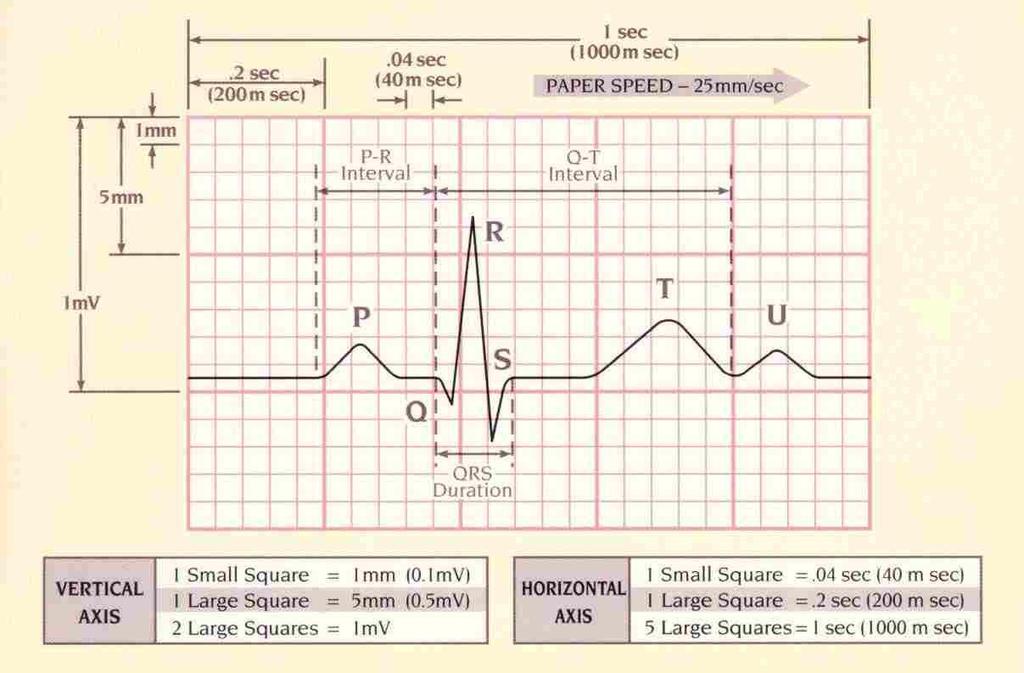
20 ECG (Electrocardiogram) 3 distinct waves are produced during cardiac cycle P wave caused by arterial depolarization QRS complex caused by ventricular depolarization T wave results from ventricular repolarization


21 Small box 1 mm 0.04 s Large box 5 mm (0.5mV) 0.20 s
22

23 The electrical activity of the heart originates in the SA node. The impulse then rapidly spreads through the right atrium to the AV node. It also spreads through the atrial muscle directly from the right atrium to the left atrium. The P-wave is generated by activation of the muscle of both atria
24 The impulse travels very slowly through the AV node, then very quickly through the bundle of His, then the bundle branches, the Purkinje network, and finally the ventricular muscle. The first area of the ventricular muscle to be activated is the interventricular septum, which activates from left to right. This generates the Q-wave.
25 Next, the left and right ventricular free walls, which form the bulk of the muscle of both ventricles, gets activated, with the endocardial surface being activated before the epicardial surface. This generates the R-wave.
26 A few small areas of the ventricles are activated at a rather late stage. This generates the S-wave.

27 Finally, the ventricular muscle repolarizes. This generates the T-wave.
28 P wave Represents atrial depolarization Impulse normally from the SA node Occurs before the QRS complex Shape Generally rounded Duration s Amplitude Usually < o.3 mv ( < 3mm) Focused on location and shape
29 P-R Interval Shape P wave and isoelectric (flat) Represents time it takes for the electrical impulse to travel from the SA node through AV node to the bundle branches Duration s Shorter PR indicates impulse didn t originate in SA node Prolonged PR indicated the Impulse is delayed as it passes through the AV node (AV-block) Focus on duration
30 QRS complex Ventricular depolarization Atrial repolarization Shape The QRS complex can take on many shapes in various ECG leads Duration s ( small boxes) Amplitude <30mm (3mV) peak R-wave to peak S-wave Focus on shape and duration
31 S-T segment S-T segment represents End of ventricular depolarization & beginning of ventricular repolarization Lowest blood supply through coronary arteries Shape Flat Depression -> MI or ischemia (> 1mm ) Elevation more than 1mm -> MI Some conditions and medications may cause S-T segment changes
32 T wave Ventricular repolarization

33 Q-T interval Time needed for ventricular depolarization-repolarization cycle Interval time varies with: Age/sex/HR QT shortens with increased HR Usually between s Abnormal duration may indicate a myocardial irregularity Focus on duration
34 Sinus rhythm Virtually all beats arise from the sinus node Sinus P wave P wave is conducted normally through the AV junction With normal sinus rhythm heart rate (atrial and ventricular) is regular and falls within the range of bpm.
35
36 ECG changes with exercise : healthy individual Tachycardia : heart rate increase is directly proportional to exercise intensity and myocardial work Rate-related shortening of QT interval S-T segment depression, upsloping, less than 1mm Exertional arrhythmias: rare, single PVCs

37 Premature heartbeats originating from the ventricles of the heart. A sign of decreased oxygenation to the heart muscle. PVCs are benign and may even be found in healthy hearts. A PVC may be perceived as a "skipped beat" or felt as palpitations in the chest. Although there are many signs and symptoms associated with PVCs, PVCs may have no symptom at all. PVCs can occur in a healthy person of any age, but are more prevalent in the elderly and in men.
38 ECG changes with exercise : an individual with myocardial ischemia and CAD Significant tachycardia : occurs at lower intensities of exercise Exertional arrhythmias : increased frequency of ventricular arrhythmias during exercise and/or recovery S-T segment depression; horizontal or downsloping depression, greater than 1mm below baseline is indicative of myocardial ischemia
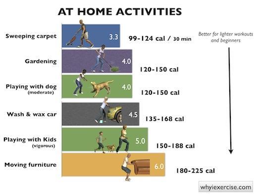
39 Determine activity levels :METs (Metabolic equivalents) MET : the amount of oxygen consumed at rest (sitting); equal to 3.5mL/kg per minute. MET levels can be estimated during ETT during steady state exercise; the max VO2 achieved on ETT is divided by resting VO2; highly predictable with standardized testing modes. Estimated MET = VO2 max resting VO2 Can be used to predict energy expenditure during certain activities.

40
41

42

43
44 Pre-test 12-lead ECG and BP in exercise postures Exercise 12-lead ECG recorded during last 15 seconds of every stage (2-3minutes) and at peak exercise or continuously BP during last minute of each stage RPE at the end of each stage
45 Post-test 12-lead ECG and BP immediately after exercise and every 1-2 minutes until stabilized near baseline Symptomatic ratings taken until return to baseline Monitoring continues until : SBP is within 20 mmhg of resting rate Arrhythmias have stabilized ST segment depression/elevation is <1mm difference from baseline levels


46 The physical therapist must have a clear understanding of the rationale for terminating any exercise test

47 Essentials of Cardiopulmonary Physical Therapy, 3 rd edition, Ellen Hillegass, Elsevier
48 Once the test is concluded, the results are written on a worksheet (documented)to provide data for the interpretation. The final summary of ETT should define weather the outcome of the test is normal or abnormal. If the outcome is abnormal, the summary should provide the reasons. The interpretation also provides valuable information regarding safety during exercise for the patient.

49 Essentials of Cardiopulmonary Physical Therapy, 3 rd edition, Ellen Hillegass, Elsevier
50 1. Take resting vital signs 2. Have patient/client stand next to a wall. Measure the height of the iliac crest and patella and mark it on the wall. Then place a piece of tape on the wall half the distance between the two. 3. On the signal go the patient/client begins stepping (not running) in place, raising each knee to the mark on the wall, for as many times as possible in the 2 minute period. 4. Only count the number of times the right knee reaches the required height. That is the score. 5. If the proper knee height cannot be maintained, ask the participant to slow down, or to stop until they can regain the proper form, but keep the stopwatch running. 6. At the end of the test, provide a cool down by asking the patient/client to walk slowly for a minute. 7. A person with impaired balance may use the back of a chair as a touch-hold for stability. Note this modification in your documentation 8. Take post exercise vital signs.

51 b0-1c5b-40f7-bd55-64a59158f7cf/805/fit_tb/fit011_1_lr10/fit011_1_lr10res/fit011_1_lr10_1_4_1.jpg
52 Rikli RE, Jones CJ (1999). Functional fitness normative scores for community residing older adults ages Journal of Aging and Physical Activity, 7, Range of scores between the 25% and 75% percentiles Age Number of steps Women Number of steps Men Scores less than 65 were associated with lower levels of functional ability
53 The original purpose of the six minute walk was to test exercise tolerance in chronic respiratory disease and heart failure. The 6-Minute Walk test is a sub maximal measure of aerobic capacity. The person may use an ambulation aid and oxygen if they do so normally The test has since been used as a performance-based measure of functional exercise capacity in other populations including healthy older adults, people undergoing knee or hip arthroplasty, fibromyalgia, and scleroderma. It has also been used with children.
54 Instructions to the Participant "The object of this test is to walk as far as possible for 6 minutes. You will walk back and forth in this hallway. Six minutes is a long time to walk, so you will be exerting yourself. You will probably get out of breath or become exhausted. You are permitted to slow down, to stop, and to rest as necessary. You may lean against the wall while resting, but resume walking as soon as you are able. You will be walking back and forth around the cones. You should pivot briskly around the cones and continue back the other way without hesitation. Now I'm going to show you. Please watch the way I turn without hesitation." (ATS, 2002) We will avoid having a conversation so that you can save your wind for walking. You can begin when I say go.
55

56
57 1. National Physical Therapy Examination, O sullivan&siegelman, TherapyEd 2. Essentials of Cardiopulmonary Physical Therapy, 3 rd edition, Ellen Hillegass, Elsevier 3. Cardiovascular and pulmonary Physical Therapy Evidence to Practice, 5 th edition, Donna Frownfelter, Elizabeth Dean, Elsevier 4. Cardiopulmonary Physical Therapy Management and Case Studies, 2 nd edition, W.Darlence Reid, Frank Chung, Kylie Hill, SLACK Inc. 5. Steele, Joel Dorman Hygienic Physiology (New York, NY: A. S. Barnes & Company, 1888) 6. PTEXAM the complete study guide, Scott M Giles, Scorebuilders 7. Khan academy,
Cardiopulmonary Physical Therapy. Haneul Lee, DSc, PT
 Cardiopulmonary Physical Therapy Haneul Lee, DSc, PT Airway Clearance Techniques Breathing Exercise Special Considerations for Mechanically Ventilated Exercise Injury Prevention and Equipment provision
Cardiopulmonary Physical Therapy. Haneul Lee, DSc, PT
 Cardiopulmonary Physical Therapy Haneul Lee, DSc, PT A comprehensive pulmonary rehabilitation program should incorporate the following components : Patient assessment and goal-setting Exercise and functional
Chad Morsch B.S., ACSM CEP
 What Is Cardiac Stress Testing? Chad Morsch B.S., ACSM CEP A Cardiac Stress Test is a test used to measure the heart's ability to respond to external stress in a controlled clinical environment. Cardiac
Mobilization and Exercise Prescription
 1 Clinicians can use this job aid as a tool to guide them through mobilization and exercise prescription with patients who have cardiopulmonary conditions. Mobilization and Exercise Prescription Therapy
Exercise Test: Practice and Interpretation. Jidong Sung Division of Cardiology Samsung Medical Center Sungkyunkwan University School of Medicine
 Exercise Test: Practice and Interpretation Jidong Sung Division of Cardiology Samsung Medical Center Sungkyunkwan University School of Medicine 2 Aerobic capacity and survival Circulation 117:614, 2008
Chapter 20 (2) The Heart
 The Heart") Chapter 20 (2) The Heart ----------------------------------------------------------------------------------------------------------------------------------------- Describe the component and function of
Chapter 20 (2) The Heart ----------------------------------------------------------------------------------------------------------------------------------------- Describe the component and function of
PROPEL: PRomoting Optimal Physical Exercise for Life* Submaximal Graded Exercise Assessment Guidelines
 PROPEL: PRomoting Optimal Physical Exercise for Life* Submaximal Graded Exercise Assessment Guidelines PROPEL: PRomoting Optimal Physical Exercise for Life* Submaximal Graded Exercise Assessment I. Foreword
Full file at
 MULTIPLE CHOICE. Choose the one alternative that best completes the statement or answers the question. 1) What electrical event must occur for atrial kick to occur? 1) A) Atrial repolarization B) Ventricular
UNDERSTANDING YOUR ECG: A REVIEW
 UNDERSTANDING YOUR ECG: A REVIEW Health professionals use the electrocardiograph (ECG) rhythm strip to systematically analyse the cardiac rhythm. Before the systematic process of ECG analysis is described
By the end of this lecture, you will be able to: Understand the 12 lead ECG in relation to the coronary circulation and myocardium Perform an ECG
 By the end of this lecture, you will be able to: Understand the 12 lead ECG in relation to the coronary circulation and myocardium Perform an ECG recording Identify the ECG changes that occur in the presence
Chapter 21: Clinical Exercise Testing Procedures
 Publisher link: thepoint http://thepoint.lww.com/book/show/2930 Chapter 21: Clinical Exercise Testing Procedures American College of Sports Medicine. (2010). ACSM's resource manual for guidelines for exercise
My Patient Needs a Stress Test
 My Patient Needs a Stress Test Amy S. Burhanna,, MD, FACC Coastal Cardiology Cape May Court House, New Jersey Absolute and relative contraindications to exercise testing Absolute Acute myocardial infarction
CASE 10. What would the ST segment of this ECG look like? On which leads would you see this ST segment change? What does the T wave represent?
 CASE 10 A 57-year-old man presents to the emergency center with complaints of chest pain with radiation to the left arm and jaw. He reports feeling anxious, diaphoretic, and short of breath. His past history
Interpreting Electrocardiograms (ECG) Physiology Name: Per:
 Physiology Name: Per:") Interpreting Electrocardiograms (ECG) Physiology Name: Per: Introduction The heart has its own system in place to create nerve impulses and does not actually require the brain to make it beat. This electrical
Interpreting Electrocardiograms (ECG) Physiology Name: Per: Introduction The heart has its own system in place to create nerve impulses and does not actually require the brain to make it beat. This electrical
EKG Abnormalities. Adapted from:
 EKG Abnormalities Adapted from: http://www.bem.fi/book/19/19.htm Some key terms: Arrhythmia-an abnormal rhythm or sequence of events in the EKG Flutter-rapid depolarizations (and therefore contractions)
Lab Activity 24 EKG. Portland Community College BI 232
 Lab Activity 24 EKG Reference: Dubin, Dale. Rapid Interpretation of EKG s. 6 th edition. Tampa: Cover Publishing Company, 2000. Portland Community College BI 232 Graph Paper 1 second equals 25 little boxes
4. The two inferior chambers of the heart are known as the atria. the superior and inferior vena cava, which empty into the left atrium.
 Answer each statement true or false. If the statement is false, change the underlined word to make it true. 1. The heart is located approximately between the second and fifth ribs and posterior to the
EKG Competency for Agency
 EKG Competency for Agency Name: Date: Agency: 1. The upper chambers of the heart are known as the: a. Atria b. Ventricles c. Mitral Valve d. Aortic Valve 2. The lower chambers of the heart are known as
Electrocardiography Abnormalities (Arrhythmias) 7. Faisal I. Mohammed, MD, PhD
 7. Faisal I. Mohammed, MD, PhD") Electrocardiography Abnormalities (Arrhythmias) 7 Faisal I. Mohammed, MD, PhD 1 Causes of Cardiac Arrythmias Abnormal rhythmicity of the pacemaker Shift of pacemaker from sinus node Blocks at different
Electrocardiography Abnormalities (Arrhythmias) 7 Faisal I. Mohammed, MD, PhD 1 Causes of Cardiac Arrythmias Abnormal rhythmicity of the pacemaker Shift of pacemaker from sinus node Blocks at different
Cardiac Cycle. Each heartbeat is called a cardiac cycle. First the two atria contract at the same time.
 The Heartbeat Cardiac Cycle Each heartbeat is called a cardiac cycle. First the two atria contract at the same time. Next the two ventricles contract at the same time. Then all the chambers relax. http://www.youtube.com/watch?v=frd3k6lkhws
Cardiovascular System Notes: Heart Disease & Disorders
 Cardiovascular System Notes: Heart Disease & Disorders Interesting Heart Facts The Electrocardiograph (ECG) was invented in 1902 by Willem Einthoven Dutch Physiologist. This test is still used to evaluate
Chapter 08. Health Screening and Risk Classification
 Chapter 08 Health Screening and Risk Classification Preliminary Health Screening and Risk Classification Protocol: 1) Conduct a Preliminary Health Evaluation 2) Determine Health /Disease Risks 3) Determine
Signal Processing of Stress Test ECG Using MATLAB
 Signal Processing of Stress Test ECG Using MATLAB Omer Mukhtar Wani M. Tech ECE Geeta Engineering College, Panipat Abstract -Electrocardiography is used to record the electrical activity of the heart over
ECG. Prepared by: Dr.Fatima Daoud Reference: Guyton and Hall Textbook of Medical Physiology,12 th edition Chapters: 11,12,13
 ECG Prepared by: Dr.Fatima Daoud Reference: Guyton and Hall Textbook of Medical Physiology,12 th edition Chapters: 11,12,13 The Concept When the cardiac impulse passes through the heart, electrical current
Chapter 9, Part 2. Cardiocirculatory Adjustments to Exercise
 Chapter 9, Part 2 Cardiocirculatory Adjustments to Exercise Electrical Activity of the Heart Contraction of the heart depends on electrical stimulation of the myocardium Impulse is initiated in the right
Graded exercise testing (GXT): extension of medical history and physical examination
: extension of medical history and physical examination") Related Readings Fletcher, G. F., Balady, G. J., Amsterdam, E. A., Chaitman, B., Eckel, R., Fleg, J., et al. (2001). Exercise standards for testing and training: A statement for healthcare professionals
Related Readings Fletcher, G. F., Balady, G. J., Amsterdam, E. A., Chaitman, B., Eckel, R., Fleg, J., et al. (2001). Exercise standards for testing and training: A statement for healthcare professionals
Disclosure. 3. ST depression indicative of ischemia is most commonly observed in leads: 1. V1-V2. 2. I and avl 3. V
 Interpreting Stress Induced Ischemia by ECG, Bundle Branch Block & Arrhythmias Disclosure Gregory S Thomas MD, MPH Medical Director, MemorialCare Heart & Vascular Institute, Long Beach Memorial Astellas
physiology 6 Mohammed Jaafer Turquoise team
 15 physiology 6 Mohammed Jaafer 22-3-2016 Turquoise team Cardiac Arrhythmias and Their Electrocardiographic Interpretation Today, we are going to talk about the abnormal excitation. As we said before,
Subject: Outpatient Phase Ii Cardiac Rehab Individualized Treatment Plan And Exercise Prescription
 CARDIAC REHAB POLICY & PROCEDURES Policy #: CR 208 Subject: Outpatient Phase Ii Cardiac Rehab Individualized Treatment Plan And Exercise Prescription Purpose: To establish guidelines for developing and
C1: Medical Standards for Safety Critical Workers with Cardiovascular Disorders
 C1: Medical Standards for Safety Critical Workers with Cardiovascular Disorders GENERAL ISSUES REGARDING MEDICAL FITNESS-FOR-DUTY 1. These medical standards apply to Union Pacific Railroad (UPRR) employees
Collin County Community College
 Collin County Community College BIOL. 2402 Anatomy & Physiology WEEK 5 The Heart 1 The Heart Beat and the EKG 2 1 The Heart Beat and the EKG P-wave = Atrial depolarization QRS-wave = Ventricular depolarization
This presentation will deal with the basics of ECG description as well as the physiological basics of
 Snímka 1 Electrocardiography basics This presentation will deal with the basics of ECG description as well as the physiological basics of Snímka 2 Lecture overview 1. Cardiac conduction system functional
Course: Exercise and Aging for Special Populations
 Copyright EFS Inc. All Rights Reserved. Course: Exercise and Aging for Special Populations Session 2: Cardiovascular and Metabolic Disease Considerations for Exercise Program Design Presentation Created
Human Anatomy and Physiology II Laboratory Cardiovascular Physiology
 Human Anatomy and Physiology II Laboratory Cardiovascular Physiology 1 This lab involves two exercises: 1) Conduction System of the Heart and Electrocardiography and 2) Human Cardiovascular Physiology:
ECG ABNORMALITIES D R. T AM A R A AL Q U D AH
 ECG ABNORMALITIES D R. T AM A R A AL Q U D AH When we interpret an ECG we compare it instantaneously with the normal ECG and normal variants stored in our memory; these memories are stored visually in
Lab #3: Electrocardiogram (ECG / EKG)
") Lab #3: Electrocardiogram (ECG / EKG) An introduction to the recording and analysis of cardiac activity Introduction The beating of the heart is triggered by an electrical signal from the pacemaker. The
Lab #3: Electrocardiogram (ECG / EKG) An introduction to the recording and analysis of cardiac activity Introduction The beating of the heart is triggered by an electrical signal from the pacemaker. The
37 1 The Circulatory System
 H T H E E A R T 37 1 The Circulatory System The circulatory system and respiratory system work together to supply cells with the nutrients and oxygen they need to stay alive. a) The respiratory system:
Contra-indications, Risks, and Safety Precautions for Stress Testing. ACSM guidelines, pg 20 7 ACSM RISK FACTORS. Risk Classifications pg 27
 Contra-indications, Risks, and Safety Precautions for Stress Testing Data to Support Stress Testing How safe is stress testing? Contra-indications Termination Criteria Ellstad Chapt 5 ACSM Chapts 3-6 Seattle
ECG interpretation basics
 ECG interpretation basics Michał Walczewski, MD Krzysztof Ozierański, MD 21.03.18 Electrical conduction system of the heart Limb leads Precordial leads 21.03.18 Precordial leads Precordial leads 21.03.18
Cardiovascular System Notes: Physiology of the Heart
 Cardiovascular System Notes: Physiology of the Heart Interesting Heart Fact Capillaries are so small it takes ten of them to equal the thickness of a human hair. Review What are the 3 parts of the cardiovascular
Sample. Analyzing the Heart with EKG. Computer
 Analyzing the Heart with EKG Computer An electrocardiogram (ECG or EKG) is a graphical recording of the electrical events occurring within the heart. In a healthy heart there is a natural pacemaker in
Lab 16. The Cardiovascular System Heart and Blood Vessels. Laboratory Objectives
 Lab 16 The Cardiovascular System Heart and Blood Vessels Laboratory Objectives Describe the anatomical structures of the heart to include the pericardium, chambers, valves, and major vessels. Describe
Understanding the 12-lead ECG, part II
 Bundle-branch blocks Understanding the 12-lead ECG, part II Most common electrocardiogram (ECG) abnormality Appears as a wider than normal S complex Occurs when one of the two bundle branches can t conduct
CORONARY ARTERIES. LAD Anterior wall of the left vent Lateral wall of left vent Anterior 2/3 of interventricluar septum R & L bundle branches
 CORONARY ARTERIES RCA Right atrium Right ventricle SA node 55% AV node 90% Posterior wall of left ventricle in 90% Posterior third of interventricular septum 90% LAD Anterior wall of the left vent Lateral
CARDIOVASCULAR SYSTEM
 CARDIOVASCULAR SYSTEM Overview Heart and Vessels 2 Major Divisions Pulmonary Circuit Systemic Circuit Closed and Continuous Loop Location Aorta Superior vena cava Right lung Pulmonary trunk Base of heart
DR QAZI IMTIAZ RASOOL OBJECTIVES
 PRACTICAL ELECTROCARDIOGRAPHY DR QAZI IMTIAZ RASOOL OBJECTIVES Recording of electrical events in heart Established electrode pattern results in specific tracing pattern Health of heart i. e. Anatomical
Chapter 9. Learning Objectives. Learning Objectives 9/11/2012. Cardiac Arrhythmias. Define electrical therapy
 Chapter 9 Cardiac Arrhythmias Learning Objectives Define electrical therapy Explain why electrical therapy is preferred initial therapy over drug administration for cardiac arrest and some arrhythmias
Paroxysmal Supraventricular Tachycardia PSVT.
 Atrial Tachycardia; is the name for an arrhythmia caused by a disorder of the impulse generation in the atrium or the AV node. An area in the atrium sends out rapid signals, which are faster than those
REtrive. REpeat. RElearn Design by. Test-Enhanced Learning based ECG practice E-book
 Test-Enhanced Learning Test-Enhanced Learning Test-Enhanced Learning Test-Enhanced Learning based ECG practice E-book REtrive REpeat RElearn Design by S I T T I N U N T H A N G J U I P E E R I Y A W A
Family Medicine for English language students of Medical University of Lodz ECG. Jakub Dorożyński
 Family Medicine for English language students of Medical University of Lodz ECG Jakub Dorożyński Parts of an ECG The standard ECG has 12 leads: six of them are considered limb leads because they are placed
Electrical Conduction
 Sinoatrial (SA) node Electrical Conduction Sets the pace of the heartbeat at 70 bpm AV node (50 bpm) and Purkinje fibers (25 40 bpm) can act as pacemakers under some conditions Internodal pathway from
Step by step approach to EKG rhythm interpretation:
 Sinus Rhythms Normal sinus arrhythmia Small, slow variation of the R-R interval i.e. variation of the normal sinus heart rate with respiration, etc. Sinus Tachycardia Defined as sinus rhythm with a rate
Relax and Learn At the Farm 2012
 Relax and Learn At the Farm 2012 Session 2: 12 Lead ECG Fundamentals 101 Cynthia Webner DNP, RN, CCNS, CCRN-CMC, CHFN Though for Today Mastery is not something that strikes in an instant, like a thunderbolt,
Electrocardiography for Healthcare Professionals
 Electrocardiography for Healthcare Professionals Kathryn A. Booth Thomas O Brien Chapter 5: Rhythm Strip Interpretation and Sinus Rhythms Learning Outcomes 5.1 Explain the process of evaluating ECG tracings
MR Advance Techniques. Cardiac Imaging. Class IV
 MR Advance Techniques Cardiac Imaging Class IV Heart The heart is a muscular organ responsible for pumping blood through the blood vessels by repeated, rhythmic contractions. Layers of the heart Endocardium
Cardiac Telemetry Self Study: Part One Cardiovascular Review 2017 THINGS TO REMEMBER
 Please review the above anatomy of the heart. THINGS TO REMEMBER There are 3 electrolytes that affect cardiac function o Sodium, Potassium, and Calcium When any of these electrolytes are out of the normal
CARDIAC CYCLE CONTENTS. Divisions of cardiac cycle 11/13/13. Definition. Badri Paudel GMC
 CARDIAC CYCLE Badri Paudel GMC CONTENTS Ø DEFINATION Ø DIVISION OF CARDIAC CYCLE Ø SUB DIVISION AND DURATION OF CARDIAC CYCLE Ø SYSTOLE Ø DIASTOLE Ø DESCRIPTION OF EVENTS OF CARDIAC CYCLE Ø SUMMARY Ø ELECTROCARDIOGRAPHY
ABCs of ECGs. Shelby L. Durler
 ABCs of ECGs Shelby L. Durler Objectives Review the A&P of the cardiac conduction system Placement and obtaining 4-lead and 12-lead ECGs Overview of the basics of ECG rhythm interpretation Intrinsic
12 Lead ECG Interpretation
 12 Lead ECG Interpretation Julie Zimmerman, MSN, RN, CNS, CCRN Significant increase in mortality for every 15 minutes of delay! N Engl J Med 2007;357:1631-1638 Who should get a 12-lead ECG? Also include
MYOCARDIALINFARCTION. By: Kendra Fischer
 MYOCARDIALINFARCTION By: Kendra Fischer Outline Definition Epidemiology Clinical Aspects Treatment Effects of Exercise Exercise Testing Exercise Rx Summary and Conclusions References Break it down MYOCARDIAL
THE CARDIOVASCULAR SYSTEM. Heart 2
 THE CARDIOVASCULAR SYSTEM Heart 2 PROPERTIES OF CARDIAC MUSCLE Cardiac muscle Striated Short Wide Branched Interconnected Skeletal muscle Striated Long Narrow Cylindrical PROPERTIES OF CARDIAC MUSCLE Intercalated
The HEART. What is it???? Pericardium. Heart Facts. This muscle never stops working It works when you are asleep
 This muscle never stops working It works when you are asleep The HEART It works when you eat It really works when you exercise. What is it???? Located between the lungs in the mid thoracic region Apex
HTEC 91. Performing ECGs: Procedure. Normal Sinus Rhythm (NSR) Topic for Today: Sinus Rhythms. Characteristics of NSR. Conduction Pathway
 Topic for Today: Sinus Rhythms. Characteristics of NSR. Conduction Pathway") HTEC 91 Medical Office Diagnostic Tests Week 3 Performing ECGs: Procedure o ECG protocol: you may NOT do ECG if you have not signed up! If you are signed up and the room is occupied with people who did
HTEC 91 Medical Office Diagnostic Tests Week 3 Performing ECGs: Procedure o ECG protocol: you may NOT do ECG if you have not signed up! If you are signed up and the room is occupied with people who did
Lab 2. The Intrinsic Cardiac Conduction System. 1/23/2016 MDufilho 1
 Lab 2 he Intrinsic Cardiac Conduction System 1/23/2016 MDufilho 1 Figure 18.13 Intrinsic cardiac conduction system and action potential succession during one heartbeat. Superior vena cava ight atrium 1
Cardiovascular System: The Heart
 Cardiovascular System: The Heart I. Anatomy of the Heart (See lab handout for terms list) A. Describe the size, shape and location of the heart B. Describe the structure and function of the pericardium
Basic EKG Interpretation. Nirja Parikh, PT, DPT
 Basic EKG Interpretation Nirja Parikh, PT, DPT Electrocardiogram (EKG) Using surface electrodes record the electrical activity of the heart electrical activity (usually) correlates to mechanical function
CRITICAL THINKING QUESTIONS AND ANSWERS AND CYCLE 2 LAB EXAM TEMPLATE. There are two main mechanisms that work in conjunction to return the blood
 CRITICAL THINKING QUESTIONS AND ANSWERS AND CYCLE 2 LAB EXAM TEMPLATE There are two main mechanisms that work in conjunction to return the blood THE CARDIAC PUMP 1) The forward pull(vis a fronte) This
Unit 6: Circulatory System. 6.2 Heart
 Unit 6: Circulatory System 6.2 Heart Functions of Circulatory System 1. The heart is the pump necessary to circulate blood to all parts of the body 2. Arteries, veins and capillaries are the structures
CRC 431 ECG Basics. Bill Pruitt, MBA, RRT, CPFT, AE-C
 CRC 431 ECG Basics Bill Pruitt, MBA, RRT, CPFT, AE-C Resources White s 5 th ed. Ch 6 Electrocardiography Einthoven s Triangle Chest leads and limb leads Egan s 10 th ed. Ch 17 Interpreting the Electrocardiogram
The Electrocardiogram
 The Electrocardiogram Chapters 11 and 13 AUTUMN WEDAN AND NATASHA MCDOUGAL The Normal Electrocardiogram P-wave Generated when the atria depolarizes QRS-Complex Ventricles depolarizing before a contraction
Outline. Electrical Activity of the Human Heart. What is the Heart? The Heart as a Pump. Anatomy of the Heart. The Hard Work
 Electrical Activity of the Human Heart Oguz Poroy, PhD Assistant Professor Department of Biomedical Engineering The University of Iowa Outline Basic Facts about the Heart Heart Chambers and Heart s The
Matters of the Heart: Comprehensive Cardiology SARAH BEANLANDS RN BSCN MSC
 Matters of the Heart: Comprehensive Cardiology SARAH BEANLANDS RN BSCN MSC Who am I? Class Outline Gross anatomy of the heart Trip around the heart Micro anatomy: cellular and tissue level Introduction
Patient Resources: Arrhythmias and Congenital Heart Disease
 Patient Resources: Arrhythmias and Congenital Heart Disease Overview Arrhythmias (abnormal heart rhythms) can develop in patients with congenital heart disease (CHD) due to thickening/weakening of their
12-Lead ECG Interpretation. Kathy Kuznar, RN, ANP
 12-Lead ECG Interpretation Kathy Kuznar, RN, ANP The 12-Lead ECG Objectives Identify the normal morphology and features of the 12- lead ECG. Perform systematic analysis of the 12-lead ECG. Recognize abnormalities
Electrocardiography Biomedical Engineering Kaj-Åge Henneberg
 Electrocardiography 31650 Biomedical Engineering Kaj-Åge Henneberg Electrocardiography Plan Function of cardiovascular system Electrical activation of the heart Recording the ECG Arrhythmia Heart Rate
PART I. Disorders of the Heart Rhythm: Basic Principles
 PART I Disorders of the Heart Rhythm: Basic Principles FET01.indd 1 1/11/06 9:53:05 AM FET01.indd 2 1/11/06 9:53:06 AM CHAPTER 1 The Cardiac Electrical System The heart spontaneously generates electrical
3/26/15 HTEC 91. EKG Sign-in Book. The Cardiac Cycle. Parts of the ECG. Waves. Waves. Review of protocol Review of placement of chest leads (V1, V2)
") EKG Sign-in Book HTEC 91 Review of protocol Review of placement of chest leads (V1, V2) Medical Office Diagnostic Tests Week 2 http://www.cvphysiology.com/arrhythmias/a013c.htm The Cardiac Cycle Represents
EKG Sign-in Book HTEC 91 Review of protocol Review of placement of chest leads (V1, V2) Medical Office Diagnostic Tests Week 2 http://www.cvphysiology.com/arrhythmias/a013c.htm The Cardiac Cycle Represents
ECG CONVENTIONS AND INTERVALS
 1 ECG Waveforms and Intervals ECG waveforms labeled alphabetically P wave== represents atrial depolarization QRS complex=ventricular depolarization ST-T-U complex (ST segment, T wave, and U wave)== V repolarization.
Cardiovascular Nursing Practice: A Comprehensive Resource Manual and Study Guide for Clinical Nurses 2 nd Edition
 Cardiovascular Nursing Practice: A Comprehensive Resource Manual and Study Guide for Clinical Nurses 2 nd Edition Table of Contents Volume 1 Chapter 1: Cardiovascular Anatomy and Physiology Basic Cardiac
CAMOSUN COLLEGE BIOLOGY 144 (2010) LABS
 LABS") LAB 8: CARDIOVASCULAR PHYSIOLOGY PART 1. HEART SOUNDS AND PULSE DETERMINATIONS Introduction Two distinct sounds can be heard during each cardiac cycle. These sounds are commonly described as lub and dup
LAB 8: CARDIOVASCULAR PHYSIOLOGY PART 1. HEART SOUNDS AND PULSE DETERMINATIONS Introduction Two distinct sounds can be heard during each cardiac cycle. These sounds are commonly described as lub and dup
Anatomy Review: The Heart Graphics are used with permission of A.D.A.M. Software, Inc. and Benjamin/Cummings Publishing Co.
 Anatomy Review: The Heart Graphics are used with permission of A.D.A.M. Software, Inc. and Benjamin/Cummings Publishing Co. Anatomy Views Label the diagrams of the heart below: Interactive Physiology Study
Practice Exercises for the Cardiovascular System
 Practice Exercises for the Cardiovascular System On the diagram below, color the oxygen-rich blood red and the oxygen-poor blood blue. Label the parts: Continued on the next page... Label the parts on
Study methodology for screening candidates to athletes risk
 1. Periodical Evaluations: each 2 years. Study methodology for screening candidates to athletes risk 2. Personal history: Personal history of murmur in childhood; dizziness, syncope, palpitations, intolerance
ECG Interpretation Cat Williams, DVM DACVIM (Cardiology)
") ECG Interpretation Cat Williams, DVM DACVIM (Cardiology) Providing the best quality care and service for the patient, the client, and the referring veterinarian. GOAL: Reduce Anxiety about ECGs Back to
ECG Interpretation Cat Williams, DVM DACVIM (Cardiology) Providing the best quality care and service for the patient, the client, and the referring veterinarian. GOAL: Reduce Anxiety about ECGs Back to
Cardiovascular Physiology
 Cardiovascular Physiology The mammalian heart is a pump that pushes blood around the body and is made of four chambers: right and left atria and right and left ventricles. The two atria act as collecting
Where are the normal pacemaker and the backup pacemakers of the heart located?
 CASE 9 A 68-year-old woman presents to the emergency center with shortness of breath, light-headedness, and chest pain described as being like an elephant sitting on her chest. She is diagnosed with a
Risk Stratification for CAD for the Primary Care Provider
 Risk Stratification for CAD for the Primary Care Provider Shimoli Shah MD Assistant Professor of Medicine Directory, Ambulatory Cardiology Clinic Knight Cardiovascular Institute Oregon Health & Sciences
DEPARTMENT NAME PRE-PARTICIPATION SCREENING THE SPORTS PHYSICAL
 PRE-PARTICIPATION SCREENING THE SPORTS PHYSICAL Michele Krenek, MSN, RN, FNP-C TCHAPP Conference, Houston, TX April 4, 2019 PRE-PARTICIPATION SPORTS SCREENING According to the AHA the definition of the
CORONARY ARTERIES HEART
 CARDIAC/ECG MODULE THE HEART CORONARY ARTERIES FIBRILLATING HEART CORONARY ARTERIES HEART PRACTICE RHYTHMS PRACTICE RHYTHMS ELECTRICAL CONDUCTION SA Node (60 100) Primary pacemaker AV Node (40 60) ***Creates
TELEMETRY BASICS FOR NURSING STUDENTS
 TELEMETRY BASICS FOR NURSING STUDENTS Accuracy of cardiac monitoring is an important component of patient safety in hospitalized patients who meet the criteria for dysrhythmia monitoring. (AACN, 2016,
Physical Fitness Biology Concepts of Biology 13.1
 Physical Fitness Biology 100 - Concepts of Biology 13.1 Name Instructor Lab Section Objectives: To gain an understanding of: What is meant by the term physical fitness Various tests used to measure an
Practice Exam Case Study
 Practice Exam 2017 Case Study A 58-year old professor presents himself to your clinic. He is apparently healthy, but his older brother had a heart attack at age 60, as did his father (age 62). He is clinically
The Electrocardiogram part II. Dr. Adelina Vlad, MD PhD
 The Electrocardiogram part II Dr. Adelina Vlad, MD PhD Basic Interpretation of the ECG 1) Evaluate calibration 2) Calculate rate 3) Determine rhythm 4) Determine QRS axis 5) Measure intervals 6) Analyze
Cardiovascular Emergencies. January 2016 Continuing Education
 Cardiovascular Emergencies January 2016 Continuing Education Cardiovascular Emergency Objectives Review the anatomy of heart structures, vessels and function. Review of the Cardiac Cycle Blood Flow through
Cardiovascular System
 Cardiovascular System The Heart Cardiovascular System The Heart Overview What does the heart do? By timed muscular contractions creates pressure gradients blood moves then from high pressure to low pressure
TEST BANK FOR ECGS MADE EASY 5TH EDITION BY AEHLERT
 Link download full: http://testbankair.com/download/test-bank-for-ecgs-made-easy-5thedition-by-aehlert/ TEST BANK FOR ECGS MADE EASY 5TH EDITION BY AEHLERT Chapter 5 TRUE/FALSE 1. The AV junction consists
PATIENT WITH ARRHYTHMIA IN DENTIST S OFFICE. Małgorzata Kurpesa, MD., PhD. Chair&Department of Cardiology
 PATIENT WITH ARRHYTHMIA IN DENTIST S OFFICE Małgorzata Kurpesa, MD., PhD. Chair&Department of Cardiology Medical University of Łódź The heart is made up of four chambers Left Atrium Right Atrium Left Ventricle
P F = R. Disorder of the Breast. Approach to the Patient with Chest Pain. Typical Characteristics of Angina Pectoris. Myocardial Ischemia
 Disorder of the Breast Approach to the Patient with Chest Pain Anthony J. Minisi, MD Department of Internal Medicine, Division of Cardiology Virginia Commonwealth University School of Medicine William
Cardiac Arrhythmias. Cathy Percival, RN, FALU, FLMI VP, Medical Director AIG Life and Retirement Company
 Cardiac Arrhythmias Cathy Percival, RN, FALU, FLMI VP, Medical Director AIG Life and Retirement Company The Cardiovascular System Three primary functions Transport of oxygen, nutrients, and hormones to
Listing Form: Heart or Cardiovascular Impairments. Medical Provider:
 Listing Form: Heart or Cardiovascular Impairments Medical Provider: Printed Name Signature Patient Name: Patient DOB: Patient SS#: Date: Dear Provider: Please indicate whether your patient s condition