OCT Findings: Lesson from Stable vs Unstable Plaques
|
|
|
- Joy Robertson
- 6 years ago
- Views:
Transcription


1 ANGIOPLASTY SUMMIT TCTAP 2010 Imaging Workshop OCT Findings: Lesson from Stable vs Unstable Plaques Giulio Guagliumi MD Ospedali Riuniti di Bergamo, Italy

2 DISCLOSURE OF FINANCIAL INTERESTS Consultant Boston Scientific Volcano Grant/ Research Support Medtronic Boston Scientific LightLab Imaging Abbott Vascular Labcoat Giulio Guagliumi Ospedali Riuniti di Bergamo, Italy

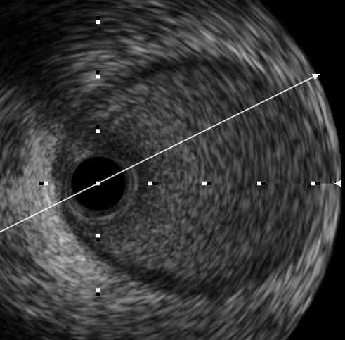
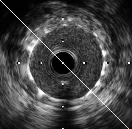
3 Calcified Artery with/without Surface Lipid Pool Ca ++ LP OFDI IVUS


4 From a Foggy Sight to a Clear Vision G. Guagliumi and M. Costa Editorial Comment JACC Interv 2009 May18; 2: 467


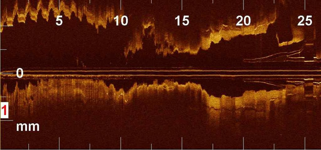
5 ACS: Focus on mid-distal LAD V. Sirbu Ospedali Riuniti di Bergamo


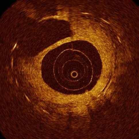
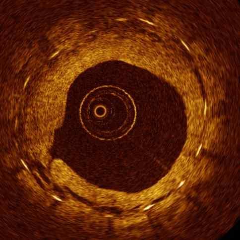
6 LAD Focus on proximal LAD LM CSA stenosis=31% CSA=6.17mm² CSA=5.10mm² CSA=8.70mm²


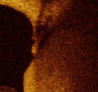
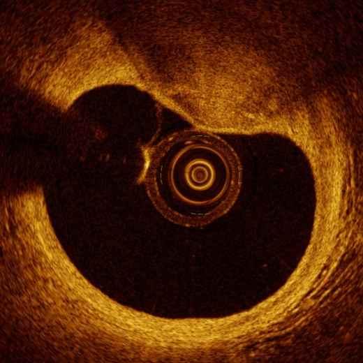
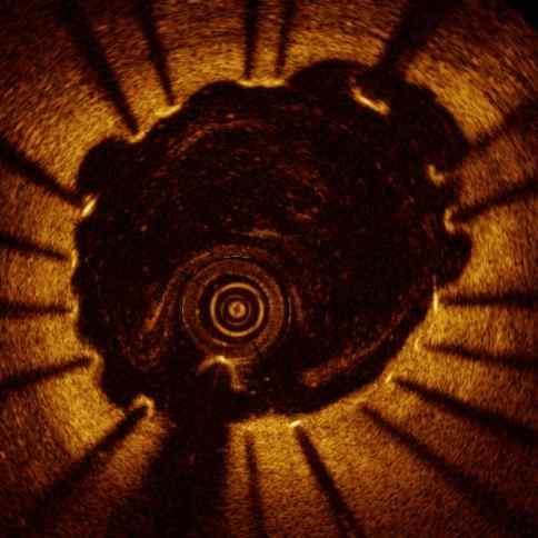
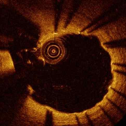
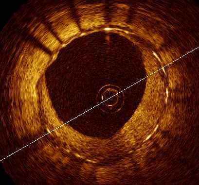
7 TCFA in Proximal LAD


8 Thin-cap fibroatheroma (TCFA) Fibrous cap Lipid plaque 30µm 100µm TCFA was defined as a plaque with fibrous cap <65µm thick. The high resolution of OCT has an ability to identify thin-fibrous cap clearly even if it is less than 100µm. Kubo et al. Expert Rev Med Dev. 2008;5:691-7
9 Clinical Presentation and Plaque Morphology in Unstable Angina 80 Class I Class II Class III % p< µm p< mm² p= % p< plaque rupture 0 cap thickness 0 MLA 0 ulceration/no cap rupture M. Mizukoshi et al. i2 ACC. 2010; JACC Poster
10 Multivessel Desease: SAP vs AMI Infarct Related Artery SAP AMI 100 % 100 % 200 µm 80 p<0.001 p= p< Plaque rupture 0 Thrombus 0 Cap Thickness Kubo T et al. Am J Cardiol 2010:105:
11 Multivessel Desease: SAP vs AMI Non Infarct Related Artery SAP AMI 100 % p= µm p= TCFA Cap Thickness Kubo T et al. Am J Cardiol 2010:105:


, plaque rupture")
 were")

 and")
 were detected by")

12 Multiple plaque rupture in AMI: 3-vessel OCT examination The culprit lesion was in LCX (#11). TCFA (6), plaque rupture (7, 8) and intracoronary thrombus (7, 8) were observed by OCT. Although the plaques in LAD (12, 13) were not unstable, TCFA (1-5) and plaque rupture (3, 4, 5, 10, 11) were detected by OCT in the non-culprit lesions of RCA and LCX. Kitabata, Kubo et al. Heart. 2008;94:544


13 ACS Multilink 3.0/25 mm 12 yrs after implant G. Guagliumi, V. Sirbu Ospedali Riuniti di Bergamo

14 LAD distally to the thrombotic stent site in VLST of BMS Ospedali Riuniti di Bergamo


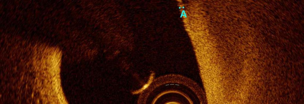
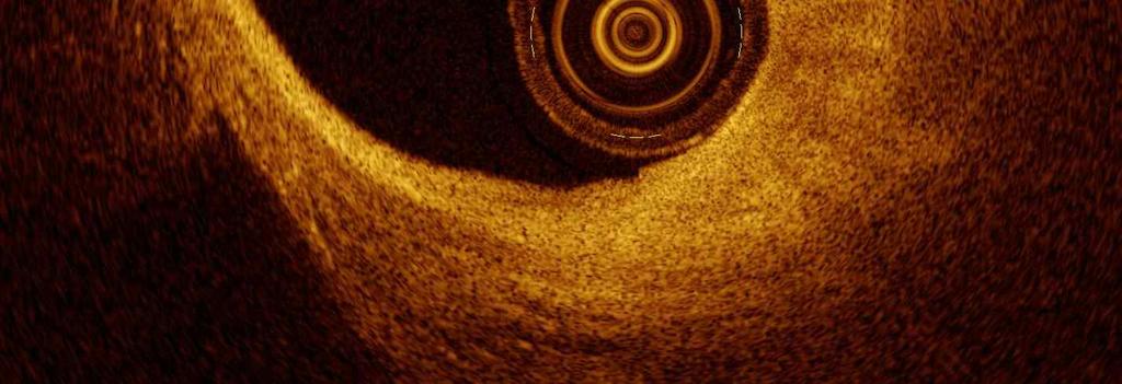
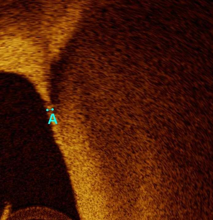
15 TCFA rupture Ospedali Riuniti di Bergamo

16 TCFA rupture 32 µm Ospedali Riuniti di Bergamo


17 Use of VH-IVUS vs OCT for detecting in-vivo TCFA Non-thin-cap IVUS-derived TCFA n=33 % necrotic-core: 15% Angle of the total NCCL 35.8º Cap thickness; 90µm Definite TCFA n= 28 % necrotic-core: 22% Cap thickness; 40µm Angle of the total NCCL 100.3º T. Sawada, et al. EHJ 2008; 29(9):
 Definite-TCFA VH +, OCT +")
 96.3 (75.6, 117.0) 0.")
 0.0005 VH % Necrotic-core 18.6 (15.7, 21.4) 20.")
 89.4 (63.6, 112.4) 0.0003 T. Sawada, et al.")
18 Combined use of IVUS-VH and OCT for detection of TCFA Non-TCFA VH +, OCT - (n=33) Definite-TCFA VH +, OCT + (n=28) P Gray-scale IVUS Plaque volume (mm 3 /cm) 65.3 (39.3, 91.4) 96.3 (75.6, 117.0) Remodelling index 1.10 (1.06, 1.13) 1.21 (1.17, 1.25) VH % Necrotic-core 18.6 (15.7, 21.4) 20.0 (17.0, 22.9) NS Total NCCL angle (degree) 54.6 (35.6, 73.5) 89.4 (63.6, 112.4) T. Sawada, et al. EHJ 2008; 29(9):
19 G. Guagliumi, V. Sirbu Ospedali Riuniti di Bergamo


20 in-vivo Association between Positive Remodeling and TFCA IVUS and OCT imaging in 54 lesions from 48 pts % of TCFA within each group 100 p < % % 5.6 % Positive Absent Negative Remodeling O. Raffael Eur Heart J 2008; 29:1721


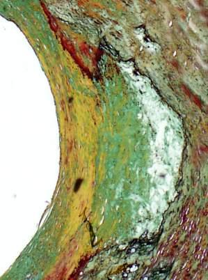
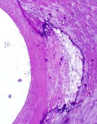
21 OFDI Rupture Site IVUS NC NC Cavity Cavity Cavity Thrombus Thrombus NC NC Courtesy R. Virmani MD
22 OCT identifies plaque rupture, cap erosion and TCFA..and Thrombus OCT vs AS vs IVUS: 30 AMI pts Finding OCT (n=30) AS (n=30) IVUS (n=30) p Fibrous cap disruption 73% * 47% 40% Fibrous cap erosion 23% * 3% 0% Thrombus 100% 100% 33% < * OCT vs AS, p<0.05. OCT vs IVUS, p<0.01. AS vs IVUS, p<0.01. Kubo et al. J Am Coll Card 2007; 50: 933
23 OCT Findings of Culprit Lesions STEMI n=40 NSTE ACS n=49 P value Plaque Rupture % Lipid Rich Plaque % (>= 2 Quadrants) TFCA % Fibrous Cap Thickness µm Thrombus % Red White None ±20 109± M. Riga i2acc 2010, JACC A190 :
24 OCT and IVUS Findings After PCI in UAP and SAP Patients (%) UAP IVUS SAP (%) Inadequate * Stent Apposition p=0.001 OCT Tissue* Protrusion Intracoronary* Thrombus OCT vs IVUS* P< Inadequate Stent Apposition Tissue Protrusion Intracoronary Thrombus T. Kubo et al J Am Coll Cardiovasc Imaging, July 2008:475-84


25 HORIZONS AMI: 2 Separated Lesions TAXUS mm mm Index Procedure 13 month FU Ospedali Riuniti di Bergamo


26 HORIZONS OCT 13 mos FU TAXUS 2.75 x 20 mm culprit lesion TAXUS mm non culprit G. Guagliumi MD, Ospedali Riuniti di Bergamo
 Stable /UA vs STEMI (%) p=0.")
 DES: Cypher, Taxus, Xience, Biolimus STEMI:")


27 Difference in DES Incomplete Strut Apposition (ISA) Stable /UA vs STEMI (%) p=0.01 Median follow-up time 9 months (range 7 to 72) DES: Cypher, Taxus, Xience, Biolimus STEMI: N=16 Stable/Unstable Angina : N=27 % Lesion with at least one ISA % Frames with at least one ISA N. Gonzalo et al JACC Int 2009; 2 (5): % of Incompletely apposed struts PSST April 2009 Page 27 of 19


28 Thrombus Naturally or Pharmacologically Remodels Overtime Delayed Healing? Late Incomplete Apposition? B1 G. Guagliumi : HORIZONS Trial 3604 AMI randomized
29 STEMI Postimplant 6 mos FU G. Guagliumi, V. Sirbu Ospedali Riuniti di Bergamo


30 OCTAMI Trial : Index RCA Endeavor 3.0/24 mm 6 mos FU OCT G. Guagliumi et al TCT Featured Clinical Research, JACC Intv 2010 May, in press
31 Coverage distribution in different cross-sections along the Stent 6-month FU (%) BMS ZES Percentage of Covered Struts Proximal Mid Distal G. Guagliumi et al OCTAMI Featured Research TCT, JACC Intv 2010, May in press


32 HORIZONS OCT: 13 Months 118 Consecutive STEMI pts enrolled in HORIZONS, 155 Taxus vs 45 BMS % p= p< Independent Core Labs Blind to the stent assignment OCT Core Lab: Case Western Reserve Univeristy, Cleveland, OH 7,748 cross-sections- 44, 121struts G. Guagliumi et al, LBT Abstracts Circulation 2008;118: , submitted
33 Difference in Uncovered Strut Frequency - DES (%) p=0.04 Median follow-up time 9 months (range 7 to 72) STEMI: N=16 Stable/Unstable Angina : N=27 % Lesion with at least one uncovered strut % Frames with at least one uncovered strut N. Gonzalo et al JACC Int 2009; 2 (5): % Uncovered struts PSST April 2009 Page 33 of 19
34 Consistent Strut Level Analysis among the OCT studies with PES HORIZONS OCT (AMI), ODESSA (Long Lesions with stent in overlap), OCTDESI (on label) 35 % Trial Taxus Stents n Struts n FU mos Percent of Struts (%) ODESSA Liberté 44 11,908 6 Horizons OCT Express , OCTDESI Liberté 19 34,474 * 6 80, mm *based on every frame analysis Strut-Lumen Distance

35 Backscattered power curves for calcific, fibrous and lipid tissues Quantitative Analysis relative power P(z)/P 0 (z) calcium fiber lipid depth (mm) Plaque type Backscattering coefficient Attenuation coefficient Calcification 4.9 ± ± 1.4 Fiber 19.2 ± ± 0.7 Lipid 29.7 ± ± 2.5 Xu C, Schmitt J et al J Biomed Optics 2008 june ; 13:


36 A. Tanaka, G. Tearney, B. Bouma J Biomed Optics Jan/Feb 2010 OFDI Instent restenosis Macrophages Courtesy R. Virmani MD

37 Plaque Characterisation Stents
38 Conclusions Today, the in vivo assessment of entire segments of coronary arteries is possible due to significant improvements in imaging acquisition methods (FD-OCT). OCT lesion findings are related to clinical presentation (SAP ACS, STEMI non STEMI) and different in culprit compared with not culprit vessel. Simultaneous assessment of culprit and not culprit lesions can track atherosclerotic changes overtime (D cap tichkness, D number of TFCA,...) evaluating progression and regression and the effects of treatment. Challenging informations like macrophage density need to be further validated Incomplete struts apposition, plaque protrusion and thrombus formation after stenting are more frequently observed in UAP vs SAP. Data from randomized OCT based studies do not entirely support > uncovered struts in STEMI vs SAP/UAP when treated with PES The major expected advances in OCT for lesion assessment are at 3 different levels: cellular (texture parameters), plaque (tissue characterization spectroscopy) and vessel (3D volumetric rendering of the pathology)
39 Case TIME to LST OCT in Late and Very Late Stent Thrombosis DES type Indication at index Overlap DES Length, mm Antiplatelet therapy SES ACS YES 28 Aspirine SES ACS YES 33 Aspirine SES ACS NO 18 Aspirine PES ACS YES 32 DAT PES SA YES 44 None SES ACS NO 33 Aspirine EVES ACS NO 18 DAT PES ACS NO 24 DAT PES ACS YES 48 Aspirine PES ACS YES 48 Aspirine SES SA YES 33 None PES ACS YES 100 Aspirine PES ACS YES 36 Aspirine PES ACS NO 24 Aspirine PES ACS YES 48 Aspirine PES ACS NO 16 Aspirine ZES ACS YES 48 Aspirine PES ACS NO 24 Aspirine Mean 579 days, ( ) 88% 36±19 mm V. Sibu ESC 2008, Young Investigator Awarded Session Thrombosis, ESC 2008

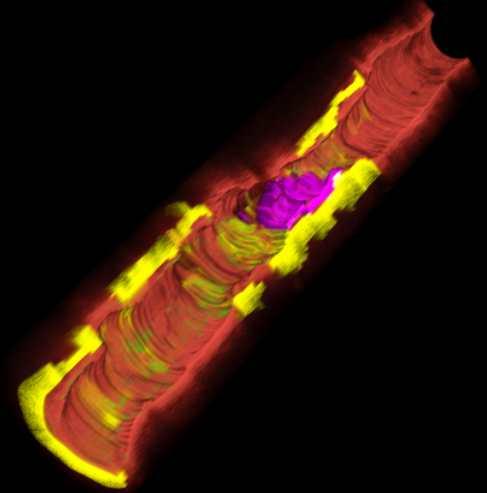
40 Artery Wall Lipid Calcium Macrophages Stent Guide Wire Thrombus Baseline Prox end Distal end 15 mos Prox end Courtesy G. Tearney MGH and B. Bouma MIT Boston Distal end
CLINICAL APPLICATIONS OF OPTICAL COHERENCE TOMOGRAPHY. Konstantina P. Bouki, FESC 2 nd Department of Cardiology General Hospital Of Nikea, Pireaus
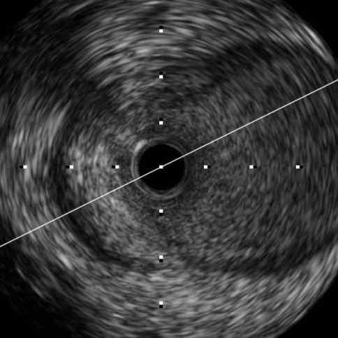
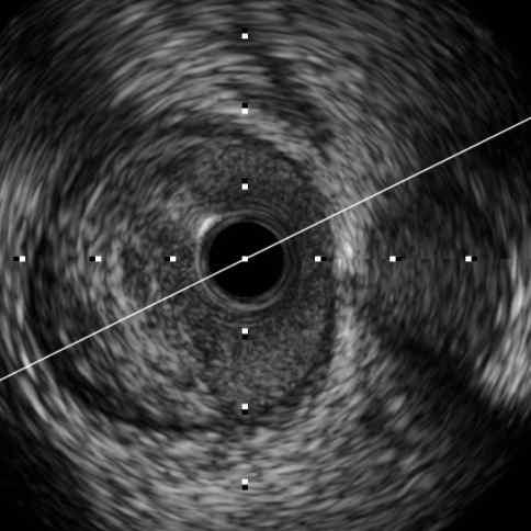
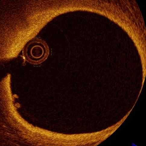
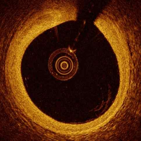
 CLINICAL APPLICATIONS OF OPTICAL COHERENCE TOMOGRAPHY Konstantina P. Bouki, FESC 2 nd Department of Cardiology General Hospital Of Nikea, Pireaus OPTICAL COHERENCE TOMOGRAPHY (OCT) IVUS and OCT IVUS OCT
OCT; Comparative Imaging Results with IVUS, VH and Angioscopy
 OCT; Comparative Imaging Results with IVUS, VH and Angioscopy Takashi Akasaka, M.D. Department of Cardiovascular Medicine Wakayama, Japan Comparison among coronary imaging techniques OCT IVUS MRI CAG Angioscopy
Clinical Value of OCT. Guidance for Coronary Stenting. Giulio Guagliumi, MD
 Clinical Value of OCT Guidance for Coronary Stenting Giulio Guagliumi, MD 100 % Endovascular Imaging Indications of use 87.5 % 75 % 57.5 % 50 % 45 % 25 % 15 % 0 Lesion morphology Stent optimization Lesion
Usefulness of OCT during coronary intervention
 Usefulness of OCT during coronary intervention Takashi Akasaka, M.D. Department of Cardiovascular Medicine Wakayama, Japan Predictors at 12 Months of Stent Thrombosis and Target Lesion Revascularization
Imaging Atheroma The quest for the Vulnerable Plaque
 Imaging Atheroma The quest for the Vulnerable Plaque P.J. de Feijter 1. Department of Cardiology 2. Department of Radiology Coronary Heart Disease Remains the Leading Cause of Death in the U.S, Causing
Can IVUS Define Plaque Features that Impact Patient Care?
 Can IVUS Define Plaque Features that Impact Patient Care? A Pichard L Satler, K Kent, R Waksman, W Suddath, N Bernardo, N Weissman, M Angelo, D Harrington, J Lindsay, J Panza. Washington Hospital Center
Assessment of Vulnerable Plaque by IVUS and VH-IVUS
 Assessment of Vulnerable Plaque by IVUS and VH-IVUS Akiko Maehara, MD Director of Intravascular Imaging & Physiology Core Laboratories Associate Director of MRI/MDCT Core Laboratory Cardiovascular Research
Imaging Overview for Vulnerable Plaque: Data from IVUS Trial and An Introduction to VH-IVUS Imgaging
 Imaging Overview for Vulnerable Plaque: Data from IVUS Trial and An Introduction to VH-IVUS Imgaging Gary S. Mintz,, MD Cardiovascular Research Foundation New York, NY Today, in reality, almost everything
Added Value of Invasive Coronary Imaging for Plaque Rupture and Erosion
 Assessment of Coronary Plaque Rupture and Erosion Added Value of Invasive Coronary Imaging for Plaque Rupture and Erosion Yukio Ozaki, MD, PhD, FACC, FESC Cardiology Dept., Fujita Health Univ. Toyoake,
OCT in the Evaluation of Vascular Healing Following DES Implantation: Will It Be a Helpful Tool to Reduce Stent Thrombosis?
 OCT in the Evaluation of Vascular Healing Following DES Implantation: Will It Be a Helpful Tool to Reduce Stent Thrombosis? Juan F. Granada, MD Medical Director, Skirball Center for Cardiovascular Research
2yrs 2-6yrs >6yrs BMS 0% 22% 42% DES 29% 41% Nakazawa et al. J Am Coll Cardiol 2011;57:
 Pathology of In-stent Neoatherosclerosis in BMS and DES 197 BMS, 103 SES, and 106 PES with implant duration >30 days The incidence of neoatherosclerosis was significantly greater in DES (31%) than BMS
Assessment of plaque morphology by OCT in patients with ACS
 Assessment of plaque morphology by OCT in patients with ACS Takashi Akasaka, M.D. Department of Cardiovascular Medicine Wakayama, Japan Unstable plaque Intima Lipid core Plaque rupture and coronary events
1st Department of Cardiology, University of Athens, Hippokration Hospital, Athens, Greece
 Konstantinos Toutouzas, Maria Riga, Antonios Karanasos, Eleftherios Tsiamis, Andreas Synetos, Maria Drakopoulou, Chrysoula Patsa, Georgia Triantafyllou, Aris Androulakis, Christodoulos Stefanadis 1st Department
Catch-up Phenomenon: Insights from Pathology
 Catch-up Phenomenon: Insights from Pathology Michael Joner, MD CVPath Institute Inc. Gaithersburg, MD USA Path Lessons learned from the BMS and DES (1 st Gen) era Neointimal Thickness [mm] In Stent Re
Left main coronary artery (LMCA): The proximal segment
: The proximal segment") Anatomy and Pathology of Left main coronary artery G Nakazawa Tokai Univ. Kanagawa, Japan 1 Anatomy Difinition Left main coronary artery (LMCA): The proximal segment RCA AV LAD LM LCX of the left coronary
Anatomy and Pathology of Left main coronary artery G Nakazawa Tokai Univ. Kanagawa, Japan 1 Anatomy Difinition Left main coronary artery (LMCA): The proximal segment RCA AV LAD LM LCX of the left coronary
as a Mechanism of Stent Failure
 In-Stent t Neoatherosclerosis e osc e os s as a Mechanism of Stent Failure Soo-Jin Kang MD., PhD. University of Ulsan College of Medicine, Heart Institute Asan Medical Center, Seoul, Korea Disclosure I
Cardiovascular Research Foundation and Columbia University Medical Center, New York.
 Virtual Histology Intravascular Ultrasound Analysis of Non-culprit Attenuated Plaques Detected by Grayscale Intravascular Ultrasound in Patients with Acute Coronary Syndromes Xiaofan Wu, Akiko Maehara,
Optical Coherence Tomography for Intracoronary Imaging
 Optical Coherence Tomography for Intracoronary Imaging Lorenz Räber Stephan Windecker Department of Cardiology Swiss Cardiovascular Center and Clinical Trials Unit Bern Bern University Hospital, Switzerland
Insights in Thrombosis and In-Stent Restenosis
 Clinical Value of OCT Insights in Thrombosis and In-Stent Restenosis Fernando Alfonso MD, PhD, FESC Interventional Cardiology. Cardiovascular Institute. Clinico San Carlos University Hospital. Madrid.
Assessment of vulnerable plaque by OCT
 Assessment of vulnerable plaque by OCT Comparison with histology and possible clinical applications Takashi Akasaka, M.D. Department of Cardiovascular Medicine Wakayama, Japan Identification of vulnerable
The PROSPECT Trial. A Natural History Study of Atherosclerosis Using Multimodality Intracoronary Imaging to Prospectively Identify Vulnerable Plaque
 The PROSPECT Trial Providing Regional Observations to Study Predictors of Events in the Coronary Tree A Natural History Study of Atherosclerosis Using Multimodality Intracoronary Imaging to Prospectively
Drug Eluting Stent DES Pathology Update What we know, what we do not know
 Drug Eluting Stent DES Pathology Update What we know, what we do not know 13 th Summit TCT Asia 25 th April 2008 Renu Virmani, MD. CVPath Institute Inc. Gaithersburg, MD Disclosure Statement of Financial
CPIS So-Yeon Choi, MD., PhD. Department of Cardiology Ajou University School of MedicineSuwon, Korea
 So-Yeon Choi, MD., PhD. Department of Cardiology Ajou University School of MedicineSuwon, Korea Coronary Artery Imaging The ideal coronary imaging technology would be capable of identifying not only vessel
Pathology of Vulnerable Plaque Angioplasty Summit 2005 TCT Asia Pacific, Seoul, April 28-30, 2005
 Pathology of Vulnerable Plaque Angioplasty Summit 25 TCT Asia Pacific, Seoul, April 28-3, 25 Renu Virmani, MD CVPath, A Research Service of the International Registry of Pathology Gaithersburg, MD Plaque
Invasive Coronary Imaging Modalities for Vulnerable Plaque Detection
 Invasive Coronary Imaging Modalities for Vulnerable Plaque Detection Gary S. Mintz, MD Cardiovascular Research Foundation New York, NY Greyscale IVUS studies have shown Plaque ruptures do not occur randomly
JACC: CARDIOVASCULAR INTERVENTIONS VOL. 2, NO. 5, PUBLISHED BY ELSEVIER INC. DOI: /j.jcin
 JACC: CARDIOVASCULAR INTERVENTIONS VOL. 2, NO. 5, 2009 2009 BY THE AMERICAN COLLEGE OF CARDIOLOGY FOUNDATION ISSN 1936-8798/09/$36.00 PUBLISHED BY ELSEVIER INC. DOI: 10.1016/j.jcin.2009.01.012 Incomplete
Next Generation Drug- eluting Stent : Will It Solve the Problem?
 Next Generation Drug- eluting Stent : Will It Solve the Problem? Yangsoo Jang, M.D., Ph.D, FACC Severance Cardiovascular Hospital Yonsei University Healthcare System Commercially Available DES An Epidemic
Culprit Lesion Remodeling and Long-term (> 5years) Prognosis in Patients with Acute Coronary Syndrome
 Prognosis in Patients with Acute Coronary Syndrome") Culprit Lesion Remodeling and Long-term (> 5years) Prognosis in Patients with Acute Coronary Syndrome Hiroyuki Okura*, MD; Nobuya Matsushita**,MD Kenji Shimeno**, MD; Hiroyuki Yamaghishi**, MD Iku Toda**,
Culprit Lesion Remodeling and Long-term (> 5years) Prognosis in Patients with Acute Coronary Syndrome Hiroyuki Okura*, MD; Nobuya Matsushita**,MD Kenji Shimeno**, MD; Hiroyuki Yamaghishi**, MD Iku Toda**,
Analysis of macrophage accumulation using optical coherence tomography one year after sirolimus, paclitaxel and zotarolimus-eluting stent
 Analysis of macrophage accumulation using optical coherence tomography one year after sirolimus, paclitaxel and zotarolimus-eluting stent implantation. Department of Cardiology, Ehime Prefectural Imabari
What Stent to Use? JASVINDAR SINGH MD, FACC
 What Stent to Use? JASVINDAR SINGH MD, FACC ASSOCIATE PROFESSOR OF MEDICINE WASHINGTON UNIVERSITY IN ST. LOUIS DIRECTOR, CARDIAC CATHETERIZATION LAB BARNES-JEWISH HOSPITAL What Stent to Use? Jasvindar
Κλινική Χρήση IVUS και OCT PERIKLIS A. DAVLOUROS ASSOCIATE PROFESSOR OF CARDIOLOGY INVASIVE CARDIOLOGY & CONGENITAL HEART DISEASE
 Κλινική Χρήση IVUS και OCT PERIKLIS A. DAVLOUROS ASSOCIATE PROFESSOR OF CARDIOLOGY INVASIVE CARDIOLOGY & CONGENITAL HEART DISEASE Conflict of interest None to declare While IVUS is the most used intravascular
A Novel Low Pressure Self Expanding Nitinol Coronary Stent (vprotect): Device Design and FIH Experience
: Device Design and FIH Experience") A Novel Low Pressure Self Expanding Nitinol Coronary Stent (vprotect): Device Design and FIH Experience Juan F. Granada, MD Medical Director, Skirball Center for Cardiovascular Research The Cardiovascular
A Novel Low Pressure Self Expanding Nitinol Coronary Stent (vprotect): Device Design and FIH Experience Juan F. Granada, MD Medical Director, Skirball Center for Cardiovascular Research The Cardiovascular
Plaque Characteristics in Coronary Artery Disease. Chourmouzios Arampatzis MD, PhD, FESC
 Plaque Characteristics in Coronary Artery Disease Chourmouzios Arampatzis MD, PhD, FESC Disclosure Statement of Financial Interest Regarding this Presentation NONE Atherosclerosis Model proposed by Stary
Drug Eluting Stents Sometimes Fail ESC Stockholm 29 Set 2010 Stent Thrombosis Alaide Chieffo
 Drug Eluting Stents Sometimes Fail ESC Stockholm 29 Set 2010 Stent Thrombosis 11.45-12.07 Alaide Chieffo San Raffaele Scientific Institute, Milan, Italy Historical Perspective 25 20 15 10 5 0 Serruys 1991
Review Article Optical Coherence Tomography Imaging in Acute Coronary Syndromes
 SAGE-Hindawi Access to Research Cardiology Research and Practice Volume 2011, Article ID 312978, 7 pages doi:10.4061/2011/312978 Review Article Optical Coherence Tomography Imaging in Acute Coronary Syndromes
OCT guidance for distal LM lesions
 OCT guidance for distal LM lesions FRANCESCO BURZOTTA INSTITUTE OF CARDIOLOGY CATHOLIC UNIVERSITY OF THE SACRED HEART ROME, ITALY LM suitability for OCT At FU in stented LM Parodi G et al. Eurointervention
Evaluation of stent placement and outcomes with optical coherence tomography
 REVIEW Evaluation of stent placement and outcomes with optical coherence tomography Optical coherence tomography (OCT) is an imaging modality based on fiberoptic technology. OCT imaging systems use optical
Integrating IVUS, FFR, and Noninvasive Imaging to Optimize Outcomes. Gary S. Mintz, MD Cardiovascular Research Foundation
 Integrating IVUS, FFR, and Noninvasive Imaging to Optimize Outcomes Gary S. Mintz, MD Cardiovascular Research Foundation COURAGE Nuclear Substudy (n=314) Death/MI according the residual ischemia (SPECT)
FESC, FACC, MAHA, MSCAI, MEAPSI, ESH
 «Απεικόνιση και φυσιολογία στο αιμοδυναμικό εργαστήριο». Eυάλωτη αθηρωματική πλάκα. Πού βρισκόμαστε? Ηλίας Α. Σανίδας MD, PhD, FESC, FACC, MAHA, MSCAI, MEAPSI, ESH Specialist Επεμβατικός Kαρδιολόγος Επιμελητής,
Technical considerations in the Treatment of Left Main Lesions Ioannis Iakovou, MD, PhD
 Technical considerations in the Treatment of Left Main Lesions Ioannis Iakovou, MD, PhD Onassis Cardiac Surgery Center, Athens, Greece Critical issues in LM PCI Anatomic variability Techniques Variability
HCS Working Group Seminars Macedonia Pallas Hotel, Friday 21 st February Drug-eluting stents Are they all equal?
 HCS Working Group Seminars Macedonia Pallas Hotel, Friday 21 st February 2014 Drug-eluting stents Are they all equal? Vassilis Spanos Interventional Cardiologist, As. Director 3 rd Cardiology Clinic Euroclinic
Béla MERKELY MD, PhD, DSc, FESC. Stent thrombosis: patophysiology, predisposing factors, definition, classification, prevention and treatment
 Semmelweis University Heart Center Budapest, Hungary Béla MERKELY MD, PhD, DSc, FESC Stent thrombosis: patophysiology, predisposing factors, definition, classification, prevention and treatment 10th Interventional
COMPARE Trial Elvin Kedhi Maasstad Ziekenhuis Rotterdam The Netherlands
 COMPARE Trial Elvin Kedhi Maasstad Ziekenhuis Rotterdam The Netherlands TCTAP 2010 Seoul, Korea Disclosures Research Foundation of the Cardiology Department has received unrestricted research grants from:
Acta Cardiol Sin 2018;34: doi: /ACS _34(2) A
 A") Original Article Acta Cardiol Sin 2018;34:124 129 doi: 10.6515/ACS.201803_34(2).20171115A Coronary Artery Disease Vascular Healing Response after Everolimus-Eluting Stent Implantation in Acute Coronary
Original Article Acta Cardiol Sin 2018;34:124 129 doi: 10.6515/ACS.201803_34(2).20171115A Coronary Artery Disease Vascular Healing Response after Everolimus-Eluting Stent Implantation in Acute Coronary
Coronary plaque erosion: a clinical case. Dr. Giampaolo Niccoli, MD, PhD, FESC Institute of Cardiology Catholic University, Rome, Italy
 Coronary plaque erosion: a clinical case, MD, PhD, FESC Institute of Cardiology Catholic University, Rome, Italy Coronary plaque erosion: a clinical case B.M. Age: 59 years Sex: female. Cardiological risk
Can We Identify Vulnerable Patients & Vulnerable Plaque?
 Can We Identify Vulnerable Patients & Vulnerable Plaque? We Know Enough to Treat High-Risk Lesions? Takashi Akasaka, MD, PhD Department of Cardiovascular Medicine, Japan Disclosure Statement of Financial
Que nos puede aportar el OCT intracoronario
 XXXI Jornadas SOLACI. 10ª Región CONOSUR LIIIº Congreso Chileno de Cardiología y Cirugía Cardiovascular Hotel Patagónico. Puerto Varas. Chile (30 Nov 1 Dic 2016) Que nos puede aportar el OCT intracoronario
malapposition assessed by OCT
 Stent t coverage and malapposition assessed by OCT Myeong-Ki Hong, M.D. Ph D Professor of Medicine Division of Cardiology, Severance Cardiovascular Hospital Yonsei University College of Medicine, Seoul,
Stents selection and optimal implantation: sizes, design, deployment Abbott Vascular. All rights reserved.
 Stents selection and optimal implantation: sizes, design, deployment Stent classification: Mechanism of expansion - Self-expanding - Balloon expandable Design - Mesh structure - Coil - Slotted tube - Ring
DESolve NX Trial Clinical and Imaging Results
 DESolve NX Trial Clinical and Imaging Results Alexandre Abizaid, MD, PhD, Instituto Dante Pazzanese, Sao Paulo, Brazil On behalf of the DESolve Nx Trial Investigators Please refer to the TCT2014 App or
OCT GUIDED TREATMENT OF CALCIFIED LESIONS RICHARD SHLOFMITZ, MD CHAIRMAN OF DEPT. OF CARDIOLOGY ST. FRANCIS HOSPITAL ROSLYN, NEW YORK
 OCT GUIDED TREATMENT OF CALCIFIED LESIONS RICHARD SHLOFMITZ, MD CHAIRMAN OF DEPT. OF CARDIOLOGY ST. FRANCIS HOSPITAL ROSLYN, NEW YORK Disclosure Statement of Financial Interest Within the past 12 months,
DID OCT change our experience on coronary arteries?
 DID OCT change our experience on coronary arteries? Istanbul June 2012 F Prati San Giovanni Hospital, Rome Rome Heart Research Use imaging modalities to.. Avoid useless procedures! MLA of 2.0 mm 2 best
Gary S. Mintz,, MD. IVUS Observations in Acute (vs Chronic) Coronary Artery Disease: Structure vs Function
 Coronary Artery Disease: Structure vs Function") Gary S. Mintz,, MD IVUS Observations in Acute (vs Chronic) Coronary Artery Disease: Structure vs Function Important IVUS Observations: Remodeling Originally used (first by Glagov) ) to explain atherosclerosis
Gary S. Mintz,, MD IVUS Observations in Acute (vs Chronic) Coronary Artery Disease: Structure vs Function Important IVUS Observations: Remodeling Originally used (first by Glagov) ) to explain atherosclerosis
BIOFREEDOM: Polymer free Biolimus A9 eluting
 TCTAP 2011 Seoul, April 27 29, 2011 BIOFREEDOM: Polymer free Biolimus A9 eluting Stents and Paclitaxel eluting stents Eberhard Grube MD, FACC, FSCAI Hospital Oswaldo Cruz - Dante Pazzanese, São Paulo,
OCT Analysis in Patients With Very Late Stent Thrombosis
 JACC: CARDIOVASCULAR IMAGING VOL. 6, NO. 6, 2013 ª 2013 BY THE AMERICAN COLLEGE OF CARDIOLOGY FOUNDATION ISSN 1936-878X/$36.00 PUBLISHED BY ELSEVIER INC. http://dx.doi.org/10.1016/j.jcmg.2013.02.006 OCT
Coronary Artery Thermography
 Coronary Artery Thermography The 10th Anniversary, Interventional Vascular Therapeutics Angioplasty Summit 2005 TCT Asia Pacific Christodoulos Stefanadis Professor of Cardiology Athens Medical School In
OCT Technology: Differences between Biodegradable and Durable Polymers: Insights from the LEADERS Trial LEADERS OCT
 OCT Technology: Differences between Biodegradable and Durable Polymers: Insights from the LEADERS Trial LEADERS OCT Substudy Carlo Di Mario, MD Peter Barlis, MD Evelyn Regar, MD Peter Juni, MD Patrick
IVUS Assessment of the Mechanism of In-stent Restenosis? Gary S. Mintz, MD Cardiovascular Research Foundation
 IVUS Assessment of the Mechanism of In-stent Restenosis? Gary S. Mintz, MD Cardiovascular Research Foundation SURE Trial: Restenosis in non-stented lesions Average of the two image slices with the smallest
High-risk vulnerable plaques. Kostis Raisakis G.Gennimatas General Hospital of Athens
 High-risk vulnerable plaques. Kostis Raisakis G.Gennimatas General Hospital of Athens Overview: 1 Definition-Pathology 2 3 Diagnostic Strategies Invasive Non Invasive Prognostic Value of Detection 4 Treatment
Clinical Application of OCT in Stent Evaluation
 Imaging & Physiology Summit 2010 in Soul #1. Basics of Image Interpretation: IVUS/VH/OCT Clinical Application of OCT in Stent Evaluation Mitsuyasu Terashima, MD, PhD, FACC Stent implantation Stent Apposition
Drug eluting stents (DES) have decreased
 have decreased") JACC: CARDIOVASCULAR IMAGING VOL. 5, NO. 11, 1 1 BY THE AMERICAN COLLEGE OF CARDIOLOGY FOUNDATION ISSN 1936-878X/$36. PUBLISHED BY ELSEVIER INC. http://dx.doi.org/1.116/j.jcmg.1.. BRIEF REPORT OCT-Verified
JACC: CARDIOVASCULAR IMAGING VOL. 5, NO. 11, 1 1 BY THE AMERICAN COLLEGE OF CARDIOLOGY FOUNDATION ISSN 1936-878X/$36. PUBLISHED BY ELSEVIER INC. http://dx.doi.org/1.116/j.jcmg.1.. BRIEF REPORT OCT-Verified
Review Article Pathologic Etiologies of Late and Very Late Stent Thrombosis following First-Generation Drug-Eluting Stent Placement
 ombosis Volume 2012, Article ID 608593, 16 pages doi:10.1155/2012/608593 Review Article Pathologic Etiologies of Late and Very Late Stent ombosis following First-Generation Drug-Eluting Stent Placement
Neointimal coverage of bare-metal and sirolimuseluting stents evaluated with optical coherence tomography
 Neointimal coverage of bare-metal and sirolimuseluting stents evaluated with optical coherence tomography B X Chen, F Y Ma, W Luo, J H Ruan, W L Xie, X Z Zhao, S H Sun, X M Guo, F Wang, T Tian, X W Chu
Bifurcation stenting with BVS
 Bifurcation stenting with BVS Breaking the limits or just breaking the struts? Maciej Lesiak Department of Cardiology University Hospital in Poznan, Poland Disclosure Speaker s name: Maciej Lesiak I have
Optical Coherence Tomography (OCT): A New Imaging Tool During Carotid Artery Stenting
: A New Imaging Tool During Carotid Artery Stenting") Chapter 6 Optical Coherence Tomography (OCT): A New Imaging Tool During Carotid Artery Stenting Shinichi Yoshimura, Masanori Kawasaki, Kiyofumi Yamada, Arihiro Hattori, Kazuhiko Nishigaki, Shinya Minatoguchi
Chapter 6 Optical Coherence Tomography (OCT): A New Imaging Tool During Carotid Artery Stenting Shinichi Yoshimura, Masanori Kawasaki, Kiyofumi Yamada, Arihiro Hattori, Kazuhiko Nishigaki, Shinya Minatoguchi
Post PCI functional testing and imaging: case based lessons from FFR React
 Post PCI functional testing and imaging: case based lessons from FFR React Joost Daemen, MD, PhD, FESC Optics in Cardiology 2018 April 21st, 2018 10.15 10.30h Disclosure Statement of Financial Interest
Index. B Bare metal stents (BMS) vs. DES, 172 OCT findings, 170, 172
 vs. DES, 172 OCT findings, 170, 172") Index A Absorbable metal stent (AMS), 189 Absorb BVS, 184 187 Acquired malapposition in DES, stent thrombosis. See also Incomplete stent apposition (ISA) coronary angiography, 155, 156 DAPT therapy, 155
Index A Absorbable metal stent (AMS), 189 Absorb BVS, 184 187 Acquired malapposition in DES, stent thrombosis. See also Incomplete stent apposition (ISA) coronary angiography, 155, 156 DAPT therapy, 155
DEB experience in Gachon Universtiy Gil Hospital (in ISR) Soon Yong Suh MD., PhD. Heart Center Gachon University Gil Hospital Seoul, Korea.
 Soon Yong Suh MD., PhD. Heart Center Gachon University Gil Hospital Seoul, Korea.") DEB experience in Gachon Universtiy Gil Hospital (in ISR) Soon Yong Suh MD., PhD. Heart Center Gachon University Gil Hospital Seoul, Korea. In-stent restenosis (ISR) Remains important issue even in the
DEB experience in Gachon Universtiy Gil Hospital (in ISR) Soon Yong Suh MD., PhD. Heart Center Gachon University Gil Hospital Seoul, Korea. In-stent restenosis (ISR) Remains important issue even in the
Intervention: How and to which extent is technology helping us?
 Cardiological Society of India Congress 12th February 2016 Chennai, India Intervention: How and to which extent is technology helping us? SIMONE BISCAGLIA MD CARDIOVASCULAR INSTITUTE, FERRARA, ITALY Introduction
Yukio Ozaki, M Okumura, TF Ismail 2, S Motoyama, H. Naruse, K. Hattori, H. Kawai, M. Sarai, J. Ishii, Jagat Narula 3
 Culprit Lesion Characteristics in Acute Coronary Syndrome and Stable Angina Assessed by Optical Coherence Tomography (OCT), Angioscopy, IVUS and Multidetector Computed Tomography (MDCT) Yukio Ozaki, M
eluting Stents The SPIRIT Trials
 Everolimus-eluting eluting Stents The SPIRIT Trials Gregg W. Stone, MD Columbia University Medical Center Cardiovascular Research Foundation Abbott XIENCE V Everolimus-eluting eluting Stent Everolimus
PCI for Long Coronary Lesion
 PCI for Long Coronary Lesion Shift of a General Idea with the Introduction of DES In the Bare Metal Stent Era Higher Restenosis Rate With Increasing Stent Length and Decreasing Stent Area Restenosis.6.4.2
Evaluation of Intermediate Coronary lesions: Can You Handle the Pressure? Jeffrey A Southard, MD May 4, 2013
 Evaluation of Intermediate Coronary lesions: Can You Handle the Pressure? Jeffrey A Southard, MD May 4, 2013 Disclosures Consultant- St Jude Medical Boston Scientific Speaker- Volcano Corporation Heart
Optical Coherence Tomography
 Optical Coherence Tomography Disclosure Information Demetrius Lopes MD The following relationships exist related to this presentation: University Grant/Research Support: Rush University Industry Grant
Left Main Intervention: Will it become standard of care?
 Left Main Intervention: Will it become standard of care? David Cox, MD FSCAI, FACC Director, Interventional Cardiology Research Associate Director, Cardiac Cath Lab Lehigh Valley Health Network Allentown,
Vulnerable Plaque Pathophysiology, Detection, and Intervention. VP: A Local Problem or Systemic Disease. Erling Falk, Denmark
 Vulnerable Plaque Pathophysiology, Detection, and Intervention VP: A Local Problem or Systemic Disease Erling Falk, Denmark Vulnerable Plaque Pathophysiology, Detection, and Intervention VP: A Local Problem
DES In-stent Restenosis
 DES In-stent Restenosis Roxana Mehran, MD Columbia University Medical Center The Cardiovascular Research Foundation DES Restenosis Mechanisms Predictors Morphological patterns Therapy approach Mechanisms
Brief History of Development of OCT
 So-Yeon Choi, MD., PhD. Department of Cardiology Ajou University School of Medicine, Suwon, Korea Brief History of Development of OCT 1990-9191 Invention of OCT by Fujimoto (USA), Tanno (Japan) 1996 Exploratory
Noninvasive Coronary Imaging: Plaque Imaging by MDCT
 Coronary Physiology & Imaging Summit 2007 Noninvasive Coronary Imaging: Plaque Imaging by MDCT Byoung Wook Choi Department of Radiology Yonsei University, Seoul, Korea Stary, H. C. et al. Circulation
Failure of positive. Recanalization and CTO formation. TCFA rupture with (fatal) thrombotic occlusion. TCFA Lipid pool
 thrombotic occlusion. TCFA Lipid pool") Vulnerable Plaque features on coronary CT Jin Ho Choi, MD, PhD Department of Internal Medicine, Emergency Medicine Samsung Medical Center, Sungkyunkwan University School of Medicine, Seoul, Korea IPS /
Vulnerable Plaque features on coronary CT Jin Ho Choi, MD, PhD Department of Internal Medicine, Emergency Medicine Samsung Medical Center, Sungkyunkwan University School of Medicine, Seoul, Korea IPS /
Left Main Intervention: Where are we in 2015?
 Left Main Intervention: Where are we in 2015? David A. Cox, MD FSCAI Director, Cardiology Research Associate Director, Cardiac Cath Lab Lehigh Valley Health Network Allentown, PA Fall Fellows Course Laa
RESTENOSIS Facing up to the problem
 RESTENOSIS Facing up to the problem Petr Kala University Hospital Brno Czech Republic ESC 2011, Paris Disclosure Scientific Advisory Boards or Education presentations fee Abbott, Boston Scientific, Cordis
Le espressioni della placca che preoccupano il clinico: progressione o vulnerabilità? CardioLUCCA Marzo 2017
 Le espressioni della placca che preoccupano il clinico: progressione o vulnerabilità? CardioLUCCA Marzo 2017 F Prati San Giovanni H. and CLI Foundation, Rome Euro Image Research Che cosa preoccupa il cardiologo
IVUS Virtual Histology. Listening through Walls D. Geoffrey Vince, PhD The Cleveland Clinic Foundation
 IVUS Virtual Histology Listening through Walls D. Geoffrey Vince, PhD Disclosure VH is licenced to Volcano Therapeutics Grant funding from Pfizer, Inc. Grant funding from Boston-Scientific Most Myocardial
Between Coronary Angiography and Fractional Flow Reserve
 Visual-Functional Mismatch Between Coronary Angiography and Fractional Flow Reserve Seung-Jung Park, MD., PhD. University of Ulsan, College of Medicine Asan Medical Center, Seoul, Korea Visual - Functional
Bifurcation Stenting: IVUS and OCT Information
 Bifurcation Stenting: IVUS and OCT Information Yoshinobu Murasato MD, PhD (New Yukuhashi Hospital) On behalf of J-REVERSE investigators October 14-15, 2011, Lisbon Proximal stent deformation induced by
Thoraxcenter, Erasmus Medical Center, Rotterdam, the Netherlands
 Hellenic J Cardiol 2015; 56: 125-135 Original Research Early and Late Optical Coherence Tomography Findings Following Everolimus-Eluting Bioresorbable Vascular Scaffold Implantation in Myocardial Infarction:
DES in primary PCI for STEMI: contra
 DES in primary PCI for STEMI: contra Philippe Gabriel Steg Department of Cardiology Hôpital Bichat Claude Bernard, AP-HP Université Paris VII Denis Diderot INSERM U-698 Paris, France Ph. Gabriel Steg -
The BIO revolution: bioadsorbable stents. Federico Conrotto Cardiologia 2 Città della Salute e della Scienza di Torino
 The BIO revolution: bioadsorbable stents Federico Conrotto Cardiologia 2 Città della Salute e della Scienza di Torino BVS stent (Abbot Vascular) Strut Material: Poly-L-Lactic acid Coating Material: Poly-D,L-lactide
ISAR-LEFT MAIN: A Randomized Clinical Trial on Drug-Eluting Stents for Unprotected Left Main Lesions
 Julinda Mehilli, MD Deutsches Herzzentrum Technische Universität Munich Germany ISAR-LEFT MAIN: A Randomized Clinical Trial on Drug-Eluting Stents for Unprotected Left Main Lesions Background Left main
PCI for Left Anterior Descending Artery Ostial Stenosis
 PCI for Left Anterior Descending Artery Ostial Stenosis Why do you hesitate PCI for LAD ostial stenosis? LAD Ostial Lesion Limitations of PCI High elastic recoil Involvement of the distal left main coronary
Non-LM bifurcation studies of importance in 2011
 7th European Bifurcation Club 14-15 October 2011 LISBON Goran Stankovic MD, PhD Non-LM bifurcation studies of importance in 2011 October 15 th : 08:00 08:10 DKCRUSH-II: A Prospective Randomized Trial of
Basics of Angiographic Interpretation Analysis of Angiography
 Basics of Angiographic Interpretation Analysis of Angiography Young-Hak Kim, MD, PhD Cardiac Center, University of Ulsan College of Medicine, Seoul, Korea What made us nervous Supervisors Stent Contrast
The Future of intravascular Is there light or sound at the end of the tunnel? Hybrid Imaging
 The Future of intravascular imaging : Is there light or sound at the end of the tunnel? Hybrid Imaging Jung Ho Heo MD, PhD Kosin University Hospital Busan, Korea From: A Heart With 67 Stents J Am Coll
Safety of Drug-Eluting Stents in Acute Coronary Syndromes
 Rotterdam, June 11 th 2012 Safety of Drug-Eluting Stents in Acute Coronary Syndromes Stephan Windecker Department of Cardiology Swiss Cardiovascular Center and Clinical Trials Unit Bern Bern University
Cottrell Memorial Lecture. Has Reversing Atherosclerosis Become the New Gold Standard in the Treatment of Cardiovascular Disease?
 Cottrell Memorial Lecture Has Reversing Atherosclerosis Become the New Gold Standard in the Treatment of Cardiovascular Disease? Stephen Nicholls MBBS PhD @SAHMRI_Heart Disclosures Research support: AstraZeneca,
Optical coherence tomography evaluation of zotarolimus-eluting stents at 9-month follow-up: comparison with sirolimus-eluting stents
 Optical coherence tomography evaluation of zotarolimus-eluting stents at 9-month follow-up: comparison with sirolimus-eluting stents J-S Kim, 1 I-K Jang, 2 J-S Kim, 1 T H Kim, 1 M Takano, 3 T Kume, 4 N
Quick guide. Core. precision guided therapy system
 Quick guide Core precision guided therapy system The Philips Volcano imaging system should only be operated by trained personnel. The following information is presented for your convenience and is not
IVUS vs FFR Debate: IVUS-Guided PCI
 IVUS vs FFR Debate: IVUS-Guided PCI Gary S. Mintz, MD Cardiovascular Research Foundation New York, NY Disclosure Statement of Financial Interest Within the past 12 months, I have had a financial interest/arrangement