Brief History of Development of OCT
|
|
|
- Lambert Osborne
- 5 years ago
- Views:
Transcription


1 So-Yeon Choi, MD., PhD. Department of Cardiology Ajou University School of Medicine, Suwon, Korea
2 Brief History of Development of OCT Invention of OCT by Fujimoto (USA), Tanno (Japan) 1996 Exploratory in vitro studies by Brezinski et al in MIT and MGH Validated the superior resolution compared to IVUS by Weissman In vivo imaging in animal (rabbit) by Fujimoto 2000 First published clinical studies by IK Jang, Bouma, Tearney, SJ Park, SW Park, KB Seung, KB Choi et al. in US and Korea 2002 Clinical trials began by Grube, Serruys, William, Suzuki 2003 CE-approved on Oct Nov KFDA approved The LightLab OCT system
3 Current Statement of OCT Korea 6 Japan 20 China 16 Brazil 8 Israel 1 South Africa 2 New Zealand 1 Italy 21 Sweden 4 Germany 6 The Netherland 5 UK 5 Austria 3 Greece/Cyprus 10 Spain 15 Switzerland 3 France 2 Poland 3 Latvia 1 Belarus 2 Denmark 1 Norway 1 Lithuania 1
4 What is OCT? Optical Coherence Tomography (OCT) is a high-resolution imaging technology that employs near-infrared light (1.3 μm) to probe micrometer-scale structures inside biological tissues. Near-infrared light μm, biologically safe Property of particle and wave Goes straightly Reflection, refraction, interference, diffraction Doppler effect


5 The Key Basic Mechanism of OCT Interferometer Schematic Getting OCT Imaging Reference Source Sample Beam Splitter Detector Analogous to sonar and radar, OCT OCT from LightLab Imaging system measures back-reflected optical LightLab ImageWire intensity i in terms of optical echoes. Helios Occlusion Balloon Catheter


6 Why Use OCT? 1. High Resolution 2. Real time image for intravascular structure Dense fiber Lipid 3. Tissue characterization PG rich Renu Virmani, MD, Erik Mont, MD AFIP OCT provides not only high resolution image. But also, it has benefit to obtain real time image for intravascular structures because of small size probe and inherently able to provide information on tissue composition.
 in clinical i l practice.")


7 Comparison of IVUS and OCT As comparing currently widely used Intravascular ultrasound (IVUS) in clinical i l practice. IVUS OCT Resolution (axial) μm μm (lateral) μm μm Size The of imaging OCT has core a higher resolution, 0.8 mm almost 10 times more 0.4 mm than IVUS. Dynamic The range scan area and penetration db depth are narrow 90 - and 100 shallow. db Also OCT requires blood clearing to avoid the attenuation by blood. Frame rate 30 frames/s 15 frames/s Scan area mm 70mm 7.0 Max. depth of penetration 4-8 mm Blood clearing not required mm Requires blood clearing
8 Potential Applications of OCT Vulnerable Plaque OCT may provide better understand the natural progression of coronary artery disease and the answers longstanding questions about the relationship between vulnerable plaque and the risk of heart attack. Therapeutic Guidance Evaluation of Therapeutic Results The technique is poised to play an important role in the guidance of therapeutic interventions and assessment of the results of medical and interventional treatment.


9 Detection of VP in OCT


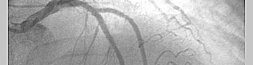
10 Case SJS M/46 STEMI AP LAO 90% of tubular stenosis at mid LAD
11 Case SJS M/46 STEMI, LAD A significant stenosis with large plaque burden characterizing mixed plaque Irregular and disrupted Irregular and disrupted contour on surface of plaque


12 Case SJS M/46 STEMI, LAD Various superfical lesion: fissuring or erosion plaque rupture intraluminal thrombus


13 Plaque Characterization by OCT OCT could well identify the features of three distinct plaque types. Fibrous High reflectivity Homogenous Finely textured Lipid-rich Low reflectivity Homogenous Diffuse margins Calcified Low reflectivity Inhomogeneous Sharp margins Isolated, strong reflections in dark background KSY M/43 SA LSJ F/62 UA CSW M/67 SA Presented in Ajou University Medical Center
14 Plaque Characterization by OCT Ex Vivo Study Excised plaques of these three types could be differentiated t d with sensitivity greater than 87% and specificity greater than 94 %. There was a good concordance in interobserver and intraobserver analysis. Sensitivity Specificity Positive predictive value Negative predictive value Fibrous Calcific Lipid pool Accuracy Statistics: Interobserver k=0.88, Intraobserver k=0.91 H Yabushita, IK Jang, et al. Circulation. 2002;106:

 Thin cap with large lipid core Endothelial denudation with superficial platelet aggregation Fissured plaque Stenosis 90% Minor")
15 Criteria for Defining Vulnerable Plaque The vulnerable plaque characterized by thin fibrous cap, extensive macrophage infiltration, and large lipid core. Circulation. 2003;108: Based on previously presented autopsy study Major criteria Active inflammation (monocyte/macrophage and T-cell infiltration) Thin cap with large lipid core Endothelial denudation with superficial platelet aggregation Fissured plaque Stenosis 90% Minor criteria Superficial calcified nodule Glistening gyellow Intraplaque hemorrhage Endothelial dysfunction Outward (positive) remodeling
16 Criteria for Defining Vulnerable Plaque OCT OCT could detect most of this criteria of VP Major criteria Active inflammation (monocyte/macrophage and T-cell infiltration) Thin cap with large lipid core Endothelial denudation with superficial platelet aggregation Fissured plaque Stenosis 90% Minor criteria Superficial calcified nodule Glistening gyellow Intraplaque hemorrhage Endothelial dysfunction Outward (positive) remodeling


17 Detection of VP in OCT Macrophage Accumulation There was a high degree of positive correlation between OCT and histological measurements of fibrous MQ density r=0.84, p< OCT Image MQ Density 0% 10% GJ Tearney, et al. Circulation 2003;107:113-9 Briain D. MacNeill, et al. J Am Coll Cardiol 2004;44:972 9


18 Detection of VP in OCT 119 lipid rich plaques in 49 patients 49 AMI; 46 ACS; 24 SAP The increases in both multi-focal and focal macrophage densities are highly correlated with symptom severity. Lipid-rich plaque was defined by lipid occupying 2 quadrants of the CSA Briain D. MacNeill, et al. J Am Coll Cardiol 2004;44:972 9
19 Detection of VP in OCT Thin Fibrous Cap 57 patients: 20 AMI, 20 ACS, 17 SAP Thin-cap fibroatheroma was defined by lipidrich plaque with cap thickness 65 μm CAS ACS AMI 0 Thin-cap Fibroatheroma Thin-cap fibroatheroma was more frequently observed in patients with AMI or ACS than SAP. IK Jang, et al. Circulation. 2005;111:1551-5


20 Detection of VP in OCT Thrombus OCT allowed us not only to estimate plaque morphology but also to distinguish thrombus from the plaque. LJS F/56 UA Thrombus PDS M/46 NSTEMI Presented in Ajou University Medical Center


21 Detection of VP in OCT Superficial lesion of Plaque OCT also provided superficial information of the plaque. Intimal Tear Plaque rupture PJK M/43 UA PDS M/46 NSTEMI LPK M/38 STEMI Presented in Ajou University Medical Center
22 Comparison among intravascular modalities for detection vulnerable plaque conventional ultrasound vs. virtual histology vs. optical coherence tomography So-Yeon Choi, Seung-Jea Tahk, Sung-Il Woo, Hyung-Mo Yang, Hong-Seok Lim, Byoung-Joo Choi, Myeong-Ho Yoon, Soo-Jin Kang, Gyo-Seung Hwang, Joon-Han Shin Department of Cardiology Ajou University School of Medicine, Suwon, Korea
23 Background Study Purpose We hypotheses that OCT might have a potential to detect vulnerable plaque by demonstrating thin fibrous cap, lipid-rich plaque and thrombi because of its high h resolution imaging i capability. We performed Intravascular Imaging studies with conventional IVUS(Grayscale), VH-IVUS and OCT in patient with coronary disease. 1. To compare qualitative and quantitative parameters of each imaging modalities between patient with stable angina pectoris (SAP) and acute coronary syndrome (ACS). 2. To compare the ability of modalities to detect specific morphology according to the given criteria i for vulnerable plaque in all lesions.
24 Methods Patients Patients undergoing cardiac catheterization were enrolled and categorized according to their clinical presentation into SAP and ACS. Inclusion Criteria Patients who have >50% coronary lesion Lesion: reference vessel size 4~2.5mm length <40mm Exclusion Criteria Cardiogenic shock Severe LV dysfunction EF <25% Previous PCI Saphenous vein graft lesion or arterial graft lesion Lesion: Left main disease, ostial disease, chronic total occlusion


25 Methods Intravascular Imaging Study Assessment for Quantitative and Qualitative Parameters Preintervention Postintervention Grayscale VH-IVUS OCT 40MHz Boston Scientific, USA Volcano Therapeutics, Inc., USA Light Lab Imaging Inc., USA The Parameters were assessed by two independent observers, and when the findings were different between the two observers, the findings in accordance with the third observer were adopted.
26 Methods Definitions Specific morphology according to the given criteria for VP Finding Grayscale VH-IVUS OCT Thin cap NA Red area being in <65μmμ contact the lumen Lipid core Echolucent area 3mm Red area 10% Low reflectivity with h omogenous, diffuse m argins 2 quadrants Thrombus Intraluminal mass NA Intraluminal mass Rupture A recess or ulceration NA A recess or ulceration /fissure with a tear detected in with a tear detected in /erosion surface surface 90%stenosis Compromise of lumen by 90% by CSA compared with proximal reference lumen SCN Echogenic material White nodule being in Isolated, strong with acoustic shadow contact the lumen reflections in dark background being in contact with the lumen Positive remodeling Lesion EEM CSA / prox. ref. EEM CSA >1.0 NA
27 Methods Definitions Thin cap fibroatheroma (TCFA) was defined as In VH-IVUS 10% Necrotic core No evident overlying fibrous tissue >40% plaque burden In OCT In OCT Lipid rich plaque ( 2 quadrants) with a Thin fibrous cap (<65 µm)
28 Results Baseline Clinical Characteristics 41 target lesions in 41 patients (24 with ACS and 17 with SAP) were enrolled. ACS SAP p value n=24 n=17 Age, yr 58±12 60± Male sex 17(71) 14(82) Risk factors Diabetes mellitus 10(42) 8(47) Hypertension 13(54) 10(59) Dyslipidemia Total cholesterol 9(37) 8(47) >220mmHg Smoking 14(58) 10(59) LV EF, % 56±18 64±
29 Results Baseline Angiographic Characteristics 41 target lesions in 41 patients (24 with ACS and 17 with SAP) were enrolled. ACS SAP p value n=24 n=17 LAD involvement 17(71) 12(71) Reference vessel, mm 3.5± ± Lesion length, mm 18.6± ± MLD, mm 107±0 1.07± ±0 1.02± %DS, % 67.8± ±
30 Results Baseline Clinical Characteristics 41 target lesions in 41 patients (24 with ACS and 17 with SAP) were enrolled. ACS SAP p value n=24 n=17 Age, yr 58±12 60± Male sex 17(71) 14(82) Risk factors Diabetes mellitus 10(42) 8(47) Hypertension 13(54) 10(59) Dyslipidemia Total cholesterol 9(37) 8(47) >220mmHg Smoking 14(58) 10(59) LV EF, % 56±18 64±
31 Results Conventional IVUS Analysis Quantitative Parameters ACS SAP p value n=24 n=17 Lesion length, mm 21.2±9.72±9 24.7± Proximal reference LA, mm ± ± VA, mm ± ± PB, % 30.2± ± Narrowest segment LA, mm 2 3.5± ± VA, mm ± ± PA, mm ± ± PB, % 72.3± ± Eccentric index 0.19± ± Remodeling index 0.89± ±
32 Results Conventional IVUS Analysis Qualitative Parameters ACS SAP p value n=24 n=17 Positive remodeling 13(58) 5(23) Soft plaque 10(42) 7(41) Lipid core at the most narrowest CSA 3(13) 1(6) within the whole lesion 6(25) 1(6) Rupture/Fissure 3(13) 0(0) Thrombus 1(4) 0(0) 0.823
33 Results Virtual Histology-IVUS Analysis Parameters in the Most Narrowest CSA ACS SAP p value n=24 n=17 Quantitative i Analysis Lumen area, mm 2 3.8± ± Vessel area, mm ± ± Plaque area, mm ± ± % Plaque burden, % 74.1± ± % Fibrous area, % 64±10 66± % Fibro-fatty area, % 16.0±8 14± % Dense calcium area, % 8±5 12± % Necrotic core area, % 16±18 13± Qualitative Analysis TCFA 9(38) 3(24) SCN 5(21) 2(12) 0.161
34 Results Virtual Histology-IVUS Analysis Parameters in the Most Largest NC CSA ACS SAP p value n=24 n=17 Quantitative i Analysis Lumen area, mm 2 4.1± ± Vessel area, mm ± ± Plaque area, mm 2 9.8± ± % Plaque burden, % 64.1± ± % Fibrous area, % 58±11 69± % Fibro-fatty area, % 19±4 17± % Dense calcium area, % 11±3 18± % Necrotic core area, % 24±21 18± Qualitative Analysis TCFA 10(42) 3(24) SCN 9(38) 3(24) 0.261
35 Results OCT Analysis Quantitative and Qualitative Parameters ACS SAP p value n=24 n=17 The Most Narrowest Segment Lumen area, mm 2 3.8± ± Intimal thickness, mm 0.23± ± TCFA 3(13) 0(0) Lipid rich plaque 3(13) 0(0) Within the Whole Lesion Intimal thickness, mm 008±1 0.08± ±0 0.23± TCFA 12(50) 2(17) Surface lesion 10(42) 3(18) Thrombus 10(42) 1(6) Lipid rich plaque 7(35) 1(6) In OCT analysis, thickness of fibrous cap, the frequency of TCFA, lipid-rich plaque and the presence of thrombus were significantly correlated with clinical presentation.
36 Results The Incidence of TCFA Comparison between ACS and SAP Thin cap fibroatheroma (TCFA) was defined as a Lipid rich plaque ( 2 quadrants) with a thin fibrous cap (<65 µm) *p<0.05 between ACS vs.sap % 80 p<0.05 between VH-IVIS vs. OCT NA * Grayscale VH-IVUS OCT ACS SAP 41 lesions of 41 patients were evaluated. VH-IVUS has a higher tendency TCFA in ACS, in OCT, TCFA was significantly correlated with clinical presentation.
37 Results Comparison among Imaging Modalities The Major Criteria for Vulnerable Plaque % * Gray Scale RF-IVUS OCT NA NA NA ThinCap Lipid Core Thrombus Rupture/Fissure AS 90% *p<0.05 among 3 modalities, p<0.05 between GS vs. OCT, p<0.05 between VH-IVUS vs. OCT The detection of lipid core is different among three groups, and thrombus and rupture of p g g p, p plaque were more frequently identified in OCT than conventional IVUS.
38 Results Comparison among Imaging Modalities The Minor Criteria for Vulnerable Plaque % Gray Scale RF-IVUS OCT NA *p<0.05 among 3 modalities p<0.05 between GS vs. OCT p<0.05 between VH-IVUS vs. OCT
39 Results Multivariate Determinants of Clinical Symptom Regression Analysis Grayscale Variables 0R 95% CI p value Positive remodeling ~ VH-IVUS %NC area in VH ~ OCT TCFA in OCT ~ Thrombus ~ Fissure/erosion ~ After adjusting for confounding factors, %NC area in VH, TCFA and thrombus in OCT were independent risk factors for predicting clinical presentation.


40 Case Case 1 PJW /M UAIIIB Mixed Plaque Eccentric PB PR(+) NC 51% Scattered DC 22% TCFA(+) Fibrofatty >2 Lipid core TCFC(+) Angiogram Grayscale VH -IVUS OCT Case 2 JMA /F SA Mixed Plaque Concentric PB Remodeling (-) NC 12% Sup. calcium TCFA(-) Superficial calcium FC 110


 NC 5%")
")


41 Case Case 3 LCS /M NSTEMI Angiogram Mixed Plaque Eccentric PB Remodeling (+) NC 5% FF 20% TCFA(-) Grayscale VH -IVUS OCT Superficial Erosion FC <65 Case 4 SJW /F UA Soft Plaque Remodeling (-) NC 2% FF 37% TCFA(-) Fibrofatty >2 Lipid core Erosion, FC<65
42 Summary The positive remodeling and presence of lipid core in IVUS are relatedtoclinicalpresentation to presentation. In VH-IVUS, the frequency of thin-cap fibroatheroma had a higher tendency in ACS and ACS had significantly smaller fibrous area and dlarger NC area at tthe largest tnccsa CSA compared dto SAP. In OCT analysis, lipid-rich plaque, thickness of fibrous cap and the presence of thrombus in OCT were significantly correlated with clinical presentation. After adjusting for confounding factors NC area in VH and TCFA After adjusting for confounding factors, NC area in VH, and TCFA and thrombus in OCT were independent risk factors for predicting clinical presentation.
43 Conclusion This study was prospectively performed to compare currently available intravascular modalities for detailed in vivo plaque morphology in patients with different clinical presentations. OCT for vulnerable plaque is feasible and provides superior contrast and resolution of arterial pathology than other modalities. Understanding natural process of atherosclerosis is needed and the role of diagnostic tools should be further studied in clinical setting.
44 Imaging g Modalities for Detection of VP OCT might allow us to identify tissue characterization more accurately than other modalities. OCT has a potential benefit to identify vulnerable plaques by detecting of thin fibrous cap, lipid core, surface pathology of plaque and presence of thrombus, and measuring the accumulation of macrophages.


45 OCT as a Tool for PCI OCT as a Tool for PCI Evaluation of Therapeutic Results
46 OCT as a Tool for PCI Ideal Intravasular Image for PCI Lesion assessment Device sizing Decision of strategies for the lesion Understanding mechanism of intervention Decision of ending of predecure Recognition of complications F/U


47 Case Pre-intervention Post-intervention Presented in Ajou University Medical Center


48 OCT as a Tool for PCI Understanding PCI Mechanism OCT provides a information to understand d the mechanism after various PCI. After Cutting Balloon After DCA LJ. Diaz-Sandoval, IK Jang et al. Cath Cardio Interv. 2005:65:492-6 Presented by Suzuki Toyohashi Heart Center, Japan


49 OCT as a Tool for PCI Evaluation Just After Stenting OCT also provides a information about the result just after stenting. Well-apposed stent Mal-apposed stent Minor prolapse of plaque after stenting BJK M/48 Cypher 4.0 x 23 mm Cypher 3.5 x 28 mm Presented by Suzuki BJK M/48 Cypher 4.0 x 23 mm


50 Evaluation of Therapeutic Results Follow-up for Complex PCI F/U after Crushing with DESs in Bifurcation A A B C D E KKN F/61 1yr F/U Crushing with Cypher at LAD-D1 D1 D B E C Presented in Ajou University Medical Center


51 Evaluation of Therapeutic Results Tissue Reaction after Stenting KHB M/66 T3528 LAD Presented in Ajou University Medical Center
52 Incomplete tissue coverage of stent t at 1 year follow-up after coronary intervention with DES: optical coherence tomography findings So-Yeon Choi, Seung-Jea Tahk, Sung-Il Woo, Hyung-Mo Yang, Hong-Seok Lim, Byoung-Joo Choi, Myeong-Ho Yoon, Soo-Jin Kang, Gyo-Seung Hwang, Joon-Han Shin Department of Cardiology Ajou University School of Medicine, Suwon, Korea
53 Background Stent thrombus following PCI is related to serious clinical outcomes including myocardial infarction and sudden cardiac death. Incomplete endothelialization of stent has been considered as one of the pathogenesis of chronic stent thrombus in DES era. OCT has outstanding high resolution property, and recent study presented absence of tissue strut coverage was properly identified by corresponding OCT cross-section on a rabbit model.
54 Methods Patient Enrollment Patients received PCI with DES for proximal stenotic lesion more than 1 year before were enrolled for this study. We excluded patient with malapposition of stent by assessing of initial intravascular ultrasound images and the instent restenosis on f/u angiogram was also excluded. Inclusion criteria Lesion: reference vessel size 2.5mm Possible lesion: bifurcation, overlapping stent Exclusion criteria Cardiogenic shock Severe LV dysfunction EF <25% Lesion: Left main disease, ostial disease, bifurcation lesion
55 Methods Measurement Parameters The OCT image was obtained by constant pullback mechanics and analyzed each 1mm slice from center of stent. The uncovered strut was defined as a protruding strut without any reflective tissue layer on the luminal side of strut. The uncovered strut index of a stent was defined as the ratio of the number of uncovered strut and the number of total observed struts. The uncovered strut index (USI) = non-endothelialization strut (n) / evaluated strut (n) (analysis: each 1mm slice from center of stent)» Cypher inch, 5 μm» TAXUS inch, 25.6 μm» Endeavor inch, 9.2 μm


56 Results Complete Tissue Coverage Cypher Taxus HDH M/44 C3533 LAD SWS F/67 T3528 LAD Presented in Ajou University Medical Center


57 Results Incomplete Tissue Coverage Cypher Taxus SJS M/70 C3533 LAD LYJ M/46 T3020 LAD Presented in Ajou University Medical Center
58 Results Incomplete Tissue Coverage 748 Cross Sectional Images patients 25 P= Age 56±11, 18 males (23 Cypher, 11 Taxus) 15 Average follow up period: 13.4±1 months 10 5 Cypher Taxus 0 Total Cypher Taxus The uncovered strut index (USI) of total observed stent strut was 0.13±0.12. The USI of Cypher stent was significantly increased than Taxus stent (Cypher 0.15±0.21, Taxus 0.04±0.08, 04±0 08 p=0.036). 036)
59 Conclusion OCT is useful for detecting the coverage of a stent with neointima following PCI. There was a difference of tissue coverage between two DESs. Although the relationship between incomplete tissue coverage of the stent t and clinical i l events is not clear, we should consider long-term antiplatelet treatment for more than one year.
60 OCT and PCI Preinterventional lesion assessment Assessment of severity and clinical impact Detect Vulnerable Plaque IVUS OCT During intervention ti Device sizing Decision of strategies for the lesion Understanding mechanism of intervention Decision of ending of predecure Recognition of complications Serial follow-up Understanding for atherosclerosis Mechanisms, prevention and Tx of restenosis Assessment for long-term complication OCT also might be used as a tool for PCI like IVUS by providings useful informations in detection VP and PCI complication and stent f/u.
61 Complications of OCT Experience in Ajou University Medical Center Total 95 patients (male 73, age 57±9), 107 lesions Successful image acquisition : 102 lesions (95%) (3 wire passage failure, 2 incomplete occlusion ) Complication number % Air embolization 0 0 Transient ischemia : ECG change with pain Pulmonary edema due to volume 0 0 overloading Ventricular arrhythmia Post PCI Infarction 0 0 Death 0 0
62 Limitation of OCT 1. OCT imaging is attenuated by blood and needs to create blood free zone. 2. Penetration through the arterial wall is in the range of 2 3 mm. The entire plaque cannot be imaged and only superficial anatomic information is obtained. 3. OCT could not detect lipid pools or calcium behind thick fibrou s caps, and by an inability to distinguish calcium deposits from lipid id pools or the opposite. 4. OCT devices has some technical limitation, so there are some 4. OCT devices has some technical limitation, so there are some limited lesions including left main disease, ostial disease, very tortuous lesion and so on.
63 Currently Evolving OCT Toward easy practice with OCT Next generation OCT has imaging i acquisition iti speeds of 10 times faster and resolution 3 times greater than the existing product. It dose not require occlusion of the patient s blood flow during the imaging procedure. Successfully completion of preclinical study on Aug Live broadcast of first-in-manin case by Dr. E Grube in TCT, 2007, This is a huge step up in ease of use.

64 Currently Evolving OCT Toward complete lesion characterization with OCT New platform modality and combine with other modality are also promising. Morphological Lesion size Backscatter/ Lesion shape Gray Scale % stenosis Cap thickness Lipid Dense fiber Biochemical composition Lipid, collagen, proteoglycans, calcium Spectroscopy Polarization Physiological Flow Doppler disturbances CFR, FFR Mechanical Plaque stiffness Elastography PG rich Renu Virmani, MD, Erik Mont, MD AFIP


65 Take Home Massages OCT is feasible as an intravascular imaging i tool and it could be conducted safely in cath Lab. OCT has a high resolution, it could assess the tissue characterization more accurately than IVUS. OCT has a potential benefit to identify vulnerable plaques and also provides superficial i information of the vessel during and after PCI. OCT has major limitations in need of blood clear zone and low penetrating depth. Evolving OCT image moves closer to becoming a powerful diagnostic tool that will provide new insights into the etiology and treatment of coronary artery disease.
CPIS So-Yeon Choi, MD., PhD. Department of Cardiology Ajou University School of MedicineSuwon, Korea
 So-Yeon Choi, MD., PhD. Department of Cardiology Ajou University School of MedicineSuwon, Korea Coronary Artery Imaging The ideal coronary imaging technology would be capable of identifying not only vessel
Invasive Coronary Imaging Modalities for Vulnerable Plaque Detection
 Invasive Coronary Imaging Modalities for Vulnerable Plaque Detection Gary S. Mintz, MD Cardiovascular Research Foundation New York, NY Greyscale IVUS studies have shown Plaque ruptures do not occur randomly
Imaging Atheroma The quest for the Vulnerable Plaque
 Imaging Atheroma The quest for the Vulnerable Plaque P.J. de Feijter 1. Department of Cardiology 2. Department of Radiology Coronary Heart Disease Remains the Leading Cause of Death in the U.S, Causing
OCT Findings: Lesson from Stable vs Unstable Plaques
 ANGIOPLASTY SUMMIT TCTAP 2010 Imaging Workshop OCT Findings: Lesson from Stable vs Unstable Plaques Giulio Guagliumi MD Ospedali Riuniti di Bergamo, Italy DISCLOSURE OF FINANCIAL INTERESTS Consultant Boston
Imaging Overview for Vulnerable Plaque: Data from IVUS Trial and An Introduction to VH-IVUS Imgaging
 Imaging Overview for Vulnerable Plaque: Data from IVUS Trial and An Introduction to VH-IVUS Imgaging Gary S. Mintz,, MD Cardiovascular Research Foundation New York, NY Today, in reality, almost everything
Added Value of Invasive Coronary Imaging for Plaque Rupture and Erosion
 Assessment of Coronary Plaque Rupture and Erosion Added Value of Invasive Coronary Imaging for Plaque Rupture and Erosion Yukio Ozaki, MD, PhD, FACC, FESC Cardiology Dept., Fujita Health Univ. Toyoake,
Declaration of conflict of interest. Nothing to disclose
 Declaration of conflict of interest Nothing to disclose Hong-Seok Lim, Seung-Jea Tahk, Hyoung-Mo Yang, Jin-Woo Kim, Kyoung- Woo Seo, Byoung-Joo Choi, So-Yeon Choi, Myeong-Ho Yoon, Gyo-Seung Hwang, Joon-Han
IVUS Analysis. Myeong-Ki. Hong, MD, PhD. Cardiac Center, Asan Medical Center University of Ulsan College of Medicine, Seoul, Korea
 IVUS Analysis Myeong-Ki Hong, MD, PhD Cardiac Center, Asan Medical Center University of Ulsan College of Medicine, Seoul, Korea Intimal disease (plaque) is dense and will appear white Media is made of
CLINICAL APPLICATIONS OF OPTICAL COHERENCE TOMOGRAPHY. Konstantina P. Bouki, FESC 2 nd Department of Cardiology General Hospital Of Nikea, Pireaus
 CLINICAL APPLICATIONS OF OPTICAL COHERENCE TOMOGRAPHY Konstantina P. Bouki, FESC 2 nd Department of Cardiology General Hospital Of Nikea, Pireaus OPTICAL COHERENCE TOMOGRAPHY (OCT) IVUS and OCT IVUS OCT
Optical Coherence Tomography (OCT): A New Imaging Tool During Carotid Artery Stenting
: A New Imaging Tool During Carotid Artery Stenting") Chapter 6 Optical Coherence Tomography (OCT): A New Imaging Tool During Carotid Artery Stenting Shinichi Yoshimura, Masanori Kawasaki, Kiyofumi Yamada, Arihiro Hattori, Kazuhiko Nishigaki, Shinya Minatoguchi
Chapter 6 Optical Coherence Tomography (OCT): A New Imaging Tool During Carotid Artery Stenting Shinichi Yoshimura, Masanori Kawasaki, Kiyofumi Yamada, Arihiro Hattori, Kazuhiko Nishigaki, Shinya Minatoguchi
Can IVUS Define Plaque Features that Impact Patient Care?
 Can IVUS Define Plaque Features that Impact Patient Care? A Pichard L Satler, K Kent, R Waksman, W Suddath, N Bernardo, N Weissman, M Angelo, D Harrington, J Lindsay, J Panza. Washington Hospital Center
OCT; Comparative Imaging Results with IVUS, VH and Angioscopy
 OCT; Comparative Imaging Results with IVUS, VH and Angioscopy Takashi Akasaka, M.D. Department of Cardiovascular Medicine Wakayama, Japan Comparison among coronary imaging techniques OCT IVUS MRI CAG Angioscopy
Assessment of plaque morphology by OCT in patients with ACS
 Assessment of plaque morphology by OCT in patients with ACS Takashi Akasaka, M.D. Department of Cardiovascular Medicine Wakayama, Japan Unstable plaque Intima Lipid core Plaque rupture and coronary events
Cardiovascular Research Foundation and Columbia University Medical Center, New York.
 Virtual Histology Intravascular Ultrasound Analysis of Non-culprit Attenuated Plaques Detected by Grayscale Intravascular Ultrasound in Patients with Acute Coronary Syndromes Xiaofan Wu, Akiko Maehara,
2yrs 2-6yrs >6yrs BMS 0% 22% 42% DES 29% 41% Nakazawa et al. J Am Coll Cardiol 2011;57:
 Pathology of In-stent Neoatherosclerosis in BMS and DES 197 BMS, 103 SES, and 106 PES with implant duration >30 days The incidence of neoatherosclerosis was significantly greater in DES (31%) than BMS
as a Mechanism of Stent Failure
 In-Stent t Neoatherosclerosis e osc e os s as a Mechanism of Stent Failure Soo-Jin Kang MD., PhD. University of Ulsan College of Medicine, Heart Institute Asan Medical Center, Seoul, Korea Disclosure I
1st Department of Cardiology, University of Athens, Hippokration Hospital, Athens, Greece
 Konstantinos Toutouzas, Maria Riga, Antonios Karanasos, Eleftherios Tsiamis, Andreas Synetos, Maria Drakopoulou, Chrysoula Patsa, Georgia Triantafyllou, Aris Androulakis, Christodoulos Stefanadis 1st Department
Usefulness of OCT during coronary intervention
 Usefulness of OCT during coronary intervention Takashi Akasaka, M.D. Department of Cardiovascular Medicine Wakayama, Japan Predictors at 12 Months of Stent Thrombosis and Target Lesion Revascularization
Left main coronary artery (LMCA): The proximal segment
: The proximal segment") Anatomy and Pathology of Left main coronary artery G Nakazawa Tokai Univ. Kanagawa, Japan 1 Anatomy Difinition Left main coronary artery (LMCA): The proximal segment RCA AV LAD LM LCX of the left coronary
Anatomy and Pathology of Left main coronary artery G Nakazawa Tokai Univ. Kanagawa, Japan 1 Anatomy Difinition Left main coronary artery (LMCA): The proximal segment RCA AV LAD LM LCX of the left coronary
Assessment of Vulnerable Plaque by IVUS and VH-IVUS
 Assessment of Vulnerable Plaque by IVUS and VH-IVUS Akiko Maehara, MD Director of Intravascular Imaging & Physiology Core Laboratories Associate Director of MRI/MDCT Core Laboratory Cardiovascular Research
Optical Coherence Tomography for Intracoronary Imaging
 Optical Coherence Tomography for Intracoronary Imaging Lorenz Räber Stephan Windecker Department of Cardiology Swiss Cardiovascular Center and Clinical Trials Unit Bern Bern University Hospital, Switzerland
Neointimal coverage of bare-metal and sirolimuseluting stents evaluated with optical coherence tomography
 Neointimal coverage of bare-metal and sirolimuseluting stents evaluated with optical coherence tomography B X Chen, F Y Ma, W Luo, J H Ruan, W L Xie, X Z Zhao, S H Sun, X M Guo, F Wang, T Tian, X W Chu
Culprit Lesion Remodeling and Long-term (> 5years) Prognosis in Patients with Acute Coronary Syndrome
 Prognosis in Patients with Acute Coronary Syndrome") Culprit Lesion Remodeling and Long-term (> 5years) Prognosis in Patients with Acute Coronary Syndrome Hiroyuki Okura*, MD; Nobuya Matsushita**,MD Kenji Shimeno**, MD; Hiroyuki Yamaghishi**, MD Iku Toda**,
Culprit Lesion Remodeling and Long-term (> 5years) Prognosis in Patients with Acute Coronary Syndrome Hiroyuki Okura*, MD; Nobuya Matsushita**,MD Kenji Shimeno**, MD; Hiroyuki Yamaghishi**, MD Iku Toda**,
Pathology of Vulnerable Plaque Angioplasty Summit 2005 TCT Asia Pacific, Seoul, April 28-30, 2005
 Pathology of Vulnerable Plaque Angioplasty Summit 25 TCT Asia Pacific, Seoul, April 28-3, 25 Renu Virmani, MD CVPath, A Research Service of the International Registry of Pathology Gaithersburg, MD Plaque
The PROSPECT Trial. A Natural History Study of Atherosclerosis Using Multimodality Intracoronary Imaging to Prospectively Identify Vulnerable Plaque
 The PROSPECT Trial Providing Regional Observations to Study Predictors of Events in the Coronary Tree A Natural History Study of Atherosclerosis Using Multimodality Intracoronary Imaging to Prospectively
Optical Coherence Tomography
 Optical Coherence Tomography Disclosure Information Demetrius Lopes MD The following relationships exist related to this presentation: University Grant/Research Support: Rush University Industry Grant
Assessment of vulnerable plaque by OCT
 Assessment of vulnerable plaque by OCT Comparison with histology and possible clinical applications Takashi Akasaka, M.D. Department of Cardiovascular Medicine Wakayama, Japan Identification of vulnerable
Review Article Optical Coherence Tomography Imaging in Acute Coronary Syndromes
 SAGE-Hindawi Access to Research Cardiology Research and Practice Volume 2011, Article ID 312978, 7 pages doi:10.4061/2011/312978 Review Article Optical Coherence Tomography Imaging in Acute Coronary Syndromes
Drug eluting stents (DES) have decreased
 have decreased") JACC: CARDIOVASCULAR IMAGING VOL. 5, NO. 11, 1 1 BY THE AMERICAN COLLEGE OF CARDIOLOGY FOUNDATION ISSN 1936-878X/$36. PUBLISHED BY ELSEVIER INC. http://dx.doi.org/1.116/j.jcmg.1.. BRIEF REPORT OCT-Verified
JACC: CARDIOVASCULAR IMAGING VOL. 5, NO. 11, 1 1 BY THE AMERICAN COLLEGE OF CARDIOLOGY FOUNDATION ISSN 1936-878X/$36. PUBLISHED BY ELSEVIER INC. http://dx.doi.org/1.116/j.jcmg.1.. BRIEF REPORT OCT-Verified
PCI for Long Coronary Lesion
 PCI for Long Coronary Lesion Shift of a General Idea with the Introduction of DES In the Bare Metal Stent Era Higher Restenosis Rate With Increasing Stent Length and Decreasing Stent Area Restenosis.6.4.2
Chapter 43 Noninvasive Coronary Plaque Imaging
 hapter 43 Noninvasive oronary Plaque Imaging NIRUDH KOHLI The goal of coronary imaging is to define the extent of luminal narrowing as well as composition of an atherosclerotic plaque to facilitate appropriate
IVUS Virtual Histology. Listening through Walls D. Geoffrey Vince, PhD The Cleveland Clinic Foundation
 IVUS Virtual Histology Listening through Walls D. Geoffrey Vince, PhD Disclosure VH is licenced to Volcano Therapeutics Grant funding from Pfizer, Inc. Grant funding from Boston-Scientific Most Myocardial
Percutaneous Intervention of Unprotected Left Main Disease
 Percutaneous Intervention of Unprotected Left Main Disease Technical feasibility and Clinical outcomes Seung-Jung Park, MD, PhD, FACC Professor of Internal Medicine Asan Medical Center, Seoul, Korea Unprotected
Gary S. Mintz,, MD. IVUS Observations in Acute (vs Chronic) Coronary Artery Disease: Structure vs Function
 Coronary Artery Disease: Structure vs Function") Gary S. Mintz,, MD IVUS Observations in Acute (vs Chronic) Coronary Artery Disease: Structure vs Function Important IVUS Observations: Remodeling Originally used (first by Glagov) ) to explain atherosclerosis
Gary S. Mintz,, MD IVUS Observations in Acute (vs Chronic) Coronary Artery Disease: Structure vs Function Important IVUS Observations: Remodeling Originally used (first by Glagov) ) to explain atherosclerosis
PCI for Left Anterior Descending Artery Ostial Stenosis
 PCI for Left Anterior Descending Artery Ostial Stenosis Why do you hesitate PCI for LAD ostial stenosis? LAD Ostial Lesion Limitations of PCI High elastic recoil Involvement of the distal left main coronary
IVUS-Guided d Provisional i Stenting: Plaque or Carina Shift. Soo-Jin Kang, MD., PhD.
 Left Main and Bifurcation Summit IVUS-Guided d Provisional i Stenting: ti Plaque or Carina Shift Soo-Jin Kang, MD., PhD. Department of Cardiology, University of Ulsan College of Medicine Asan Medical Center,
Insights in Thrombosis and In-Stent Restenosis
 Clinical Value of OCT Insights in Thrombosis and In-Stent Restenosis Fernando Alfonso MD, PhD, FESC Interventional Cardiology. Cardiovascular Institute. Clinico San Carlos University Hospital. Madrid.
OCT Technology: Differences between Biodegradable and Durable Polymers: Insights from the LEADERS Trial LEADERS OCT
 OCT Technology: Differences between Biodegradable and Durable Polymers: Insights from the LEADERS Trial LEADERS OCT Substudy Carlo Di Mario, MD Peter Barlis, MD Evelyn Regar, MD Peter Juni, MD Patrick
So-Yeon Choi, MD., PhD. Department of Cardiology Ajou University School of Medicine, Korea
 So-Yeon Choi, MD., PhD. Department of Cardiology Ajou University School of Medicine, Korea Anatomical vs Functional Significance? Coronary Angiogram Treadmill Test Tc-99m-MIBI SPECT MDCT Coronary Angiogram
Yukio Ozaki, M Okumura, TF Ismail 2, S Motoyama, H. Naruse, K. Hattori, H. Kawai, M. Sarai, J. Ishii, Jagat Narula 3
 Culprit Lesion Characteristics in Acute Coronary Syndrome and Stable Angina Assessed by Optical Coherence Tomography (OCT), Angioscopy, IVUS and Multidetector Computed Tomography (MDCT) Yukio Ozaki, M
Can We Identify Vulnerable Patients & Vulnerable Plaque?
 Can We Identify Vulnerable Patients & Vulnerable Plaque? We Know Enough to Treat High-Risk Lesions? Takashi Akasaka, MD, PhD Department of Cardiovascular Medicine, Japan Disclosure Statement of Financial
Analysis of macrophage accumulation using optical coherence tomography one year after sirolimus, paclitaxel and zotarolimus-eluting stent
 Analysis of macrophage accumulation using optical coherence tomography one year after sirolimus, paclitaxel and zotarolimus-eluting stent implantation. Department of Cardiology, Ehime Prefectural Imabari
Between Coronary Angiography and Fractional Flow Reserve
 Visual-Functional Mismatch Between Coronary Angiography and Fractional Flow Reserve Seung-Jung Park, MD., PhD. University of Ulsan, College of Medicine Asan Medical Center, Seoul, Korea Visual - Functional
Pathology of Coronary Artery Disease
 Pathology of Coronary Artery Disease Seth J. Kligerman, MD Pathology of Coronary Artery Disease Seth Kligerman, MD Assistant Professor Medical Director of MRI University of Maryland Department of Radiology
Carotid Intravascular Imaging Technique and Indication
 Nurse and Technician Forum Carotid Intravascular Imaging Technique and Indication Gianmarco de Donato Assistant Professor Vascular and Endovascular Surgery University of Siena - Italy Disclosure Speaker
A Novel Low Pressure Self Expanding Nitinol Coronary Stent (vprotect): Device Design and FIH Experience
: Device Design and FIH Experience") A Novel Low Pressure Self Expanding Nitinol Coronary Stent (vprotect): Device Design and FIH Experience Juan F. Granada, MD Medical Director, Skirball Center for Cardiovascular Research The Cardiovascular
A Novel Low Pressure Self Expanding Nitinol Coronary Stent (vprotect): Device Design and FIH Experience Juan F. Granada, MD Medical Director, Skirball Center for Cardiovascular Research The Cardiovascular
Evaluation of stent placement and outcomes with optical coherence tomography
 REVIEW Evaluation of stent placement and outcomes with optical coherence tomography Optical coherence tomography (OCT) is an imaging modality based on fiberoptic technology. OCT imaging systems use optical
Noninvasive Coronary Imaging: Plaque Imaging by MDCT
 Coronary Physiology & Imaging Summit 2007 Noninvasive Coronary Imaging: Plaque Imaging by MDCT Byoung Wook Choi Department of Radiology Yonsei University, Seoul, Korea Stary, H. C. et al. Circulation
Plaque Characteristics in Coronary Artery Disease. Chourmouzios Arampatzis MD, PhD, FESC
 Plaque Characteristics in Coronary Artery Disease Chourmouzios Arampatzis MD, PhD, FESC Disclosure Statement of Financial Interest Regarding this Presentation NONE Atherosclerosis Model proposed by Stary
Failure of positive. Recanalization and CTO formation. TCFA rupture with (fatal) thrombotic occlusion. TCFA Lipid pool
 thrombotic occlusion. TCFA Lipid pool") Vulnerable Plaque features on coronary CT Jin Ho Choi, MD, PhD Department of Internal Medicine, Emergency Medicine Samsung Medical Center, Sungkyunkwan University School of Medicine, Seoul, Korea IPS /
Vulnerable Plaque features on coronary CT Jin Ho Choi, MD, PhD Department of Internal Medicine, Emergency Medicine Samsung Medical Center, Sungkyunkwan University School of Medicine, Seoul, Korea IPS /
OCT in the Evaluation of Vascular Healing Following DES Implantation: Will It Be a Helpful Tool to Reduce Stent Thrombosis?
 OCT in the Evaluation of Vascular Healing Following DES Implantation: Will It Be a Helpful Tool to Reduce Stent Thrombosis? Juan F. Granada, MD Medical Director, Skirball Center for Cardiovascular Research
FFR and IVUS Guided DES Implantation in Long Diffuse Lesions
 FFR and IVUS Guided DES Implantation in Long Diffuse Lesions Can We Reach Optimal DES Expansion With Conventional Stent Delivery System in Long Diffuse Lesion? Seung-Jea Tahk, MD., PhD. Ajou University
Evaluation of Intermediate Coronary lesions: Can You Handle the Pressure? Jeffrey A Southard, MD May 4, 2013
 Evaluation of Intermediate Coronary lesions: Can You Handle the Pressure? Jeffrey A Southard, MD May 4, 2013 Disclosures Consultant- St Jude Medical Boston Scientific Speaker- Volcano Corporation Heart
Catch-up Phenomenon: Insights from Pathology
 Catch-up Phenomenon: Insights from Pathology Michael Joner, MD CVPath Institute Inc. Gaithersburg, MD USA Path Lessons learned from the BMS and DES (1 st Gen) era Neointimal Thickness [mm] In Stent Re
Basics of Angiographic Interpretation Analysis of Angiography
 Basics of Angiographic Interpretation Analysis of Angiography Young-Hak Kim, MD, PhD Cardiac Center, University of Ulsan College of Medicine, Seoul, Korea What made us nervous Supervisors Stent Contrast
Effect of Intravascular Ultrasound- Guided vs. Angiography-Guided Everolimus-Eluting Stent Implantation: the IVUS-XPL Randomized Clinical Trial
 Effect of Intravascular Ultrasound- Guided vs. Angiography-Guided Everolimus-Eluting Stent Implantation: the IVUS-XPL Randomized Clinical Trial Myeong-Ki Hong, MD. PhD on behalf of the IVUS-XPL trial investigators
Medical sciences 1 (2017) 1 9
 1 9") Medical sciences 1 (2017) 1 9 TISSUE CHARACTERISTICS OF CULPRIT CORONARY LESIONS IN ACUTE CORONARY SYNDROME AND TARGET CORONARY LESIONS IN STABLE ANGINA PECTORIS: VIRTUAL HISTOLOGY AND INTRAVASCULAR ULTRASOUND
Medical sciences 1 (2017) 1 9 TISSUE CHARACTERISTICS OF CULPRIT CORONARY LESIONS IN ACUTE CORONARY SYNDROME AND TARGET CORONARY LESIONS IN STABLE ANGINA PECTORIS: VIRTUAL HISTOLOGY AND INTRAVASCULAR ULTRASOUND
The PROSPECT Trial. A Natural History Study of Atherosclerosis Using Multimodality Intracoronary Imaging to Prospectively Identify Vulnerable Plaque
 The PROSPECT Trial Providing Regional Observations to Study Predictors of Events in the Coronary Tree A Natural History Study of Atherosclerosis Using Multimodality Intracoronary Imaging to Prospectively
Analysis of neointimal coverage after silolimus-eluting stent implantation using optical coherence tomography.
 Analysis of neointimal coverage after silolimus-eluting stent implantation using optical coherence tomography. Division of Cardiology, Department of Internal Medicine, Fasculty of Medicine, Kinki University,
Intervention: How and to which extent is technology helping us?
 Cardiological Society of India Congress 12th February 2016 Chennai, India Intervention: How and to which extent is technology helping us? SIMONE BISCAGLIA MD CARDIOVASCULAR INSTITUTE, FERRARA, ITALY Introduction
malapposition assessed by OCT
 Stent t coverage and malapposition assessed by OCT Myeong-Ki Hong, M.D. Ph D Professor of Medicine Division of Cardiology, Severance Cardiovascular Hospital Yonsei University College of Medicine, Seoul,
Drug Eluting Stent DES Pathology Update What we know, what we do not know
 Drug Eluting Stent DES Pathology Update What we know, what we do not know 13 th Summit TCT Asia 25 th April 2008 Renu Virmani, MD. CVPath Institute Inc. Gaithersburg, MD Disclosure Statement of Financial
Intracoronary Optical Diagnostics
 The MIT Faculty has made this article openly available. Please share how this access benefits you. Your story matters. Citation As Published Publisher Lowe, Harry C., Jagat Narula, James G. Fujimoto, and
Cover Page. Author: Wang, Ancong Title: Automatic quantification of intravascular optical coherence tomography Issue Date:
 Cover Page The handle http://hdl.handle.net/1887/29690 holds various files of this Leiden University dissertation Author: Wang, Ancong Title: Automatic quantification of intravascular optical coherence
Shockwave Intravascular Lithotripsy System treatment of calcified lesions: Intravascular OCT analysis
 Shockwave Intravascular Lithotripsy System treatment of calcified lesions: Intravascular OCT analysis Andrew Holden, MBChB, FRANZCR, EBIR Director of Interventional Radiology Auckland, New Zealand LINC
Intravascular Ultrasound
 May 2008 Beth Israel Deaconess Medical Center Harvard Medical School Intravascular Ultrasound Matthew Altman, HMS III Gillian Lieberman, MD BIDMC Department of Radiology Presentation Overview 1. Patient
Assessment of Culprit Lesion Morphology in Acute Myocardial Infarction
 Journal of the American College of Cardiology Vol. 50, No. 10, 2007 2007 by the American College of Cardiology Foundation ISSN 0735-1097/07/$32.00 Published by Elsevier Inc. doi:10.1016/j.jacc.2007.04.082
FFR and intravascular imaging, which of which?
 FFR and intravascular imaging, which of which? Ayman Khairy MD, PhD, FESC Associate professor of Cardiovascular Medicine Vice Director of Assiut University Hospitals Assiut, Egypt Diagnostic assessment
What is the Optimal Triple Anti-platelet Therapy Duration in Patients with Acute Myocardial Infarction Undergoing Drug-eluting Stents Implantation?
 What is the Optimal Triple Anti-platelet Therapy Duration in Patients with Acute Myocardial Infarction Undergoing Drug-eluting Stents Implantation? Keun-Ho Park, Myung Ho Jeong, Min Goo Lee, Jum Suk Ko,
Abstract Background: Methods: Results: Conclusions:
 Two-Year Clinical and Angiographic Outcomes of Overlapping Sirolimusversus Paclitaxel- Eluting Stents in the Treatment of Diffuse Long Coronary Lesions Kang-Yin Chen 1,2, Seung-Woon Rha 1, Yong-Jian Li
DESolve NX Trial Clinical and Imaging Results
 DESolve NX Trial Clinical and Imaging Results Alexandre Abizaid, MD, PhD, Instituto Dante Pazzanese, Sao Paulo, Brazil On behalf of the DESolve Nx Trial Investigators Please refer to the TCT2014 App or
Revascularization after Drug-Eluting Stent Implantation or Coronary Artery Bypass Surgery for Multivessel Coronary Disease
 Impact of Angiographic Complete Revascularization after Drug-Eluting Stent Implantation or Coronary Artery Bypass Surgery for Multivessel Coronary Disease Young-Hak Kim, Duk-Woo Park, Jong-Young Lee, Won-Jang
Ischemic heart disease
 Ischemic heart disease Introduction In > 90% of cases: the cause is: reduced coronary blood flow secondary to: obstructive atherosclerotic vascular disease so most of the time it is called: coronary artery
OCT. molecular imaging J Jpn Coll Angiol, 2008, 48: molecular imaging MRI positron-emission tomography PET IMT
 48 6 CT MRI PET OCT molecular imaging J Jpn Coll Angiol, 2008, 48: 456 461 atherosclerosis, imaging gold standard computed tomography CT magnetic resonance imaging MRI CT B intima media thickness IMT B
Side Branch Occlusion
 Side Branch Occlusion Mechanism, Outcome, and How to avoid it From COBIS II Registry Hyeon-Cheol Gwon Cardiac&Vascular Center, Samsung Medical Center Sungkyunkwan University School of Medicine SB occlusion
Coronary Artery Imaging. Suvipaporn Siripornpitak, MD Inter-hospital Conference : Rajavithi Hospital
 Coronary Artery Imaging Suvipaporn Siripornpitak, MD Inter-hospital Conference : Rajavithi Hospital Larger array : cover scan area Detector size : spatial resolution Rotation speed : scan time Retrospective
FFR-guided Jailed Side Branch Intervention
 FFR-guided Jailed Side Branch Intervention - Pressure wire in Bifurcation lesions - Bon-Kwon Koo, MD, PhD Seoul National University Hospital, Seoul, Korea Bifurcation Lesions Bifurcation Lesions Still
Vulnerable Plaque Pathophysiology, Detection, and Intervention. VP: A Local Problem or Systemic Disease. Erling Falk, Denmark
 Vulnerable Plaque Pathophysiology, Detection, and Intervention VP: A Local Problem or Systemic Disease Erling Falk, Denmark Vulnerable Plaque Pathophysiology, Detection, and Intervention VP: A Local Problem
Optical coherence tomography evaluation of zotarolimus-eluting stents at 9-month follow-up: comparison with sirolimus-eluting stents
 Optical coherence tomography evaluation of zotarolimus-eluting stents at 9-month follow-up: comparison with sirolimus-eluting stents J-S Kim, 1 I-K Jang, 2 J-S Kim, 1 T H Kim, 1 M Takano, 3 T Kume, 4 N
Intravascular Ultrasound (IVUS)
") University Heart Journal Vol. 10, No. 2, July 2014 Intravascular Ultrasound (IVUS) S MOKADDAS HOSSAIN (SADI) Labaid Cardiac Hospital, Dhaka, Bangladesh Address of Correspondence: Dr. S Mokaddas Hossain
University Heart Journal Vol. 10, No. 2, July 2014 Intravascular Ultrasound (IVUS) S MOKADDAS HOSSAIN (SADI) Labaid Cardiac Hospital, Dhaka, Bangladesh Address of Correspondence: Dr. S Mokaddas Hossain
Que nos puede aportar el OCT intracoronario
 XXXI Jornadas SOLACI. 10ª Región CONOSUR LIIIº Congreso Chileno de Cardiología y Cirugía Cardiovascular Hotel Patagónico. Puerto Varas. Chile (30 Nov 1 Dic 2016) Que nos puede aportar el OCT intracoronario
LM stenting - Cypher
 LM stenting - Cypher Left main stenting with BMS Since 1995 Issues in BMS era AMC Restenosis and TLR (%) 3 27 TLR P=.282 Restenosis P=.71 28 2 1 15 12 Ostium 5 4 Shaft Bifurcation Left main stenting with
Coronary Artery Thermography
 Coronary Artery Thermography The 10th Anniversary, Interventional Vascular Therapeutics Angioplasty Summit 2005 TCT Asia Pacific Christodoulos Stefanadis Professor of Cardiology Athens Medical School In
State of the Art. Advances in Cardiovascular Imaging. ESC Congres Stockholm September 1, 2010 Frank E. Rademakers, MD, PhD, FESC
 State of the Art Advances in Cardiovascular Imaging ESC Congres Stockholm September 1, 2010 Frank E. Rademakers, MD, PhD, FESC Coronary Artery Disease Content Patho Physiology Imaging requirements Economical
Κλινική Χρήση IVUS και OCT PERIKLIS A. DAVLOUROS ASSOCIATE PROFESSOR OF CARDIOLOGY INVASIVE CARDIOLOGY & CONGENITAL HEART DISEASE
 Κλινική Χρήση IVUS και OCT PERIKLIS A. DAVLOUROS ASSOCIATE PROFESSOR OF CARDIOLOGY INVASIVE CARDIOLOGY & CONGENITAL HEART DISEASE Conflict of interest None to declare While IVUS is the most used intravascular
CHAPTER (2) THE VULNERABLE PLAQUE
 THE VULNERABLE PLAQUE") CHAPTER (2) THE VULNERABLE PLAQUE UNSTABLE OR HIGH RISK ATHEROSCLEROTIC PLAQUE - Definition and Composition - Plaque Destabilization and Disruption - Fate of Disrupted Plaque - Clinical Presentation -
CHAPTER (2) THE VULNERABLE PLAQUE UNSTABLE OR HIGH RISK ATHEROSCLEROTIC PLAQUE - Definition and Composition - Plaque Destabilization and Disruption - Fate of Disrupted Plaque - Clinical Presentation -
Clinical Value of OCT. Guidance for Coronary Stenting. Giulio Guagliumi, MD
 Clinical Value of OCT Guidance for Coronary Stenting Giulio Guagliumi, MD 100 % Endovascular Imaging Indications of use 87.5 % 75 % 57.5 % 50 % 45 % 25 % 15 % 0 Lesion morphology Stent optimization Lesion
Evaluation of a new micromesh carotid stent with Optical Coherence Tomography.
 Evaluation of a new micromesh carotid stent with Optical Coherence Tomography. Technical case report. J. Lemoine,S. Myla,Z. Chati,R.Aslam, M.Amor Clinic Louis Pasteur,Essey les Nancy.France Disclosure
Plaque Shift vs. Carina Shift Prevalence and Implication
 TCTAP 2013 Fellowship Course Left Main and Bifurcation PCI: Bifurcation PCI Plaque Shift vs. Carina Shift Prevalence and Implication Soo-Jin Kang, MD., PhD. Department of Cardiology, University of Ulsan
Radiation Safety Abbott Vascular. All rights reserved.
 Radiation Safety More and more complex cases are performed Complexity Index and Fluoroscopy Time 2 3 Collimators / Distances The intensity of scattered radiation is a function of exposed field size Use
Appearance of Lipid-Laden Intima and Neovascularization After Implantation of Bare-Metal Stents
 Journal of the American College of Cardiology Vol. 55, No. 1, 2010 2010 by the American College of Cardiology Foundation ISSN 0735-1097/10/$36.00 Published by Elsevier Inc. doi:10.1016/j.jacc.2009.08.032
Upgrade of Recommendation
 Challenges in LM PCI Decision-making process for stenting Young-Hak Kim, MD, PhD, Heart Institute, University of Ulsan College of Medicine Asan Medical Center, Seoul, Korea Upgrade of Recommendation for
MULTIVESSEL PCI. IN DRUG-ELUTING STENT RESTENOSIS DUE TO STENT FRACTURE, TREATED WITH REPEAT DES IMPLANTATION
 MULTIVESSEL PCI. IN DRUG-ELUTING STENT RESTENOSIS DUE TO STENT FRACTURE, TREATED WITH REPEAT DES IMPLANTATION C. Graidis, D. Dimitriadis, A. Ntatsios, V. Karasavvides Euromedica Kyanous Stavros, Thessaloniki.
Integrated Use of IVUS and FFR for LM Stenting
 Integrated Use of IVUS and FFR for LM Stenting Gary S. Mintz, MD Cardiovascular Research Foundation Four studies have highlighted the inaccuracy of angiography in the assessment of LMCA disease Fisher
대한심장학회춘계학술대회 Satellite Symposium
 대한심장학회춘계학술대회 Satellite Symposium Coronary Plaque Regression and Compositional Changes by Lipid-Lowering Therapy: IVUS Substudy in Livalo (Pitavastatin) in Acute Myocardial Infarction Study (LAMIS) Livalo
Post PCI functional testing and imaging: case based lessons from FFR React
 Post PCI functional testing and imaging: case based lessons from FFR React Joost Daemen, MD, PhD, FESC Optics in Cardiology 2018 April 21st, 2018 10.15 10.30h Disclosure Statement of Financial Interest
IVUS vs FFR Debate: IVUS-Guided PCI
 IVUS vs FFR Debate: IVUS-Guided PCI Gary S. Mintz, MD Cardiovascular Research Foundation New York, NY Disclosure Statement of Financial Interest Within the past 12 months, I have had a financial interest/arrangement
Cover Page. The handle holds various files of this Leiden University dissertation.
 Cover Page The handle http://hdl.handle.net/1887/64938 holds various files of this Leiden University dissertation. Author: Liu, S. Title: Optical coherence tomography for coronary artery disease : analysis
actually rupture! Challenges to the vulnerable plaque concept
 An Update on the Pathogenesis of the Acute Coronary Syndromes Peter Libby Brigham & Women s Hospital Harvard Medical School ADVANCES IN HEART DISEASE University of California San Francisco December 20,
High-risk vulnerable plaques. Kostis Raisakis G.Gennimatas General Hospital of Athens
 High-risk vulnerable plaques. Kostis Raisakis G.Gennimatas General Hospital of Athens Overview: 1 Definition-Pathology 2 3 Diagnostic Strategies Invasive Non Invasive Prognostic Value of Detection 4 Treatment